THE ISOLATION AND CHARACTERIZATION OF THE LYMPHOCYTE RECEPTOR FOR ANTIGEN
by
Peter Louis Ey
A Thesis submitted for the
degree of
Doctor of Philosophy
The Australian National University
Statement
The investigations described in this thesis constitute my own original work and were
carried out by myself, except where otherwise stated.
Peter Ey,
l l l
Department of Microbiology, John Curtin School of Medical
Research,
lV
Acknowledgements
I would like to thank my supervisor, Professor G.L. Ada, firstly for accepting me as a student in his laboratory, and also for his invaluable advice and constant encouragement during the course of this work. He was always willing to
discuss problems and offer suggestions or advice, for which I am most grateful.
I should also like to acknowledge the stimulating and helpful discussions I have had with colleagues in the
Departments of Microbiology, Experimental Pathology, and
Immunology, and to thank them and members of the John Curtin School in general for their interest in my work and welfare. Persons deserving particular mention include Dr. R. Scollay and Professor B. Morris for the provision of sheep lymph; Mr. K. Clarke for excellent technical assistance; Miss K.D. Lattin and Mrs. E. Weil for secretarial assistance; Mr. R. Westen
for his excellent photographic services; and Mrs. J. McKendry for typing this thesis.
Table of Contents
Page St ate men t . . . . 111
Acknowledgements . . . . 1V
.
Chapter 1. General introduction... 1
Chapter 2. Materials and methods . . . • . . 22
Chapter 3. Selective labeling of plasma membranes by nonenzymic radioiodination of intact
sheep and chicken red blood c e l l s . . . 37
Chapter 4. The isolation of plasma membranes from
sheep lymphocytes . . . 48
Chapter 5. Irnrnunoglobulins on the surface of sheep lymphocytes: Class and cellular
d . 1 s r t 1 'b u ion . . . . t ' 64
Chapter 6. Irnrnunoglobulins on the surface of sheep lymphocytes: Class, size and fate during
incubation of lymphocytes at 37°c 71
Chapter 7. Measurement of net antigen binding to lymphoid cell populations: A possible means of identifying the lymphocyte
receptor for antigen on the basis of its
ability to bind an ti gen. . . . . . . . 8 4
Chapter 8. General discussion and conclusions... 93
Chapter 1
1.1 Antigen-binding cells (ABC)
1.1.1 1.1.2 1.1.3 1.1.4
Detection of ABC
Irnmunocompetence of ABC Incidence of T and B ABC
Nature of ABC receptors for antigen
1.2 Lymphocyte surface-irnrnunoglobulins
Page
3 3 5 6 8
9
1.2.1 Methods of detection 9
1.2.2 Nature of Ig+ and Ig- lymphocytes 9 1.2.3 Class and origin of surface-lg 10
1.2.4 Lymphocyte surface-IgM 12
1.2.5 Lymphocyte membrane modulation
( "patching" and "capping") 14 1.2.6 Synthesis of IgM by B lymphocytes 16 1.2.7 T lymphocyte surface-lg 18
1.3 Aims and outline of thesis 20
1.4 Communications 21
2
This thesis is concerned with lymphocyte
surface-irnrnunoglobulins. These molecules, in particular IgM, now seem certain to be the receptors for antigen on nonthymus- and
possibly also on thymus-derived lymphocytes. However, this has still to be proved unequivocally for either kind of cell.
The first part of this introduction concerns antigen-binding cells, their biological significance, and the nature of their reaction with antigen. These phenomena provide direct evidence for the existance of receptors for antigen. The
second part considers the characteristics of lymphocyte surface-immunoglobulins.
The ability of animals to mount an immune response depends upon antigen-mediated reactions between lymphocytes and one of at least two types of cell, macrophages or dendritic
follicular cells (Nossal and Ada,1971). The role of macrophages and dendritic cells is nonspecific, in that neither is capable of binding antigen to its surface without the help of cytophilic antibodies (Nelson,1969; Nossal and Ada,1971; Feldman and
Nossal,1972). These cells are involved in antigen manipulation, such as its degradation to smaller antigenic units (macrophages only) or its localization on their surfaces. In contrast,
lymphocytes are essential for specific recognition of antigen and are themselves responsible for tolerance or immunity
(humeral and/or cell-mediated) and are capable of transferring these phenomena to lethally-irradiated recipients (Warner,
Szenberg and Burnet,1962; Gowans and Uhr,1966; Miller and Mitchell,1968; Weigle, Chiller and Habicht,1972).
In the mouse and chicken, two kinds of lymphocyte have been distinguished by their different immunocompetent abilities and also, in the mouse, by their surface antigens
(Roitt, Greaves, Torrigiani, Brostoff and Playfair,1969; Raff,1971).
Thymus-derived lymphocytes ("T" cells) can be activated by antigen to (a) initiate cell-mediated immune responses and/or (b) co-operate with B lymphocytes in the initiation of humeral immunity. In mice, T lymphocytes bear the theta (8) alloantigen on their surface (Reif and Allen, 19 6 4) .
3 precursors of antibody-forming cells and are derived from the bursa (birds) or bone marrow (mice). B lymphocytes have large quantities of imrnunoglobulin (Ig) on their surface (mouse:
Unanue, Grey, Rabellino, Campbell and Schmidtke,1971; chicken: Rabellino and Grey,1971; Kincade, Lawton and Cooper,1971).
1.1 Antigen-binding cells (ABC)
It now seems certain that within any population of imrnunocompetent cells from normal animals, only a proportion of cells can specifically bind any one antigen (see reviews: Sulitzeanu,1968; Ada,1970; Roelants,1972a). This proportion, which depends on the nature, and concentration of the antigen used, seems to have an upper limit of 5-10 % although in most instances i t is less than 1 %. Antigen-binding is termed
specific if (a) i t occurs at
o
0c
in the presence of anti-metabolites (e.g. sodium azide) at concentrations whichprevent phagocytic uptake of antigen, and (b) i t is inhibited by the homologous or serologically-related, but not unrelated, antigens.
1.1.1 Detection of ABC
ABC have been detected by the binding of either macroscopic or soluble antigens. Macroscopic antigens are useful both for the detection of ABC and for their separation from other cells. Because of their size, however, few particles can be bound by any one ABC and the number or density of
antigen receptor sites on ABC cannot be determined. Normal or antigen-coated red blood cells (RBC) have been used extensively and form rosette-like clusters with ABC, which are consequently termed "rosette-forming cells" (RFC) (Zaalberg, 1964; Laskov, 1968; Biozzi et a l . , 1968; Greaves and Moller,1970; Wilson,1971;
Bankhurst and Wilson,1971; Wilson and Miller,1971; Moller and Sjoberg,1972; Hogg and Greaves,1972; Ashman and Raff,1973).
Synthetic beads and fibres coated with antigen have also proved useful, especially for the removal (depletion) of ABC from cell populations (Baker, Bernstein, Pasanen and Landy,1966; Abdou and Richter,1969; Wigzell,1970; Henry, Kimura and Wofsy,1972; Rutishauser, Millette and Edelman,1972).
4
density of receptors on different ABC. Radiolabeled antigens are most commonly used and ABC, after incubation in vitro with labeled antigen, are detected by light or electron microscope autoradiography. This method, first applied to the detection of ABC by Naor and Sulitzeanu(1967) has since been used by numerous workers using a variety of antigens and animals (see reviews: Sulitzeanu,1968; Ada,1970; Roelants,1972a). The proportion of cells identified as binding an antigen depends on factors such as the concentration and specific radioactivity of the antigen, duration of exposure of autoradiographs, number of cells counted, and the criteria used to designate a cell
positive (Ada,1970). Enzymes have also been employed, their catalytic activity being used to identify cells which bind them as antigens (Modabber and Sercarz,1970; Rotman and Cox,1971).
The results obtained using either type of antigen (soluble or macroscopic) are qualitatively similar and in general show that:
(a) Antigen binds specifically to the surface of ABC, in the presence or absence of antimetabolites and at
temperatures~
o
0c
(Mandel, Byrt and Ada,1969; Ada,1970; Dwyer and Mackay,1972; Bankhurst and Wilson,1971).(b) ABC which bind one antigen do not bind unrelated antigens (Naor and Sulitzeanu,1969; Byrt and Ada,1969; Ada and Cooper,1971; Laskov,1968).
(c) Immunized animals show an increased incidence of cells binding the homologous or related antigens, but not those binding unrelated antigens (Naor and
Sulitzeanu,1969; Humphrey and Keller,1970; Ada and Cooper,1971; Laskov,1968).
(d) Essentially all ABC are lymphocytes. Macrophages '
bind antigen specifically under some circumstances
but this has been shown to involve cytophilic antibodies (Nossal and Ada,1971).
(e) Some anti-immunoglobulin reagents inhibit the binding of antigen to ABC (considered later).
(f) A hierarchy of AEC is usually observed, i . e . some cells appear to bind more antigen than others (Ada,1970). This suggests that any two ABC binding the same antigen may have (i) dissimilar numbers of the same receptor or
5
is supported by the changes in antibody heterogeneity
and affinity observed during humoral responses to various
antigens (Werblin and Siskind,1972).
Quantitatively, the results obtained using macroscopic
or soluble antigens to detect ABC will depend on the conditions
used to label cells and on the criteria used in each case to
identify ABC. In mice immune to fowl gamma globulin (FGG),
Bankhurst and Wilson (1971) found that 0.8 % of splenic lymphoid
cells were labeled (in autoradiographs) by 125 I-labeled FGG,
whereas only 0.16 % formed rosettes with FGG-coated RBC. Of these RFC, only 59 % were labeled. Thus, in this study at
least, i t is clear that each method detected different
although overlapping populations of ABC. It was suggested by
these authors that this differential detection of RFC and ABC
is due to the fact that a RFC has only to bind 10-15 erythrocytes
in order to be detected whilst an ABC may have to bind at least
4,000 molecules of radiolabeled antigen (Byrt and Ada,1969), i.e. cells with very low receptor densities may be detected as RFC
provided their receptors are of sufficient affinity to remain
bound to the RBC. For example, if an ABC must be linked to an
erythrocyte by at least 100 receptors (an arbitrary figure)
for the two to remain associated, then a cell possessing 2,000
receptors of high affinity might, with only 50 % of its receptors
saturated, bind up to 10 RBC and be detected as a RFC, but may
not be detected as an ABC by autoradiography. In contrast, a
cell with many receptors (say 50,000) of low affinity may bind
sufficient radioactive antigen to be labeled but may not be
able to retain bound RBC.
1.1.2 Immunocompetence of ABC
At least some ABC appear necessary for the full
expression of both humeral and/or cell-mediated immunity, as
evidenced by radiolabeled antigen-mediated "suicide" and by
specific depletion of ABC on columns of antigen-coated beads.
Prolonged exposure of lymphoid cells to 125 I-labeled
antigen of high specific radioactivity decreases their ability
to transfer antibody responsiveness to lethally-irradiated
recipients (Ada and Byrt,1969; Basten, Miller, Warner and Pye,
1971; Roelants and Askonas,1971; Unanue,197la,b). Mice injected
6
response against the same antigen upon subsequent challenge (Humphrey and Keller,1970). Antibody responsiveness to
serologically unrelated antigens is unaffected and treatment of cells with either unlabeled or nonradioactive 127 I-labeled antigen results in normal antibody responsiveness. Both T and B lymphocytes can be inactivated. Exposure to 125 I-labeled antigen specifically diminishes the ability of T cells to (a) co-operate in a humeral response (Basten et al.,1971; Roelants and Askonas ,1971) or to (b) transfer cell-mediated immunity
(Cooper and Ada,1972), and of B cells to develop into antibody-forming cells (Basten et al.,1971). It is thought that only those cells which bind the most antigen are inactivated (Ada,
1970) although the proportion of ABC which these cells constitute is at present unknown.
The functional importance of ABC is further demon-strated by .the reduced immunocompetence of lymphoid cell
populations after passage through columns of antigen-coated beads (Wigzell and Anderson,1969; Abdou and Richter,1969; Wigzell,1970; Henry, Kimura and Wofsy,1972). No loss of immunocompetence is observed if the beads are coated with unrelated antigens, or if excess antigen (the same as that on the beads) is included in the medium used to wash the cells through the column.
1.1.3 Incidence of T and B ABC
What proportion of ABC are thymus-derived? Estimates have relied on changes in the incidence of ABC effected by
anti-8 serum (Raff,1969) and complement, or identification of thymus-derived ABC (T ABC) by labeling with fluorescent anti-8 antibody. The validity of these methods requires that T ABC contain 8 antigen in amounts detectable by immunofluorescence
or by anti-8-induced cytolysis, and also that anti-8 sera contain no other antibodies which might affect nonthymus-derived ABC.
The proportion of ABC identified as thymus-derived may also depend on the method used to detect ABC. It was mentioned earlier that Bankhurst and Wilson (1971) detected fewer RFC
7
amongst the labeled RFC. If this result is interpreted according to the hypothesis presented earlier (p. 5), then T ABC must
possess fewer receptors than B ABC and most of the cells labeled by 125 I-labeled FGG would be B cells. Unfortunately the effect of anti-8 treatment on either labeled cells or RFC as separate populations was not reported.
Because of these factors, estimates of the proportion of T cells in ABC populations vary considerably. In addition, the proportion may change upon immunization if Tor B ABC
increase disproportionately in number.
In the spleens of unprimed mice, the estimated proportion of SRBC-specific RFC which are anti-8 sensitive
ranges from< 20 % to 48 % (Greaves and Moller,1970; Wilson and Miller,1971; Ashman and Raff,1973). Roelants(1972b), using
Maia squinado haemocyanin and also the synthetic polypeptide 'TIGAL', estimated the proportion of ABC which were lysed by
anti-8 serum and complement to be 20-30 % in spleens of unprimed mice. Most of these cells bound fewer molecules of antigen
than did anti-8-resistant ABC. In immune mice, however, 8-positive ABC (i.e. ABC labeled by fluorescent anti-8 antibody) bound as many antigen molecules as did 8-negative ABC (Roelants, Forni and Pernis,1973). After immunization, both the incidence of ABC for the immunogen and the proportion of ABC identified as T cells are reported to increase (Bankhurst and Wilson,1971; Wilson and Miller,1971; Roelants,1972b).
It is significant to this discussion that the
frequency of ABC detected by autoradiography in spleens from unimmunized normal, neonatally-thymectomized, or congenitally-athymic mice is similar (Dwyer, Mason, Warner and Mackay,1971). As considered above, this technique selectively detects those ABC possessing the most receptors. Thus, if T cells possess
fewer receptors than B cells such that T ABC are not readily detected by autoradiography, then the above result would be likely. The similar incidence of ABC in each type of mouse strongly suggests that this technique does detect mainly nonthymus-derived ABC.
Finally, i t should be noted that the incidence of
8
serum and complement was O % for E. coli lipopolysaccharide (a thymus-independent antigen) and 31 % for sheep RBC or the
hapten 4-hydroxy-3,5-dinitrophenyl acetic acid (NNP), both
T-dependent antigens (Moller and Sjoberg,1972). It is not known
whether significant variation occurs between different
T-dependent antigens.
1.1.4 Nature of ABC receptors for antigen
The binding of radiolabeled antigens to ABC from
mice is significantly reduced by polyvalent anti-immunoglobulin
sera (Byrt and .Ada,1969; Dwyer and Warner,1971; Roelants, Forni and Pernis,1973) and generally by anti-light or anti-µ but not
by anti-y-chain sera (Warner, Byrt and Ada,1970; Roelants,
Forni and Pernis,1973). A similar situation is found in man
(Dwyer and Mackay,1970; 1972). However, anti-y-chain sera are
reported more effective than anti-µ against ABC from guinea
pigs (Davie and Paul,1971; Davie, Rosenthal and Paul,1971) and
in one instance in mice (Unanue,197la).
Anti-immunoglobulin reagents , particularly those
directed against light (L) orµ heavy chains, also inhibit
rosette formation (Greaves and Moller,1970; Hogg and Greaves,
1972; Moller and Sjoberg,1972; Ashman and Raff,1973) and
depletion of immunocompetent cells by antigen-coated beads
(Wigzell,1970; Rutishauser, Millette and Edelman,1972). The
bulk of ABC detected in these experiments are probably B cells
(see above) and as anti-Lor anti-µ-chain sera usually reduce
the number of detectable ABC in the mouse and man by> 70 %, the receptor for antigen on B ABC is almost certainly closely
associated with (if not) an immunoglobulin, probably IgM.
Several results indicate that the receptors on T ABC
are similar to those of B cells. Firstly, 8-positive RFC are
significantly suppressed by polyvalent anti-immunoglobulin
sera (Ashman and Raff,1973) and by anti-µ-chain sera but not by
sera against other immunoglobulin heavy chains (Hogg and
Greaves,1972). Binding of radiolabeled antigens to 8-positive
ABC is also significantly inhibited by polyvalent anti-Ig,
anti-µ or anti-L-chain sera (Roelants, Forni and Pernis,1973).
Secondly anti-L-chain sera prevent the inactivation (by 1 2
5r-labeled antigen) of immunocompetent Band T cells in mice
1.2 Lymphocyte surface-irnmunoglobulin
1.2.1 Methods of detection
9
Techniques for demonstrating cell surface-lg all rely
at some stage on the recognition of Ig molecules by anti-Ig antibodies. Many of these techniques measure the binding of
anti-Ig reagents to live lymphocytes, either directly by
irnmunofluorescence, irnmunoautoradiography, or rosette formation
(using anti-lg-coated RBC) or indirectly by measuring secondary effects such as lymphocyte transformation, complement-mediated cytolysis, altered electrophoretic mobility, suppression of
immune responses, or reduction of antigen binding. Alternatively, antibody consumption tests which measure the capacity of
lymphocytes to neutralize anti-Ig antibody activity (assayed by radioimmunoassay or by passive haemagglutination) can be used. More recently, surface-Igs have been radiolabeled (biosynthet-ically, or using lactoperoxidase) and extracted from cells prior to their identification by serological and chemical means.
Individual cells which can be shown by t hese methods to have large quantities of Ig on their surface will be hereafter abbreviated as "Ig+", whereas cells which cannot be readily shown to bear surface-lg will be termed "Ig-".
1.2.2 Nature of Ig+ and Ig- lymphocytes
Studies showing that anti-Ig antibodies are mitogenic for lymphocytes (reviews: Sell,1970; Greaves,1970), alter their electrophoretic mobility (Bert, Massaro, Di Cossano and Maja,
1969), and inhibit their antigen-binding capacity (above) provided substantial indirect evidence for the existence of
lymphocyte surface-lg.
In 197P, Raff et al. reported that mice contained two distinct populations of peripheral lymphocytes, one
thymus-derived and labeled by fluorescent anti-8 antibody, the other thymus-independent and labeled by fluorescent or 125 r-labeled anti-mouse-lg antibody (Raff,1970; Raff, Sternberg and Taylor, 1970). This was confirmed in adult-thymectomized, lethally-irradiated CBA mice reconstituted with syngeneic bone marrow cells and, in some cases, 4 weeks later with syngeneic or
10
12 weeks after irradiation indicated that essentially all Ig
lymphocytes were thymus-derived and that the majority of Ig+
lymphocytes were B cells. This conclusion is also supported
by the high incidence of Ig+ lymphocytes in thoracic duct lymph
of congenitally-athymic mice (94 %) compared to that of normal
mice (24 %) (Bankhurst and Warner,1972), and by the selective
cytolysis of B lymphocytes by anti-K-chain serum (Miller, Sprent,
Basten and Warner,1972; Takahashi, Old, McIntire and Boyse,
1971).
The incidence of Ig+ lymphocytes in various tissues
of mice is shown in Table 1.1. In chickens, Ig+ lymphocytes are
bursa-derived (Rabellino and Grey,1971) and constitute nearly
100 % of bursal lymphocytes in day-old birds (Kincade, Lawton
and Cooper,1971). Thus in these two species most and possibly
all mature B lymphocytes are Ig+. In contrast, thymocytes
and T lymphocytes are Ig-, i . e . they do not possess readily-detectable surface-lg.
Ig+ lymphocytes are also found in the rabbit (Pernis, Forni and Amante,1970,1971; Davie, Paul, Mage and Goldman,1971),
rat (Avrameas and Guilbert,1971; Unanue, Perkins and Karnovsky, 1972a,b), man (Coombs, Feinstein and Wilson,1969; Biberfeld, Biberfeld and Perlman,1971; Frohland and Natvig,1971; Heller,
Bhoopalam, Yakulis and Costea,1971; Pernis, Forni and Amante,
1971; Grey, Rabellino and Pirofsky,1971; Johansson and Klein,
1970), sheep (Chapter 5) and amphibia (Du Pasquier, Weiss and Loor,1972). The incidence of Ig+ cells in lymphoid tissues in
these animals is similar to that found in mice, being lowest
in thymus and highest in spleen.
1.2.3 Class and origin of surface-immunoglobulins
The po'ssibili ty that surface-Igs may be humor al Igs adsorbed by the cells on which they are detected is of
particular concern in studies of lymphocyte surface-lg, as
mouse B lymphocytes have been shown to possess on their
surfaces receptors which bind the Fe moiety of Ig molecules
(Basten, Miller, Sprent and Pye,1972; Paraskevas, Lee, Orr and
Israels,1972). Ig bound in this manner is termed 'cytophilic'.
To determine whether cytophilic Igs contribute significantly
to the Ig present on Ig+ lymphocytes, some workers have
Table 1.1
Incidencea) in mouse tissues of lymphocytes bearing 8 antigen or Ig determinants on their surface membranes
ANTI- 9 ANTI-IMMUNOGLOBULIN
'
Polyvalent3) Anti- K 4)
MOUSE 1) . 2)
STATUS TISSUE Fluor. Lys1.s Fluor. ARG Lysis
NORMAL Thymus 95-100 100 0 0.2 0
Bone marrow - 0 - 15 30
Blood - 71 -
-
-NORMAr Thoracic 85-90 88
-
14 15NUDEb , 5) duct lymph
-
-
-
94-NORMAL Peripheral 78-85 61 19 17 40 ATXBMc) ' 6 )
6) lymph
-
- 71-78-
-ATXBM + thymus node
-
-
32-35 --NORMAL Spleen 30-50 31 35 41 50
ATXBM6)
6
II
-
- 85-100-
-ATXBM + thymus ) II
-
-
53-55 --. 4) ....
Anti-µ Lysis
0 20
-10
-30
-40
-a) Percentage of lymphocytes binding antibody as detected by immunofluorescence {Fluor), immunoautoradiography {ARG), or complement-mediated cytolysis {Lysis).
b) Nude mice: congenitally-athymico
c) ATXBM: adult-thymectomized, lethally-irradiated, bone marrow-reconstituted mice. Data taken from: (1) Raff,1970; (2) Raff,1971; (3) Raff, Sternberg and Taylor,1970;
11
found that almost all Ig+ rabbit lymphocytes exhibit allelic
exclusion for both a and b locus allotypes, indicating that these cells almost certainly synthesized their own surface-lg
(Pernis, Forni and Amante,1970,1971; Davie, Paul, Mage and
Goldman,1971). Furthermore, the only class of surface-lg
detected was IgM, although both IgM and IgG were demonstrated
intracellularly (Pernis, Forni and Amante,1970,1971).
Unfortunately, the contribution of cytophilic Ig has
not been evaluated in many studies and i t is therefore difficult to ascertain the origin, and consequently the significance, of
the various classes of surface-lg which are reported. The
presence on the surface of lymphocytes of determinants belonging to more than one Ig class was first indicated by the mitogenic
properties of antibodies directed against different Ig light
and heavy chains (Sell,1970; Sell, Lowe and Gell,1970; Greaves,
1970), and determinants characteristic of Kand A light chains
and ofµ, y, a and (in humans) o heavy chains have since been demonstrated by direct labeling of lymphocytes with fluorescent
or radiolabeled antibodies (Pernis, Forni and Amante,1971;
Grey, Rabellino and Pirofsky,1971; Frohland and Natvig,1971;
Jones, Torrigiani and Roitt,1971; Bankhurst and Warner,1972;
Rowe, Hug, Faulk, McGormick and Gerber,1973).
In some cases single lymphocytes are reported to bear
surface-lg of only one class, although different cells may bear
different classes (Jones, Torrigiani and Roitt,1971; Rabellino,
Colon, Grey and Unanue,1971; Frohland and Natvig,1971; Grey,
Rabellino and Pirofsky,1971). This is usually judged by
determining whether the total proportion of cells labeling for
all heavy chains equals or exceeds that labeling for light
chains. In other instances, single cells are found to possess
surface-Igs of more than one class (Bankhurst and Warner,1971;
Nossa!, Warner, Lewis and Sprent,1972; Chapter 5). However,
in nearly all of these studiesi more lymphocytes are heavily
labeled by antibodies directed againstµ chains than by those
directed against other Ig heavy chains. In many of these
reports, some classes of surface-lg may arise by cytophilic
binding. The clarification of this point is important in
determining whether a single lymphocyte is able to synthesize
12
classes of Ig can function in the surface membrane as a
receptor for antigen. Single cells which simultaneously
syn-thesize more than one class of Ig (as evidenced by the
demonstration of more than one Ig in their cytoplasm) do occur,
but are infrequent in populations of normal or immune lymphoid
cells (Nossal et al.,1963; Costea et al.,1967; Takahashi et al., 1968; Pernis, Forni and Amante,1971).
1.2.4 Lymphocyte surface-IgM
In every vertebrate so far examined, antibodies
specific for light orµ heavy chains bind in greater amounts to
a higher proportion of lymphocytes than do antibodies directed
against other Ig heavy chains. Furthermore the only Ig
determinants detected by immunofluorescence on the surface of
blood lymphocytes (obtained from lymphocytic leukemia patients
who each possessed a greater-than-normal proportion of Ig+
lymphocytes and for whom these cells were Ig class-tested) were
those ofµ heavy chains (29/34 cases; no heavy chain determinants
were detected in the other 5), K light chains (26/34) and\
light chains (8/34) (Johansson and Klein,1970; Klein, Eskeland,
Inoue, Strom and Johansson,1970; Grey, Rabellino and Pirofsky,
1971; Pernis, Forni and Amante,1971). The IgM+ cells from
several of these patients, none of whom exhibited monoclonal
hypergammaglobulinemia, could not be shown to secrete IgM
(Johansson and Klein,1970).
Fractionation of the cells from one patient indicated
that most of the IgM determinants were situated in the plasma
membrane (Eskeland, Klein, Inoue and Johansson,1971). Using
fluorescent anti-µ-chain antibody to label cells, i t was found
that 90-100 % of blood lymphocytes from this patient were IgM+
(Johansson and Klein,1970). Furthermore, by using intact cells
to neutralize anti-µ-chain antibody (assayed by agglutination
of IgM-coated RBC), i t was estimated that 10 6 lymphocytes had
the equivalent of 25 ng of IgM on their surfaces (Klein et al., 1970; Eskeland et al.,1971). All the membrane IgM solubilized
during subcellular fractionation was shown by sedimentation
centri-fugation to be present as a 7S molecule, in contrast to serum IgM
which is a 19S molecule (Eskeland et al.,1971). Eacn cell was est-imated to have approximately 80,000 7S IgM molecules exposed on its
13
have been calculated for mouse lymphocytes (Rabellino, Colon,
Grey and Unanue,1971; Grey, Colon, Campbell and Rabellino,1972;
Unanue, Grey, Rabellino, Campbell and Schmidtke,1971).
Lactoperoxidase-catalyzed radioiodination of intact
lymphocytes has also been used to reveal the presence of IgM
on the cell surface. This procedure is reported to label only
surface membrane components (Marchalonis, Cone and Santer,1971;
Baur, Schenkein and Uhr,1972). After washing, the labeled
cells are disrupted, carrier Ig is added and Igs are precipitated
from the cell extracts with lg-specific antisera. The washed
precipitates are redissolved in dissociating solvents and
examined for radiolabeled components by chromatography and/or
gel electrophoresis.
Thus, light andµ heavy chains have been isolated from
the surface of human lymphoma cells (Baur, Vitetta, Sherr,
Schenkein and Uhr,1971) and mouse spleen cells (Baur et a l . ,
1971; Vitetta, Baur and Uhr,1971; Marchalonis, Cone and Atwell,
1972; Grey, Kubo and Cerottini,1972). Little labeled y chain
was isolated from mouse spleen cells (Vitetta, Baur and Uhr,
1971) although y and light chains could be isolated from the
surface of IgG-secreting mouse myeloma cells (Baur, Schenkein
and Uhr,1972). These results indicate that IgM is the major Ig
on the surface of mouse spleen cells. Furthermore, surface-IgM
isolated without reduction from mouse spleen cells is virtually
all 7S (Vitetta, Baur and Uhr,1971; Vitetta and Uhr,1972a,b),
as is the IgM on the surface of human leukemia cells (Eskeland
et al.,1971).
The use of [3Hl-tyrosine to biosynthetically label
cell proteins, and 125
r
to label surface-proteins, hasindicated that spleen cell populations from unimrnunized mice
(a) synthesize and secrete both IgM (mainly 7S, but some 19S)
and IgG and (b) incorporate a proportion of the 7S IgM but
virtually no IgG into their surface membranes (Vitetta and Uhr,
1972a,b). Similarly, Parkhouse (1973) has reported that
although IgG and IgM are synthesized by mouse lymph node cells
(2/1, wt/wt), IgG only is secreted and 30-35 % of the cells
possess surface-IgM, as indicated by labeling with fluorescent anti-µ-chain antibody.
14
7S IgM is the only labeled Ig released by radioiodinated spleen cells at 37°c (Vitetta and Uhr,1972a,b). A variety of labeled membrane components seem to be released (Cone, Marchalonis
and Rolley,1971) and< 5 % of the released radioactivity can be precipitated with anti-Ig sera. The release of these
components is temperature-dependent, but there is uncertainty as to whether cellular metabolism is involved (Cone, Marchalonis and Rolley,1971; Vitetta and Uhr,1972a,b). Surface-IgM may be released associated noncovalently with other membrane components
(Vitetta and Uhr,1972a,b).
1.2.5 Lymphocyte membrane modulation ("patching" and "capping")
Numerous investigators using fluorescent anti-Ig antibodies to label intact lymphocytes often noted that cells did not always label in a uniform manner. In some cases, the cells were labeled uniformly, giving a ring-like effect; in others, a patchy distribution was observed; and in yet others the label was concentrated at one pole of the cell, in the
form of a "cap". Membrane modulation is not unique to anti-Ig reagents but may be induced by incubating lymphocytes (and
other cell types, e.g. Leonard,1973) with multivalent antibodies directed against cell-surface components or with certain
agglutinins (Taylor et al.,1971; Loar, Forni and Pernis,1972; Unanue, Perkins and Karnovsky,1972a,b; de Petris and Raff,1972, 1973; Elson, Singh and Taylor,1973; Hutteroth, Cleve and Litwin, 1973).
The rapidity at which cap formation and subsequent
endocytosis occur depends on a number of factors and the process may be stopped artifically at each of the visually-distinct
stages. Cap formation in mouse splenic lymphocytes results
from two independent processes (Taylor et al.,1971; Loar, Forni and Pernis,1972):
(a) Patch formation
This is thought to involve diffusion of membrane
components in the plane of the membrane and their cross-linkage via divalent antibodies. Monovalent rabbit (anti-mouse-Ig)
15
antibody, which cross-links them ("piggy-back" effect). These
observations correlate with the ability of divalent, but not monovalent Fab, antibodies to stimulate transformation of human
or rabbit lymphocytes (Woodruff, Reid and James,1967; Fanger, Hart, Wells and Nisonoff,1970). However, anti-Ig capping can
occur without causing stimulation (Elson, Singh and Taylor, 1973).
Conditions that inhibit diffusion of membrane
components should inhibit patch formation. Thus the rate of
patch formation is markedly decreased by (a) low temperatures
(< 20°c), which possibly increase the viscosity of membrane
phospholipids, and (b) Concanavalin A or high concentrations of sugars such as galactose or mannose, which may interact with
and restrict the freedom of membrane glycoproteins (Loor, Forni and Pernis,1972). Antimetabolites, such as sodium
azide,2,4-dinitrophenol, or inhibitors of protein synthesis have little to no effect (Taylor et al.,1971; Loor, Forni and Pernis,1972;
Hutteroth, Cleve and Litwin,1973). Excess antibody can inhibit
patch formation by saturating membrane components so that they cannot be crosslinked (Taylor et al.,1971). The efficiency of
patch formation presumably depends on the antibody concentration
and the density, size and mobility of the membrane antigens.
Mono-determinant surface-antigens should not mediate patch
formation except via a "piggy-back" antibody.
(b) Cap formation
Capping, which involves the aggregation of patches,
does not occur via simple diffusion but as a result of cell
motility. It is markedly temperature dependent in the range 15-2S0 c and is completely inhibited by antimetabolites, such
as sodium azide or 2,4-dinitrophenol which prevent
energy-production (Taylor et al.,1971; Loor, Forni and Pernis,1972;
de Petris and Raff,1973). It is thought that during cell
movement mobile membrane components flow forward whilst
immobilized regions, such as "patches", accumulate and
cross-link further at the tail, or uropod, of the cell to form a "cap".
Endocytosis, which begins during cap formation, may be partial or complete, depending on the nature of the
16
1972a,b). Surface-lg, once capped, is rapidly and completely
endocytosed (Loor, Forni and Pernis,1972; Wilson, Nossal and
Lewis,1972; Unanue, Perkins and Karnovsky,1972a,b).
Incubation of mouse spleen cells in the presence of, or after
pulse-labeling with anti-Ig antibody has indicated that the
cell-bound antibody disappears from the cell surface within
30-60 min. at 37°c (Loor, Forni and Pernis,1972; Elson, Singh
and Taylor,1973; Hutteroth, Cleve and Litwin,1973). A similar
disappearance of surface-lg occurs during incubation of spleen
cells with E. coli lipopolysaccharide (Melchers and Andersson, 1973), and with antigen (Diener and Paetkau,1972; Raff,
Feldman and de Petris,1973). Replacement of surface-lg by the
lymphocytes requires approximately 5-20 h (Loor, Forni and
Pernis,1972; Elson, Singh and Taylor,1973; Hutteroth, Cleve and
Litwin,1973; Melchers and Andersson,1973). These experiments
strongly suggest that IgM+ lymphocytes do synthesize their own
surface Ig.
1.2.6 Synthesis of IgM by B lymphocytes
Recently Melchers and Andersson (1973) have utilized
the capping phenomenon to aggregate surface-lg on spleen cells
of congenitally-athymic (nu/nu) mice so that the Ig is insoluble
in detergent extracts of the cells. By pulse-labeling cells
with [3Hl-leucine,-mannose,-galactose or -fucose and 'chasing'
in nonradioactive medium for various periods before capping all
the surface-lg and dissolving the cells with Nonidet P-40,
these investigators were able to measure the synthesis of IgM
and its secretion into the medium or its incorporation into the
surface membrane. Their findings are summarized below:
(a) Unstimulated nu/nu mouse spleen cells
(i) Populations of unstimulated B cells synthesize
small amounts of IgM but do not synthesize IgG in detectable quantities.
(ii) A proportion of newly-synthesized intracellular
7S IgM (IgMs) sequentially acquires carbohydrate residues
during its passage to the cell surface where i t is polymerized
and immediately secreted as 19S IgM. The synthesis and
secretion of this IgM has a half-life of 4-8 h.
rapidly-17 secreted acquires only a fraction of the carbohydrate residues found in secreted 19S IgM and is incorporated into the plasma membrane as surface-IgM. This IgM is not polymerized,
contains almost no galactose, fucose or N-acetylneuraminic acid residues, and has an apparent half-life of 30-80 h.
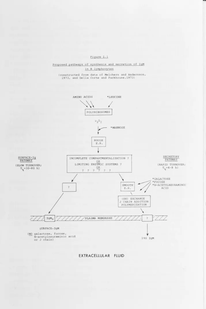
These processes are represented diagramatically in Fig 1.1.
(b) Lipopolysaccharide-stimulated nu/nu mouse spleen cells
(i) The surface-IgM of B cells cultured in vitro in the presence of mitogenic concentrations of E. coli
lipopolysaccharide (LPS) becomes aggregated and disappears from the cell surface during the first 30 min. of culture.
This IgM, which presumably caps and becomes endocytosed by the cells, is slowly digested by proteases. Such cells remain
devoid of surface-IgM until 25-30 h of LPS stimulation. (ii) After 10-15 h stimulation with LPS, total
cellular protein and IgM synthesis increases. IgMs molecules reappear on the lymphocyte surface after 25-30 h stimulation, in greater numbers than were originally present. A similar phenomenon is observed after capping surface-lg with either antigen (Diener and Paetkau,1972) or anti-Ig antibody (Loor, Forni and Pernis,1972). As judged by irnmunofluorescence, more than half the cells have increased quantities of surface-IgM.
(iii) Simultaneous with the reappearance of surface-IgM, secretion of 19S IgM (half-life, 4-8 h) is begun on a scale greatly exceeding that of unstimulated cells. The
secretion of large numbers of 19S IgM molecules is thought to be responsible for the increased numbers of IgMs molecules on the cell surfaces. Pokeweed mitogen has also been observed to selectively increase IgM synthesis in mouse B lymphocytes
(Parkhouse, Janossy and Greaves,1972).
Surface-IgM thus differs from secreted 19S IgM in two important ways. Firstly surface-IgM lacks the semiterminal
galactose and terminal fucose and N-acetylneuraminic acid
carbohydrate residues found in all secreted 19S IgM. Secondly surface-IgM molecules are 7S IgM subunits (IgMs).
SURFACE-lg PATHWAY (SLOW TURNOVER;
[image:25.755.15.730.11.1084.2]T~=30-80 h)
Figure 1.1
Proposed pathways of synthesis and secretion of IgM in B lymphocytes
(constructed from data of Melchers and Andersson, 1973, and Della Corte and Parkhouse,1973)
AMINO ACIDS *LEUCINE
~\
I
I
POLYRIBOSOMES j*MANNOSE
INCOMPLETE COMPARTMENTALIZATION? OR
LIMITING ENZYMIC SYSTEMS? OR
? ? ? ? ? ?
SECRETORY PATHWAY
(RAPID TURNOVER; T~=4-8 h)
*GALACTOSE
I
D
\
§
- ; ; ;*FUCOSE
~~ *N-ACETYLNEURAMINIC
I
ACID... ' ... ·· ... ·· .. J
IgMs [·· .. ·· ... ·· .. ·· ... ·· .. ·· ... ·· .. ··~LASMA MEMBRANESURFACE-IgM
(NO galactose, fucose, N-acetylneuraminic acid or J chain)
EXTRACELLULAR
\
(SH) EXCHANGE J CHAIN ADDITION
POLYMERIZATION
\
...
·· ...
·
l
?l
t .· .
·_ .· .. ·19S IgM
18
molecules may alter their conformation and allow polymerization
and secretion. However IgMs molecules lacking a full
carbo-hydrate complement can be polymerized in vitro by the covalent
addition of a joining (J) chain in the presence of a
disulphide-exchange system (Della Corte and Parkhouse,1973). Nevertheless,
the addition of these residues may increase the affinity of the
IgMs molecule for the disulphide-exchange system so that the
IgM is rapidly and efficiently polymerized. Furthermore, IgMs
molecules are probably very hydrophobic, considering their
stability in the plasma membrane. It is possible that the
addition of carbohydrate residues may decrease the hydrophobic
nature of IgMs. Thus, if these residues are added in the
proximity of both the cell surface and a disulphide-exchange
system, altered IgMs molecules would be rapidly polymerized
and secreted. Molecules acquiring only an incomplete
carbo-hydrate complement, perhaps as a result of a rate-limiting
enzyme system or because of prior compartmentalization, would
not be readily polymerized and would remain as stable entities
within the surface membrane. These aspects warrant further
investigation.
1.2.7 T Lymphocyte surface-lg
It now seems likely that T lymphocytes also contain
surface-IgM, but in quantities considerably less than found on
B cells. This conclusion is based on the following evidence:
(a) Anti-L-chain antibody has been shown to prevent
radioactive 'suicide' of irnmunocompetent T cells by 125r-labeled
antigens (Basten et al.,1971; Cooper and Ada,1972) and also to
(Mason and Warner, inhibit graft-versus-host reactions in mice
1970; Riethmuller, Rieber and Seeger,1971). Furthermore, both
anti-L-chain and anti-µ-chain sera can prevent the binding of
antigens by thymus-derived ABC (Hogg and Greaves,1972; Roelants, Forni and Pernis,1973; Ashman and Raff,1973).
(b) Nearly all Ig- rat thoracic duct lymphocytes can
specifically bind 1 2 5 r-labeled anti-rat-Fab antibodies, but
in quantities (200-3,000 molecules/cell) considerably less than
bound by Ig+ (B?) lymphocytes (20,000-150,000 molecules/cell)
(Jensenius and Williams,1973a). A similar phenomenon has been
observed for mouse spleen cells (Nossal, Warner, Lewis and
19
(c) Radioactive Ig possessing antibody activity is
released at 37°c by radioiodinated thymus-derived mouse thoracic
duct lymphocytes activated against histocompatability antigens
(Cone, Sprent and Marchalonis,1972).
These findings indicate that the antigen receptors
on both irnmunocompetent and antigen-binding T lymphocytes are
closely associated with bothµ and light Ig chains, and also
that most peripheral T lymphocytes have small amounts ofµ and light chains, probably as lgM, on their surfaces. At present,
the possibility that this T lymphocyte surface lgM is cytophilic
cannot be excluded (Webb and Cooper,1973).
There have been numerous reports concerning
surface-Ig on thymus cells. These cells have attracted attention
mainly for their unlikely contamination by rg+ (B) lymphocytes,
although< 5 % are irnmunocompetent (Blomgren and Andersson,1971)
and the majority may not be typical of mature T lymphocytes.
A small proportion (< 3 %) of mouse thymocytes are labeled
heavily by anti-Ig antibodies, but these cells are reported to be
not typical of thymocytes, B lymphocytes or plasma cells (Bankhurst and Warner,1971; Harnmerling and Rajewsky,1971;
Perkins, Karnovsky and Unanue,1972; Kirov and Ada, to be
published) and are deficient in
e
and TL (thymocyte) antigensbut positive for surface-lg and the plasma cell alloantigen PC.l
(Vitetta, Uhr and Boyse,1973). In addition, the Ig synthesized
and secreted by these lg+ cells (lgG and 19S IgM) accounts
completely for the Ig produced by whole thymus (Vitetta, Uhr
and Boyse,1973) and may also account for the isolation ofµ
and light Ig chains from radioiodinated thymocytes (Marchalonis,
Atwell and Cone,1972; Marchalonis, Cone and Atwell,1972).
Others, using similar techniques, have not detected surface-lg '
on thymocyte populations (Grey, Kubo and Cerottini,1972;
Vitetta, Bianco, Nussenzweig and Uhr,1972). Radioirnmunoassays
have indicated that mouse thymus cell populations contain very
little surface or total Ig compared to spleen cell populations,
(Gnanue, Grey, Rabellino, Campbell and Schmidtke,1971; Grey, .
Colon, Campbell and Rabellino,1972; Grey, Kubo and Cerottini,
20
1.3 Aims and outlines of thesis
This thesis describes studies aimed at elucidating
the identity of antigen-receptors on B lymphocytes. Isolation
and direct characterization of these receptors for antigen is
necessary to determine unequivocally whether they are
immunoglobulin and if so, whether and in what way they differ
from humoral immunoglobulins. It is also particularly
important to determine whether the receptors interact with any
membrane component(s) which might cause the lymphocyte to be
stimulated, perhaps by the activation of an enzymic process,
after the receptors have bound antigen.
The problem was approached initially by isolating
the lymphocyte plasma membrane in a state free of intracellular
material, especially Ig. Once this was achieved i t was intended
to fractionate the membrane into its various components and to characterize them. Thus, i t was hoped to identify the receptors
and any component(s) which might be associated with them.
In order to distinguish membrane components from
intracellular material which could adsorb to the membrane during
its purification, a method was developed for labeling, with
radioactive iodide, the plasma membranes of intact cells
(Chapter 3).
The results of studies on sheep lymphocyte plasma
membranes are described in Chapter 4. Sheep lymphocytes were
chosen for this work because they were available in pure form
and in large numbers. The membranes were fragmented under a
variety of conditions, and using a number of membrane markers
their distribution in density gradients was investigated. One
finding of this study was that the detergent Nonidet P-40 can '
be used to dissolve the plasma membrane without dissociating
membrane-bound antibodies from the surface antigens to which
they are attached. This use of NP40 was subsequently extended
to study the complexes formed between anti-Ig antibodies and
lymphocyte surface-lg (Chapter 6).
During these investigations i t was discovered that
anti-Ig antibodies bound in considerable amounts to intact
sheep lymphocytes. Because surface-lg is likely to be the
receptor for antigen (see 1.1.4), this phenomenon was
21
proportion (20-30 %) of sheep lymph cells were labeled by anti
-L, anti-µ, and (to a lesser extent) by anti-y-chain antibodies
(Chapter 5). Furthermore, by labeling the lymphocytes with
anti-lg antibodies and then dissolving them in NP40 i t was
found that IgM was present on the cell surface in both 7S and
19S forms (Chapter 6). The fate of the (surface-lg/anti-lg)
complexes during incubation of labeled cells at 37°c was also
determined (Chapter 6).
Thus, as had been found in lymphocyte populations of
other vertebrates, a proportion of sheep lymphocytes possessed
large quantities of Ig on their surface. Were these molecules
the "true" receptors for antigen? Experiments were initiated
with the aim of determining this, and preliminary studies
investigating the ability of anti-lg antibody to inhibit the
net binding of antigens to populations of mouse spleen, lymph
node and sheep lymph cells are described in Chapter 7. It is
hoped that an extension of this approach will enable the
receptors for a particular antigen to be isolated from a
population of lymphocytes and identified.
1. 4 Communications
Some of the work contained in this thesis has been
presented at meetings and has been published, or is to be
submitted for publication, as follows:
1. Ey, P.L. (1971). Immunoglobulins on the sheep lymphocyte
surface. Abstr. Ann. Meet. Aust. Soc. Immunol.,
Melbourne, p. 7.
2. Ey, P.L. (1973). Immunoglobulins on the surface of sheep
lymphocytes. I. Class and cellular distribution. Eur . J.
Immunol. 3 : 37.
3. Ey, P.L. (1973). Immunoglobulins on the surface of sheep
lymphocytes. II. Class, size and fate during incubation
of lymphocytes at 37°c. Eur. J. Immunol. (in press).
4. Ey, P.L. A nonenzymic method for selective radioiodination
of the plasma membranes of intact red blood cells.
(in preparation).
5. Ada, G.L. and Ey, P.L. Lymphocyte receptors for antigen
Chapter 2
2.1 General
2.1.1
2.1.2
2.1.3
2.1.4
2.1.5
2.1.6
2.1.7
2.1.8
2.1.9
2.1.10
2.1.11
2.1.12
2.1.13
Media and reagents
Protein determination
5'-Nucleotidase assay
Polyacrylamide gel electrophoresis
Irnrnunoelectrophoresis
Radio iodination
Autoradiography
Radioirnrnunoassay
Gradient centrifugation
Antigens
Animals
Cell suspensions
Antibody titrations
22
2.2 Irnrnunoglobulins, antisera and antibodies
2.2.1 Sheep IgG and serum macroglobulin
2.2.2 Reduction of sheep IgG: Separation of y and L chains
2.2.3 Sheep IgM
2.2.4 Mouse IgG and IgM
2.2.5 Normal rabbit IgG
2.2.6 Rabbit antisera and antibodies
2.2.7 Purity of sheep Igs and specificity of rabbit
anti-sheep-Ig sera
2.2.8 Rabbit anti-sheep-Ig Fab fragments: Preparation
2.1 General
2.1.1
2.1.2
2.1.3
2.1.4
2.1.5
2.1.6
2.1.7
2.1.8
2.1.9
2.1.10
2.1.11
Media and reagents
Protein determination
5'-Nucleotidase assay
Polyacrylamide gel electrophoresis
Irnrnunoelectrophoresis
Radio iodination
Autoradiography
Radioirnrnunoassay
Gradient centrifugation
Antigens
Animals
2.1.12 Cell suspensions
2.1.13 Antibody titrations
22
2.2 Irnrnunoglobulins, antisera and antibodies
2.2.1 Sheep IgG and serum macroglobulin
2.2.2 Reduction of sheep IgG: Separation of y and L chains
2. 2. 3 Sheep IgM
2. 2. 4 Mouse IgG and IgM
2.2.5 Normal rabbit IgG
2.2.6 Rabbit antisera and antibodies
2. 2. 7 Purity of sheep Igs and specificity of rabbit
anti-sheep-Ig sera
2.2.8 Rabbit anti-sheep-Ig Fab fragments: Preparation
23
2.1 General
2.1.1 Media and reagents
The following solutions were prepared using
double-distilled water by the Department of Microbiology Media
Service, and stored at 4°c before use. Saline: 0.9 % (w/v)
NaCl dispensed in 150 ml or 500 ml aliquots and autoclaved at
112°c for 15 min. Alsever's solution: NaCl (21 g), D-glucose
(102.5 g), sodium citrate dihydrate (40 g) and citric acid
(4 g) dissolved in water to a final volume of 5 litres. The
solution was adjusted to pH 6.1 with 10 % citric acid,
dispensed in 100 ml aliquots and autoclaved at 112°c for 20
min. Dulbecco's balanced salt solution (DBSS) was made by
combining three solutions, each prepared separately (Dulbecco
and Vogt,1954): Phosphate-buffered saline (PBS) contained
NaCl .(8.0 g), KCl (0.2 g), Na 2HP04 (1.15 g), KH 2Po4 (0.2 g) and
water to 800 ml. It had a pH of 7.4, was dispensed in 400 ml
aliquots and autoclaved at 121°c for 20 min; PBS2 consisted of
cacl 2 (0.1 % w/v) in 50 ml aliquots; PBS3 was MgC1 2 .6H20 (0.1 % w/v) in 50 ml aliquots. Both PBS2 and PBS3 wer e autoclaved at 112°c for 15 min. DBSS (500 ml) was prepared by combining
PBS solutions 1, 2 and 3. Eagle's minimal essential medium
(Cat. No. F-15, Grand Island Biological Co., N.Y.)
filter-sterilized, was obtained from Dr. A.J. Cunningham. Foetal
calf serum (FCS) was obtained from the Commonwealth Serum
Laboratories (Parkville, Victoria).
Reagents were obtained as follows: Papain (2X
crystallized, Cat. No. P-3125) and
Tris(hydroxymethyl)amino-methane (Tris) ("Sigma 7-9") (Sigma Chemical Co.); 2-Mercaptoethanol
(2ME) and N,N-Diethylaminoethyl(DEAE)-cellulose (Eastman '
Organic Chemicals, N.Y.); Ammonium sulphate (Mallinckrodt
Chemical Works, U.S.A.); Nonidet P-40 (NP40; Shell Petroleum
Co.). All other reagents were of laboratory or analytical
grade.
2.1.2 Protein determination
Protein concentrations were usually estimated from
absorbance at 280 nm (A 280 ). The extinction coefficient
24
13, based on values published for rabbit and human Igs (Williams and Chase,1968). Otherwise, where indicated in the text,
protein was measured using Folin's reagent (Lowry et al.,1951). 2.1.3 5'-Nucleotidase assay
5'-Nucleotidase (5'-ribonucleotide phosphohydrolase; EC 3.1.3.5) activity was assayed by the method of Michell and Hawthorne (1965). Samples (0.4 ml) were mixed with 0.2 ml of assay solution to give final concentrations of 100 rnM KCl, 10 rnM MgC1 2 , 10 rnM L (+) NaK-tartrate, 50 rnM Tris-HCl, pH 7.5 and 5 rnM adenosine 5'-monophosphate (Calbiochem.). Blanks
contained all but the nucleotide. Assay mixtures were incubated in a 37°c water bath and the reaction stopped by placing
samples in ice and adding 0.2 ml iced trichloroacetic acid
(1.5 M). The standard reaction time w.as 15 min. Inorganic
phosphate released by the nucleotidase was measured by optical density at 820 nm (A 820 ) using the molybdate/ascorbic acid
method of Ames and Dubin (1960).
2.1.4 Polyacrylamide gel electrophoresis Two gel systems were employed, one using
non-dissociating conditions and run at pH 8.9 (Davis, 1964), the
other using dissociating conditions and run at pH 3.5 in 9 M urea (Parish and Marchalonis,1970). Gels from both systems
were stained in a solution consisting of 7 % (v/v) acetic acid, 0.25 % (w/v) amido black and 0.5 % (w/v) HgC1 2 . Destaining was
performed electrophoretically.
2.1.5 Irnrnunoelectrophoresis
This technique was performed by the method of
Scheidegger (1955). The gels contained 1.5 % agar in barbital buffer, pH 8.2 (µ
=
0.05).2.1.6 Radioiodination
Soluble proteins were labeled with carrier-free Na
!
125rj (IMS3; The Radiochemical Centre, Amersham) or with Na !_131r J (Code llBl, Australian Atomic Energy Commission, Lucas Heights, Sydney) by direct oxidation with chloramine-T(Greenwood, Hunter and Glover,1963; Ada, Nossal and Pye,1964). To 5-50 µl of protein solution (0.2-5 mg/ml in DBSS or Tris
25
chloramine-T to a final concentration of 0.1-0.5 rnM. After
5-10 min. at room temperature, Na 2s 2
o
5 was added in 3 to 4-fold molar excess with respect to chloramine-T, followed by"carrier" NaI to 10 rnM. The resulting solution was diluted
(usually 10 to 20-fold with DBSS or Tris buffer containing
10 % (v/v) foetal calf serum (FCS), or normal rabbit serum
(NRS) and dialyzed at room temperature against buffer containing
15 rnM NaN 3 until most unreacted iodine had been removed.
Labeling efficiencies were generally 70-95 % and specific
activities of 1-50 µCi/µg protein were used. Iodination had
no measurable effect on antibody activity as labeled antibody
retained its ability to bind specifically to the antigen
against which i t was prepared (Fig 2.6). Henceforth, unless
stated otherwise, "labeled" refers to 125 r-labeled.
Cells were iodinated in either of two ways: (a)
Using chloramine-T oxidation as described above, but replacing
the protein solution with a cell suspension. After reaction,
labeled cells were washed by centrifugation (500 g, 10 min.)
and resuspension in isotonic media. (b) Using lactoperoxidase.
This enzyme was purified by Mr. K. Clarke from fresh skimmed
cows milk by (NH 4 )2so4 precipitation and carboxymethylcellulose chromatography as described by Marchalonis (1969). The purified
enzyme (E 412;E280
=
0.44) was stored at -20°c in 0.2 ml aliquots of 0.5 mg/ml. Details for both methods are given in the text.For direct measurement of radioactivity, samples
were counted individually to within 5 % error in a Packard
Model 3002 Tri-Carb Scintillation Spectrometer. Counting
efficiency for 12 51 was approximately 60 %.
2.1.7 Autoradiography
This technique was carried out according to Byrt and
Ada (1969). Briefly, cell smears were dipped in Kodak NTB-2
photographic emuls~on at 42°c, dried vertically and stored in
the presence of anhydrous caso
4 at 4°c for 1-28 days. After development, the slides were stained in 5 % Giemsa.
2.1.8 Radioirnrnunoassay
In this technique, the precipitation of labeled
antigen (routinely, 1-10 ng) is tested by mixing antigen with
26
precipitation versus antiserum dilution is thus derived. In
addition, soluble substances similar or related to the labeled
antigen can be tested for their ability to inhibit precipitation
of a constant amount of labeled antigen by a given dilution of
antiserum. The amount of inhibitor present in a preparation
can thus be estimated.
The method is described in detail elsewhere (Parish,
Wistar and Ada,1969). Two diluents were used: BSA-dil, which
was 3 % (w/v) bovine serum albumin in 0.05 M Tris-HCl,pH 7.4
containing 15 mM NaN 3 ; and S-dil, which was BSA-dil containing
10 % (v/v) NRS. In some experiments, Nonidet P-40 (NP40) was
included in the diluents at 0.5-5 % (v/v) final concentration.
At these concentrations, this nonionic detergent dissolves the
cell plasma membrane but has no effect on the formation of
(Ig/anti-Ig antibody) complexes as measured by this technique.
A typical inhibition curve is shown (Fig 2.1) in which 44 ng
of sheep IgG or 3800 ng of sheep IgM was needed to inhibit by
50 % the precipitation of 125r-labeled sheep IgG by rabbit
anti-sheep-y-chain serum. Thus the IgM preparation contained
1. 2 % (w/w) IgG.
Similarly, inhibitors present on particulate supports
can be detected and measured. To estimate the class and amount
of Ig chain equivalents exposed on the surface of lymphocytes
and able to bind anti-lg antibody, washed lymphocytes (10 8
cells/ml) in DBSS containing 1 % NRS and 15 mM NaN 3 were
diluted serially in twofold steps with the same medium.
Portions (50 µl) of each dilution were mixed with 10 µl of
diluted antiserum (anti-µ or anti-y) and incubated overnight
at 4°c with gentle agitation. Assays were done in the presence
and absence of NP40 (1 % v/v). As standards, dilutions of IgM
or IgG were used instead of cells. After incubation, the cells
were pelleted and 20 µl of each supernatant was mixed with 50
µl of 125 r-labeled, IgM or IgG and treated as for the direct
precipitation assay (Parish, Wistar and Ada,1969). Controls
included cells plus diluent (for minimal precipitation) and
diluent plus antiserum (maximum precipitation).
2.1.9 Gradient centrifugation
This was performed using freshly-prepared linear
100 C
0
-
0-
Q.u
C1) ~ Q. E :::> E )(
50
0
E ~
0
C
0
-_J:) J::. C
~
0
0
10
44 ng
100 1,000
Amount of Inhibitor (ng}
lgG
lgM
3,800 ng
10,000
Figure 2.1: Radioimmunoassay curves illustrating
the ability of unlabeled sheep IgG or IgM to 125
inhibit the precipitation of I-labeled IgG
[image:37.738.11.718.22.954.2]27
gradients of 10 % to 30 % (w/v) sucrose were employed. For
equilibrium density centrifugation, gradients varying between
20 % and 60 % (w/v) sucrose were used. Details are provided
in the text. Samples were layered on to gradients immediately
prior to centrifugation. Fractionation of gradients was
accomplished usually by tube bottom puncture, but otherwise
by collection via a fine glass tube inserted through the
gradient. Sucrose concentrations were calculated by comparing
the refractive indexes of collected fractions with those of
solutions of known sucrose concentration. Densities were
estimated by reference to published tables (Dawson, Elliott,
Elliott and Jones,1969).
2.1.10 Antigens
Monomeric flagellin (MON), molecular weight 40,000
daltons, and its particulate derivative, polymerized flagellin
(POL), were prepared from flagella of Salmonella adelaide SW
1338 (H antigen: f,g) or of
s.
typhimurium SL 870 (H antigen:1,2) as described by Nossal and Ada (1971). Haemocyanin (HCY),
a pentamer of molecular weight approximately 450,000, was
crystallized from the haemolymph of the Southern Australian
crayfish, Jasus lalandii (Moore, Henderson and Nichol, 1968).
It was stored at -20°c as a 100 mg/ml solution in water.
TMVP, the unit protein of tobacco mosaic virus, strain vulgare,
was obtained through the courtesy of Dr. E. Benjamini.
2.1.11 Animals
Sheep were randomly bred merino virgin ewes or wethers.
Inbred CBA/H mice and outbred chickens and rabbits were obtained
from the John Curtin School of Medical Research animal colony.
2.1.12 Cell Suspensions
2.1.12.1 Red Cells
Sheep red blood cells (SRBC) were prepared from blood
drawn by venipuncture. Red cells were washed at least 4 times
in saline and the buffy coat removed after each centrifugation.
Chicken red cells (CRBC) were prepared as for SRBC.
2.1.12.2 Lymphoid cells