JOURNALOFVIROLOGY,Apr.1968,p.393-399 Copyright ©1968 AmericanSocietyforMicrobiology
Vol.2,No. 4 PrinitedinU.S.A.
Ultrastructure of
Lymphocystis Virus
LUTZ 0. ZWILLENBERG AND KEN WOLF
DepartmentofHygiene and Medical Microbiology, UniversityofBerne,CH-3008Berne,Switzerland; and Bureau
of Sport Fisheries and Wildlife, Eastern Fish DiseaseLaboratory, Kearneysville, West Virginia 25430
Received forpublication26December1967
Lymphocystisvirusobtained frombluegills(Lepomis macrochirus) wascultured in
thepermanentbluegillcelllineBF-2andexaminedbyelectronmicroscopy in
ultra-thin sections of cell cultures and in negative-contrast preparations from cells and from centrifuged culture medium. Accordingtonegative-contrast preparations, the icosahedral virions have an overall diameter close to but not exceeding 300 m,u. Delicate filaments seemtoissuefromthevertices. In collapsed virions, anordered arrayofmorphological unitswasseen.Positivelycontrasted virionsin ultrathin
sec-ticns showa shell withthree dark (heavy metal-stained) layers alternatingwithand
separated by two clear layers. The acquisition of an additional outer membrane duringrelease from the cell,asfound in African swine fevervirus, wasnever seen.
Morphologically, lymphocystis virus is consideredtobeclosely relatedtoTipula iri-descent virus.
Lymphocystis is a virus disease of various teleost fish species, a disease known for halt a century (19, 20). The ultrastructure of the
lymphocystis virus has been investigated by
Walker (16), Walker and Weissenberg (17), and Walker and Wolf (18), but the technique
did not allow high resolution. Moreover, an examination by means of negative staining seemed necessary in order to supplement the
information fromultrathinsections.
MATERIALS AND MErHODS
Lymphocystis virusfrom bluegills (Lepomis
macro-chirus) wasinoculated ontomonolayersofBF-2cells,
apermanentfibroblast-like cell line derived from blue-gill fry and propagated in the Eastem Fish Disease Laboratory. Theinfected culturesweresent toBerne for electron microscopic processing. They were kept
at roomtemperature in the dark.After21 days,when
the cytopathic effect typical for lymphocystis virus (21) had become welldeveloped,someof the cultures
were washed with Millonig's phosphate buffer and fixed in situ with4%reagentgrade Formalindissolved in thesamebuffer. Thecell sheetwasthenscraped off with a rubber wiper and centrifuged. Pieces of the pellet werepostfixed in 2% osmiumtetroxide, dehy-drated in ethyl alcohol, treated overnight with 1% uranyl acetate in ethyl alcohol, washed again in
ethyl alcohol, and embedded in Durcupan ACM
(FLUKAAktiengesellschaft,BuchsSG,Switzerland), with propylene oxide as an intermediate solvent. Ultrathin sections were cut with glass knives on an LKB Ultrotome III, collected on grids coated with collodium and carbon,poststained with lead citrate, and examined in a Philips EM 200 electron micro-scope.
Other cultures were examined 10 to25 days after inoculation,either with theunmodified negative-stain-ing technique described by Zwillenberg and Burki (24), or by using distilled water containing about 0.01%bovine serum albuminas asuspension medium instead of the 5% ammonium acetate solution. In other cases, thesupernatant medium waswithdrawn from theculture andcentrifugedat2,500 X gfor10
min at roomtemperature. Thesedimentwas washed
withMillonig'sphosphate buffer(samecentrifugation speed and time) and suspended either in 5%
am-monium acetate solution or in distilled water, both containing about0.01% bovineserumalbumin, prior
tomixing with2% sodium phosphotungstate solution (pH 7.0).
RESULTS
In ultrathin sections of infected BF-2 cells, virusparticles withanoverall diameteraveraging 250 m, were found in the cytoplasm, either in more or less coherent groups or surrounding
clear areas ofviroplasm (Fig. 1). The particles had a shell with a hexagonal profile (Fig. 2 and 3). In this profile, a central dark (electron opaque) line, about 4.8 m,u in thickness, was
separated by a clear (electron-translucent)
space from an outer and inner dark line. These lines were about 2
my
thick and about 12 m,u apart from each other. The shells were either filled with a granular or fibrillar mass, or theywere empty. Some shells had a more or less crumpled appearance (Fig. 2). A few shellswere not closed but showed a more or less wide gap. In many instances a fuzzy halo, about 40 m,u inthickness, was surrounding theshells (Fig. 3).
393
on November 11, 2019 by guest
http://jvi.asm.org/
Special attention was paid to any images sug-gesting the passage of particles through the cell membrane and indicating an acquisition of an
additional outer membrane during the passage. Suchimageswere never seen.
When negatively stained specimens were pre-pared, with 5% ammonium acetate solution as the suspension medium, icosahedral virus par-ticles of an overall diameter of about 300 mp were observed in an apparently intact state, the ridges of the icosahedra being clearly set offby an outerelectron-translucent band (width about 6mMA) andaninnerelectron-opaqueline (Fig.4).
When distilled water was used, a large propor-tion ofthe particles had collapsed and showed
an ordered array ofmorphological units along the facets of the icosahedra. These units were
resolved with difficulty and their images
de-teriorated rapidly under electron bombardment.
They gave the impression of rings with a diam-eterofabout 5 m,u.Theywere arrangedin rows, some ofthem nearly radial and some ofthem
seemingly crossing each other under an oblique angle (Fig. 5 and 6). The electron-translucent margin ofthe collapsed particles was separated fromtheabove rowsbya dark line. Themargin
was often fragmented. The fragments showed a
close relation to the rows of morphological units. They seemed to be their extension.
Sediment from centrifuged culture medium containednot only single virions,butmoreoften
clusters ofthem. In one instance, these clusters werehuge. The clustered virionsweresometimes
not in close contact, but seemed attached to each other by means of
filaments,
which hadbecomeentangled. Suchfilaments could be better
studiedinoccasionalfree-lying virionsorin
small,
looseclusters (Fig.4).Thefilamentshadawidth
ofthe order of4m,u anda
length
of200 to 300m,u. Although various cell debris were present in the preparations,
comparable
filaments were not found outside virion clusters or outside theimmediate
vicinity
of virions. In those cases,in which
crowding
of virions and filaments wasnotserious, sothat thecourseofsingle filaments
could be followed to the contours of presump-tive virions of origin, such filaments seemed to issue preferentially from angles in the virion contours or from identifiable vertices of
appar-ently intact virions (Fig. 4 and 5). Comparable filaments were not seen in specimens prepared fromwhole cells.
DISCUSSION
The findings in ultrathin sections of infected BF-2 cells accord well with those of previous investigators (16-18). In addition, they show more detail of the virus shell and possibly a morphological substrate for the osmiophilia (19) and the ether-sensitivity (21) of the virus. In fact, the hexagonal profiles are reminiscent
of those quintuple-layered membranelike struc-tures (with three electron-opaque and two elec-tron-translucent layers after a comparable
fixa-tion and staining procedure), which are the
re-sult of lipoprotein aggregation in degenerating cells. For fish cells, such structures have been
pictured in remnants of erythrocytes ingested
bytroutspleen reticulum cells (23). This
morpho-logical resemblance does not prove a structural
similarity but lends support to the supposition that thelymphocystis virusshellcontains alipidic component. However, both the very regular,
ico:ahedral
shape of the shell and the imagesproduced by negative staining show that theshell is certainly different from an ordinary
lipopro-tein membrane and that an ordered array of capsomeresisintegrated in the shell. The precise nature of this integration is not clear.
While the granular image of any dark line in the sections must be interpreted with caution on account ofpossible phase-contrast effects of the carbon substrate, the fragmentation of the margin
of collapsed, negatively stained virions is cer-tainly real. As a purely tentative interpretation, the heavycentral line in the sections and the elec-tron-translucent marginal band of negatively contrasted virions may be correlated with the capsomeres, and these may be backed, both to the outside and the inside, by lipoprotein layers. The greater virion diameters, as compared to
former ultrastructural studies, are explained, at
least in part, by the stronger shrinkage of the methacrylate used in those studies. Even so, there was evidently some shrinkage and knife compression in our material also. The diameter incollapsed negatively stained virionsis
certainly
exaggerated by flattening. However, the greater diameter of noncollapsed negatively stainedvirions,ascomparedtosectionedmaterial,cannot be ascribed to flattening only. The actual virion diameter should therefore be quite close to but not in excessof 300 m,u.
Though there is no absolute guarantee that
FIG 1. UltrathinsectionofaBF-2cellinfected withlymphocystisvirus. Virusparticles surroundingclearspaces.
X 14,000.
FIG. 2. Lymphocystis virus inthecytoplasm ofaBF-2cell.Noteparticlecontentandstructureof the shell. X
174,000.
on November 11, 2019 by guest
http://jvi.asm.org/
ULTRASTRUCTURE OF LYMPHOCYSTIS VIRUS
',*tf*
I.
0.22^
395
VOL. 2,1968
.4414..t.
..z.
w
11I.I *V.#14n, "-.
A
"'Cl*-L
v i*
..10. 0
., I :k
A
on November 11, 2019 by guest
http://jvi.asm.org/
1,~ ~ ~4
L4A
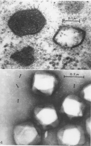
FIG. 3. Lymphocystisvirusinthecytoplasmofa BF-2cell.Notestructure ofthe shell and fuzzyhalo around the particles. X 250,000.
FIG. 4. Negatively stainedlymphocystis virusfromculturemedium; suspendedin5% ammonium acetate.
Ar-rowspointtofilaments. X150,000.
396
.: .:... 4.
.."
4:ioi. I..,fv
on November 11, 2019 by guest
http://jvi.asm.org/
[image:4.485.63.441.23.627.2]ULTRASTRUCTURE OF LYMPHOCYSTIS VIRUS
the observed filaments are viral appendages, both the visual observations on the microscope screen and the recorded images created this impression, %Ahich was strengthened by the lack of comparable filaments outside the immediate vicinity of virions. If the filaments are actually appendages issuing from virion vertices, this would constitute a certain parallel to the fibers of adenoviruses(9,12,15). Uptothepresent time, there is no evidence that lymphocystis virus has hemagglutinating or hemadsorbing properties (21). Africanswinefever virus, whichis morpho-logically similar, shows the phenomenon of hemadsorption (5).
Although African swine fever virus andTipula iridescent virus are morphologically related (1), there is at least one property, which separates them: the porcine virus acquires an additional membrane during its passage through the cell membrane (4, 6). This has not been observed either in Tipula iridescent virus (2, 3, 22) or in
lymphocystis virus. It has been observed in morphologically similarfrogviruses (7,8, 10,11). Our results suggest that lymphocystis virus should be placed taxonomically close to Tipula
iridescent virus. Other comparable viruses are Sericesthis iridescent virus (14) and the gecko virus formerly named Pirhenmocyton tarentolae
(13).
LITERATURE CITED
1. ALMEIDA, J. D., A. P. WATERSON, AND W.
PLOWRIGHT. 1967. The morphological
charac-teristics of African swine fevervirus and its
re-semblance to Tipula ilidescent virus. Arch. Ges. Virusforsch. 20:392-396.
2. BIRD, F. T. 1961. The development of Tipula
iridescent virusin the cranefly, Tipula paludosa Meig., and the wax moth, Galleria melloniella
L. Can. J.Microbiol. 7:827-830.
3. BIRD, F. T. 1962. On the development of the
Tipula iridescent virus particle. Can. J.
Micro-biol. 8:533-534.
4. BREESE, S. S.,JR., ANDC.J. DEBOER. 1966.
Elec-tron microscope observations of African swine fever virus in tissue culture cells. Virology 28:
420-428.
5. BREESE,S. S., JR., AND W. R. HEss. 1966. Electron
microscopy ofAfrican swine fever virus
hemad-sorption. J. Bacteriol. 92:272-274.
6. BREESE, S. S., JR., S. S. STONE, C. J. DEBOER,AND
W. R. HEss. 1967. Electron microscopy of the
interaction of African swine fever virus with
ferritin-conjugated antibody. Virology 31:508-513.
7. CAME, P. E., AND P. D. LUNGER. 1966. Viruses
iso-lated from frogs and their relationship to the
Lucke tumor. Arch. Ges. Virusforsch.
19:464-468.
8. DARLINGTON, R. W., A. GRANOFF, AND D. C.
BREEZE. 1966. Viruses anJ renal carcinoma of
Rania pipielts. 11. Ultrastructural studies and sequential development of virus isolated from
normaland tumortissue. Virology 29:149-156.
9. GINSBERG, H. S., H. G.PEREIRA,R.C. VALENTINE,
AND W. C. WILCOX. 1966. A proposed
termi-nology forthe adenovirus antigens and virion
morphological subunits. Virology 28:782-783.
10. GRANOFF, A., P.E. CAME, AND K. A. RAFFERTY,
JR. 1965. Theisolation and properties of viruses
from Ra,ia pipienis: their possible relationship to the renal adenocarcinoma of the leopard
frog. Ann. N.Y. Acad.Sci. 126:237-255.
11. LUNGER, P. D., AND P. E. CAME. 1966.
Cyto-plasmic viruses associated with Lucke tumor
cells.Virology 30:116-126.
12. NORRBY, E. 1966. The relationship between
solu-bleantigensand the virion of adenovirus type 3. I. Morphological characteristics. Virology
28:236-248.
13. STEHBENS, W. E., AND M. R. L. JOHNSTON. 1966. Theviral structure ofPirhemocvton tarenitolae.
J.Ultrastruct.Res.15:543-554.
14. STEINHAUS, E. A., AND R. LEUTENEGGER. 1963. Icosahedral virus from a scarab (Sericesthis).
J. Insect Pathol.5:266-270.
15. VALENTINE, R. C., AND H. G. PEREIRA. 1965.
Antigens and structure of the adenovirus. J. Mol. Biol. 13:13-20.
16. WALKER, R. 1962. Finestructureof lymphocystis
virus offish. Virology18:503-505.
17. WALKER, R., AND R. WEISSENBERG. 1965.
Con-formityoflightand electron microscopic stud-iesonvirusparticledistributioninlymphocystis
tumor cells of fish. Ann. N.Y. Acad. Sci. 126:
375-395.
18. WALKER,R., AND K. WOLF. 1962. Virus arrayin
lymphocystis cells of sunfish. Am. Zoologist 2:566.
19. WEISSENBERG, R. 1965. Fiftyyearsof research on thelymphocystis disease of fishes (1914-1964).
Ann.N.Y.Acad.Sci. 126:362-374.
20. WOLF, K.1966.The fish viruses. Advan. Virus Res.
12:35-101.
21. WOLF, K., M.GRAVELL,ANDR. G.MALSBERGER.
1966. Lymphocystisvirus:isolation and
propa-gationincentrarchid fish cell lines. Science 151: 1004-1005.
397
VOL. 2, 1968
on November 11, 2019 by guest
http://jvi.asm.org/
i.S: ..
:n st
.r. ;.;
..
0.-2r
--/
5
398
on November 11, 2019 by guest
http://jvi.asm.org/
ULTRASTRUCTURE OF LYMPHOCYSTIS VIRUS
22. XEROS, N. 1964.Development of Tipula iridescent virus (TIV). J. Insect Pathol. 6:261-283.
23. ZWILLENBERG, H. H. L.,ANDL.0.ZWILLENBERG.
1963. Uber den Erythrocytenabbau in der Forellenmilz unter besonderer
Berucksichti-gung der Erythrocytenfeinstruktur. Z. Zell-forsch. 60:313-324.
24. ZWILLENBERG, L.O.,ANDF. BURKI. 1966.Onthe
capsid structure of some small feline and bovine RNAviruses. Arch. Ges. Virusforsch. 19:373-384.
FIG. 5. Collapsed, negativelystainedlymphocystisvirus;suspendedindistilledwater.Arrowpoints tofilament. X 145,000.
FIG. 6. Collapsed,negatively stainedlymphocystis virus;suspendedindistilledwater.Arrowdenotesarea where
rowsof subunitscanbediscerned. X 311,000.
399
VOL.2,1968