Under consideration for publication in J. Plasma Phys.
Measurement of the Angle, Temperature
and Flux of Fast Electrons Emitted from
Intense Laser-Solid Interactions
D. R. Rusby
1,2, L. A. Wilson
1, R. J. Gray
2, R. J. Dance
2, N. M. H.
Butler
2, D. A. MacLellan
2, G. G. Scott
1, V. Bagnoud
3, B. Zielbauer
3,
P. McKenna
2, D. Neely
1,21STFC Rutherford Appleton Laboratory, Oxfordshire OX11 0QX, UK 2
SUPA Department of Physics, University of Strathclyde, Glasgow G4 0NG, UK
3PHELIX Group, Gesellschaft fur Schwerionenforschung, D-64291 Darmstadt, Germany
(Received ?; revised ?; accepted ?. - To be entered by editorial office)
High-intensity laser-solid interactions generate relativistic electrons, as well as high-energy (multi-MeV) ions and X-rays. The directionality, spectra and total number of electrons that escape a target-foil is dependent on the absorption, transport and rear side sheath conditions. Measuring the electrons escaping the target will aid in improv-ing our understandimprov-ing of these absorption processes and the rear-surface sheath fields that retard the escaping electrons and accelerate ions via the Target Normal Sheath Ac-celeration (TNSA) mechanism. A comprehensive Geant4 study was performed to help analyse measurements made with a wrap-around diagnostic that surrounds the target and uses differential filtering with a FUJI-film image plate detector. The contribution of secondary sources such as x-rays and protons to the measured signal have been taken into account to aid in the retrieval of the electron signal. Angular and spectral data from a high-intensity laser-solid interaction are presented and accompanied by simulations. The total number of emitted electrons has been measured as 2.6×10¹³ with an estimated
total energy of 12±1J from a 100µm Cu target with 140J of incident laser energy during a 4×10²⁰ W/cm2interaction.
1. Introduction
When a high-intensity laser ( I>1018W/cm2) pulse interacts with a solid target,
elec-trons are accelerated on the front surface and travel through the target where they will be emitted from the rear surface of the target, accelerating protons and heavy ions with them (McKennaet al.(2004)). The source of these electrons is the initial absorption processes that occur at the front surface. At intensities ∼1016 W/cm2 the dominant absorption processes are resonance absorption and Brunel heating (Brunel (1987); Wilks & Kruer (1997)). The latter is particularly dependent on minimal pre-plasma (scale length ( Ls)< laser wavelength (λ)). Both of these processes accelerate the electrons perpendicular to the target surface whereas for higher intensities (> 1018 W/cm2) the ponderomotive
provides new insight into the front and rear surface field evolution processes.
Many significant measurements of the angular and spectral distributions of the elec-trons emitted from the rear surface and bremsstrahlung from internal elecelec-trons stopping within the target have been made and reported in the past (Hatchettet al.(2000); Ed-wardset al. (2002); Schwoereret al. (2001); Chen et al.(2013); Norreys et al.(1999)). However, these measurements are often made at a single point as opposed to a contin-uous angular distribution. These techniques can be very susceptible to beam pointing or non-uniformities that may arise during the laser-plasma interaction. Measurements of the entire beam escaping the target can yield improvements in the knowledge of such interactions.
In this paper, we have used an angular wrap-around stack previously introduced by Gray et al. (2011) to measure the total forward distribution of electrons escaping the target. The diagnostic is a differentially filtered cylindrical stack with the target posi-tioned in the centre. Significant improvements in the understanding of the diagnostic sensitivity have been made using simulations of both electron and x-ray absorption and scattering to infer spectral information about the escaping electrons. The diagnostic has also been used on a laser-solid interaction experiment with the aim of measuring the escaping electron distribution.
2. Design
The wrap-around stack is a 270o cylindrical diagnostic designed to provide angular information about the escaping particles/radiation from the target which is positioned at its centre, as shown in Figure 1. Multiple layers of Fuji BAS-TR image plate (IP) which sits inbetween 0.85mm thick Fe filtering is used to infer information on the emit-ted spectra. To initially ensure that the measured signal on the IP layer was primarily electrons, simulations were conducted using the ion stopping code SRIM (Ziegleret al. (2010)) to calculate the minimum filtering required to stop any protons accelerated from the target contributing to the signal. During solid-target interaction, maximum proton energies of 30MeV were measured with a separate diagnostic, which require the first layer of Fe filtering to be 1.7mm thick as shown in Figure 1. The filtering design, highlighted in Figure 1, separates the electron signals spectrally. The diagnostic is usually positioned above or below the horizontal axis to enable other diagnostics to monitor the target si-multaneously, and given the depth of 50mm of the plates; this also provides a vertical angular distribution over ∼ 30o. The open side of the diagnostic enables the focusing laser light to reach the target.
3. Simulations
em-Figure 1.Schematic of the diagnostic arrangement of Fuji BAS-TR Image Plate (IP) between 0.85mm Fe filters used in the wrap-around stack that covers 270 degrees around the target.
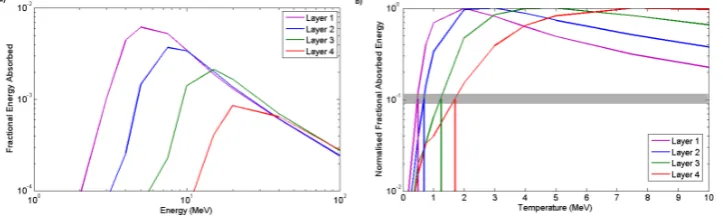
Figure 2.(a) The fractional absorption in the IP layers from mono-energetic electrons incident onto the array of Fe filters as a function of energy. (b) The fractional absorption of Relativistic Maxwellian electron distributions normalised to the maximum of each layer. The shaded region represents where the signal has dropped to 10% on that layer. A temperature extraction is unreliable below this region due to the contribution of X-rays.
ployed. The stack arrangement was built as shown in Figure 1 and the simulations were performed using 106 electrons, with each run using a single energy between 1MeV to
100MeV. Separating the energy deposited on each layer was achieved by Geant4 provid-ing the position and amount of energy accumulated from the simulation results. Dividprovid-ing the energy in each layer of IP by the total input energy yields the fractional deposited energy in each layer. The resulting response curve of the diagnostic for mono-energetic electrons is shown in Figure 2 (a). Due to the heavy filtering required to stop proton contamination, the threshold energy for electrons is approximately 2MeV.
An escaping relativistic Maxwellian electron distribution is assumed as the output to represent the experimental escaping electron distribution. The output from these sim-ulations can be compared to experimental data to find the temperature of the escaped electron distribution. The normalised fractional absorbed energy from an incident rela-tivistic Maxwellian electron distribution is show in Figure 2 (b).
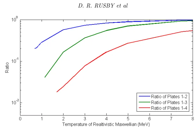
[image:3.595.98.460.276.384.2]Figure 3.The ratios of layer 1 to each sequential layer for the total energy absorbed by the image plate as a function of temperature.
As well as the target being a source of protons, it will also generate many bremsstrahlung x-rays as the electrons pass through it. These x-rays will make a simultaneous contri-bution to the IP signal regardless of the design/arrangement of layers. To estimate the impact of the x-ray signal, electrons were sent into a thin target in Geant4 creating a bremsstrahlung spectrum. An example target of 100µm-thick Cu was used. The spectra and numbers of electrons and x-rays that reached the back of the target were recorded. These were both sent separately into the wrap-around stack and the absorption of en-ergy into the IP layers was recorded, similar the previous simulations. The total enen-ergy absorbed was found by summing the absorption for the x-rays and electrons, with the escaping electron numbers reduced to 10% and 5% to act as upper and lower bound for the expected escaping electron fractions (Linket al. (2011); Myattet al.(2007); Fiorini et al.(2014)). The electrons which reflux inside the target are not intrinsically included in Geant4. Previous studies of the influence of refluxing on x-ray emission suggest an increase of a factor of 2 (Fioriniet al.(2014)) for an escaping fraction of 10 %. Including this consideration of refluxing, the signal due to x-rays makes up<5 % for the first two layers of the diagnostic at a temperature of 1.5MeV; this increases to up to 20 % for the later layers. As the temperature increases the x-ray contribution decreases as more elec-trons are able to penetrate the deeper layers of the diagnostic. Below these temperatures, which are beyond the working range of the diagnostic, the x-ray contribution increases to values above 50 %.
4. Experimental Data
The wrap-around stack was installed on an experiment at the PHELIX laser system at GSI in Darmstadt Bagnoud et al. (2010), which is capable of delivering up to 140J of 1µm radiation pulse length of ∼ 700fs onto a 4µm focal spot, achieving intensities of 3.9×10²⁰ W/cm2. The contrast of the laser a nanosecond before the main pulse
is approximately 10−7. The S-polarized laser pulse was focused at 20 degrees onto a
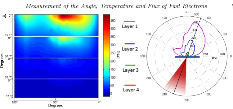
Figure 4.(a) PSL signal from the remapped layers of IP between the Fe filtering from a 140J shot onto a 100um Cu target. (b) a polar plot of the data. The peak emmission apears to be close to laser axis.
The first IP subtends a larger solid angle than the sequential layers and therefore the data have been remapped to enable pixel to pixel ratio comparison. The measurements taken on the angular wrap-around stack are shown in Figure 4 along with a polar plot showing the incoming laser. It is quite clear to see that the majority of the electrons are directed along the laser axis as is expected from interactions of this intensity (Malka & Miquel (1996); Wilks & Kruer (1997)).
The digitalisation process of the IP converts the dose to PSL (Photo-Stimulated Lumi-nescence) which is also a linear representation of the signal. Using the earlier simulations showing that only electrons above 2MeV reach the IP layers and calibrations by Tanaka et al.(2005), where the numbers of electrons per PSL were reported, the total number of electrons can be calculated. Summing the total PSL signal on the first layer of IP leads to the incident electron signal absorbed being∼8×10¹⁰ on layer 1.
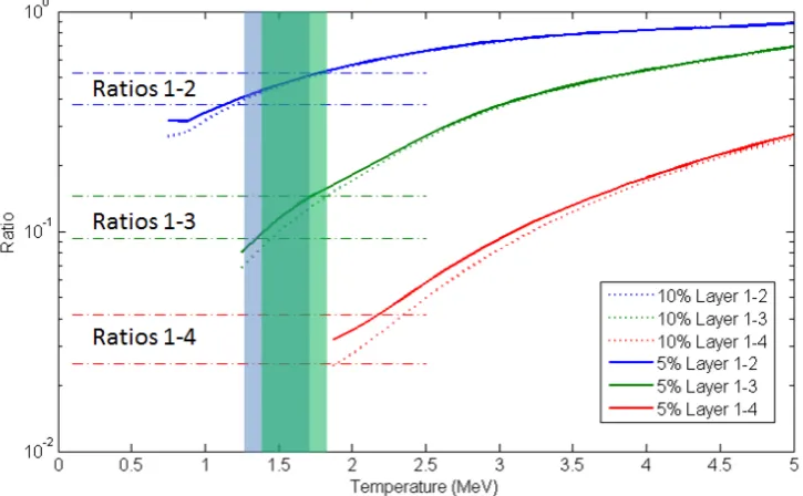
The signal ratio from this data can be calculated by dividing the signal in first layer by the signal in any of the following layers pixel by pixel. An example of the ratio measured from IP layer 1 to IP layers 2 and 3 is show in Figure 5, together with an angular plot of the PSL data from layers 1,2 and 3. The upper and lower ratios obtained from each comparison are plotted with the expected ratios as a function of temperature produced from the simulations. This is shown on Figure 6 with the bounds shown as horizontal lines intercepting the simulated ratios. Ratios corresponding to the escaping electron fractions of 10% and 5% are also shown but do not differ significantly. The overlapping shaded regions represent where data crosses the 10% ratios, which is between 1.4 and 1.7MeV. The working range of layers 1-4 set in section 3 puts the experimental data out of range as can be seen in Figure 5.
For the temperatures of 1.4 and 1.7MeV, the number of electron escaping the target can be estimated from the previously quoted total absorbed electrons by multiplying by the known absorption fraction for these temperatures from the simulations shown in Figure 2 (b). This yields an incident electron number of∼2.6×10¹³. This value assumes
Figure 5.Angular profiles of the data shown in Figure 4(solid-lines,left-axis) with the ratios of layers 1-2 and 1-3 (dotted-lines,right-axis) The ratios do not change quickly over the entire angular range. The maximum and minimum of the ratios are taken and used as upper and lower bounds in Figure 6.
[image:6.595.105.468.404.628.2]distribution, however it has been shown that the internal electrons can have a dual-temperature distribution by monitoring the x-ray spectra (Chen et al. (2009); Zulick et al. (2013)). Knowing this, future experiments using this diagnostic are planned in conjunction with simultaneous x-ray and electron spectrometer measurement which will provide a more accurate temperature diagnostic.
5. Conclusions
The response of a cylindrical electron diagnostic, designed to provide angular and spectral information regarding the electrons escaping from a solid target has been as-sessed. The first layers of filtering eliminates any proton contribution to the first layer and any sequential layers. Using Monte-Carlo simulations, the response of each IP layer of the diagnostic has been analysed for mono-energetic and relativistic Maxwellian elec-tron distributions. Using the energy absorbed for these given elecelec-tron distributions, an electron temperature where the diagnostic can be reliable has been found for each layer of IP. Experimentally the diagnostic has been used to measure half an escaping electron beam with ∼2.6×10¹³ electrons with a temperature between 1.4 and 1.7MeV from a 3.9×10²⁰ W/cm2 interaction. Future experiments using this diagnostic are planned in conjunction with x-ray and electron spectrometers to generate a more complete picture of the interaction and help provide better estimates of the total energy of the escaping electrons.
The authors gratefully acknowledge the expert assistance of the PHELIX laser opera-tions team and funding from EPSRC (Grant Nos. EP/J003832/1 and EP/K022415/1). This work has been carried out within the framework of the EUROfusion Consortium and has received funding from the EURatom research and training programme 2014-2018 (grant agreement No 633053) and from LASERLAB-EUROPE (grant agreement no. 284464, EC’s Seventh Framework Programme). The views and opinions expressed herein do not necessarily reflect those of the European Commission. Data associated with research published in this paper is accessible at http://dx.doi.org/10.15129/f774dd94-1861-47d5-b01e-04dda4a97292.
REFERENCES
Brunel, F.1987 Not-So-Resonant, Resonant Absorption.Physical Review Letters 59(1). Chen, C. D., Kemp, A. J., Perez, F., Link, A., Beg, F. N., Chawla, S., Key, M. H.,
McLean, H., Morace, A., Ping, Y., Sorokovikova, A., Stephens, R. B., Streeter, M., Westover, B. & Patel, P. K.2013 Comparisons of angularly and spectrally resolved Bremsstrahlung measurements to two-dimensional multi-stage simulations of short-pulse laser-plasma interactions.Physics of Plasmas20(5), 052703.
Chen, C. D., Patel, P. K., Hey, D. S., Mackinnon, A. J., Key, M. H., Akli, K. U., Bar-tal, T., Beg, F. N., Chawla, S., Chen, H., Freeman, R. R., Higginson, D. P., Link, A., Ma, T. Y., MacPhee, A. G., Stephens, R. B., Van Woerkom, L. D., Westover, B. & Porkolab, M.2009 Bremsstrahlung and Kαfluorescence measurements for inferring conversion efficiencies into fast ignition relevant hot electrons.Physics of Plasmas 16(8), 082705.
Courtois, C., Compant La Fontaine, A., Landoas, O., Lidove, G., Meot, V., Morel, P., Nuter, R., Lefebvre, E., Boscheron, A., Grenier, J., Aleonard, M. M., Gerbaux, M., Gobet, F., Hannachi, F., Malka, G., Scheurer, J. N. & Tarisien, M.2009 Effect of plasma density scale length on the properties of bremsstrahlung x-ray sources created by picosecond laser pulses.Physics of Plasmas 16(1), 013105.
Edwards, R. D., Sinclair, M. A., Goldsack, T. J., Krushelnick, K., Beg, F. N., Clark, E. L., Dangor, A. E., Najmudin, Z., Tatarakis, M., Walton, B., Zepf, M., Leding-ham, K. W. D., Spencer, I., Norreys, P. A., Clarke, R. J., Kodama, R., Toyama, Y. & Tampo, M. 2002 Characterization of a gamma-ray source based on a laser-plasma accelerator with applications to radiography.Applied Physics Letters80(12), 2129–2131.
Fiorini, F., Neely, D., Clarke, R.J. & Green, S. 2014 Characterization of laser-driven electron and photon beams using the Monte Carlo code FLUKA.Laser and Particle Beams 32(02), 233–241.
Gray, R. J., Yuan, X. H., Carroll, D. C., Brenner, C. M., Coury, M., Quinn, M. N., Tresca, O., Zielbauer, B., Aurand, B., Bagnoud, V., Fils, J., Kuhl, T., Lin, X. X., Li, C., Li, Y. T., Roth, M., Neely, D. & McKenna, P. 2011 Surface transport of energetic electrons in intense picosecond laser-foil interactions. Applied Physics Letters 99(17), 171502.
Hatchett, Stephen P., Brown, Curtis G., Cowan, Thomas E., Henry, Eugene A., Johnson, Joy S., Key, Michael H., Koch, Jeffrey A., Langdon, A. Bruce, Lasinski, Barbara F., Lee, Richard W., Mackinnon, Andrew J., Pennington, Deanna M., Perry, Michael D., Phillips, Thomas W., Roth, Markus, Sangster, T. Craig, Singh, Mike S., Snavely, Richard A., Stoyer, Mark A., Wilks, Scott C. & Yasuike, Kazuhito2000 Electron, photon, and ion beams from the relativistic inter-action of Petawatt laser pulses with solid targets.Physics of Plasmas7(5), 2076–2082.
Link, A, Freeman, R. R., Schumacher, D. W. & Van Woerkom, L. D.2011 Effects of target charging and ion emission on the energy spectrum of emitted electrons.Physics of
Plasmas18(5), 053107.
MacLellan, D. A., Carroll, D. C., Gray, R. J., Booth, N., Burza, M., Desjarlais, M. P., Du, F., Gonzalez-Izquierdo, B., Neely, D., Powell, H. W., Robinson, A. P. L., Rusby, D. R., Scott, G. G., Yuan, X. H., Wahlstr¨om, C.-G. & McKenna, P. 2013 Annular Fast Electron Transport in Silicon Arising from Low-Temperature Resistivity. Physical Review Letters 111(9), 095001.
Produced by an Ultrarelativistic Laser Pulse on a Solid Target.Physical Review Letters 77(1), 75–78.
McKenna, P., Ledingham, K., Yang, J., Robson, L., McCanny, T., Shimizu, S., Clarke, R., Neely, D., Spohr, K., Chapman, R., Singhal, R., Krushelnick, K., Wei, M. & Norreys, P. 2004 Characterization of proton and heavier ion acceleration in ultrahigh-intensity laser interactions with heated target foils.Physical Review E 70(3), 036405. McKenna, P., Robinson, A. P. L., Neely, D., Desjarlais, M. P., Carroll, D. C., Quinn,
M. N., Yuan, X. H., Brenner, C. M., Burza, M., Coury, M., Gallegos, P., Gray, R. J., Lancaster, K. L., Li, Y. T., Lin, X. X., Tresca, O. & Wahlstr¨om, C.-G. 2011 Effect of Lattice Structure on Energetic Electron Transport in Solids Irradiated by Ultraintense Laser Pulses.Physical Review Letters 106(18), 185004.
Myatt, J., Theobald, W., Delettrez, J. A., Stoeckl, C., Storm, M., Sangster, T. C., Maximov, A. V. & Short, R. W.2007 High-intensity laser interactions with mass-limited solid targets and implications for fast-ignition experiments on OMEGA EP. Physics of
Plasmas14(5), 056301.
Norreys, P. A., Santala, M., Clark, E., Zepf, M., Watts, I., Beg, F. N., Krushelnick, K., Tatarakis, M., Dangor, A. E., Fang, X., Graham, P., McCanny, T., Singhal, R. P., Ledingham, K. W. D., Creswell, A., Sanderson, D. C. W., Magill, J., Machacek, A., Wark, J. S., Allott, R., Kennedy, B. & Neely, D.1999 Observation of a highly directional γ-ray beam from ultrashort, ultraintense laser pulse interactions with solids.Physics of Plasmas6(5), 2150–2156.
P´erez, F., Kemp, G. E., Regan, S. P., Barrios, M. A., Pino, J., Scott, H., Ayers, S., Chen, H., Emig, J., Colvin, J. D., Bedzyk, M., Shoup, M. J., Agliata, A., Yaakobi, B., Marshall, F. J., Hamilton, R. A., Jaquez, J., Farrell, M., Nikroo, A. & Fournier, K. B. 2014 The NIF x-ray spectrometer calibration campaign at Omegaa).
Review of Scientific Instruments85(11), 11D613.
Quinn, M N, Yuan, X H, Lin, X X, Carroll, D C, Tresca, O, Gray, R J, Coury, M, Li, C, Li, Y T, Brenner, C M, Robinson, a P L, Neely, D, Zielbauer, B, Aurand, B, Fils, J, Kuehl, T & McKenna, P 2011 Refluxing of fast electrons in solid targets irradiated by intense, picosecond laser pulses.Plasma Physics and Controlled Fusion53(2), 025007.
Santala, M. I. K., Zepf, M., Watts, I., Beg, F. N., Clark, E., Tatarakis, M., Krushel-nick, K., Dangor, A. E., Wilks, S. C., Machacek, A. C., Wark, J. S., Allott, R., Clarke, R. J. & Norreys, P. A.2000 Effect of the Plasma Density Scale Length on the Direction of Fast Electrons in Relativistic Laser-Solid Interactions.Physical Review Letters 84(7), 1459–1462.
Schwoerer, H., Gibbon, P., D¨usterer, S., Behrens, R., Ziener, C., Reich, C. & Sauer-brey, R.2001 MeV X Rays and Photoneutrons from Femtosecond Laser-Produced
Plas-mas.Physical Review Letters 86(11), 2317–2320.
Tanaka, Kazuo A., Yabuuchi, Toshinori, Sato, Takashi, Kodama, Ryosuke, Kitagawa, Yoneyoshi, Takahashi, Teruyoshi, Ikeda, Toshiji, Honda, Yoshihide & Okuda, Shuuichi2005 Calibration of imaging plate for high energy electron spectrometer.Review of Scientific Instruments76(1), 013507.
Wilks, Scott C & Kruer, William L1997 Absorption of Ultrashort , Ultra-Intense Laser Light by Solids and Overdense Plasmas.IEEE Journal of Quantum Electronics 33(11), 1954–1968.
Ziegler, James F., Ziegler, M.D. & Biersack, J.P.2010 SRIM The stopping and range of ions in matter (2010).Nuclear Instruments and Methods in Physics Research Section B:
Beam Interactions with Materials and Atoms 268(11-12), 1818–1823.