Copyright© 1973 AmericanSocietyforMicrobiology Printed in U.S.A.
Measles
Virus Ribonucleic Acid and Protein
Synthesis: Effects of 6-Azauridine and
Cycloheximide
on
Viral
Replication
ALLEN PORTNERl AND ROBERT H. BUSSELL
DepartmentofMicrobiology, The University of Kansas, Lawrence, Kansas66044
Received forpublication30August1972
Cycloheximide and 6-azauridine were employed tostudy the time course of
measles virus protein and nucleic acid syntheses in AV3 cells. Synthesis of ribonucleic acid (RNA) essential for infectivitywasfirstdetected at6hr and
in-creased concurrently with the formation of essential protein. Maximum levels
ofvirus-specific RNA and protein were presentby 18hr, a time whenonly 5%
ofprogeny virus was detected. Essential RNA and protein syntheses preceded
the formation of infectious virusbyatleast 10 to 12 hr. The timecourseof RNA
and protein syntheses essential for the formation of complement-fixing (CF) antigen and salt-dependent agglutinin (SDA) was also determined. RNA
syn-thesis essentialforthe formation ofSDAwasfirst detectedat2 hr andwas
pres-ent maximally by 6 hr, whereas SDA-protein increased concurrently with the protein essential for infectivity. This suggested that the last protein essential for infectivity may be SDA. RNA synthesis essential for the formation of CF antigen was first detectedat 4hr, while CF-protein increased at 5 hrand
pre-ceded SDA-protein and protein essential for infectivity by approximately3 hr.
Reversal of inhibitionofprotein synthesis by cycloheximideindicated thatearly protein synthesis (1 to 3hr) was required for the formation of infectious virus.
The data suggest that the relatively long eclipse period observed with measles virus is relatedtoalong maturation period rather thantolate formation ofearly proteins, viral RNA, orstructural proteins.
There is much information on the physical and biological properties ofmeasles virus (16, 18, 28), but knowledge of the mechanism of replication is limited. Prior studies have re-vealed the general characteristics of the growth cycle (6, 16, 17) and the sequence of formation of infectious virus, hemagglutinin, complement-fixing antigen, and
hemolysin
(19). The present study was undertaken to determine the time course of ribonucleic acid (RNA) and protein synthesis necessary for the formation of infectious virus, salt-depend-ent agglutinin (24), and complement-fixing activity in measles virus-infected cells. An inhibitor of RNA synthesis, 6-azauridine (10), was used to determine the time course of RNA synthesis. Cycloheximide, a protein synthesis inhibitor (8), was used to determine the time course ofviral protein synthesis.IPresent address: LaboratoryofVirology, St. Jude
Chil-dren's ResearchHospital andthe University of Tennessee, Memphis,Tenn. 38101.
46
MATERIALS AND METHODS Cells and media. The AV3 continuous human amnioncell line (21) was used. The cells were grown
in monolayers in 32-ounce (.95-liter) prescription bottles in medium 199 containing 0.25% lactalbumin hydrolysate and 15% heat-inactivated (60 C, 30min) newborn calf serum, and sodium bicarbonate was usedtoadjust the pH to 7.2 to 7.4 (GM). Eagle
mini-mum essential medium (MEM) with 5% newborn agammacalf serum wasusedfor maintenance (MM). Dialyzed newborn agamma calf serum was used when theeffect of6-azauridine was determined.
Virus. The Edmonston strain of measles virus
(7) wasused throughout this study. Stock virus was prepared in 48-hr cultures of AV3 cells. The virus inoculum (3 ml, 2.0 x 10⁵ PFU/ml) was adsorbed at
36 C for 2 hr. After adsorption, 45 ml of MM was added, and the cultures were incubated at 36 C for
48 hr. The MM was exchanged, and the cultures
were incubated at 31 C for an additional 24 hr
be-fore they were harvested by freezing and thawing threetimes.
Infection of cells. Approximately 1.8 x 10⁶ cells
were inoculated into 2-ounce (5.8-ml) prescription
on November 10, 2019 by guest
http://jvi.asm.org/
MEASLES VIRUS RNA AND PROTEIN SYNTHESIS
bottles and incubated for 24 to 48 hr at 36 C. The GM wasremoved, and the cells were incubated with 0.5ml of virus for 2 hr at 36C. The input multiplicity of infection(MOI)wasapproximately 5 to 10 plaque-forming units (PFU) per cell unless otherwise indi-cated. The inoculum was removed, and the cells were washed with 5 ml of phosphate-buffered saline (PBS) containing 1% agamma calf serum and rein-cubated with 5 ml of MM at 36 C. The time after the 2-hr adsorption period was regarded as zero time for these experiments. For some experiments, tube cultures were seeded with 5 x 10⁵ cells and inoculated with 0.2 ml of virus. All other conditions were as described above.
Plaque assay. Monolayers of AV3 cells, 70 to
80% complete, in 2-ounce prescription bottles were washed, and two cultures were inoculated with 0.5 mlofeach virusdilution.Adsorptionwas at 36C for 2 hr. Unadsorbed virus was decanted, and the monolayers were overlayed with 5 ml of sodium carboxymethylcellulose (NaCMC) medium. Cultures wereincubatedat36C for3to4days, atwhich time primary plaques developed. The overlay medium was decanted, and the monolayers were fixed with 8 ml ofFormalin(10%inPBS, pH7.2). Afteratleast
2hrat roomtemperature, the fixativewasdecanted,
and the monolayers were covered with 4 ml of Giemsa stain. Thirty minutes later the stain was
decanted, and the monolayers were washed three times with tap water and air dried. Plaques were
counted withadissecting microscope (12x), and the titerwas reportedasplaque-formingunitsper milli-liter. The NaCMC overlay (final concentration, 1%) was prepared by dissolving 10 g of powdered high viscosity NaCMC (General Biochemicals) in 840 ml of distilledwaterandmixingat 4C for24hr. The solution was autoclaved at 116 C for 10min. After
cooling, Eagle MEM and a final concentration of 5% newbom calfserum was added.
Salt-dependent agglutinin. Samples were as-sayed for salt-dependent agglutinin (SDA) (24) by
employing a microtechnique (27). Undiluted virus
samples were adjusted to 0.8 M citrate with 3 M sodium citrate.The diluentwas 0.8M sodiumcitrate in PBS. A 1% suspension of rhesus monkey erythro-cytes was employed. The SDA titer, expressed as the reciprocal ofthe final dilution of virus causing agglutination, wasreadafter 2hrat 37C.
Complement fixation. A microtechnique was
used for the titration of complement-fixation (CF)
antigen (4). Approximately two optimal units of monkey anti-measles antiserum and five 50% end-point units of commercial guinea pig complement were employed. The end points were considered to
be the last dilution ofantigen producing30% or less
hemolysis.
Chemicals. 6-Azauridine (AUR) and
cyclohexi-mide were purchased from Sigma Chemical Com-pany, St. Louis,Missouri.
Cell viability determination. Cells were diluted
1:2 in 0.5% trypan blue and counted in a hemo-cytometer. Cells which excluded trypan blue were considered viable.
RESULTS
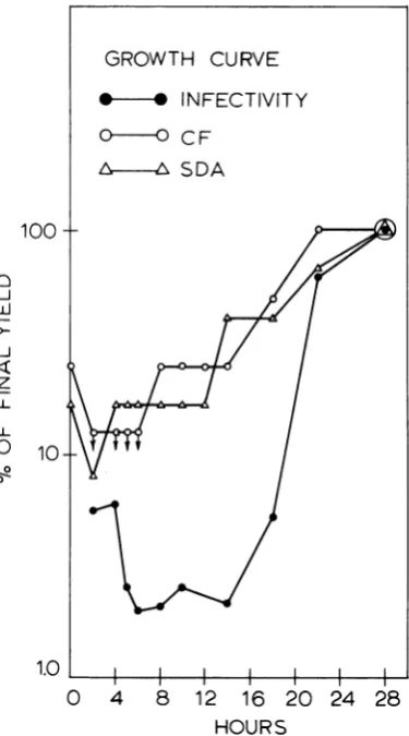
Measles virus single cycle growth curve and the appearance of viral antigens. The growth characteristics ofmeasles virus and the appearance ofviral antigens in AV3 cells were
determined to serve as base lines for the
in-hibition studies. AV3 cells were inoculated with measles virus. Cell cultures were har-vested at intervals up to 28 hr and then
ti-trated for infectious virus, CF antigen, and SDA (Fig. 1). A significant increase in infec-tious virus was observed between 14 and 18 hr, and virus titers continuedto increase until 28 hr. An increase in CF activity was detected
at 8 hr, and titers continued to increase
GROWTH
CURVE-s*INFECTIVITY
0-0 CF
L.~-~ SDA
100
t
J -J
z LL
L-0
B0
10-1.0
0 4 8 12 16
20
2428
HOURS
FIG. 1. Growthcurveof measles virus in AV3 cells.
Monolayerswere infected withaninput multiplicity
of5 to 10PFUpercell;viruswasadsorbedat36Cfor
2 hr, washed three times, and incubated with MM.
Virus was harvestedat the indicated times and
as-sayedforinfectivity, CFantigen,andSDA. Absolute maximum values (28hr) were: infectivity, 1.9 x 10⁶
PFU/ml;CFantigen,1to4;SDA,1 to 6.
VOL. 1l, 1973 47
on November 10, 2019 by guest
http://jvi.asm.org/
[image:2.495.251.439.245.583.2]throu throu
tivel)
meas (6, 1 SDA Efl tion of cy infec after centr fectiN were The centr Addi 5 to per r
expel
if cy
infec to pr
igh 22 hr. SDA activity increased from 12 AV3 cells were incubated in MM containing Lgh 28 hr. These results confirm the rela- 0.5 ,g/ml for either 6 or 24 hr. There was no
y
long
eclipse
periods
observed with alteration in cellmorphology.
The cells were;les
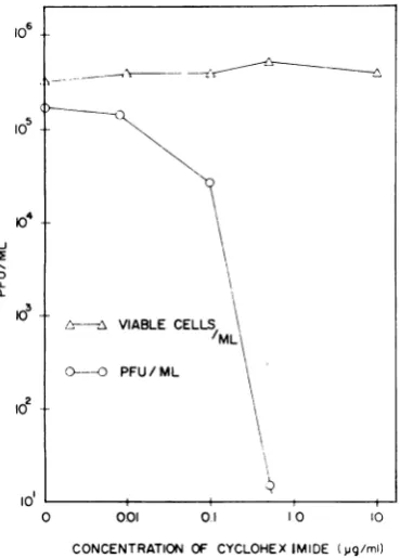
virus in various cell culture systems washed six times to remove the inhibitor and 6, 19) and show that CF antigen and then were infected. Cultures were harvested appear before infectious virus. at 24 hr and assayed. Virus yields in cultures fect of cycloheximide on virus replica- pretreated for 24 hr were 5.5 x 101 PFU/ml andcell viability. Various concentrations (control yields were 6.0 x 10⁵PFU/ml). Virus cloheximide were added to measles virus- yields in cultures pretreated for 6 hr were ted AV3 cells (MOI 0.2) immediately 2.1 x 101 PFU/ml (control yields were 2.2 x virus adsorption to determine the con- 105 PFU/ml). This indicated that the effectrations
of cycloheximide required to ef- of cycloheximide (0.5 ,ug/ml) on the capacity vely block virus replication. Virus yields of the cells to produce virus was readilyre-determined by plaque assay at 24 hr. versible.
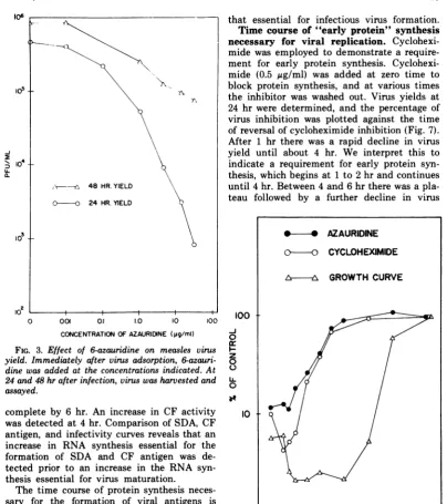
drug inhibited virus replication at con- Effect of AUR on virus replication. Ex--ations that were not cytotoxic (Fig. 2). periments were performed to determine the tional experiments employing an MOI of concentration of AUR to be used in subsequent 10 showed that 0.5
qg
of cycloheximide inhibitor experiments. After virus adsorption nl inhibited virus replication by 98%. An (MOI 1), medium containing different con-riment was also performed to determine centrations of AUR was added. Virus yields cloheximide added to the cells prior to were determined by plaque assay at 24 and tion would alter their subsequent ability 48 hr. Measles virus replication was sensitive roduce virus in the absence of the drug. to AUR; 98% inhibition occurred in thepres-ence of 5
gg/ml
at 24 hr (Fig. 3). However, at 48 hr, there was a decline in the amount of inhibition indicating some loss of inhibitionon continued incubation. No alterations in cell morphology or viability were observed with 5,ugofAUR per ml.
Time course of viral RNA and protein synthesis necessary for the formation of infectious virus. AUR or cycloheximide was added at various times after infection of AV3 cells to inhibit either RNA or protein syn-thesis. Virus yields at 28 hr were plotted against the time of addition of the inhibitors (Fig. 4). Comparison with the viral growth curve reveals that an increase in the synthe-£v-25 VIABLE CELLS sis of an essential RNA was detected at 5 to 6
/ML
hr and preceded the appearance of matureO----O PFU/ML virus by
approximately
10 hr. An increase inprotein synthesis necessary for viral infec-tivity was detected between 4 and 7 hr and preceded the appearance of mature virus by approximately 9 hr. These results also show that nearly maximal levels of RNA and
pro-CYCOE tein essential for viral
infectivity
werepresent
o 001 0.1 IO 1O several hours before the appearance of
infec-CONCENTRATION OF CYCLOHEXIMIDE (pg/ml) tious virus.
Time course of RNA and protein syn-2. Effect ofcycloheximide on virus yield and thesis essential for the production of virus
uiability. Immediately after virus adsorption * ml
heximidewas added at the concentrationsindi-
antign.
thetimetcou
of RNsnthesis
At 24 hr, virus was harvested and
assayed
essental for the formaton of CFantigen
(CF-sond group of cultures was not infected, and RNA) and SDA (SDA-RNA) iS shown inheximide was added as indicated above. At Fig. 5. The
infectivity
curve is the same as theviable cell counts wereperformed by staining azauridine curve in Fig. 4. An increase in
trypanblue. SDA activity was detected at 2 hr and was
l0
4
-J ILJ
0.
3
le
10
FIG
cell X
cyclo)
cated A sec
cyclol
24 hr,
withi
48
on November 10, 2019 by guest
http://jvi.asm.org/
[image:3.495.65.250.321.579.2]MEASLES VIRUS RNA AND PROTEIN SYNTHESIS
o6
10c5
-j
U-
a-2
0 0.01 0.1 1.0 10
CONCENTRATION OFAZAURIDNE (pg/mi)
100
FIG. 3. Effect of 6-azauridine on measles virus
yield. Immediately after virus adsorption,
6-azauri-dine was addedat the concentrations indicated. At
24and 48 hrafter infection, viruswasharvested and
assayed.
complete by 6 hr. An increase in CF activity
was detected at4 hr. Comparison of SDA, CF antigen, and infectivity curves reveals that an
increase in RNA synthesis essential for the formation of SDA and CF antigen was
de-tected prior to an increase in the RNA
syn-thesis essential for virus maturation.
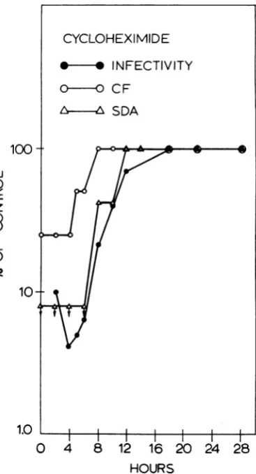
The time course of protein synthesis neces-sary for the formation of viral antigens is
shown in Fig. 6. The infectivity curve is the same as the cycloheximide curve in Fig. 4.
An increase in protein synthesis essential for SDA was detected at 8 hr and was
com-plete by 12 hr. Protein synthesis essential for CF activity showed an initial rise at 5 hr and was complete by 8 hr. Comparison of SDA, CF antigen, and infectivity curves reveals
that protein synthesis essential forthe
forma-tion ofCFantigenwasinitiated priortoprotein synthesis necessary for SDA and infectious
virus. Proteinsynthesisessential for the
forma-tion ofSDA activity appears to coincide with
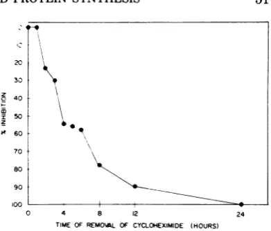
that essential for infectious virus formation. Time course of "early protein" synthesis necessary for viral replication. Cyclohexi-mide was employed to demonstrate a require-ment for early protein synthesis. Cyclohexi-mide (0.5 ,g/ml) was added at zero time to block protein synthesis, and at various times the inhibitor was washed out. Virus yields at 24 hr were determined, and the percentage of virus inhibition was plotted against the time ofreversalofcycloheximide inhibition (Fig. 7). After 1 hr there was a rapid decline in virus yield until about 4 hr. We interpret this to indicate a requirement for early protein syn-thesis, which begins at 1 to 2 hr and continues until 4hr. Between 4 and6hr therewas a pla-teau followed by a further decline in virus
100
I -j
0 z 0 0
1.0
O 4 8 12 16 20 24 28
HOURS
FIG. 4. Measles virus yield as a function of the
timeof addition of6-azauridine andcycloheximide.
AV3 cells were infected as described in Fig. 1. At
the indicatedtimes, 6-azauridine(5ug/ml) or
cyclo-heximide (0.5 gg/ml) was added. Virus was
har-vestedat28hr,and virusyieldswereplotted against the timeofadditionoftheinhibitor.Alsoincludedis
the control growth curveof virus in untreated
cul-tures. Absolute maximum value (28 hr) was:
infec-tivity, 1.9 x 10⁶ PFU/ml. -A
,I- 48 HR. YIELD
O ---O 24 HR.YIELD
1 - *- AZAURIDINE
o-(o
CYCLOHEXIMIDEL~-A GROWTH CURVE
49
VOL. 11, 1973
on November 10, 2019 by guest
http://jvi.asm.org/
[image:4.495.46.451.70.526.2] [image:4.495.257.447.258.560.2]DISCUSSION
RNA and protein synthesis essential for
AZAURIDINE measles virus infectivity were detected at 5 to
6 hr and 6to 8hr, respectively, and bothwere
*@
* INFECTIVITY at near maximum levels before significantO-O CF amounts of new infectious virus were
pro-duced. The synthesis of this essential RNA
S DA and protein preceded virus maturation by
approximately 10 to 12 hr throughout the
1(
A z growth~cycle.
We shouldemphasize
that the100 methods employed detect only the last
essen-Oi / ; /tial RNA and protein needed to confer
infec-0
tivity
on thevirus.F-
W tlCycloheximide
appeared
to affect SDA8 andviral infectivity withasimilar timecourse.
IL
0
o
0 CYCLOHEXIMIDE
i0
0* * INFECTIVITYO-O CF
6-- SDA
1.0~~~~~~~~~0
1.0L
I
I
I
IC2
0
48
12
16
20 24
28
8
HOURS
FIG. 5. Virus yield, SDA, and CF activity as a o
functionofthe timeofadditionof6-azauridine. Pro- e cedureasinFig.4.Infectivity curve same asazauri- o
dine curve inFig.4.Controlequalsyieldat28hr(no 10- 1 6-azauridine added). Absolute maximum values (28
hr) were: infectivity, 1.9 x 10⁶ PFU/ml; CF anti- T
gen, 1to8;SDA, 1to6.
yield. In other
experiments,
higher
concentra-tions of
cycloheximide
wereemployed
to morerapidly and
effectively
block proteinsynthesis,
allowing a better determination of the time course of early proteinsynthesis.
Cyclohexi-mide (5Ag/ml)
was added at various times and then removed from all cultures at 5 hr.Virus yields were determined at 2-hr intervals 0 4 8 12 16 20 24 28
and plotted against the time of harvest.
Fig.
8 HOURSshows that inhibitor added at zero time and
2 hr
..
displaced the early portion ofthe curvesFIG.
6. Virus yield, SDA, and CF activity as ahrdisplaced the early portion ofthe
curvesfunction
of the time of addition of cycloheximide.to the right.
However,
when inhibitor was Procedure asin
Fig.
4.Infectivity
curve same ascy-added at 3 and 4
hr,
thegrowth
curves were cloheximidecurve in Fig. 4. Control equals yield at similar to that of the control. This confirms 28 hr(nocycloheximide). Absolute maximum values that early protein synthesis occurs and is es- (28 hr) were: infectivity, 1.9x 10⁶PFU/ml;
CF anti-sentially completeby
3 hr. gen, 1 to8;SDA, I to6.50
PORTNER AND BUSSELL J. VIROLon November 10, 2019 by guest
http://jvi.asm.org/
[image:5.495.268.452.249.589.2]MEASLES VIRUS RNA AND PROTEIN SYNTHESIS
This suggests that the protein responsible for SDA activity may be the last protein
needed for the formation of infectious virus.
However, RNA synthesis essential for SDA
ac-tivity was detected very early in the growth
cycle (approximately 2hr) and wascompleted
by 6 hr. Thus, the inhibition of RNA synthesis does not affect the formation of SDA activity
with the same time course as the formation
of infectious virus and CF activity. This
sug-gests that more than one RNA species may
be involved in the formation of these viral constituents. We should point out that the times given for the appearance of various
activities (infectivity, CF, SDA) in the in-hibitor experiments are only approximations
because we do not know how long it takes the inhibitors to block virus-specific RNA and protein synthesis. What we wish to emphasize
in these experiments is the relative time
course of the various activities and not the
precise times given.
Multiple species ofvirus-specific RNA have been observed in cells infected with New-castle disease virus (NDV) and Sendai virus (1, 2, 13). In addition, quantitative and quali-tative differences have been recognized in
these RNAs that are synthesized early and
late during the growth cycle (14). If SDA repre-sents a precursor of a portion of the viral
envelope (24), then one of the first processes
detected in measles-infected cells was the
synthesis of RNA essential for the formation of a virus-specific envelope protein.
How-ever, this is difficult to reconcile with the
cycloheximide inhibition experiments that indicated that the actual formation of SDA coincided with the synthesis of the last
pro-tein required for virus infectivity. Although the time course of SDA-RNA appears to differ from that of CF-RNA and RNA essential for infectivity, their rates of appearance are
nearly thesame.
RNA synthesis essential forCF activity was
first detected at 3 hr and preceded the for-mation of protein essential for CF activity by
2 hr. We made no attempt to distinguish
between the various kinds of CF antigens. However, the S antigen (internal nucleopro-tein component) was the major CF
compo-nentfoundearlyinthegrowth cycleofmeasles
virus (20). Thus, the early increase in CF
ac-tivity we observed is probably due to the
production of S antigen.
Experiments were performed to determine
whether early protein synthesis was essential
for the replication of measles virus.
Experi-z
m
20
30
40
50
60
70
80
90
100
0 4 8 12
TIME OF REMO\AL OF CYCLOHEXIMIDE (HOURS)
FIG. 7. Virus yield as a function of the time of
reversal of cycloheximide inhibition. AV3 cells were
infectedas described in Fig. 1. The MM contained
0.5 usg of cycloheximide per ml. At the indicated
times, the cultures were washed sixtimestoreverse
cycloheximide inhibition. Virus was harvested at
24 hr. Zero time represents virus yield from
un-treated cultures (5.5 x 10⁵ PFU/ml).
ments involving the reversal of the effects of cycloheximide indicated that early protein(s) synthesis was required and takes place
dur-ing the first 3 hr after virus adsorption (Fig.
7 and 8). The early protein(s) was formed
prior to the proteins essential for CF antigen, SDA, and infectious virus. The synthesis of early and late proteins in cells infected with
myxo- and paramyxoviruses has been
demon-strated by others (25, 26, 29, 30). These stud-ies indicated that the late proteins represent
virus structural protein, whereas the early protein was essential for viral nucleic acid
synthesis as well as structural protein
synthe-sis. An RNA-dependent RNA polymerase has been observed in cells infected with Sendai virus (15) and has also been found in purified NDV and Sendai virus virions (11, 22). Cycloheximide blocked the synthesis of Sendai virus 57S RNA but not the 18S and 35S RNAs (23). This suggested that the 18S and 35S RNAs were formed by the
poly-merase carried by the virion, whereas the 57S
RNA could be synthesized by a different
en-zyme. Since similar species of RNA ('18,
35, and 57S) have been found in measles-in-fected cells (Bussell and Robinson,
manu-script in preparation), it is possible that the early protein we have reported in this paper is also involved in the synthesis of measles 57S RNA.
One of the most striking differences
be-tween the growth cycles of measles and the
40'IR
VOL. 11, 1973 51
,-I
on November 10, 2019 by guest
http://jvi.asm.org/
[image:6.495.253.449.64.231.2]not due to lengthy delays in the formation of early protein(s) or RNA and protein. While measles RNA andprotein syntheses appear to be somewhat slower, this does not entirely account for the longer eclipse period. The results indicate that the rate-limiting step in the formation of infectious measles virus is not the rate of synthesis of essential RNA or protein, but is some subsequent stage such as the maturation process.
The experiments employing AUR to
measure the time course of RNA synthesis essential for infectivity do not give any clue as to the species of RNA that is primarily in-volved. This RNA could be the parental RNA or some other species of RNA needed for the synthesis of other viral components. The latter could be virus-specific comple-mentary RNA similar to that described in NDV- andSendai virus-infected cells (1, 2, 13).
ACKNOWLEDGMENTS
Wewishto expressourappreciationtoLyndel Kingand
Mary Portner for excellent technical assistance. This in-vestigation was supported by Public Health Service re-searchgrantAI 07143 from the National Institute ofAllergy and Infectious Diseases, Public Health Service Training Grant 5T GM-703 from the National Institute of General 12 14 16 18 20 22 24 Medical Sciences, and NASA Training Grant NSG(T)-55.
HOURS
FIG. 8. The inhibitionof protein synthesis and de-lay in appearance ofinfectious virus after reversal.
AV3 cells were infected as described in Fig. 1. At
various times, cycloheximide was added (5 osg/ml)
andthen removed fromallcultures 5hrafter
adsorp-tion. Virus yieldswere determinedat 2-hr intervals
and plotted against the time ofharvest.
structurally related myxoviruses and
para-myxoviruses is the length of the eclipse period. Considerable variation in the eclipse period of measles virus has been observed (16);
how-ever, in all cases it was considerably longer
than that observed with myxoviruses and paramyxoviruses such as fowl plague (3, 25),
NDV (9, 12, 26, 30), influenza A, strain
WSN (9), and SV5 (5). It is difficult to
com-pare our data with those cited above because
of differences in either the inhibitors
em-ployed or the methods employed to
deter-mine sequential synthesis of viral
compo-nents. However, the results obtained with measles virus differ in one major respect
and that is the lag observed between the synthesis of the various virus components
and virus maturation. Thus, the relatively long eclipse period appears to be mainly due
to a relatively long maturation process and
LITERATURE CITED
1. Blair, C. D., andW. S.Robinson. 1968.Replication of Sendai virus. I. Comparison of the viral RNA and virus specific RNA synthesis with Newcastle dis-ease virus. Virology 35:537-549.
2. Bratt, M. A., and W. S. Robinson. 1967. Ribonucleic acid synthesis in cells infected with Newcastle dis-ease virus. J. Mol. Biol. 23:1-21.
3. Borland, R.,and B. W. J. Mahy.1968.Deoxyribonucleic acid-dependent ribonucleic acid polymerase activity incells infected with influenzavirus.J. Virol.2:33-39. 4. Casey, H.L. 1965.Adaptionof LBCF methodto micro-technique. Standardized diagnostic complement-fixation method andadaption to micro test. Public Health Monograph No. 74, Public Health Service PublicationNo. 1228.
5. Choppin, P. W., and K. V. Holmes. 1967. Replication of SV5 RNA and theeffects of superinfection with polio-virus. Virology33:442-451.
6. DeJong,J.C.,and K. C.Winkler. 1970. The multiplica-tion ofmeasles virus inhumanamnioncellsin vitro. J. Gen. Virol.7:13-17.
7. Endersi J. F., and T. C. Peebles. 1954. Propagationin tissues cultures of cytopathogenic agents from pa-tients with measles. Proc. Soc. Exp. Biol. Med. 86:
277-286.
8. Ennis, H. L., and M. Lubin. 1964. Cycloheximide:
as-pects of inhibition of protein synthesis in
mamma-lian cells. Science146:1474-1476.
9. Granoff, A., and D. W. Kingsbury. 1964. Effect of actinomycin D on the replication ofNewcastle dis-ease and influenza viruses, p. 96-115. In G. E. W. Wolstenholme and J.Knight (ed.), Cellularbiologyof myxovirusinfections. Little, BrownandCo., Boston. 10. Handschumacher, R. E. 1960. Orotidylic acid
decar-i0d
05+---J
IL a.
CYCLOHEXIMIDE ADDED (TIME IN HOURS)
O-O 0
*--4 2
A-A 3
L] ] 4
A- -A CONTROL / /
/7
~~ /~//'/
/'
/
/ .
104-
-id'
j
1
on November 10, 2019 by guest
http://jvi.asm.org/
[image:7.495.61.252.59.374.2]MEASLES VIRUS RNA AND-PROTEIN SYNTHESIS
boxylase: inhibitorstudies with 6-azauridine-5'-phos-phate. J. Biol. Chem. 235:2917-2919.
11. Huang, A. S., D. Baltimore, and M. A. Bratt. 1971. Ribonucleic acid polymerase in virions of New-castle disease virus: comparison with the vesicular stomatitis virus polymerase. J. Virol. 7:389-394. 12. Kingsbury, D. W. 1962. Use ofactinomycin D to
un-mask RNA synthesis induced by Newcastle disease virus. Biochem. Biophy. Res. Commun. 9:156-161. 13. Kingsbury. D. W. 1966. Newcastle disease virus RNA. II. Preferential synthesis ofRNA complementary to viral RNA bv chick embryo cells. J. Mol. Biol. 18: 204-214.
14. Kingsburv, D. W. 1970. Replication and functions of myxovirus ribonucleic acids. Prog. Med. Virol. 12: 49-77.
15. Mahy, B. W. J., J. E. Hutchinson, and R. D. Barry. 1970. Ribonucleic acid polymerase induced in cells infected with Sendaivirus.J.Virol. 5:663-671. 16. Matumoto, M. 1966. Multiplicationofmeasles virus in
cell culture. Bacteriol. Rev. 30:152-176.
17. Matumoto, M., M. Arita, and M. Oda. 1965.
Enhance-mentof measles virusreplication by actinomycin D. Jap.J.Exp.Med.35:319-329.
18. Norrby, E. 1967. Measles, p. 1-53. In A. P. Waterson (ed.), Recent advances in medical microbiology. Little Brown andCo., Boston.
19. Numazaki, Y., and D. T. Karzon. 1966.Density separ-ablefractions during growthofmeasles virus. J. Im-munol. 97:458-469.
20. Numazaki, Y., and D. T. Karzon. 1966.Solubleantigen ofmeasles virus. J. Immunol. 97:470-476.
21. Rivadenino, J. C., and F. C. Robbins. 1958. On the origin of variants ofhuman amniotic cells in tissue culture. Amer. J. Dis. Child. 96:435.
22. Robinson, W. S. 1971. Ribonucleic acid polymerase activity in Sendai virionsandnucleocapsid. J. Virol. 8:81-86.
23. Robinson, W. S. 1971.Sendai virusRNAsynthesisand nucleocapsid formation in the presenceof cyclohexi-mide. Virology44:494-502.
24. Schluederberg, A., and M. Nakamura. 1967. A salt-dependent hemagglutinating particle from measles infected cells. Virology 33:297-306.
25. Scholtissek, C., R. Rott, P. Hausen, H. Hausen, and W. Schafer. 1962. Comparative studies ofRNA and protein synthesis withamyxovirusandasmall poly-hedralvirus.Cold Spring Harbor Symp. Quant. Biol. 27:245-256.
26. Scholtissek, C., and R. Rott. 1965. Metabolic changes inchick fibroblastsafterinfectionwith Newcastle dis-ease virus. Nature (London) 206:729-730.
27. Severs, J. L. 1962. Application of a microtechnique
to viral serological investigations. J. Immunol. 88: 320-:329.
28. Waterson, A. P. 1965. Measles virus. Arch. Gesamte Virusforsch. 16:57-80.
29. White, D. O., H. M. Day, E. J. Batchelder, I. M.
Cheyne, and A. J. Wainsbrough. 1965. Delay in the multiplicationofinfluenzavirus.Virology25:289-302. 30. Wilson, D. E., andP. LoGerfo. 1964.Inhibitionof ribo-nucleic acid synthesis in Newcastle diseasevirus in-fected cellsby puromycin and 6-azauridine. J. Bac-teriol. 88:1550-1555.
VOL. 11, 1973 53