City, University of London Institutional Repository
Citation
:
Wang, Z., Slabaugh, G. G., Unal, G. B., Zhou, M. and Fang, T. (2007). An
information-theoretic detector based scheme for registration of speckled medical images.
Paper presented at the IEEE International Conference on Systems, Man and Cybernetics,
2007, 07-10-2007 - 10-10-2007, Montreal, Canada.
This is the accepted version of the paper.
This version of the publication may differ from the final published
version.
Permanent repository link:
http://openaccess.city.ac.uk/4409/
Link to published version
:
http://dx.doi.org/10.1109/ICSMC.2007.4413864
Copyright and reuse:
City Research Online aims to make research
outputs of City, University of London available to a wider audience.
Copyright and Moral Rights remain with the author(s) and/or copyright
holders. URLs from City Research Online may be freely distributed and
linked to.
City Research Online:
http://openaccess.city.ac.uk/
publications@city.ac.uk
An Information-Theoretic Detector Based Scheme for Registration of
Speckled Medical Images
Zhe Wang
1,
Student Member, IEEE
, Greg Slabaugh
2,
Member, IEEE
, Gozde Unal
2,
Member, IEEE
,
Mengchu Zhou
1,
Fellow, IEEE
, and Tong Fang
2,
Member, IEEE
1
New Jersey Institute of Technology
Department of Electrical and Computer Engineering
Newark, NJ 07102
2
Siemens Corporate Research
Intelligent Vision and Reasoning Department
Princeton, NJ 08540
email: zw27@njit.edu, greg.slabaugh@siemens.com, gozde.unal@siemens.com,
zhou@njit.edu, tong.fang@siemens.com
Abstract— Several studies dealt with medical ultrasound registration. Their similarity metrics relied on pixel-to-pixel intensity comparisons. Hence, they are not well suited to the case of speckled images. To better handle the speckle noise, our previous work proposed an information-theoretic feature detector-based registration approach. This work aims to extend it to the cases where the image speckle model is Rayleigh or normalized Fisher-Tippett distributed. Using speckle modeling based on these distributions, a speckle-specific information-theoretic feature detector is constructed and applied to pro-vide feature images. Those feature images are then registered using differential equations, the solution of which provides a transformation to bring the images into alignment. Compared to standard gradient-based techniques, the experimental results demonstrate the effectiveness of our method, particularly for low contrast ultrasound images.
Index Terms— Speckle Image, Image registration, informa-tion theory, biomedical image processing
I. INTRODUCTION
Within the current clinical setting, medical image registra-tion is a crucial component of a large number of applicaregistra-tions, including disease detection, analysis, and treatment. Image registration seeks to bring two or more images of the same scene into alignment.
Medical ultrasound images are a very important diagnostic tool for physicians. Ultrasound has many advantages, as it is fast, portable, cheap and safe. However, speckle noise degrades severely the quality of medical B-mode ultrasonic images, and renders the registration of ultrasound speckled images as a difficult problem. Speckle is an interference pattern resulting from the coherent accumulation of random scattering in a resolution cell of the ultrasound beam.
By concealing fine structures, the speckle has a detrimental effect to the current image registration algorithms, especially those based on comparing images on a pixel-to-pixel basis, since for ultrasound images, two corresponding pixels can have very different intensity levels due to the speckle.
In recent years, many registration algorithms have been proposed. However, few of them are designed specifically
for ultrasound speckle images [1]. Ultrasound-specific regis-tration algorithms are presented in [2], [3]. They are based on pixel-to-pixel comparison between images. When the displacement is large, or images are taken of the same region from different scans, or different transducers, etc., these methods face difficulty, since the correlation of the speckle no longer exists. Unlike such previous work, our method relies on distribution-to-distribution comparisons. Consequently, it is significantly more robust.
The fully formed speckle is known to be a multiplicative Rayleigh distribution in the envelope detected image and Fisher-Tippett (doubly exponential) distribution in the log-compressed image. In our previous work, we use sym-metrized Kullback-Liebler distance (J-divergence) of Fisher-Tippett distributed variables for comparing regions in a log-compressed ultrasound image in the context of feature detection [4]. However, since intensity of speckle is always non-negative, normalized Fisher-Tippett distribution is more suitable for speckle image modeling. Furthermore, some speckle images are not log-compressed and thus, Fisher-Tippett can no longer be applied. This paper expands our previous work to the cases of Rayleigh and normalized Fisher-Tippett distributed speckle images.
II. STATISTICAL MODELING OF SPECKLE
A. Rayleigh Case
Fully formed speckle is typically assumed when the num-ber of scatters per cell is greater than ten [5]. It has been shown that the speckle in inphase/quadrature image,QI(x,y),
has a complex Gaussian distribution,
p(QI) =
1 2πσ2e
−|QI(x,y)|2/(2σ2) (1)
Since QI is complex, its magnitude is taken to produce a
real image. The distribution in this magnitude imageI(x,y)
is Rayleigh [6],
p(I) =I(x,y)
σ2 e−
I(x,y)2/2σ2
where I(x,y) is real. To obtain the maximum likelihood Rayleigh estimator, we differentiate the log likelihood of Eq.(2), with respect toσ, and set this expression to zero to determine the maximum likelihood estimate (MLE) ofσ2,
σ2=
ΩI(x,y)2dxdy
2Ωdxdy (3)
With the above equation, we can calculate the MLE ofσ2
from the image intensities in the region assuming Rayleigh distribution. Note that this distribution is fully described by this one parameter.
B. Normalized Fisher-Tippett Case
LetI(x,y)denote a pixel intensity in the decorrelated log magnitude IQ image at the location (x,y). The normalized Fisher-Tippett PDF for a pixel’s intensity can be written as
p(I(x,y)) =2e2σ12e
2I(x,y)−ln 2σ2−e2I(x,y)−ln 2σ2
(4) where σ2 denotes the normalized Fisher-Tippett parameter of the reflectivity samples. For a regionΩ in the image, the log likelihood can then be expressed as
=
Ω
ln 2+ 1
2σ2+2I(x,y)−ln 2σ
2−e2I(x,y)−ln 2σ2
dxdy. (5) Next, we find an expression for σ2that is the maximum
likelihood estimator of the normalized Fisher-Tippett distri-bution, by taking the derivative ofand setting the expression equal to zero,
∂
∂σ =
Ω
−σ13− 4σ
2σ2+
e2I(x,y)−ln 2σ2
4σ
2σ2
dxdy=0. (6) We solve forσ2as
σ2=
Ω(e2I(x,y)−1)dxdy
2Ωdxdy (7)
Thus, given a regionΩwith area given byΩdxdy, we can compute the maximum likelihood value of the normalized Fisher-Tippett distribution from the image intensities in the region.
III. TWO-STAGE APPROACH
Our scheme consists of two steps, which are fully de-scribed in [4]. We give a short introduction in this section for self-completeness. For each image to be registered, we first apply our information-theoretic feature detector to the images and for each image compute an edge map which is robust to noise but still captures the significant edges in the image. Afterwards, we register these feature maps using a sum of squared differences (SSD) similarity metric, which is used to guide differential equations that update the registration.
A. Feature detection
The feature detector we employ is based on a statistical comparison of regions in a speckle image [7]. As mentioned above, fully formed speckle in the magnitude image can be modeled using a Rayleigh or normalized Fisher-Tippett distribution. The Rayleigh distribution has the form in Eq.(2), whereσ2denotes the Rayleigh parameter of the reflectivity samples, while normalized Fisher-Tippett distribution is de-scribed in Eq.(4) andσ2 fully describes the distribution.
Given a region Ω inside an ultrasound image, we can statistically estimate the Rayleigh distribution using the maximum likelihood estimator as in Eq.(3) and normalized Fisher-Tippett distribution using Eq.(7).
Our feature detector is based on information-theoretic comparison of two regions in an ultrasound image. That is, given two Rayleigh or normalized Fisher-Tippett distri-butions coming from different regions in the image, one parameterized byσ1 and the other byσ2, we compute the
J-divergence, or symmetrized Kullback-Liebler distance, as a measure of the degree of difference between the distributions. The J-divergence of two Rayleigh distributed variables was derived in [7] as
J=−1+ σ
2 1
2σ22+
σ2 2
2σ12 (8) As stated above, the normalized Fisher-Tippett distribu-tion models the intensities of fully formed speckle in the log magnitude image, which is the image that is typically presented to the user of an ultrasound machine. The Fisher-Tippett distribution is given by Eq.(4). Next, we derive an analytic expression for the Kullback-Liebler distance of two regions described by Fisher-Tippett distributions, as
D(p||q) =
plnp
qdI (9)
It can be simplified as
D(p||q) =ln
σ2 2 σ2 1
−1+σ
2 1
σ2 2
. (10)
Thus, the J-divergence of two Fisher-Tippett distributed variables is then
J=−1+ σ
2 1
2σ22+
σ2 2
2σ12. (11) Note that this is exactly the same expression for the J-divergence of two Rayleigh distributed variables. Eq.(11) is a similarity metric for ultrasound image registration of log magnitude IQ images.
Our feature detector has two sliding windowsw1andw2.
They are placed on either side of a pixel. Given the set of pix-els in w1, Rayleigh or normalized Fisher-Tippett parameter
σ2
1 andσ22are determined using Eq.(3) or Eq.(7). Then, we
compute J-divergence between these two distributions using Eq.(8) or Eq.(11) as a measure of how different the regions are. When the windows are placed to the left and to the right of the pixel, this gives a horizontal distance map Jx(x,y)
direction, except that the values are non-negative. This can be repeated in theydirection. Here, we define a feature map
F(x,y)as
F(x,y) =
Jx(x,y)2+Jy(x,y)2. (12)
This feature detector is much less distracted by the speckle compared to the gradient estimator, yet still detects the important edges in the image.
For each image to be registered, it is applied to transform the image into a feature-detected image that contains the important edges needed for registration while simultaneously mitigating false responses due to the speckle. These feature-detected images are then passed to the registration algorithm, to be described next.
B. Registration
LetT(x,y)be the transformation between the two feature detected images, F1 and andF2. Our goal is to estimate the
parameters of the transformation so that the feature images become aligned. To accomplish this, we minimize an energy function based on the sum of square differences between two feature maps,
E(T(x,y)) =
[F1(x,y)−F2(T(x,y))]2dxdy, (13)
where the transformation is applied to the second image. Starting with an initial guess, we can iteratively update the transformation using PDEs based on a Gauss-Newton opti-mization [8] to minimize the energy functional in Eq.(13). Upon convergence, the transformation is a local optimum of the energy.
IV. EXPERIMENTAL RESULTS
Although there do exist speckled images which are not log-compressed, display ultrasound images usually go through a log-compression process. For completeness, we discuss the Rayleigh case in the above section. However, the experiments mainly focus on normalized Fisher-Tippett distribution.
In the experiments, we generate synthetic speckle images to study the registration performance as the image contrast is diminished. There are two images at each contrast level. The ground truth registration parameters are (5,5) for the translation and 5◦for the rotation. The images of normalized Fisher-Tippett distribution and their feature detection results are shown in Fig.2. For comparison, a standard edge map formed with a difference of Gaussian filter is also created. It is obvious that the normalized Fisher-Tippett feature detector produces cleaner edge as it robustly identifies the important features without many false detections due to speckle noise. Fig.1 shows the registration error, both in translation and rotation. It is denoted as the squared error of the estimated parameter compared to the ground truth value. We can easily notice that the registration error of the gradient-based edge maps (solid blue curves) quickly increases as the contrast is diminished, while the registration of the proposed method
(dashed red curve) is significantly lower. It is similar to Fisher-Tippett case in [4].
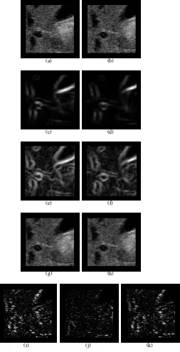
[image:4.612.333.532.182.508.2]For the region image extracted from an abdominal part ultrasound image, we applied our method again to examine its effectiveness, depicted in Fig.3. Since there is no ground truth, we compute the sum of squared differences (SSD) be-tween the original and registered images. The SSD decreased by 51.14% for the proposed method while it decreased only by 12.73% with the standard gradient scheme.
Fig. 1. Registration error as a function of diminishing contrast. The top figure shows the translational error while the bottom one shows rotational error.
V. CONCLUSION
In this article, we use speckle statistical model based on normalized Fisher-Tippett distribution to construct a speckle-specific information-theoretic feature detector and utilize these feature maps for speckle image registration. Since the similarity metric is distribution to distribution comparison, rather than pixel-pixel intensity comparison, it is more robust to uncorrelated speckle noise. Our experiment results also demonstrate the effectiveness of this method.
REFERENCES
Fig. 2. Feature detection in synthetic ultrasound images. The leftmost column shows the original synthetic ultrasound images with diminishing contrast. The middle column shows the feature detection with information-theoretic method, while the rightmost column shows the feature detection with the difference of Gaussian filter.
(a) (b)
(c) (d)
(e) (f)
(g) (h)
[image:5.612.322.575.123.618.2](i) (j) (k)
[2] B. Cohen and I. Dinstein, New Maximum Likelihood Motion Estima-tion Schemes for Noisy Ultrasound Images,Pattern Rec., vol. 35, no. 2, pp. 455-463, 2002.
[3] D. Boukerroui, J.A. Noble, and M. Brady, Velocity Estimation in Ultrasound Images: a Block Matching Approach, inProc. Information Processing in Medical Imaging, 18th International Conference, pp. 586-598, IPMI 2003, Ambleside, UK.
[4] Z. Wang, G.Slabaugh, G. Unal and T. Fang, Registration of Ultrasound Images using an Information-Theoretic Feature Detector,IEEE Inter-national Symposium on Biomedical Imaging, ISBI 2007, Washington DC, USA.
[5] V. Dutt and J. Greenleaf, Statistics of the Log- Compressed Envelope,
Journal of the Acoustical Society of America, vol. 99, no. 6, pp. 3817-3825, 1996.
[6] J. Goodman, Speckle Phenomena: Theory and Applications, 1st ed. Work in Progress, 2005.
[7] Slabaugh, G. and G. Unal and T. Chang, Information-Theoretic Feature Detection in Ultrasound Images,Proc. IEEE International Conference of the Engineering in Medicine and Biology Society, pp.2638-2642, EMBC 2006, NYC, USA.
[8] R. Frackowiak, Human Brain Function,Academic Press, San Diego, second edition, 1997.