R E S E A R C H A R T I C L E
Open Access
NEDD4 expression is associated with breast
cancer progression and is predictive of a
poor prognosis
Lingfeng Wan
1,2, Tao Liu
1,2, Zhipeng Hong
2,3, You Pan
1, Steven T. Sizemore
2, Junran Zhang
2*and Zhefu Ma
1,4*Abstract
Background:A role for neural precursor cell-expressed developmentally downregulated gene 4 (NEDD4) in
tumorigenesis has been suggested. However, information is lacking on its role in breast tumor biology. The purpose of this study was to determine the role of NEDD4 in the promotion of the growth and progression of breast cancer (BC) and to evaluate the clinicopathologic and prognostic significance of NEDD4.
Methods:The impact of NEDD4 expression in BC cell growth was determined by Cell Counting Kit-8 and colony
formation assays. Formalin-fixed paraffin-embedded specimens were collected from 133 adjacent normal tissues (ANTs), 445 BC cases composed of pre-invasive ductal carcinoma in situ (DCIS,n= 37), invasive ductal carcinomas (IDC,n= 408, 226 without and 182 with lymph node metastasis), and 116 invaded lymph nodes. The expression of NEDD4 was analyzed by immunohistochemistry. The association between NEDD4 expression and clinicopathological characteristics was analyzed by chi-square test. Survival was evaluated using the Kaplan–Meier method, and curves were compared using a log-rank test. Univariate and multivariate analyses were performed using the Cox regression method.
Results:NEDD4 promoted BC growth in vitro. In clinical retrospective studies, 16.5% of ANTs (22/133) demonstrated positive NEDD4 staining. Strikingly, the proportion of cases showing NEDD4-positive staining increased to 51.4% (19/37) in DCIS, 58.4% (132/226) in IDC without lymph node metastasis, and 73.1% (133/182) in BC with lymph node metastasis (BCLNM). In addition, NEDD4-positive staining was associated with clinical parameters, including tumor size (P= 0.030), nodal status (P= 0.001), estrogen receptor status (P= 0.035), and progesterone receptor status (P= 0.023). Moreover, subset analysis in BCLNM revealed that high NEDD4 expression correlated with an elevated risk of relapse (P= 0.0276). Further, NEDD4 expression was an independent prognostic predictor. Lastly, the rates for 10-year overall survival and disease-free survival were significantly lower in patients with positive NEDD4 staining than those in BC patients with negative NEDD4 staining BC (P= 0.0024 andP= 0.0011, respectively).
Conclusions:NEDD4 expression is elevated in BC and is associated with BC growth. NEDD4 correlated with
clinicopathological parameters and predicts a poor prognosis. Thus, NEDD4 is a potential biomarker of poor prognosis and a potential therapeutic target for BC treatment.
Keywords: Breast cancer, NEDD4, Prognosis, Tumor progression
© The Author(s). 2019Open AccessThis article is distributed under the terms of the Creative Commons Attribution 4.0 International License (http://creativecommons.org/licenses/by/4.0/), which permits unrestricted use, distribution, and reproduction in any medium, provided you give appropriate credit to the original author(s) and the source, provide a link to the Creative Commons license, and indicate if changes were made. The Creative Commons Public Domain Dedication waiver (http://creativecommons.org/publicdomain/zero/1.0/) applies to the data made available in this article, unless otherwise stated.
* Correspondence:Zhang.9543@osu.edu;mazhefu@hotmail.com 2Department of Radiation Oncology, The Ohio State University, Arthur G.
James Comprehensive Cancer Center and Richard L. Solove Research Institute, 460 West 12th Ave, Columbus, OH 43210, USA
1Department of Breast Surgery, The First Affiliated Hospital, Sun Yat-sen
University, No.58 of Zhongshan 2nd Road, Yuexiu District, Guangzhou 510080, China
Background
Breast cancer (BC) is the most common cancer in women worldwide, with the incidence increasing in re-cent years, particularly in developing countries, due to increased life expectancy and the adoption of a western lifestyle [1]. Moreover, BC is the second most common cause of mortality due to cancer, accounting for about 14% of all cancer deaths [2]. Although the combined ef-fects of earlier detection and a range of improvements in treatment have reduced the mortality rate of BC, the in-cidence for BC is estimated to be increasing globally [3]. Therefore, prevention and therapy of BC remain major public health concerns. The identification of new factors contributing to BC development will be the key to dis-covering novel targets for BC treatment.
Invasive ductal carcinoma (IDC) is the most common subtype of BC, comprising approximately 60–75% of all breast carcinomas [3]. The current mode for BC develop-ment involves a sequential progression from hyperplasia, atypical ductal hyperplasia, ductal carcinoma in situ (DCIS) to ultimately IDC, and eventually metastasis [4,5]. Various criteria have been used in the clinic to predict the progres-sion of BC. The tumor–node–metastasis (TNM) system is a worldwide classification that describes the stages of BC, often based on tumor size, regional lymph node involve-ment, and the absence or presence of distant metastasis [6]. TNM stage classifications were developed not only to better understand the clinical behavior of BC, but to also predict the prognosis of similar groups of patients with BC. In addition, several markers have been widely used to pre-dict a BC prognosis in clinical practice, such as the estro-gen receptor (ER), progesterone receptor (PR), and human epidermal growth factor receptor 2 (Her2) status, as well as several clinicopathologic features, such as tumor size, histologic grade, and lymph node involvement [7,8]. Based on the frequently used biomarkers, ER, PR, Her2, and Ki-67, BC is classified into four specific molecular subtypes: luminal A, luminal B, Her2-enriched, and triple negative breast cancer (TNBC) in which ER, PR, and Her2 are all negative [8]. Each subtype of BC responds differently to specific treatments and predicts the prognosis.
E3 ligases are critical components in the ubiquitination cascade, responsible for substrate recognition and modi-fication with specific polyubiquitin chains. The HECT E3 ligase family plays a critical role in regulatory and di-verse cellular pathways, operating in and leading to tumor initiation, progression, migration, and resistance to anticancer therapies [9]. NEDD4 E3 protein family members share a similar domain composition: an N-terminal C2 domain, two to four WW domains, and a catalytic HECT domain at the C-terminus [9, 10]. NEDD4 is the product of the neural precursor cell-expressed, developmentally downregulated 4 gene [11]. The cellular function of NEDD4 was initially found to
be associated with the regulation of the turnover of the epithelial sodium channel (ENaC) [12]. Subsequently, NEDD4 was demonstrated to be an oncogene due to its role in the negative regulation of the well-known tumor suppressor phosphatase and tensin homolog (PTEN) [13]. Elevated NEDD4 levels and PTEN degradation are observed in various types of human cancer lines [14]. However, no such relationship was observed in BC tissue [15]. In addition, the proto-oncogenic functions of NEDD4 can be attributed to its ability to stabilize the mouse double minute 2 homolog (Mdm2) that exerts its oncogenic activity primarily by suppressing p53 [16]. Moreover, NEDD4 acts as a negative regulator of insulin-like growth factor 1 receptor (IGF-1R) signaling by binding to the adaptor protein, Crb10 [17,18]. Many other signaling pathways are also regulated by NEDD4, such as the Wnt [19] and notch pathways [20]. In support of its role in promoting oncogenic signaling, NEDD4 over-expression correlates with cell proliferation and trans-formation [13, 21, 22]. NEDD4 overexpression was detected in malignant gastric, colorectal, and lung cancer cells [21,23,24]. In addition, aberrant NEDD4 expression has been implicated in pathogenesis and is associated with an adverse prognosis in gastric cardia adenocarcinoma tu-mors [25]. Given the role of NEDD4 in cancer growth, progression, and its poor prognosis, NEDD4 as a target is considered to be a promising therapeutic strategy for the treatment of human malignancies [26].
Although NEDD4 expression in BC has been detected in previous studies [15, 27, 28], to our knowledge, the expression of NEDD4 in BC and normal tissue have never been compared. In addition, the role of NEDD4 in BC progression and its prognostic value in BC remains unclear in a clinical context. The goal of this study was to determine the role of NEDD4 in BC growth and pro-gression and to delineate the clinical relevance of NEDD4 to human BC.
the subset of BC cases with invaded lymph nodes sug-gested that positive NEDD4 staining is associated with a higher relapse of recurrence compared to a NEDD4-negative staining subset. Lastly, NEDD4 expression is associated with lower overall survival (OS) and disease-free survival (DFS). We concluded that NEDD4 pro-motes BC growth. NEDD4 expression is associated with BC progression and is a potential biomarker for a poor prognosis. Our study suggests a novel molecular thera-peutic target for BC treatment.
Methods
Patients and tissue samples
Two cohorts were analyzed in this study: a retrospective cohort of 297 formalin-fixed paraffin-embedded (FFPE) tumor tissues from patients with early stage I–III BC who enrolled in our study from January 1, 2004, to De-cember 31, 2008, including 26 DCIS and 271 IDC cases, as well as 116 FFPE invaded lymph node tissues from patients with BC who had enrolled in this cohort. The secondary cohort consisted of 148 patients with stage I– III BC tumors, which comprised 11 DCIS and 137 IDC cases, as well as 133 paired adjacent normal tissues (ANTs) as negative controls. Of note, 126 cases had BC with lymph node metastasis in the first cohort, and 56 cases made up the second cohort. All samples were taken from the First Affiliated Hospital of Sun Yat-sen University, Guangzhou, China.
ER, PR, Her2, Ki-67, and p53 status were determined from pathology reports of the Pathology Department of the First Affiliated Hospital of Sun Yat-sen University. ER- or PR-positive tissue was defined as more than 10% of tissue staining positive [29]. In the case of Her-2, a fluorescence in situ hybridization assay was performed to evaluate gene amplification in the event of equivocal Her-2 protein expression by IHC. Each patient’s age at diagnosis, menstruation status, tumor grade, tumor size, and nodal status were obtained from medical records. All samples were examined by two independent patholo-gists. Histological type was based on the TNM system (American Joint Committee on Cancer Classification, 8th, http://www.cancerstaging.org). DFS was defined as the time after surgery to the date of clinical relapse (with histopathology confirmation or radiological evidence of tumor recurrence), a second cancer, or death. Overall survival (OS) was defined as the time from surgery until death from any cause. Uniform guidelines for post-operative follow-up procedures have been described pre-viously [30]. The follow-up deadline was September 28, 2018.
This study was approved by the ethics committee of the First Affiliated Hospital of Sun Yat-sen University. Inclusive criteria are summarized as follows: (1) All pa-tients recruited had unilateral BC and were histologically
diagnosed. Adjacent normal breast tissue was selected from an area more than 5 cm from the edge of the tumor. (2) Any patient who had distant metastasis or re-ceived preoperative radiotherapy, chemotherapy, hormo-nal therapy, or any other anticancer therapy before surgery was excluded. (3) Patients with serious compli-cations, such as heart disease, cerebrovascular disease, diabetes, or other malignant diseases, were excluded. (4) Complete clinicopathological data for further analysis were available. (5) All patients were followed up through medical appointments or by telephone.
Assignment of BC subtype
ER, PR, Her2, and Ki-67 were used to approximate BC subtypes [29]. ER-positive and/or PR-positive and Ki-67≤20% samples were considered luminal A BC. ER-positive and/or PR-ER-positive and Ki-67 > 20% samples were considered luminal B BC. Her2 positive (independ-ent of ER and PR status) was considered Her2-enriched BC. ER-negative, PR-negative, and Her2-negative sam-ples were considered TNBC. Luminal A and luminal B BC were hormone receptor positive. Her2-enriched and TNBC were hormone receptor negative.
Tissue microarray construction
The second cohort of tissue samples consisting of 148 cases was prepared for tissue microarray (TMA). All cases were initially selected from paraffin-embedded tumor tissues, and then, sections were reviewed to con-firm and select areas for the coring of corresponding blocks. Duplicate tissue cores (1.5-mm diameter) were taken from the central cellular areas of each tumor. The original cohort of cases was arrayed across four blocks. Serial 4-μm sections were cut from TMA blocks.
IHC
washing steps were performed with PBS alone, along with PBS with 0.1% Tween. To ensure consistent IHC evaluation, slides from a reference tumor previously defined as positive were included in each staining procedure.
IHC scoring
IHC scoring was performed in a blinded fashion by two independent pathologists. We determined NEDD4 stain-ing in tissues in accordance with an immunoreactive score (IRS) proposed by Remmele and Stegner [31]. IHC scores were determined according to the staining inten-sity (SI: 0, negative; 1, weak; 2, moderate; 3, strong) and the percentage of positive cells (PP: 0, < 5%; 1, 5–10%; 2, 11–50%; 3, 51–80%; 4, > 81%). An overall immunoreac-tive score (IRS) was derived by multiplying SI and PP. Slices scoring at least 3 points were classified as showing positive overexpression.
Cell lines
Human breast cancer cell lines were kindly provided by the Stem Cell Bank, Chinese Academy of Sciences (Shanghai, China). Cell lines were cultured with DMEM (high glucose; Invitrogen, Waltham, MA, USA) sup-plemented with 10% fetal bovine serum (FBS; Gibco, Waltham, MA, USA) in a humidified atmosphere.
Small interfering RNA transfections
NEDD4 small interfering (si) RNAs were purchased from RIBOBIO (siRNA#1: TGGCGATTTGTAAACCGAA; siRNA#2: GTGCAAATCATCAGGTTAT; Guangzhou, China). Lipofectamine IMAX (Invitrogen) was used for siRNA transfection. Cells transfected with non-targeting siRNA were used as controls. Transfection efficiencies were validated using quantitative reverse transcription (qRT)–PCR and western blotting.
Proliferation and colony formation assays
For proliferation, a Cell Counting Kit-8 (CCK8; Dojindo, Kumamoto, Japan) assay was used. Cells were plated into 96-well cell culture clusters at a concentration of 1000 cells/well in a volume of 100μL after transfections. CCK8 reagents were then added and incubated for 2 h at 37 °C. The absorbance at 450 nm was measured with a microplate reader. For colony formation assays, cells were plated into 6-well cell culture clusters at a concen-tration of 1000 cells/well in a volume of 2 mL after transfections. After 14 days incubation at 37 °C, cells were fixed with paraformaldehyde for 15 min and stained with 0.5% crystal violet for 30 min. Plates were then washed several times with water, and images of the optical density of colonies were scanned using an optical density scanning analysis system (GS-800; Bio-Rad,
Hercules, CA, USA). The number of colonies (> 50 cells) was counted using Image J software.
Wound healing and transwell assays
For the wound healing assay, cells were plated into a 12-well cell culture cluster at a concentration of 5 × 10⁵ cells/mL. Twenty-four hours later, the cells reached about 90% confluence in a monolayer. A 10-μL pipette tip was then used to scratch a line in the cell monolayer, and the medium was replaced with 1 mL DMEM. The cells were incubated for the indicated times, and images taken under an optical microscope. The gap closure area was measured by Image J software. For transwell assays, cells were plated into an upper transwell chamber at a concentration of 1 × 10⁴cell/mL in a volume of 200μL DMEM. The lower chamber of the transwell was filled with 600μL DMEM plus with 10% FBS. The cells were incubated for the indicated times. After the completion of migration, the cells in the upper chamber were re-moved with a cotton tip. The cells on the bottom side of the transwell membrane were fixed with 4% paraformal-dehyde for 15 min and stained with 0.5% crystal violet for 30 min. Cells were photographed under an optical microscope (DMi8; Leica, Wetzlar, Germany), and the numbers of migrated cells were determined by Image J software.
Western blot analysis
Total protein was extracted in RIPA lysis buffer. Proteins extracted from cells were resolved using 10% sodium dodecyl sulfate–polyacrylamide (SDS–PAGE) gel electro-phoresis, then transferred to a polyvinylidene fluoride membrane (Millipore, Burlington, MA, USA), blocked in 5% non-fat milk (Sigma–Aldrich, St Louis, MI, USA) for 2 h, and blotted with primary antibody (anti-NEDD4, Cell Signaling Technology, Danvers, MA, USA, 1:1000; anti-IGF-1R, Abcam, 1:1000; anti-PTEN, Cell Signaling Tech-nology, 1:500; anti-p-Aktser473, Cell Signaling Technology, 1:2000; anti-GAPDH, Cell Signaling Technology, 1:2000; anti-β-actin, Sigma–Aldrich, 1:5000) overnight at 4 °C. The next day, membranes were incubated with the appropriate HRP-conjugated secondary antibody for 1 h at room temperature. Blots were visualized with an ECL detection kit (Millipore) and analyzed using Image J software.
Cell line authentication
The authentication of each cell line was confirmed by a 100% match to the reference short tandem repeat profile of the respective cell lines from ATCC.
Statistical analysis
mean ± standard deviation (SD) derived from at least three independent experiments. Associations between NEDD4 expression and clinicopathologic data were evaluated using a chi-square test. A comparison of NEDD4 IHC scores be-tween two groups were performed using a Mann–Whitney test. The effects of NEDD4 knockdown or overexpression on cell behavior were examined using attest (two groups) or ANOVA (more than two groups). Survival was calcu-lated using the Kaplan–Meier method, and differences be-tween groups were tested by log-rank test. Univariate and multivariate analyses were undertaken using Cox regression analysis. Correlation analyses were analyzed using the Spearman correlation test. A two-tailed value of P< 0.05 was regarded as statistically significant. Data for GSE20685 (microarray-based molecular subtyping of breast cancer) was acquired throughOncomine.com
Results
NEDD4 promotes BC growth in vitro
NEDD4 has a role in promoting the growth of hepato-cellular [32] and bladder cancer cell lines [33]. To deter-mine the role of NEDD4 in BC growth, we first examined how NEDD4 expression affects cell growth using BC cell lines from different BC subtypes. NEDD4 was first knocked down by two independent siRNAs in five BC cell lines: luminal A (MCF7, T47D), luminal B (ZR-75-1), and TNBC (MDA-MB-231, BT549; Fig. 1a).
Depletion of NEDD4 significantly inhibited cell prolifer-ation in all tested BC cell lines by CCK8 cell prolifera-tion assay (Fig.1b). This result was further confirmed by colony formation assay. The depletion of NEDD4 in these BC cell lines reduced colony formation compared to control cells with intact NEDD4 expression (Fig. 1c). The impact of NEDD4 on cell growth was verified by MTT cell proliferation assay. A similar result was ob-served using short hairpin RNAs targeting different cod-ing regions of NEDD4 (Additional file 1: Figure S1). Thus, our results suggest that NEDD4 promotes cell proliferation in BC cell lines.
NEDD4 is highly expressed in human BC tumors
Given the role of NEDD4 in promoting BC growth, it is expected that NEDD4 is highly expressed in BC. To de-termine if NEDD4 is highly expressed in human BC tis-sue, 445 patients diagnosed with early primary BC that were from 2 cohorts of BC were studied. The first cohort was composed of 297 patients with complete clinical and pathologic features and available follow-up data from the period 2004–2008; the median follow-up was 127 months (range 19–171 months). The second cohort was composed of 148 patients with complete clinical and pathologic features as well as 133 paired ANTs from 2014 to 2018. All patients were female. The median age at diagnosis for the 445 patients from both cohorts was
Fig. 1NEDD4 facilitates proliferation in BC cell lines.aKnockdown of NEDD4 resulted in reduced NEDD4 protein expression in BC cell lines.b
[image:5.595.58.537.429.682.2]49 years (range from 23 to 80 years). 11.2% of patients were 35 years of age or under at diagnosis, and 40.9% had lymph node metastasis at the time of surgery. In this group, primary therapy included surgical resection in all cases followed by adjuvant hormone, chemotherapy, and radiation therapy in 57.1% (254), 67.6% (301), and 27.6% (123) cases; 13.7% (61) did not receive any form of sys-temic therapy. The clinicopathologic characteristics of cohorts are provided in Additional file2: Table S1.
NEDD4 expression was detected by IHC in all samples prepared for TMAs and FFPE slides using an antibody previously reported to be specific for NEDD4 [13]. Con-sistent with a previous report [15, 27], NEDD4 staining was found in the cytoplasm of cancer cells. Of all 445 samples, 63.8% of tumor samples were positive for NEDD4 staining, which is similar to a previous report in which NEDD4-positive expression was observed in 55% of BC tumor samples [15] (Fig. 2a). In contrast, only 16.5% of adjacent tissue samples showed positive
immunoreactivity for NEDD4 (Fig. 2a). Representative samples of different NEDD4 IHC staining grades in both normal and tumor tissues are shown in Fig. 2b. Thus, the difference in NEDD4 immunoreactivity between tumor samples and adjacent tissues was significant (Fig. 2a,χ2= 94.872,P< 0.001). The median NEDD4 IRS of BC tumors was significantly higher compared to that of ANT (Fig.2c,P< 0.001). Our study is the first to sys-temically compare NEDD4 expression in breast cancer and normal tissues. Of all 445 cases, 66.5% (296) were ER positive, including 5.7% (17) DCIS and 94.3% (279) IDC. Noticeably, the median NEDD4 IRS of ER-positive tumors was significantly higher compared to that of ER-negative tumors (Fig. 2d, P< 0.001), indicating that NEDD4 expression is associated with ER expression in BC. In support of our result, analysis of the 327 BC pa-tient samples in GSE20685 confirms that NEDD4 mRNA expression is significantly higher in ER-positive vs. ER-negative BC (Additional file 3: Figure S2; P<
Fig. 2NEDD4 is highly expressed in BC.aThe percentage of BC tumors with positive NEDD4 staining was compared to that of ANTs (χ2test, χ2= 94.872,P< 0.001).bRepresentative IHC images of different NEDD4 staining grades in BC tumors and paired ANT.cThe median IRS of BC
[image:6.595.59.539.330.674.2]0.001). Together, the results of Fig. 2 suggest that NEDD4 expression is high in human BC, particularly in ER-positive BC tissue.
Although it is not clear how NEDD4 expression is up-regulated in human BC, it has been demonstrated that there is a correlation between NEDD4 protein and mRNA expression in human BC [15]. In support of this, analysis of The Cancer Genome Atlas (TCGA) dataset suggests a strong correlation between NEDD4 protein expression and mRNA in human BC (Additional file 4: Figure S3). Thus, it is most likely that NEDD4 is highly expressed in BC and is related to the transcriptional regulation of NEDD4.
NEDD4 is associated with clinicopathological features in BC
Next, NEDD4 protein expression was compared with several clinicopathologic variables in BC, such as age, menstruation status, tumor size, histological grade, lymph node involvement, and molecular subtypes. High NEDD4-expressing BC was associated with a large tumor size (χ2= 8.973,P= 0.030) and a high incidence of lymph node invasion (χ2= 10.111,P= 0.001; Table1). In addition, high NEDD4 expression correlated with an ER-positive status (χ2= 4.451, P= 0.035) and a PR-positive status (χ2= 5.197,P= 0.023; Table1). For molecular sub-type, 68.8% (44/64) of BC displayed NEDD4-positive staining in luminal A tumors and 66.3% (177/267) in lu-minal B tumors, both of which were ER/PR positive. In addition, 58.9% (43/73) of Her2-enriched and 48.8% (20/ 41) of triple negative tumors showed NEDD4-positive staining. However, the difference in NEDD4 expression between hormone receptor positive, Her2-enriched, and triple negative tumors did not reach statistical signifi-cance (Table 1, χ2= 6.161, P= 0.104). In addition, NEDD4 expression showed no obvious relationship with other well-known clinicopathological variables, such as age, menstruation status, tumor grades, Her2 status, or Ki-67 status (P> 0.05, respectively; Table 1). Together, the results from Table1 suggested that NEDD4 expres-sion was associated with the clinicopathological charac-teristics of tumor size, nodal status, and ER and PR expression.
NEDD4 is associated with progression of BC
In order to determine if NEDD4 expression is associated with BC progression, we subgrouped BC samples based on tumor progression and advantage. We evaluated NEDD4 expression levels in DCIS (n= 37), IDC (n= 226) without lymph node metastasis, and BC with lymph node metastasis tumors (BCLNM, n= 182). NEDD4-positive expression occurred in 51.4% of DCIS, 58.4% of IDC, and 73.1% of BCLNM samples (Fig.3a). An appar-ent increase in the rate of NEDD4-positive staining
along with progression of the disease occurred, although the difference between DCIS and IDC was not signifi-cant (P= 0.421). All subtypes of BC in different stages of BC development displayed increased NEDD4 expression compared to ANT (Fig. 3a, DCIS vs. ANT, χ2= 19.166, P< 0.001; IDC vs. ANT, χ2= 62.183, P< 0.001; BCLNM vs. ANT,χ2= 98.271,P< 0.001; IDC vs. DCIS,χ2= 0.647, P= 0.421; BCLNM vs. DCIS, χ2= 6.835, P= 0.009; BCLNM vs. IDC, χ2= 9.530, P= 0.002). Additionally, as the median NEDD4 IRS of ANT, DCIS, IDC, and BCLNM gradually increased, differences between indi-cated subtypes of BC tissues reached statistical signifi-cance (Fig. 3b, DCIS vs. ANT, P= 0.007; IDC vs. ANT, P< 0.001; BCLNM vs. ANT, P< 0.001; IDC vs. DCIS, P= 0.01; BCLNM vs. DCIS, P< 0.001; BCLNM vs. IDC, P= 0.016). Representative IHC images of ANT, DCIS, IDC, and BCLNM are shown in Fig. 3c. In addition, NEDD4 IRS was elevated in the IDC subtype compared with DCIS, regardless of the status of ER (Fig.3d). How-ever, the rate of NEDD4-positive expression was high in the ER-positive group in both IDC or DCIS subgroups, which further supports the results shown in Fig. 2 and Additional file 3: Figure S2, where NEDD4 expression was associated with ER expression. These results strongly suggest that NEDD4 expression gradually in-creases during breast tumor progression. A representa-tive sample of NEDD4 staining is displayed in Fig.3e, in which NEDD4 staining intensity was enhanced in area with IDC compared to an area with DCIS from the same tumor sample (Fig.3e, *DCIS;#IDC).
In support of the hypothesis that NEDD4 correlated with tumor progression, the NEDD4 expression rate in TNM stage I was 52.7%, which then increased up to 64.3% in stage II and 77.5% in stage III (Fig. 3f, stage II vs. I,χ2= 4.402,P= 0.042; stage III vs. І,χ2= 12.215,P< 0.001; stage III vs. II, χ2= 4.998, P= 0.025). The differ-ence between the median NEDD4 IRS score of each sub-type of BC in TNM stages was significant (Fig.3g, stage II vs.І,P= 0.003; stage III vs.І,P< 0.001; stage III vs. II, P= 0.023). Representative NEDD4 IHC images of each TNM stage in BC are presented in Fig.3h. Our study is the first to systemically compare NEDD4 expression in BC tissue at different stages of cancer progression. To-gether, these results suggested that NEDD4 is associated with BC progression and that elevated NEDD4 expres-sion may play an important role in BC development.
High NEDD4 expression is associated with a poor prognosis in BC
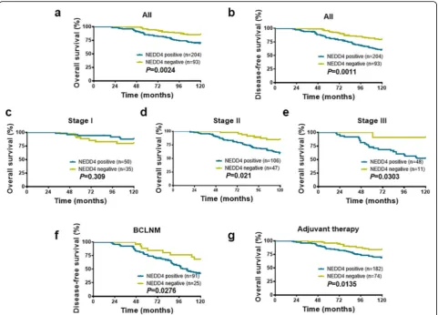
those patients had complete follow-up data. As shown in Fig.4, patients with positive NEDD4-expressing BC had a shorter OS (Fig. 4a,P= 0.0024) and DFS (Fig.4b,P= 0.0011) than patients with negative NEDD4-expressing BC.
The association of NEDD4 expression with OS in each TNM stage BC was further analyzed with the Kaplan– Meier analysis. As shown in Fig. 4c–e, NEDD4 expres-sion was inversely associated with OS in TNM stages II and III patients (Fig.4d, e,P= 0.021,P= 0.0303, respect-ively), but not stageІpatients (Fig.4c,P= 0.309). These results clearly show that patients with NEDD4-positive staining BC in stages II and III had a lower survival rate during follow-up, suggesting that NEDD4 expression may be a feasible index for predicting a poor survival
rate in patients with BC. Additionally, in the subgroup of BC patients with invaded lymph nodes, those with NEDD4-positive staining showed a high relapse during the follow-up period of 10 years compared with the NEDD4-negative expression group (Fig. 4f, P= 0.0276). These results suggested that NEDD4 expression is asso-ciated with a poor prognosis and is linked to a high risk of relapse in BC.
[image:8.595.58.542.98.519.2]Next, we investigated the relationship between NEDD4 expression and rates of OS in the following subsets of patients with BC: those who received or did not receive adjuvant therapy, ER positive or negative, and Her2 posi-tive or negaposi-tive. NEDD4 expression was associated with a low OS rate in patients who received adjuvant therapy (Fig.4g,P= 0.0135). Interestingly, a statistical correlation
Table 1Associations between clinicopathological characteristics and NEDD4 expression
Parameter Status Total NEDD4 positive NEDD4 negative χ2 P
Age at diagnosis (years) ≤35 50 36 14 1.632 0.201
> 35 395 248 147
Menopausal status Premenopausal 240 158 82 0.914 0.339
Postmenopausal 205 126 79
Tumor size (cm) T1a/b 67 36 31 8.973 0.030
T1c 127 73 54
T2 220 153 67
T3 31 22 9
Tumor grade 1 39 25 14 2.358 0.308
2 204 120 84
3 144 89 55
Unknown 58 50 8
Nodal status Positive 182 132 50 10.111 0.001
Negative 263 152 111
ER status Positive 296 199 97 4.451 0.035
Negative 149 85 64
PR status Positive 306 206 100 5.197 0.023
Negative 139 78 61
Her2 status Positive 120 68 52 3.641 0.056
Negative 325 216 109
Ki-67 status ≤20% 133 82 51 0.385 0.535
> 20% 312 202 110
P53 status Positive 299 190 109 0.291 0.590
Negative 104 63 41
Unknown 42 31 11
Molecular subtype Luminal A 64 44 20 6.161 0.104
Luminal B 267 177 90
Her2+ 73 43 30
TNBC 41 20 21
Tumor size: 0.1 cm < T1a/b < 1 cm; 1 cm≤T1c < 2 cm; 2 cm≤T2 < 5 cm; 5 cm≤T3
between NEDD4 expression and OS was not found in ER-positive breast tumors (Additional file 5: Figure S4a, P= 0.0865), while high NEDD4 expression was associ-ated with a lower OS rate in ER-negative patients (Add-itional file 5: Figure S4b, P= 0.0204). Additionally, no association between NEDD4 expression and OS was ob-served in Her2-positive BC patients (Additional file 5: Figure S4c,P= 0.0702), which is consistent with a previ-ous publication showing that NEDD4 expression was
not associated with clinical outcomes in Her2-positive BC patients [27]. However, an association was found be-tween NEDD4 expression and OS in patients with Her2-negative BC (Additional file5: Figure S4d,P= 0.0355).
In support of our results, analyses of the 327 BC patient samples in GSE20685 revealed that NEDD4 mRNA ex-pression is highly prognostic of OS (P= 0.04835) and dis-tant metastasis-free survival (P= 0.0033) in the ER-negative patient population (Additional file6: Figure S5c,
Fig. 3NEDD4 promotes BC progression.aThe proportion of ANT, DCIS, IDC, and BCLNM tissues showing NEDD4-positive staining is presented. NEDD4-positive expression was more frequent with tumor progression (χ2test).bThe median IRS was significantly increased in ANT, DCIS, IDC,
and BCLNM tumors (Mann–Whitney test).cRepresentative IHC images of ANT, DCIS, IDC, and BCLNM are presented. The staining intensity appears higher in invasive lesions.dThe median IRS is higher in IDC compared with DCIS of the same ER status (Mann–Whitney test).eStaining showing that NEDD4 expression is stronger in IDC than that in DCIS in the same tumor sample (*DCIS area,#IDC area).fThe proportions of TNM
stages I, II, and III showing NEDD4-positive staining are presented. NEDD4-positive expression is more frequent with TNM stage (χ2test).gThe
[image:9.595.62.542.86.500.2]d). Intriguingly, NEDD4 expression is not prognostic of survival in the ER-positive patient population of this co-hort, nor is NEDD4 prognostic when survival is examined in combined ER-negative and ER-positive populations of the same cohort (Additional file6: Figure S5a,b,e,f). These results suggest that the prognostic utility of NEDD4 may be strongest in ER-negative BC.
Lastly, we used a Cox proportional hazard model to determine the prognostic value of NEDD4. NEDD4 im-munoreactivity, patient’s age, tumor size, histological grade, and nodal status were chosen as risk variables since all are potential factors affecting a poor prognosis for BC. Hazard ratios are indicated in Table2. In univar-iate and multivarunivar-iate analyses, NEDD4 expression, tumor grade, and nodal status were three independent factors related to the OS rate of BC (Table 2 (a); HR = 2.353, 95% CI = 1.550 to 3.572,P< 0.001; HR = 2.003, 95% CI = 1.333 to 3.007, P= 0.001; HR = 2.105, 95% CI = 2.212 to 4.360, P< 0.001, univariate analyses, respectively; HR =
2.134, 95% CI = 1.394 to 3.268, P< 0.001; HR = 2.186, 95% CI = 1.443 to 3.310,P< 0.001; HR = 2.678, 95% CI = 1.818 to 3.970, P< 0.001, multivariate analyses, respect-ively). With regard to DFS, using a Cox regression model, we found that NEDD4 expression, lymph nodal status, and tumor grade were three independent factors related to DFS (Table2 (b); HR = 2.407, 95% CI = 1.667 to 3.475,P< 0.001; HR = 3.762, 95% CI = 2.771 to 5.108, P< 0.001; HR = 1.752, 95% CI = 1.222 to 2.510,P= 0.002, univariate analyses, respectively; HR = 2.185, 95% CI = 1.507 to 3.162, P< 0.001; HR = 3.289, 95% CI = 2.310 to 4.683, P< 0.001; HR = 1.796, 95% CI = 1.246 to 2.589, P= 0.002, multivariate analyses, respectively). Last, uni-variate and multiuni-variate Cox regression analyses sug-gested that NEDD4 expression and tumor grades were independent risk factors for relapse in BC patients with invaded lymph nodes (Table2(c); HR = 2.512, 95% CI = 1.460 to 4.321, P= 0.001; HR = 1.814, 95% CI = 1.162 to 2.932, P= 0.009; HR = 1.798, 95% CI = 1.162 to 2.780,
[image:10.595.58.540.87.433.2]P= 0.008, univariate analyses, respectively; HR = 2.834, 95% CI = 1.636 to 4.911,P< 0.001; HR = 2.033, 95% CI = 1.293 to 3.197, P= 0.002; HR = 1.861, 95% CI = 1.143 to 3.029, P= 0.013, multivariate analyses, respectively). To-gether, the data presented in Fig. 4and Table 2 suggest that NEDD4 expression is inversely associated with a poor prognosis in BC. NEDD4 expression is an inde-pendent predictive factor for OS and DFS, particularly in BC patients with invaded lymph node metastasis.
Given that the rate of NEDD4-positive staining is the highest in the BCLNM subgroup and that NEDD4 expres-sion is associated with OS and DFS, particularly in this sub-group, this suggests that NEDD4 may promote metastasis. To determine the role of NEDD4 in cancer cell migration in vitro, we conducted wound healing and transwell assays. We found that similar wound healing recovery was noted
in cells, with or without knockdown of NEDD4, using two different cell lines, MDA-MB-231 and T47D. NEDD4 knockdown (Additional file7: Figure S6a,b) had no obvious effect on the wound healing process compared to control cells with intact NEDD4. In addition, we also measured mi-gration by transwell assay. Consistent with wound healing assay results, NEDD4 knockdown had no effect on cell mi-gration in the transwell assay (Additional file7: Figure S6c). Thus, NEDD4 is not essential for cell migration in vitro. As tumor metastasis is a complicated multistep process, it may be that NEDD4 influences the metastatic process via some mechanism other than cell migration.
[image:11.595.55.537.100.504.2]NEDD4 expression is associated with IGF-1R/Akt pathway To determine the potential molecular mechanisms by which NEDD4 promotes breast tumor growth and
Table 2Univariate and multivariate analyses
a. Univariate and multivariate analyses of associations between clinical parameters, NEDD4 status, and overall survival
Overall survival parameter Comparison Univariate Multivariate
HR (95% CI) P HR (95% CI) P
Age at diagnosis (years) > 35 vs.≤35 0.648 (0.373–1.123) 0.122 0.887 (0.506–1.556) 0.676
Tumor size (cm) T2 vs. T1 1.087 (0.554–2.135) 0.808 2.166 (1.067–4.397) 0.032
T3 vs. T1 1.333 (0.692–2.567) 0.391 1.625 (0.840–3.143) 0.149
Tumor grade 2 vs. 1 2.003 (1.333–3.007) 0.001 2.186 (1.443–3.310) 0.000
3 vs. 1 0.728 (0.493–1.074) 0.109 1.015 (0.664–1.554) 0.944
NEDD4 expression Post vs. neg 2.353 (1.550–3.572) 0.000 2.134 (1.394–3.268) 0.000
Nodal status Post vs. neg 2.105 (2.212–4.360) 0.000 2.678 (1.818–3.970) 0.000
b. Univariate and multivariate analyses of associations between clinical parameters, NEDD4 status, and disease-free survival
Disease-free survival parameter Comparison Univariate Multivariate
HR (95% CI) P HR (95% CI) P
Age at diagnosis (years) > 35 vs.≤35 0.671 (0.408–1.105) 0.117 0.957 (0.577–3.167) 0.865
Tumor size (cm) T2 vs. T1 0.784 (0.466–1.379) 0.399 1.501 (0.831–2.713) 0.178
T3 vs. T1 1.205 (0.703–2.065) 0.497 1.482 (0.0.863–2.546) 0.154
Tumor grade 2 vs. 1 1.752 (1.222–2.510) 0.002 1.796 (1.246–2.589) 0.002
3 vs. 1 0.635 (0.453–0.89) 0.008 1.057 (0.723–1.543) 0.776
NEDD4 expression Post vs. neg 2.407 (1.667–3.475) 0.000 2.185 (1.507–3.167) 0.000
Nodal status Post vs. neg 3.762 (2.771–5.108) 0.000 3.289 (2.310–4.683) 0.000
c. Univariate and multivariate analyses of associations between clinical parameters and NEDD4 status and disease-free survival of BCLNM
Disease-free survival parameter Comparison Univariate Multivariate
HR (95% CI) P HR (95% CI) P
Age at diagnosis (years) > 35 vs.≤35 1.334 (0.697–2.550) 0.384 1.328 (0.688–2.561) 0.397
Tumor size (cm) T2 vs. T1 1.455 (0.753–2.811) 0.265 1.142 (0.553–2.356) 0.720
T3 vs. T1 1.127 (0.612–2.077) 0.701 1.074 (0.529–1.991) 0.821
Tumor grade 2 vs. 1 1.814 (1.162–2.932) 0.009 2.033 (1.293–3.197) 0.002
3 vs. 1 1.798 (1.162–2.780) 0.008 1.861 (1.143–3.029) 0.013
NEDD4 Post vs. neg 2.512 (1.460–4.321) 0.001 2.834 (1.636–4.911) 0.000
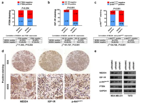
progression, we determined the expression of NEDD4 along with PTEN, IGF-1R, and p-Akt by IHC using a TMA that consisted of 148 samples of early-stage pri-mary invasive breast cancer. Positive PTEN staining was found in 55% (44/80) of BC samples with positive NEDD4 staining, which is comparable to the rate of 45.6% (31/68) observed in BC tissue samples with nega-tive NEDD4 staining (Fig. 5a). This suggests a lack of correlation between NEDD4 and PTEN expression in human BC tissue. As for IGF-1R expression, 85% (68/ 80) of NEDD4-positive staining breast carcinomas were found to stain positive for IGF-1R whereas only 26.5% (18/68) of BC tissue with negative NEDD4 staining stained positive for IGF-1R (Fig. 5b). A similar pattern was also observed for p-AktSer473 staining. Seventy-five percent (60/80) of NEDD4-positive BC samples stained
positively for p-AktSer473 whereas it was only 26.5% (18/ 68) in NEDD4-negative BC samples (Fig.5c). IHC stain-ing revealed that IGF-1R and p-AktSer473 levels were found to be consistently higher in areas of high NEDD4 protein levels (Fig.5d). In support of the results obtained from IHC staining, knocked down NEDD4 led to de-creased IGF-1R and p-AktSer473, even in the T47D cells that harbor an activating PI3K mutation that is constitu-tively active [34] (Fig. 5e). As for PTEN protein, only dramatic NEDD4 knockdown by siRNA #1 led to in-creased PTEN; however, no significant alteration in PTEN protein expression was observed in cells with moderate NEDD4 knockdown by siRNA#2 (Fig. 5e). Given that NEDD4 knockdown by both siRNAs resulted in the slow growth of BC (Fig. 1) and that there is no correlation between NEDD4 and PTEN protein
Fig. 5NEDD4 expression is associated with IGF-1R/Akt pathwayaThe expression of NEDD4 and PTEN in BC tissue. A TMA containing 148 BC tissue samples was immunohistochemically stained with anti-NEDD4 and anti-PTEN.bThe expression of NEDD4 and IGF-1R in BC tissue. A TMA containing 148 BC samples was immunohistochemically stained with anti-NEDD4 and anti-IGF-1R.cThe expression of NEDD4 and p-Aktser473in
BC tissue. A TMA containing 148 BC samples was immunohistochemically stained with anti-NEDD4 and anti-p-Aktser473.dRepresentative
immunostaining patterns for serial sections of the same tumor for NEDD4, IGF-1R, and p-Aktser473.eProtein expression level of indicated proteins
[image:12.595.58.537.291.643.2]expression in BC tissue samples (Fig.5a), the results dis-played in Fig. 5 suggest that NEDD4 promotes BC pro-gression via the activation of IGF-1R/Akt signaling, perhaps independently of PTEN.
Discussion
NEDD4 expression is associated with BC growth and progression
Our study is the first to systemically assess the associ-ation of NEDD4 with BC cell growth, and progression/ prognosis in early-stage BC. We found that NEDD4 is required for the proliferation of BC (Fig. 1), which sup-ports the observation that NEDD4 expression was ele-vated in human BC tumor tissue compared to normal BC tissue (Fig. 2). In addition, NEDD4 expression was associated with tumor size, TNM stage, nodal status, and ER and PR status (Table 1). However, NEDD4 ex-pression is not associated with a Her2, Nottingham Histologic Grade, and Ki-67 [15]. Of note, although NEDD4 expression correlated with ER status (Fig. 2d, Fig. 3d, Additional file 3: Figure S2), NEDD4 seems to have no influence on OS in ER-positive patients (Fig.3a, Additional file 5: Figure S4a, Additional file 6: Figure S5e). This may be explained by the fact that in general, most patients with ER-positive tumors were sensitive to hormone therapy and CDK4/6 inhibitors [35]; ER-positive patients usually showed satisfactory outcomes after hormone therapy [36,37], which overshadowed the effect of NEDD4 overexpression. Our study is consistent with a previous publication showing that NEDD4 ex-pression is associated with the status of ER exex-pression [15]. However, the nature of the association between ER and NEDD4 expression remains unknown and needs to be determined in future.
Cumulative evidence suggests that NEDD4 is linked to tumor progression in several human cancers, such as gastric carcinoma, hepatoma carcinoma, bladder cancer, and prostate cancer [25,32,33, 38]. Based on our stud-ies, NEDD4 expression gradually increases in line with BC progression, from normal tissues, DCIS, IDC without lymph node metastasis to BCLNM tumors, which is a well-established clinical model for BC progression [5]. In addition, gradually increased NEDD4 expression was ob-served in stages I, II, and III BC samples. Thus, our stud-ies strongly suggest that NEDD4 expression is associated with BC progression. Indeed, this hypothesis is further supported by previous studies that showed high NEDD4 expression promoted tumor progression in lung cancer [39] and gastric cardia adenocarcinoma [25].
High NEDD4 expression is a biomarker for poor outcomes Previous work has demonstrated that NEDD4 is associ-ated with poor survival in gastric carcinoma [25] and hepatocellular cancer [40]. In our study, we found that
high NEDD4 expression was associated with a poor post-surgery prognosis in patients with BC, as reflected by their OS and DFS. Therefore, NEDD4 expression may serve as a predictive biomarker of a poor prognosis for BC. Interestingly, no correlation between NEDD4 expression and OS was found in the datasets from TCGA (as of September 23, 2019, data not shown). Sev-eral reasons may exist for this discrepancy, including the heterogenous expression of NEDD4 in tumors, intratu-moral heterogeneity, and significant variability in the NEDD4 antibodies and scoring systems used. To reach a firm conclusion may require additional investigations in future.
We also found that NEDD4 expression is an independ-ent factor for a poor prognosis along with two well-known predictive factors, tumor grade, and nodal status (Table 2 (a, b)) but independent from established prog-nostic factors such as tumor size, margin status, and menstruation status [41]. Further analysis in a subset of BCLNM suggested that patients with NEDD4-positive staining correlated with a high risk of relapse (Fig.4f). In addition, the highest proportion of NEDD4-positive staining was found in BCLNM tissues (Fig.3a). An ana-lysis of GSE20685 indicated that high NEDD4 expres-sion is associated with lower distant metastasis-free survival in women with ER-negative BC (Additional file6: Figure S5d). Such results point to the role of NEDD4 in BC metastases. However, no obvious role for NEDD4 in cell migration was observed in in vitro assays suggesting that NEDD4 exerts its influence on BC metastasis through some other mechanism.
In general, ER-negative tumors are more aggressive and metastatic [42] compared to ER-positive tumors. NEDD4 expression is associated with lower OS in a sub-set of ER-negative patients. An identical trend was also observed in a Her2-negative subgroup, whereas NEDD4 expression was not associated with OS in a subset of BC that was Her2 positive. This is in line with a previous study showing that NEDD4 expression did not predict the efficiency of adjuvant trastuzumab therapy in Her2-positive BC patients [27]. The current treatments for ER-negative and/or Her2-negative BC tumors mainly rely on traditional cytotoxic therapies, which directly or indirectly cause cell DNA damage. Therefore, NEDD4 expression may be a predictor of a poor prognosis in the subset of patients with ER-negative/Her2-negative BC.
The mechanisms by which NEDD4 expression is associated with BC growth and progression
and poor prognosis. NEDD4 is reported to increase PI3K–Akt activity during embryonic development via the maintenance of cell surface IGF-1R protein levels. NEDD4 also regulates Akt via PTEN. NEDD4 negatively regulates PTEN by promoting its poly-ubiquitination and subsequent degradation. Increasing levels of NEDD4 significantly reduced PTEN expression, and potentiated cell proliferation and prostate/bladder tumor formation, suggesting an oncogenic role for NEDD4-1 in regulating PTEN functions [13]. Besides degradation, NEDD4 is also involved in the monoubiquitination of PTEN and its ensuing relocalization to the nucleus [43]. However, sub-sequent studies have shown no difference in the stability and localization of PTEN in two different strains of NEDD4-deficient mice [23]. In addition, NEDD4 is over-expressed in colorectal cancer and promotes colonic cell growth independently of the PI3K/PTEN/AKT pathway [44]. Therefore, regulation of PTEN by NEDD4 may de-pend on the model system and cellular context used. Our studies are consistent with a previous report de-scribing how NEDD4 expression is not associated with PTEN in human breast carcinoma [15]. Our results sug-gest that NEDD4 may promote BC growth and progres-sion via an IGF-1R/Akt pathway, even in the cells that harbor an activating PI3K mutation [34]. Although IGF-1R is likely involved in the NEDD4-mediated Akt activa-tion, additional mechanisms cannot be excluded. For in-stance, Akt activation can be blunted by phosphatases that inhibit Akt activity by dephosphorylation [45]. Therefore, NEDD4 could target phosphatases that dir-ectly dephosphorylate Akt, leading to increase of p-Akt, without greatly increased PTEN. The mechanisms by which NEDD4 promotes IGF-1R expression remain un-known. Although NEDD4 regulates IGF-1R in a positive manner by regulating the function of the adaptor pro-tein, Grb10 [17], the latter appears not to be a direct ubiquitination substrate of NEDD4 [46]. Thus, it is most likely that NEDD4 plays an important role in cell prolif-eration via the activation of IGF-1R signaling by the in-hibition of Grb10. NEDD4 may not directly ubiquitinate IGF-1R, but controls the activity of other E3 ligase pro-teins, which are responsible for Grb10 ubiquitination and degradation. It would be interesting to know if NEDD4 promotes cell proliferation via the facilitation of IGF-1R signaling by the regulation of Grb10 in BC cells.
Despite accumulated findings pointing to tumor-promoting functions, the roles of NEDD4 in cancer ap-pear to be more complex. While NEDD4 functions as an oncogene in most cancers [21,28,44,47,48], it can also act as a tumor suppressor in some tumors [14, 19, 49]. NEDD4 was recently found to suppress the growth of neuroblastoma and pancreatic cancers by targeting Myc and RAS oncoproteins for ubiquitination and degrad-ation [14, 49]. Therefore, NEDD4 may act as an
oncogene in a cellular context-based manner. A recent study also suggested that low NEDD4 expression was closely related to worse outcomes in multiple myeloma [50]. NEDD4 can distinctly regulate degradative ubiquiti-nation of different types of protein substrates in various cancer models, which leads to the promotion or sup-pression of tumorigenesis. Interestingly, a very recent study suggests that NEDD4 is required for the proteaso-mal degradation of PIP5Kα, which acts upstream of PI3K/Akt signaling by supplying the PI3K substrate, PIP2, and promoting BC cell proliferation [51]. The na-ture of the regulation of NEDD4 in targeting PIP5K for degradation in BC growth and progression requires fur-ther study.
Lastly, the roles of NEDD4 in the DNA damage re-sponse may contribute to the poor prognosis of patients with BC showing a high expression of NEDD4. It has been demonstrated that loss of NEDD4 increased the percentage of G1-arrested cells following a DNA-damaging insult and reduced the cell growth rate, which depends on p53, an important factor of the DNA dam-age response [52]. NEDD4 contributes to DNA damage– induced cell-cycle arrest and the inhibition of p53-dependent cell growth [52]. Interestingly, Zhou et al. showed that NEDD4 overexpression can sensitize lung cancer cells to apoptosis induced by the DNA-damaging drug, etoposide [53]. Given that DNA damage response and repair are important in any response to genotoxic chemotherapeutic drugs and ionizing radiation, and the resistance/outcomes of treatment by such modalities, it would be of interest to examine how NEDD4 affects sig-naling induced by DNA damage caused by chemother-apy and ionizing radiation.
The therapeutic potential of targeting the ubiquitin system has been demonstrated by the approval, by the Food and Drug Administration, of the proteasome inhibi-tor, Velcade, for clinical use. Theoretically, targeting E3 ligases is better than targeting the proteasome because E3 ligases represent the last step of the enzymatic cascade that determines a high degree of specificity and selectivity toward target substrates in cells [54]. Thus, NEDD4 may be a promising target for new cancer therapy.
Several limitations exist in our study. For instance, some subgroups of BC cases were on a relatively small scale and this may have affected our results. The patho-logical classification of patients with BC using NEDD4 IHC staining scores should be further investigated with an increased number of cases, such as with a more ap-propriate number of DCIS samples. In addition, TNM stage IV tumors were not included in this study.
Conclusions
associated with BC progression. Importantly, the upreg-ulation of NEDD4 is associated with a poor prognosis. Investigating the precise role of NEDD4 in BC growth and progression will increase our knowledge of the bio-logical function of NEDD4. Our study has uncovered a new potential target in BC in that targeting E3 ligases of-fers a promising therapeutic approach for this disease.
Supplementary information
Supplementary informationaccompanies this paper athttps://doi.org/10. 1186/s13058-019-1236-7.
Additional file 1: Figure S1.NEDD4 facilitates proliferation in BC cell lines.
Additional file 2: Table S1.Clinicopathological characteristics of patient samples and expression of NEDD4 in BC.
Additional file 3: Figure S2.Comparison of NEDD4 mRNA expression in the ER+ and ER- populations of GSE20685.
Additional file 4: Figure S3.Correlation of NEDD4 protein and mRNA in TCGA. Utility of NEDD4 expression in the overall, ER-, and ER+ populations of GSE20685.
Additional file 5: Figure S4.Prognostic impact of NEDD4 in BC with different ER and Her2 statuses.
Additional file 6: Figure S5.Kaplan–Meier analysis of the prognostic utility of NEDD4 expression in the overall, ER-, and ER+ populations of GSE20685.
Additional file 7: Figure S6.NEDD4 is not essential for BC migration in in vitro assays.
Abbreviations
ANT:Adjacent normal tissue; BC: Breast cancer; BCLNM: Breast cancer with lymph node metastasis; DCIS: Ductal carcinoma in situ; DFS: Disease-free survival; ER: Estrogen receptor; GAPDH: Glyceraldehyde-3-phosphate dehydrogenase; Her2: Human epidermal growth factor receptor 2; IHC: Immunohistochemistry; IRS: Immunoreactive score; IDC: Invasive ductal carcinoma; IGF-1R: Insulin-like growth factor 1 receptor; NEDD4: Neural precursor cell expressed-developmentally downregulated 4; OS: Overall survival; PBS: Phosphate-buffered saline; PR: Progesterone receptor; PTEN: Phosphatase and tensin homolog; TCGA: The Cancer Genome Atlas; TMA: Tissue microarray; TNM: Tumor–node–metastasis; TNBC: Triple negative breast cancer
Acknowledgements
The authors thank all patients, clinicians, and pathologists who participated in this study.
Authors’contributions
ZFM, JRZ, and LFW were responsible for the conception and experimental design. LFW, TL, and ZPH performed the experiments. YP and LFW constructed the TMAs. YP, TL, and LFW collected the clinical data. ZFM, JRZ, and LFW analyzed and interpreted the data. JRZ, ZFM, and LFW were responsible for the writing and/or revision of the manuscript. ZFM and JRZ provided the study supervision. STS analyzed the public datasets. All authors read and approved the final manuscript.
Funding
The work described was supported by a National Natural Science Foundation of China grant (31571452 and 31271503), a Guangdong Provincial Natural Science Foundation of China grant (S2012010008368), a startup fund from The First Affiliated Hospital of Sun Yat-sen University and scholarships from the Chinese Scholarship Council (to Z. Ma), and a startup fund from The Ohio State University Arthur G. James Comprehensive Cancer Center and Richard J. Solove Research Institute, The Ohio State University Wexner Medical Center (to J. Zhang).
Availability of data and materials
All data generated or analyzed during this study are included in this published article and its supplementary information files.
Ethics approval and consent to participate
All study participants agreed and signed a written informed consent. The present study was approved by the Ethical Review Board of the First Affiliated Hospital of Sun Yat-Sen University. All specimens were obtained under the protocols outlined by the institutional review board.
Consent for publication
Not applicable.
Competing interests
The authors declare that they have no competing interests.
Author details
1
Department of Breast Surgery, The First Affiliated Hospital, Sun Yat-sen University, No.58 of Zhongshan 2nd Road, Yuexiu District, Guangzhou 510080, China.2Department of Radiation Oncology, The Ohio State University, Arthur G. James Comprehensive Cancer Center and Richard L. Solove Research Institute, 460 West 12th Ave, Columbus, OH 43210, USA. 3Department of Breast Surgery, Affiliated Quanzhou First Hospital of Fujian
Medical University, Quanzhou 362000, China.4Department of Breast Surgery and Plastic Surgery, Cancer Hospital of China Medical University, 44 Xiaoheyan Road, Dadong District, Shenyang 110042, China.
Received: 16 December 2018 Accepted: 28 November 2019
References
1. Partridge AH. Risk communication and decision-making in the prevention of invasive breast cancer. Breast. 2017;34(Suppl 1):S55–7.
2. DeSantis C, Ma J, Bryan L, Jemal A. Breast cancer statistics, 2013. CA Cancer J Clin. 2014;64(1):52–62.
3. Bandyopadhyay S, Bluth MH, Ali-Fehmi R. Breast carcinoma: updates in molecular profiling 2018. Clin Lab Med. 2018;38(2):401–20.
4. Polyak K. Breast cancer: origins and evolution. J Clin Invest. 2007;117(11): 3155–63.
5. Hong YK, McMasters KM, Egger ME, Ajkay N. Ductal carcinoma in situ current trends, controversies, and review of literature. Am J Surg. 2018; 216(5):998–1003.
6. Cserni G, Chmielik E, Cserni B, Tot T. The new TNM-based staging of breast cancer. Virchows Arch. 2018;472(5):697–703.
7. Patani N, Martin LA, Dowsett M. Biomarkers for the clinical management of breast cancer: international perspective. Int J Cancer. 2013;133(1):1–13. 8. Cadoo KA, Traina TA, King TA. Advances in molecular and clinical subtyping
of breast cancer and their implications for therapy. Surg Oncol Clin N Am. 2013;22(4):823–40.
9. Zou X, Levy-Cohen G, Blank M. Molecular functions of NEDD4 E3 ubiquitin ligases in cancer. Biochim Biophys Acta. 2015;1856(1):91–106.
10. Chen C, Matesic LE. The Nedd4-like family of E3 ubiquitin ligases and cancer. Cancer Metastasis Rev. 2007;26(3–4):587–604.
11. Kumar S, Harvey KF, Kinoshita M, Copeland NG, Noda M, Jenkins NA. cDNA cloning, expression analysis, and mapping of the mouse Nedd4 gene. Genomics. 1997;40(3):435–43.
12. Staub O, Gautschi I, Ishikawa T, Breitschopf K, Ciechanover A, Schild L, Rotin D. Regulation of stability and function of the epithelial Na+ channel (ENaC) by ubiquitination. EMBO J. 1997;16(21):6325–36.
13. Wang XJ, Trotman LC, Koppie T, Alimonti A, Chen ZB, Gao ZH, Wang JR, Erdjument-Bromage H, Tempst P, Cordon-Cardo C, et al. NEDD4-1 is a proto-oncogenic ubiquitin ligase for PTEN. Cell. 2007;128(1):129–39. 14. Zeng T, Wang Q, Fu J, Lin Q, Bi J, Ding W, Qiao Y, Zhang S, Zhao W, Lin H,
et al. Impeded Nedd4-1-mediated Ras degradation underlies Ras-driven tumorigenesis. Cell Rep. 2014;7(3):871–82.
15. Chen Y, van de Vijver MJ, Hibshoosh H, Parsons R, Saal LH. PTEN and NEDD4 in human breast carcinoma. Pathol Oncol Res. 2016;22(1):41–7. 16. Shadfan M, Lopez-Pajares V, Yuan ZM. MDM2 and MDMX: alone and
17. Cao XR, Lill NL, Boase N, Shi PP, Croucher DR, Shan H, Qu J, Sweezer EM, Place T, Kirby PA, et al. Nedd4 controls animal growth by regulating IGF-1 signaling. Sci Signal. 2008;1(38):ra5.
18. Monami G, Emiliozzi V, Morrione A. Grb10/Nedd4-mediated multiubiquitination of the insulin-like growth factor receptor regulates receptor internalization. J Cell Physiol. 2008;216(2):426–37.
19. Lu C, Thoeni C, Connor A, Kawabe H, Gallinger S, Rotin D. Intestinal knockout of Nedd4 enhances growth of Apc (min) tumors. Oncogene. 2016;35(45):5839–49.
20. Sakata T, Sakaguchi H, Tsuda L, Higashitani A, Aigaki T, Matsuno K, Hayashi S. Drosophila Nedd4 regulates endocytosis of notch and suppresses its ligand-independent activation. Curr Biol. 2004;14(24):2228–36. 21. Amodio N, Scrima M, Palaia L, Salman AN, Quintiero A, Franco R, Botti G,
Pirozzi P, Rocco G, De Rosa N, et al. Oncogenic role of the E3 ubiquitin ligase NEDD4-1, a PTEN negative regulator, in non-small-cell lung carcinomas. Am J Pathol. 2010;177(5):2622–34.
22. Dai B, Pieper RO, Li D, Wei P, Liu M, Woo SY, Aldape KD, Sawaya R, Xie K, Huang S. FoxM1B regulates NEDD4-1 expression, leading to cellular transformation and full malignant phenotype in immortalized human astrocytes. Cancer Res. 2010;70(7):2951–61.
23. Fouladkou F, Landry T, Kawabe H, Neeb A, Lu C, Brose N, Stambolic V, Rotin D. The ubiquitin ligase Nedd4-1 is dispensable for the regulation of PTEN stability and localization. Proc Natl Acad Sci U S A. 2008;105(25):8585–90. 24. Kim SS, Yoo NJ, Jeong EG, Kim MS, Lee SH. Expression of NEDD4-1, a PTEN
regulator, in gastric and colorectal carcinomas. Apmis. 2008;116(9):779–84. 25. Sun A, Yu G, Dou X, Yan X, Yang W, Lin Q. Nedd4-1 is an exceptional
prognostic biomarker for gastric cardia adenocarcinoma and functionally associated with metastasis. Mol Cancer. 2014;13(1):248.
26. Ye X, Wang L, Shang B, Wang Z, Wei W. NEDD4: a promising target for cancer therapy. Curr Cancer Drug Targets. 2014;14(6):549–56. 27. Luhtala S, Staff S, Kallioniemi A, Tanner M, Isola J. Clinicopathological and
prognostic correlations of HER3 expression and its degradation regulators, NEDD4-1 and NRDP1, in primary breast cancer. BMC Cancer. 2018;18(1):1045. 28. Jung S, Li C, Jeong D, Lee S, Ohk J, Park M, Han S, Duan J, Kim C, Yang Y,
et al. Oncogenic function of p34SEI-1 via NEDD41mediated PTEN ubiquitination/degradation and activation of the PI3K/AKT pathway. Int J Oncol. 2013;43(5):1587–95.
29. Hunt KK, Karakas C, Ha MJ, Biernacka A, Yi M, Sahin AA, Adjapong O, Hortobagyi GN, Bondy ML, Thompson PA, et al. Cytoplasmic Cyclin E predicts recurrence in patients with breast Cancer. Clin Cancer Res. 2017; 23(12):2991–3002.
30. Yang X, Pan Y, Qiu Z, Du Z, Zhang Y, Fa P, Gorityala S, Ma S, Li S, Chen C, et al. RNF126 as a biomarker of a poor prognosis in invasive breast cancer and CHEK1 inhibitor efficacy in breast cancer cells. Clin Cancer Res. 2018; 24(7):1629–43.
31. Friedrichs K, Gluba S, Eidtmann H, Jonat W. Overexpression of p53 and prognosis in breast cancer. Cancer. 1993;72(12):3641–7.
32. Zheng H, Ke X, Li D, Wang Q, Wang J, Liu X, Deng M, Deng X, Xue Y, Zhu Y, et al. NEDD4 promotes cell growth and motility in hepatocellular carcinoma. Cell Cycle. 2018;17(6):728–38.
33. Wen W, Li J, Wang L, Xing Y, Li X, Ruan H, Xi X, Xiong J, Kuang R. Inhibition of NEDD4 inhibits cell growth and invasion and induces cell apoptosis in bladder cancer cells. Cell Cycle. 2017;16(16):1509–14.
34. Hollestelle A, Elstrodt F, Nagel JH, Kallemeijn WW, Schutte M. Phosphatidylinositol-3-OH kinase or RAS pathway mutations in human breast cancer cell lines. Mol Cancer Res. 2007;5(2):195–201.
35. Brufsky AM, Dickler MN. Estrogen receptor-positive breast cancer: exploiting signaling pathways implicated in endocrine resistance. Oncologist. 2018; 23(5):528–39.
36. Louie MC, Sevigny MB. Steroid hormone receptors as prognostic markers in breast cancer. Am J Cancer Res. 2017;7(8):1617–36.
37. Thorat MA, Cuzick J. Preventing invasive breast cancer using endocrine therapy. Breast. 2017;34(Suppl 1):S47–54.
38. Zhang Y, Goodfellow R, Li Y, Yang S, Winters CJ, Thiel KW, Leslie KK, Yang B. NEDD4 ubiquitin ligase is a putative oncogene in endometrial cancer that activates IGF-1R/PI3K/Akt signaling. Gynecol Oncol. 2015;139(1):127–33. 39. Shao G, Wang R, Sun A, Wei J, Peng K, Dai Q, Yang W, Lin Q. The E3
ubiquitin ligase NEDD4 mediates cell migration signaling of EGFR in lung cancer cells. Mol Cancer. 2018;17(1):24.
40. Hang X, Zhu S, Di H, Wu Z, Chu K, Wang J, Xin H, Yu G, Peng H, Miao X, et al. NEDD4 depletion inhibits hepatocellular carcinoma growth via targeting PTEN. Cell Physiol Biochem. 2016;39(2):768–79.
41. O'Brien KM, Cole SR, Tse CK, Perou CM, Carey LA, Foulkes WD, Dressler LG, Geradts J, Millikan RC. Intrinsic breast tumor subtypes, race, and long-term survival in the Carolina Breast Cancer Study. Clin Cancer Res. 2010;16(24): 6100–10.
42. Dunnwald LK, Rossing MA, Li CI. Hormone receptor status, tumor characteristics, and prognosis: a prospective cohort of breast cancer patients. Breast Cancer Res. 2007;9(1):R6.
43. Trotman LC, Wang X, Alimonti A, Chen Z, Teruya-Feldstein J, Yang H, Pavletich NP, Carver BS, Cordon-Cardo C, Erdjument-Bromage H, et al. Ubiquitination regulates PTEN nuclear import and tumor suppression. Cell. 2007;128(1):141–56.
44. Eide PW, Cekaite L, Danielsen SA, Eilertsen IA, Kjenseth A, Fykerud TA, Agesen TH, Bruun J, Rivedal E, Lothe RA, et al. NEDD4 is overexpressed in colorectal cancer and promotes colonic cell growth independently of the PI3K/PTEN/AKT pathway. Cell Signal. 2013;25(1):12–8.
45. Franke TF. PI3K/Akt: getting it right matters. Oncogene. 2008;27(50):6473–88. 46. Morrione A, Valentinis B, Resnicoff M, Xu S, Baserga R. The role of
mGrb10alpha in insulin-like growth factor I-mediated growth. J Biol Chem. 1997;272(42):26382–7.
47. Yim EK, Peng G, Dai H, Hu R, Li K, Lu Y, Mills GB, Meric-Bernstam F, Hennessy BT, Craven RJ, et al. Rak functions as a tumor suppressor by regulating PTEN protein stability and function. Cancer Cell. 2009;15(4):304–14.
48. Singh G, Odriozola L, Guan H, Kennedy CR, Chan AM. Characterization of a novel PTEN mutation in MDA-MB-453 breast carcinoma cell line. BMC Cancer. 2011;11:490.
49. Liu PY, Xu N, Malyukova A, Scarlett CJ, Sun YT, Zhang XD, Ling D, Su SP, Nelson C, Chang DK, et al. The histone deacetylase SIRT2 stabilizes Myc oncoproteins. Cell Death Differ. 2013;20(3):503–14.
50. Huang X, Gu H, Zhang E, Chen Q, Cao W, Yan H, Chen J, Yang L, Lv N, He J, et al. The NEDD4-1 E3 ubiquitin ligase: a potential molecular target for bortezomib sensitivity in multiple myeloma. Int J Cancer. 2019.https://doi. org/10.1002/ijc.32615. [Epub ahead of print]
51. Tran MH, Seo E, Min S, Nguyen QT, Choi J, Lee UJ, Hong SS, Kang H, Mansukhani A, Jou I, et al. NEDD4-induced degradative ubiquitination of phosphatidylinositol 4-phosphate 5-kinase alpha and its implication in breast cancer cell proliferation. J Cell Mol Med. 2018;22(9):4117–29. 52. Xu C, Fan CD, Wang X. Regulation of Mdm2 protein stability and the p53
response by NEDD4-1 E3 ligase. Oncogene. 2015;34(3):281–9.
53. Zhou WH, Xu J, Zhao YC, Sun Y. SAG/RBX2 is a novel substrate of NEDD4-1 E3 ubiquitin ligase and mediates NEDD4-1 induced chemosensitization. Oncotarget. 2014;5(16):6746–55.
54. Fajner V, Maspero E, Polo S. Targeting HECT-type E3 ligases - insights from catalysis, regulation and inhibitors. FEBS Lett. 2017;591(17):2636–47.
Publisher’s Note