R E S E A R C H A R T I C L E
Open Access
A different immunologic profile characterizes
patients with HER-2-overexpressing and
HER-2-negative locally advanced breast cancer:
implications for immune-based therapies
Elena Muraro
1, Debora Martorelli
1, Elisa Turchet
2, Gianmaria Miolo
2, Simona Scalone
2, Elisa Comaro
1,
Renato Talamini
3, Katy Mastorci
1, Davide Lombardi
2, Tiziana Perin
4, Antonino Carbone
4, Andrea Veronesi
2,
Diana Crivellari
2and Riccardo Dolcetti
1*Abstract
Introduction:The clinical efficacy of trastuzumab and taxanes is at least partly related to their ability to mediate or promote antitumor immune responses. On these grounds, a careful analysis of basal immune profile may be capital to dissect the heterogeneity of clinical responses to these drugs in patients with locally advanced breast cancer undergoing neoadjuvant chemotherapy.
Methods:Blood samples were collected from 61 locally advanced breast cancers (36 HER2- and 25 HER2+) at diagnosis and from 23 healthy women. Immunophenotypic profiling of circulating and intratumor immune cells, including regulatory T (Treg) cells, was assessed by flow cytometry and immunohistochemistry, respectively. Serum levels of 10 different cytokines were assessed by multiplex immunoassays. CD8+T cell responses to multiple tumor-associated antigens (TAA) were evaluated by IFN-g-enzyme-linked immunosorbent spot (ELISPOT). The Student’s t test for two tailed distributions and the Wilcoxon two-sample test were used for the statistical analysis of the data. Results:The proportion of circulating immune effectors was similar in HER2+patients and healthy donors, whereas higher percentages of natural killer and Treg cells and a lower CD4+/CD8+T cell ratio (with a prevalence of naïve and central memory CD8+T cells) were observed in HER2-cases. Higher numbers of circulating CD8+T cells specific for several HLA-A*0201-restricted TAA-derived peptides were observed in HER2+cases, together with a higher prevalence of intratumor CD8+T cells. Serum cytokine profile of HER2+patients was similar to that of controls, whereas HER2- cases showed significantly lower cytokine amounts compared to healthy women (IL-2, IL-8, IL-6) and HER2+cases (IL-2, IL-1b, IL-8, IL-6, IL-10).
Conclusions:Compared to HER2- cases, patients with HER2-overexpressing locally advanced breast cancer show a more limited tumor-related immune suppression. This may account for the clinical benefit achieved in this subset of patients with the use of drugs acting through, but also promoting, immune-mediated effects.
Keywords:Breast cancer, neoadjuvant therapy, HER2, trastuzumab, antitumor immune responses, tumor-associated antigens
* Correspondence: rdolcetti@cro.it
1
Cancer BioImmunotherapy Unit, Centro di Riferimento Oncologico, IRCCS -National Cancer Institute, via Franco Gallini 2, Aviano (PN), 33081, Italy Full list of author information is available at the end of the article
Introduction
Preoperative or neoadjuvant chemotherapy (NC) is cur-rently considered to be the standard of care for locally advanced and inoperable breast cancer. One of the main advantages of this approach is the reduction of tumor size, which increases the possibility of performing smal-ler resections of operable tumors with better cosmetic outcomes [1,2]. Other potential benefits of NC include an early assessment of response to chemotherapy and the possibility of obtaining prognostic/predictive infor-mation, based on the pathologic response to therapy [3]. Although breast cancers that overexpress human epi-dermal growth factor receptor-2 (HER2) are character-ized by a poor prognosis [4,5], higher rates of complete responses are currently achieved in HER2+ patients by standard chemotherapy, mainly in association with tras-tuzumab [6,7], in comparison with HER2- patients. Like other monoclonal antibodies used in anticancer therapy, the activity of trastuzumab is largely dependent on immuno-mediated mechanisms. In fact, besides trigger-ing antibody-dependent cytotoxicity (ADCC), trastuzu-mab also enhances HLA class I-restricted presentation of endogenous HER2 antigen via the proteasome path-way, and sensitizes HER2-overexpressing tumors to kill-ing by MHC class I-restricted HER2-specific cytotoxic T lymphocytes (CTLs) [7]. Intriguingly, other drugs used in NC regimens have also been shown to enhance antigen-specific immune responses in both in vitroand animal models. In particular, taxanes have immunosti-mulatory effects against tumor cells and suppress cancer not only through inhibition of cell division [8,9]. Indeed, hosts immune functions are highly enhanced after doce-taxel treatment [10], and paclidoce-taxel plays a positive role in controlling tumor growth, probably through the induction of IL-8 [8]. Furthermore, taxanes induce macrophage-mediated tumor death, stimulate the pro-duction of pro-inflammatory cytokines (TNF-a, IL-12, and IL-1), and increase lymphokine activated killer (LAK) cell and natural killer (NK) cell antitumor activity [10,11].
Given the evidence that tumor cells may be immuno-genic, more than 60 TAAs have been identified and, as observed for other tumors, breast cancer cells were also shown to express TAAs [12,13]. Moreover, convincing data demonstrate that spontaneous antitumor responses to TAAs may harness host’s immune system to fight against cancer, underscoring the need of a retained or only minimally compromised immunological proficiency particularly in patients treated with chemotherapeutic regimens including immunomodulating drugs. Neverthe-less, only limited information is available on the extent of spontaneous T cell responses to breast cancer-asso-ciated antigens in patients with locally advanced tumors.
Considering that breast cancer patients may show different types and extent of tumor-related immune dysfunctions [11,14,15], we reasoned that the efficiency of the host immune system could influence the responses to current NC regimens. Therefore, in the present study, we have carried out an extensive immu-nologic profiling of patients with locally advanced breast cancer at the time of diagnosis, as a first step towards a better understanding of the possible role of antitumor immune responses in mediating the clinical outcome of NC. The results presented herein demon-strate that patients with HER2+ and HER2- breast can-cer have a different basal immunologic profile. In particular, our data are consistent with a more limited tumor-related immune suppression in patients with HER2-overexpressing tumors, an observation that may at least in part account for the clinical benefit achieved in this subset of patients by drugs acting through immune-mediated effects.
Materials and methods
Patients and healthy donors
Peptide selection and synthesis
A total of 13 immunogenic HLA-A*0201 nonamer (9-mer) peptides, derived from different breast cancer-associated antigens (survivin, mammaglobin-A, HER2, mucin-1, taxol-resistence associated gene 3, and bcl-XL) were selected for the study. The HLA-A*0201-restricted Flu matrix 1 (M158-66) peptide (GILGFVFTL) was used as the positive control. All peptides were produced by fluorenyl-methoxycarbonil synthesis (Primm, Milan, Italy) and purity (>95%) was determined by reverse-phase high-performance liquid chromatography and verified by mass spectral MALDI-TOF analysis. Peptides were dissolved in DMSO at a concentration of 2.5 mg/ml and stored at -70°C until use. Work stocks for each peptide were prepared in PBS at a final concentration of 500μg/ml and stored frozen.
Flow cytometry
The following fluorescent-conjugated monoclonal anti-bodies were used: a-CD3 fluorescein isothiocyanate (FITC) or phycoerythrin-texasred (ECD; mouse immu-noglobulin (Ig) G1, clone UCHT1), a-CD4 phycoery-thrin-cyanine5 (PC5; mouse IgG1, 13B8.2), a-CD8 phycoerythrin-cyanine7 (PC7; mouse IgG1, SFCI2IThy2D3), a-CD16 FITC (mouse IgG1, 3G8), a -CD19 FITC (mouse IgG1 k, J3-119), a-CD25 ECD (mouse IgG2a, B1.49.9), and a-CD45RA ECD (mouse IgG1, 2H4LDH11LDB9) all from Beckman Coulter (Fullerton, CA, USA); a-CD56 phycoerythrin (PE; mouse IgG1 k, B159) and a-CD197 PE (CCR7, rat IgG2a k, 3D12) purchased from BD Biosciences (Becton Dickinson, Franklin Lakes, NJ, USA); a-CD4 PC7 (mouse IgG1 k, RPA-T4),a-CD127 PC5 (mouse IgG1, eBioRDR5), and a-FoxP3 PE (Rat IgG2a k, PCH101) from eBioscience (San Diego, CA, USA). Properly labelled isotypic antibodies were used as negative con-trols. All antibodies were used in an appropriate volume of 10% rabbit serum (Dako, Glosdrup, Denmark) and PBS (Biomerieux, Marcy l’Etoile, France) to reduce non-specific signal. Intracellular FoxP3 was determined using the eBioscience FoxP3 Staining Buffer Set (eBioscience, San Diego, CA, USA) according to the manufacturer’s instructions. Briefly, after surface molecules staining, cells were fixed and permeabilized with fixation/permea-bilization buffer for 30 minutes at 4°C, washed twice, and labelled with FoxP3 antibody in the presence of 2% rabbit serum in PBS at 4°C for at least 30 minutes and, after two washes, cells were re-suspended in PBS with 1% paraformaldehyde. At least 5 × 10⁴ cells for surface markers and 1 × 10⁶cells for intracellular staining were acquired. Cytofluorimetric analysis was performed with a Cytomics FC500 (Beckman Coulter, Fullerton, CA, USA) and data were analyzed with CXP software (Beck-man Coulter, Fullerton, CA, USA).
IFN-gELISPOT assay
The interferon (IFN)-grelease enzyme-linked immuno-sorbent spot (ELISPOT) assay was performed using a commercial kit (Human IFN gamma ELISPOT; Thermo scientific, Rockford, IL, USA), according to manufac-turer’s instructions. The assay was carried out using autologous peptide-pulsed monocytes as antigen pre-senting cells (APCs) and isolated CD8+ T lymphocytes as responders. Monocytes, isolated by a two hour plastic adherence step from patient’s PBMCs, were loaded with 10 μg/ml of each 9-mer peptide in complete medium, supplemented with 5μg/ml of humanb2-microglobulin, and incubated for two hours at 37°C with 5% CO2.
[image:3.595.57.290.102.463.2]Puri-fied effectors were obtained by immunomagnetic enrich-ment protocols using the human CD8+ T cell isolation kit II (Miltenyi Biotec, Bergisch Gladbach, Germany),
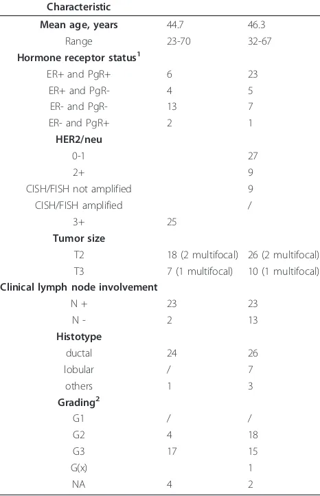
Table 1 Patients’characteristics
Characteristic
Mean age, years 44.7 46.3
Range 23-70 32-67
Hormone receptor status1
ER+ and PgR+ 6 23
ER+ and PgR- 4 5
ER- and PgR- 13 7
ER- and PgR+ 2 1
HER2/neu
0-1 27
2+ 9
CISH/FISH not amplified 9
CISH/FISH amplified /
3+ 25
Tumor size
T2 18 (2 multifocal) 26 (2 multifocal) T3 7 (1 multifocal) 10 (1 multifocal) Clinical lymph node involvement
N + 23 23
N - 2 13
Histotype
ductal 24 26
lobular / 7
others 1 3
Grading2
G1 / /
G2 4 18
G3 17 15
G(x) 1
NA 4 2
1
Hormone receptor status is significantly different between HER2+ and HER2-patients, with HER2-overexpressing cancers being mostly ER- and PgR- (P= 0.01).
CISH, chromogenicin situhybridization; ER, estrogen receptor; FISH, fluorescencein situhybridization; HER-2, human epidermal growth factor receptor-2; PgR, progesterone receptor.
2
and then cultured with peptide-loaded monocytes (50,000 cells/well) at 1:1 effector:target ratio. FLU M158-66 and unstimulated monocytes were used as positive and negative controls, respectively. Cells were seeded onto ELISPOT capture plates in triplicates and incubated for 48 hours at 37°C with 5% CO2. All plates
were evaluated by a computer-assisted ELISPOT reader (Eli.Expert, A.EL.VIS GmbH, Hannover, Germany). The number of spots in negative control wells (range of 0 to 5 spots) was subtracted from the number of spots in sti-mulated wells. Responses were considered significant if a minimum of five IFN-gproducing cells were detected in the wells.
Cytokine detection
Levels of IL-1a, IL-1b, IL-2, IL-6, IL-8, IL-10, IL-12p70, TNF-a, and granulocyte macrophage colony-stimulating factor (GM-CSF) were evaluated using the SearchLight® multiplex arrays (Food and Drug Administration approved, Aushon Biosystems, TEMA Ricerca, Bologna, Italy) according to the manufacturer’s instructions. Briefly, custom human 8-plexarray and human 1-plexarray (for GM-CSF detection) with pre-spotted cytokine-specific antibodies were used. Standards or pre-diluted samples were added in duplicate and, after one hour of incubation at room temperature and three washes, biotinylated anti-body reagent was added to each well. After 30 minutes incubation at room temperature and three washes, block solution was added to stabilize the signal. The addition of Streptavidin-HRP Reagent and SuperSignal®Substrate, and the acquisition of luminescent signal with a cooled CCD (Charge Coupled Device) camera, together with data analysis and processing, were performed by TEMA Ricerca laboratories’customer service (Bologna, Italy).
Transforming growth factor (TGF)-b1 serum levels were assessed through DRG TGF-b1 ELISA (DRG Instruments GmbH, Marburg, Germany) under instructions. Pre-diluted samples and standards underwent appropriate acidification and neutralization before testing. Briefly, pre-treated standards, controls and samples were dispensed into wells in duplicate and plates were incubated overnight at 4°C. After three washes, antiserum was added to wells and incubated for 120 minutes at room temperature, plate was rinsed three times and anti-mouse biotin (enzyme conjugate) was dispensed and incubated for 45 minutes. After three washes, enzyme complex was added to wells, then plates were incubated 45 minutes and washed three times. After the addition of substrate solution for 15 min-utes, the reaction was stopped and the adsorbance at 450 ± 10 nm was determined with a microtiter plate reader (Bio-Tek Instruments, Winooski, VT, USA). As there is an extremely variable range of normal values reported in the literature, healthy women serum levels were taken as references.
Immunohistochemistry
Considering that the diagnostic biopsy is not fully repre-sentative of the whole tumor mass, the lymphoid infiltrate was investigated in 40 primary advanced breast carcino-mas from a separate series of patients who underwent sur-gical resection during the past decade at our institution. Twenty cases were HER-2+(3+) and 20 HER-2-(0 or 1+). All specimens were routinely fixed in 10% buffered forma-lin, embedded in paraffin and then stained with H&E for histological examination. For immunohistochemical ana-lyses, 2 to 3 µm serial sections of primary tumors were processed with automated immunostainer Benchmark XT (Ventana, Tucson, AZ, USA), and staining was carried out with the following antibodies: CD8 (clone SP57, Ventana Medical System, Tucson, AZ, USA); FoxP3 (clone 259D/ C7, BD Pharmingen, Franklin Lakes, NJ, USA) diluted 1:100 e TiA-1 (clone TiA-1, Bioreagents, Golden, CO, USA) diluted 1:100. Nuclear counterstaining was accom-plished with Harris’hematoxylin. Omission of the primary antibody was used as a negative control. The results for staining were evaluated with reference to the number of unequivocally stained lymphoid cells. Ten randomly cho-sen reprecho-sentative microscopic fields were counted at 40x original magnification.
Statistical analysis
Chi-square test was used to compare hormone receptor (HR) expression and grading within HER2-and HER2+ populations. Data obtained from multiple independent experiments were expressed as mean and standard devia-tion for immunophenotypic analysis and ELISPOT assays; cytokine box plots were obtained with SigmaPlot. The Stu-dent’sttest for two tailed distributions and the Wilcoxon two-sample test were used for the statistical analysis of the data. Odds ratio and 95% confidence intervals in multivari-ate analysis were performed to assess the possible influence of clinico-pathological variables on the immunological cor-relations observed: immunological variables (cytokine levels) were divided into quartiles according to their con-centrations and then stratified for HR expression (estrogen receptor (ER)- progesterone receptor (PgR)- and ER+ and/ or PgR+), and tumor grading (G2 or G3). Wilcoxon rank-test was used to compare the distribution of intratumor CD8+
and FoxP3+cells between HER2+and HER2-cases. Results were considered to be statistically significant when
P≤0.05 (two-sided).
Results
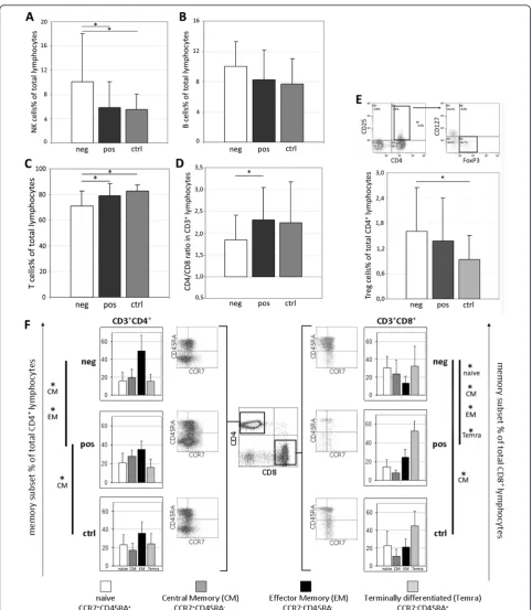
HER2+and HER2-patients exhibit a different distribution of circulating and intratumor immune cells
17 healthy women, who were considered as controls. A significantly lower percentage of CD3+ T cells (Figure 1c) was observed in HER2-patients with respect to both HER2+ cases (P= 0.028) and controls (P = 0.0003). In parallel, among the CD3-cell populations, higher num-bers of CD16+CD56+ NK cells were detected in HER2 -cases compared with HER2+ patients (P = 0.049) and healthy donors (P= 0.025; Figure 1a). Interestingly, no major difference in the distribution of circulating CD3 -cells was observed between HER2-overexpressing patients and controls. The percentage of B cells (Figure 1b) was not significantly different among the three groups investigated, even if HER2- (n = 14) patients showed slightly higher numbers of CD3-CD19+ cells than HER2+ patients (n = 15) and donors (n = 13; P= 0.07).
When the CD3+population was considered separately, HER2-patients showed significantly higher percentages of CD8+ T cells (P = 0.028; data not shown) and a lower CD4+/CD8+ ratio (Figure 1d;P = 0.046) when compared with HER2+ cases. Because of the different contribution of memory subsets in mediating antitumor immune responses [17-19], the differentiation state of T cell was investigated through the combined analysis of the chemokine receptor CCR7 and the CD45RA iso-form, to distinguish CCR7+CD45RA+ naïve, CCR7
+
CD45RA-central memory (CM), CCR7-CD45RA- effec-tor memory (EM), and CCR7-CD45RA+ terminally dif-ferentiated (Temra) cells [20]. Although the three studied groups showed a similar distribution of memory CD3+ T cell subsets (not shown), separate analysis of the CD4+and CD8+ compartments disclosed remarkable differences. In fact, compared with controls, HER2+ patients carried a higher percentage of CM CD4+ T cells (P = 0.003), whereas HER2- cases showed signifi-cantly higher numbers of CM CD8+T lymphocytes (P= 0.022). Moreover, a higher percentage of EM CD4+cells (P = 0.023), at the expense of the CM subset (P = 0.023), was found in HER2-cases compared with HER2+ patients (Figure 1f). Finally, the two groups of breast cancer patients showed a completely different memory sharing among CD8+ cells, with a prevalence of naïve (P
= 0.002) and CM cells (P = 0.005) in HER2-cases and higher percentages of EM (P = 0.005) and Temra cells (P= 0.012) in HER2+patients.
Several studies argued the unfavorable involvement of circulating regulatory T cells (Tregs) in cancer progres-sion, demonstrating the presence of increased numbers of CD4+
CD25highFoxP3+especially in metastatic cancers [21]. Considering that this immunophenotypic charac-terization is unsuitable for uniquely defining this specia-lized T cell subset, we used IL-7 receptor (CD127) down-regulation as a further feature indicative of sup-pressive functions [22] and identified Tregs as CD4
+
CD25highCD127lowFoxP3+ cells. The analysis showed that total numbers of so determined Tregs were not sig-nificantly different between patients and controls. Nevertheless, considering the two groups of patients separately, HER2-exhibited a significantly higher per-centage of circulating Treg cells (P= 0.02) when com-pared with healthy donors (Figure 1e).
Characterization of the lymphoid infiltrate in an unre-lated series of locally advanced breast cancers disclosed a significantly higher prevalence of intratumor CD8+ T cells in HER2+ cases (median 1000, range: 730 to 1880) as compared with the HER2- subgroup (median 234, range: 117 to 890, P = 0.04). TiA-1+ cells were also more abundant in HER2+ tumors and only rarely detected in the HER2-subgroup (not shown). Conver-sely, the median numbers of FoxP3+cells was higher in HER2+ cases (170, range: 50 to 508) than in HER2 -tumors (25, range: 10 to 108,P= 0.04; Figure 2).
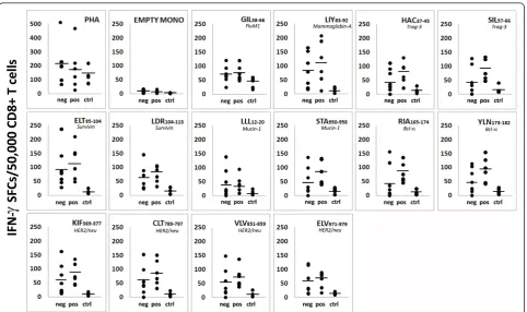
HER2+patients display enhanced CD8+T cell responses to different TAA-derived epitopes compared with both HER2-patients and healthy donors
Spontaneous CD8+ T cell responses to 13 TAA-derived peptides (Her2, muc-1, mam-A, trag-3, survivin, bcl-xL;
Table 2) were evaluated by IFN-gELISPOT assay in six HER2+and seven HER2-HLA-A*0201+patients and five HLA-A*0201+ age-matched healthy women. IFN-g -secreting CD8+T cells were detected in all samples (Fig-ure 3), although higher numbers of CD8+T cells speci-fic for all epitope peptides investigated were observed in both HER2+and HER2-patients compared with healthy donors (bothP<0.002). Notably, the number of circulat-ing TAA epitope-specific CD8+ T cells was higher in HER2+ cases compared with HER2-(P<0.005), particu-larly against peptides derived from trag-3, muc-1, and bcl-xL (Figure 3). Empty monocytes were considered as
negative controls and the number of spots was usually at the background level (<10 SFC/50,000 CD8+ cells). No significant differences were found between patients and donors against Flu M1 GIL58-66 peptide, used as
positive control (49<SFC/50,000 CD8+ cells>76), and similar levels of responses were also observed against PHA (167<SFC/50,000 CD8+ cells>221), confirming a retained T cell responsiveness.
HER2+patients show similar serum cytokine profile in
respect to donors
Figure 1Immunophenotypic characterization of peripheral blood lymphocytes. Percentages of (a) natural killer cells (NK; CD3-CD16+CD56+; neg = 20, pos = 17, ctrl = 17), (b) B cells (CD3-CD19+; neg = 14, pos = 15, ctrl = 15), (c) T lymphocytes (CD3+CD19-; neg = 20, pos = 17, ctrl = 17), (d) CD4+/CD8+ratio (CD3+CD4+/CD3+CD8+; neg = 20, pos = 17, ctrl = 17), and(e)regulatory T cells (Treg; CD3+CD4
+CD25highCD127lowFoxP3+; neg = 20, pos = 15, ctrl = 17) assessed in HER2-(neg), HER2-overexpressing (pos) patients and age-matched healthy
women (ctrl). (f) Differentiation (memory) status of CD3+CD4+and CD3+CD8+lymphocytes was investigated through CCR7 and CD45RA
that HER2-patients carried significantly lower amounts of IL-2 (P = 0.0222), IL-8 (P = 0.009), and IL-6 (P = 0.016) with respect to donors (Figure 4), whereas the cytokine profile of HER2+cases was almost superimpo-sable to that of healthy women (Figure 4). Notably, the most evident differences emerged from the comparison between the two groups of patients, with HER2- cases showing significantly reduced levels of IL-2 (P = 0.0229), IL-1b(P = 0.0207), IL-8 (P= 0.007), IL-6 (P= 0.0001), and IL-10 (P= 0.0247; Figure 4), independently
by other clinical-pathological parameters such as HR expression and tumor grading (P-trend in multivariate analysis: IL-1b P = 0.002, IL-2 P = 0.004, IL-6 P = 0.004, IL-8P= 0.02, IL-10P= 0.02; Table 3). No differ-ences were found in IL-1a, TNF-a, IL-12p70, GM-CSF, and TGF-bserum concentrations.
Discussion
Evidence accumulated so far indicates that the immune system can influence the initiation and development of cancer and it is widely believed that T lymphocytes represent the most potent antitumor effector cells. In this respect, there is an urgent need to develop thera-peutic approaches able to preserve or only minimally impair immune functions since ADCC-promoting thera-peutic antibodies, such as trastuzumab, and cancer vac-cines are being increasingly used as adjuvant and neoadjuvant treatment modalities [23]. Furthermore, also the therapeutic efficacy of some “conventional” drugs, such as doxorubicin and paclitaxel, involves immunomediated mechanisms [11,24]. On these grounds, we considered it clinically relevant to exten-sively investigate at diagnosis the immunological profile of locally advanced breast cancer patients who are can-didates for NC including immunomodulating drugs. The present study provides baseline immunological data that may constitute a reference for an informative monitor-ing of immune responses durmonitor-ing NC (ongomonitor-ing study). Provided the feasibility of monitoring antitumor responses during therapy and considering that sera from breast cancer patients should represent a valuable dis-covery tool to identify potential targets involved in breast cancer progression [25], we focused mainly on peripheral blood as the easiest accessible way to detect and measure immune-changes.
It is worth considering that most studies reporting analyses of systemic immunologic parameters in breast cancer patients included extremely variable series of cases, thus obtaining wide ranges of values and often conflicting results. Our study does not suffer from this limitation, being based on a relatively homogeneous group of patients including only locally advanced can-cers and excluding metastatic patients, which are the main contributors to outliers [26,27].
Previous data reported a significant increase in circu-lating B lymphocytes and NK cells in breast cancer patients in comparison with control groups [14], with a lower total number of T lymphocytes [15]. Interestingly, in our study, HER2+
[image:7.595.58.290.88.262.2]patients retained a normal distribu-tion in NK cells, and T and B lymphocytes if compared with healthy donors, whereas HER2- cases displayed lower percentages of T cells and higher numbers of NK and, to a lesser extent, of B cells. The increased NK cell numbers and activity reported in pre-treated patients
Figure 2Composite figure showing CD8 and FoxP3 expression in lymphoid cells infiltrating representative HER2-or HER2+
breast carcinomas. (a) Few CD8+cells are present within the
tumor, and infiltrate tumor nests in a HER2-case.(b)Some FoxP3+
cells infiltrate a HER2-breast carcinoma.(c)CD8+cells are numerous
and surround tumoral cords in a HER2+breast carcinoma.(d)
Higher numbers of FoxP3+cells infiltrate a HER2+case.(a to d)
Immunohistochemical stain; paraffin section; Hematoxylin counterstain;(a and c)20× original magnification,(b and d)40× magnification. HER2: human epidermal growth factor receptor-2.
Table 2 Library of immunogenic peptide epitopes used to evaluate CD8+ T cell responses to breast-cancer-associated antigens
Mammaglobin-A LIY83-92 LIYDSSLCDL 34
Trag-3 HAC37-45 HACWPAFTV 35
SIL57-66 SILLRDAGLV 35
Survivin ELT95-104 ELTLGEFLKL 36
LDR104-113 LDRERAKNKI 36
Mucin-1 LLL12-20 LLLLTVLTV 33
STA950-958 STAPPVHNV 33
Bcl-xL RIA165-174 RIAAWMATYL 13
YLN173-182 YLNDHLEPWI 13
Her2/neu KIF369-377 KIFGSLAFL 12
CLT789-797 CLTSTVQLV 12
VLV851-859 VLVKSPNHV 12
[image:7.595.57.291.554.712.2]were related to time to treatment failure [14] and were proposed to be the result of activation of the innate immunity by the tumor or dependent on a defective reg-ulation of NK cells in these patients [28]. Conversely, Dewan and colleagues observed significantly lower NK cell activity in PBMC from breast cancer patients, as compared with that of healthy individuals. Intriguingly, this defect was more pronounced in HER2- breast can-cer patients [15], suggesting an underlying NK dysfunc-tion in this subgroup. Further, we noticed that HER2+ patients showed an increased CD4/CD8 ratio with respect to HER2- cases, a feature previously associated with a better chance of responding to NC [14]. How-ever, opinions regarding which T-cell subset provides the best tumor protection, especially among memory sub-populations, are still controversial. Indeed experi-mental evidence suggests that central (TCM) and effector
(TEM) memory T cells can each confer a protective
advantage [17-19], with TCM providing a reservoir of
antigen-specific T cells, ready to expand and replenish the periphery upon secondary challenge, and TEM
dis-playing a more activated phenotype capable of granzyme
B and perforin expression, IFN-gsecretion, and tumor-specific killing in vitro [17]. Compared with healthy donors, our data disclosed a higher percentage of CD4+ TCM (CCR7+CD45RA-) in HER2+ patients and higher
numbers of CD8+ TCMin HER2-cases. This observation
may suggest the existence, in both groups, of an active, though predominantly memory T-cell driven, antitumor response which may benefit and respond to recall anti-gens from a cancer vaccine. Interestingly, in our study the main variations in memory subsets distribution were found between HER2- and HER2+ patients. Although the CD4+ T cell population of HER2+cases disclosed a favorable shift to the TCM phenotype, in these same
patients CD8+ T lymphocytes revealed a predominance of TEMand terminally differentiated cells. In contrast,
HER2- patients exhibit mainly CCR7+ CD8+ T cells (naïve and TCM). It should be considered that peripheral
CD8+T cells expressing effector functions against viral [29,30] or tumor antigens [31] are almost uniformly CCR7-and are endowed with full effector capacities, far greater than naïve or TCMcells. Moreover, as the
[image:8.595.58.541.87.373.2]differ-entiation to terminal effector cells is related to increased
Figure 3CD8+T cell responses to multiple breast cancer-associated antigenic epitopes assessed by IFN-g-ELISPOT
(interferon-g-Enzyme Linked Immunosorbent Spot). All tests were performed using CD8+purified T cells as effectors and autologous peptide-loaded
Figure 4Serum cytokine profile. Interleukin (IL)-2, IL-12p70, IL-1a, IL-1b, IL-8, IL-6, IL10 (neg = 36, pos = 25, ctrl = 23), Tumor necrosis factor-a (TNF-a; neg = 33, pos = 23, ctrl = 23), granulocyte-macrophage colony-stimulating factor (GM-CSF; neg = 33, pos = 23, ctrl = 19) and
cytolytic potential of CD8+ T cells, we hypothesize that the extent of maturation might be due to an effective antitumor response [18].
Pre-existing T cell responses to TAA have been reported in patients with solid tumors [32]; however, these responses usually involve a low frequency of anti-gen-specific T cells, not detectable in the majority of patients [18]. In this regard, literature data on circulat-ing tumor antigen-specific T cells in breast cancer patients are still conflicting, probably because of the pre-dominant focus on single epitopes [18]. Circulating T cells able to recognize CD8+ epitopes of HER2 [12], MUC-1 [33], mammaglobin-A [34], Trag-3 [35], survivin [36], or bcl-xL [13] have been described in distinct
papers, but the evaluation of multiepitopic antitumor responses is still lacking. We therefore assessed the amount of IFN-g-secreting CD8+ T cells specific for a broad spectrum of HLA-A*0201 peptides derived from Her2, muc-1, mam-A, trag-3, survivin, and bcl-xL.
Nota-bly, we found increased IFN-g release to all screened epitopes in the global cohort of patients if compared with healthy donors, demonstrating the existence of spontaneous T cell responses against multiple TAA in locally advanced breast cancer patients. The ability to stimulate the generation of antitumor CD8+ T cells seemed to be more pronounced in HER2+cancers, espe-cially towards Her2-, trag-3-, muc-1-, and bcl-xL-derived
epitopes. This peculiarity may be useful in the design and optimization of vaccine strategies, which could take
advantage of host’s pre-existing antitumor immune response. Moreover, the increased numbers of TAA-spe-cific circulating CD8+ T cells characterizing HER2+ patients may positively contribute to the clinical efficacy of trastuzumab, which is able to sensitize HER2-overex-pressing tumors to the killing by HER2-specific CTLs [7], and may enhance the antigen-specific immune responses promoted by doxorubicin and paclitaxel [37]. Our findings at the systemic level are also consistent with the demonstration of a significantly higher preva-lence of CD8+T lymphocytes infiltrating HER2+tumors that could contribute to a better clinical outcome [38]. This suggests that HER2 overexpression may be asso-ciated with enhanced immunogenicity of tumor cells and/or with a less immunosuppressive microenviron-ment. Further characterization of the activation state of lymphocytes infiltrating these tumors is, however, required to draw definitive conclusions in this respect.
[image:10.595.57.540.111.319.2]It is well recognized that tumors may down-regulate the immune response to tumor antigens by inducing several immune suppressor mechanisms, including Treg recruitment. Increased numbers of Tregs have been cor-related with greater disease burden and poorer overall survival [39]. In particular, Treg cells are augmented in the peripheral blood and within the tumor microenvir-onment in patients with breast carcinomas [40]. Our analysis of FoxP3 expression in intratumor lymphocytes disclosed significantly higher prevalence of FoxP3+ cells in HER2+ tumors. This may be a homeostatic
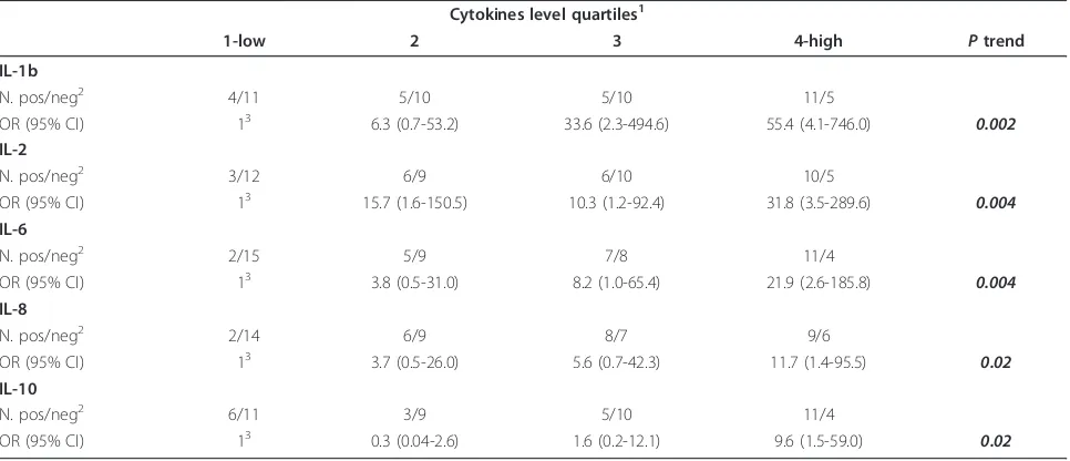
Table 3 Odds ratio (OR) and 95% confidence interval (CI) adjusted for hormone receptor expression and tumor grading according to cytokines levels in HER+ and HER- patients
Cytokines level quartiles1
1-low 2 3 4-high Ptrend
IL-1b
N. pos/neg2 4/11 5/10 5/10 11/5
OR (95% CI) 13 6.3 (0.7-53.2) 33.6 (2.3-494.6) 55.4 (4.1-746.0) 0.002 IL-2
N. pos/neg2 3/12 6/9 6/10 10/5
OR (95% CI) 13 15.7 (1.6-150.5) 10.3 (1.2-92.4) 31.8 (3.5-289.6) 0.004
IL-6
N. pos/neg2 2/15 5/9 7/8 11/4
OR (95% CI) 13 3.8 (0.5-31.0) 8.2 (1.0-65.4) 21.9 (2.6-185.8) 0.004
IL-8
N. pos/neg2 2/14 6/9 8/7 9/6
OR (95% CI) 13 3.7 (0.5-26.0) 5.6 (0.7-42.3) 11.7 (1.4-95.5) 0.02
IL-10
N. pos/neg2 6/11 3/9 5/10 11/4
OR (95% CI) 13 0.3 (0.04-2.6) 1.6 (0.2-12.1) 9.6 (1.5-59.0) 0.02
1
Quartiles were: IL-1b:≤0.5/0.6-1.7/1.8-4.2/≥4.3; IL-2:≤2.2/2.3-5.8/5.9-15.8/≥15.9; IL-6:≤1.6/1.7-3.4/3.5-6.5/≥6.6; IL-8:≤1.7/1.8-3.7/3.8-6.2/≥6.3; IL-10:≤ 0.4/0.5-1.4/1.5-3.3/≥3.4 pg/ml.
2
pos, HER2+; neg, HER2-; 3
Reference category.
consequence of the higher content of infiltrating CD8+ T cells detected in these cancers, or may reflect an active local recruitment of Tregs. It is worth considering in this respect that conflicting results have been reported with regard to Treg frequencies in HER2- ver-sus HER2+ breast cancers [27,41], discrepancies that could be due in part to tumor staging, but also to the different and often partial phenotypic markers used to identify regulatory T cells. This suppressor cell subset is often identified as CD4+CD25high cells [27] or as CD4
+
FoxP3+ lymphocytes [41], even if these markers are unable to uniquely define a regulatory T cell phenotype. Therefore, the use of FoxP3 as a single immunohisto-chemical marker to identify Tregs may have overesti-mated the number of Tregs in the HER2+ subgroup. In fact, this approach can not discriminate true FoxP3+ Treg cells from T lymphocytes activated by local stimuli and therefore transiently expressing FoxP3 without being endowed of immunosuppressive functions. To overcome these limitations, we have bona fide consid-ered as circulating Tregs only cells expressing the CD4
+
CD25highCD127lowFoxP3+phenotype, as the down-reg-ulation of the IL-7 receptor (CD127) is associated with suppressive functions [22]. This extended phenotypic definition did not disclose significant differences in Treg distribution between the whole group of breast cancer patients and controls, but it revealed higher numbers of circulating Tregs in HER2- patients with respect to donors. Tregs exert their suppressor activity by inhibit-ing T cell proliferation, NK cell-mediated cytotoxicity [42], and TAA-specific immunity [43]. On these grounds, the nearly physiological number of circulating Tregs displayed by stage II and III HER2+breast cancer patients may imply a favorable background for NK-involving therapy such as monoclonal antibodies (trastu-zumab), and may further benefit from the spontaneous enhanced antitumor T cell responses.
The likely retained immune proficiency of HER2+ patients is supported by an apparently unchanged cyto-kine profile, as sharpened by the comparison with serum cytokine levels of healthy women, which displays no significant differences. It is widely accepted that solid tumors are associated with a pathologic shift toward the T-helper type 2 cytokine pattern; whereas T-helper 1-induced inflammation inhibits tumor growth. In breast cancer patients, depressed serum levels of IL-2, GM-CSF, IFN-g and enhanced TNF-a and IL-6 amounts were reported in comparison with controls [11] and some of these immune dysfunctions are also present in early-stage tumors. In our study, we noticed significantly lower levels of IL-2 and IL-8, but also of IL-6 in HER2 -patients with respect to healthy women. Increased serum levels of IL-6 were previously reported in pro-gressive recurrent breast cancer patients [44], especially
in the presence of metastasis [26], conversely no meta-static cases were enrolled in our cohort. Interestingly, the comparison of cytokine layouts between HER2-and HER2+ patients disclosed pathogenically relevant differ-ences between the two groups. In particular, HER2 -patients showed lower levels of IL-2, previously asso-ciated with relapse of the disease [45,46], reduced amounts of the pro-inflammatory cytokines IL-1b and IL-6 (repressors of in vitro cell cycle progression) [47], and reduced levels of IL-8, recently reported also in the
in vitrocomparison of HER2-and HER2+breast cancers cell lines and in serum samples from metastatic breast cancer patients [25]. Finally, the pleiotropic cytokine IL-10, which may exert tumor-promoting activity or con-siderable antitumor effects, at low and high concentra-tions, respectively [46], was detected at lower amounts in HER2-patients. On the other hand, the higher levels of IL-2 in HER2+ patients may be consistent with an activation of T cells by TAA-derived peptides [48]. Notably, the differences in the cytokine levels observed between HER2-and HER2+ cases were independent of the clinical-pathological features showed by the two cohorts of patients (Table 3).
The different immunologic profile of patients with HER2-and HER2+ tumors highlights the importance of considering them as two distinct populations not only with regard to tumor characteristics, but also concerning their immune status. Our analysis, however, may show some limitations mainly due to the quite large number of immunological factors considered. This multipara-metric approach has considerably restricted the global case series, thus limiting the possibility to make compar-isons of adequate statistical power between subpopula-tions of HER2-and HER2+patients. Larger series should be therefore investigated to conclusively rule out the possible influence of distinct clinico-pathological vari-ables on the immunological correlations observed. Moreover, the research of TAA memory T-cell responses was confined at the peripheral immune com-partment; an in situ comparative survey is needed to confirm our data. An interesting issue that needs further investigation is the assessment of whether the higher percentage of effector memory CD8+T cells observed in HER2+ patients correlates with the enhanced response against TAA noticed in this population. In perspective, therefore, the characterization of relevant immune para-meters may also have a prognostic value, as recently emphasized by the finding that a decreased expression of immune response-associated genes was associated with poor prognosis, particularly in HER2+ cases [49].
immunological profile at diagnosis may be useful to individualize the most promising therapeutic choice or to further contribute in the therapeutic schedule’s design. Furthermore, since some chemotherapeutic drugs display beneficial effects on host immune func-tions [11,50], our results suggest that a careful immune-monitoring of breast cancer patients during NC may be useful to predict the response to therapy and to obtain a better prognostic definition.
Conclusions
In conclusion, our data indicate that, compared with HER2-cases, patients with HER2-overexpressing, locally advanced breast cancer show a more limited tumor-related immune suppression. This may account for the clinical benefit achieved in this subset of patients with the use of drugs acting through immune-mediated mechanisms. These findings also provide the rationale for further studies aimed at assessing the possible pre-dictive and/or prognostic role of immune markers in locally advanced breast cancer patients undergoing NC.
Abbreviations
ADCC: antibody-dependent cell cytotoxicity; APCs: antigen presenting cells; CISH: chromogenicin situhybridization; CTLs: cytotoxic T lymphocytes; ELISPOT: enzyme-linked immunosorbent spot; ER: estrogen receptor; FISH: fluorescencein situhybridization; FITC: fluorescein isothiocyanate; HER-2: human epidermal growth factor receptor-2; GM-CSF: granulocyte macrophage colony-stimulating factor; H&E: hematoxylin and eosin; HR: hormone receptor; IFN: interferon; IG: immunoglobulin; IL: interleukin; LAK: lymphokine activated killer; mam-A: mammaglobin-A; muc-1: mucin 1; NC: neoadjuvant chemotherapy; NK: natural killer cell; PBMCs: peripheral blood mononuclear cells; PBS: phosphate-buffered saline; PC: phycoerythrin-cyanine; PE: phycoerythrin; PgR: progesterone receptor; Tregs: regulatory T cells; TAA: tumor-associated antigens; TCM: central memory T cells; TEM:
effector memory T cells; TTemra: terminally differentiated T cells; TGF-β1:
transforming growth factor-β1; TNF-α: tumor necrosis factor-α; Trag-3: taxol-resistance associated gene 3; ULN: upper limit of normal.
Acknowledgements
The authors thank Miss S. Colussi for editing the manuscript and S. Bergamin, for his technical support. The study was supported in part by grants from the Associazione Italiana per la Ricerca sul Cancro (contract 10301 to R.D.).
Author details
1
Cancer BioImmunotherapy Unit, Centro di Riferimento Oncologico, IRCCS -National Cancer Institute, via Franco Gallini 2, Aviano (PN), 33081, Italy.
2Division of Medical Oncology C, Centro di Riferimento Oncologico, IRCCS
-National Cancer Institute, via Franco Gallini 2, Aviano (PN), 33081, Italy.
3Epidemiology and Biostatistics Unit, Centro di Riferimento Oncologico,
IRCCS - National Cancer Institute, via Franco Gallini 2, Aviano (PN), 33081, Italy.4Division of Pathology, Centro di Riferimento Oncologico, IRCCS
-National Cancer Institute, via Franco Gallini 2, Aviano (PN), 33081, Italy.
Authors’contributions
EM conceived the study, performed most of immunological experiments and drafted the manuscript. DM participated in the design of the study, performed ELISPOT experiments and contributed to draft the manuscript. ET, GM, SS, and DL collected and analyzed clinical data. EC and KM performed part of the experiments. RT carried out all statistical evaluations. TP and AC performed histopathological diagnosis and immunohistochemistry, and performed data analysis. AV reviewed and approved the final manuscript. DC
conceived the study and reviewed the manuscript. RD conceived and designed the study, performed data analysis and interpretation and reviewed the manuscript. All authors read and approved the final version of the manuscript for publication.
Competing interests
The authors declare that they have no competing interests.
Received: 9 August 2011 Revised: 16 September 2011 Accepted: 23 November 2011 Published: 23 November 2011
References
1. Mauri D, Pavlidis N, Ioannidis JP:Neoadjuvant versus adjuvant systemic treatment in breast cancer: a meta-analysis.J Natl Cancer Inst2005,
97:188-194.
2. Rouzier R, Mathieu MC, Sideris L, Youmsi E, Rajan R, Garbay JR, Andre F, Marsiglia H, Spielmann M, Delaloge S:Breast-conserving surgery after neoadjuvant anthracycline-based chemotherapy for large breast tumors.
Cancer2004,101:918-925.
3. Ferriere JP, Assier I, Cure H, Charrier S, Kwiatkowski F, Achard JL, Dauplat J, Chollet P:Primary chemotherapy in breast cancer: correlation between tumor response and patient outcome.Am J Clin Oncol1998,21:117-120. 4. Borg A, Tandon AK, Sigurdsson H, Clark GM, Ferno M, Fuqua SA,
Killander D, McGuire WL:HER-2/neu amplification predicts poor survival in node-positive breast cancer.Cancer Res1990,50:4332-4337. 5. Paik S, Hazan R, Fisher ER, Sass RE, Fisher B, Redmond C, Schlessinger J,
Lippman ME, King CR:Pathologic findings from the National Surgical Adjuvant Breast and Bowel Project: prognostic significance of erbB-2 protein overexpression in primary breast cancer.J Clin Oncol1990,
8:103-112.
6. Valabrega G, Montemurro F, Aglietta M:Trastuzumab: mechanism of action, resistance and future perspectives in HER2-overexpressing breast cancer.Ann Oncol2007,18:977-984.
7. Kono K, Sato E, Naganuma H, Takahashi A, Mimura K, Nukui H, Fujii H:
Trastuzumab (Herceptin) enhances class I-restricted antigen presentation recognized by HER-2/neu-specific T cytotoxic lymphocytes.Clin Cancer Res2004,10:2538-2544.
8. Lee LF, Hellendall RP, Wang Y, Haskill JS, Mukaida N, Matsushima K, Ting JP:
IL-8 reduced tumorigenicity of human ovarian cancer in vivo due to neutrophil infiltration.J Immunol2000,164:2769-2775.
9. Tong AW, Seamour B, Lawson JM, Ordonez G, Vukelja S, Hyman W, Richards D, Stein L, Maples PB, Nemunaitis J:Cellular immune profile of patients with advanced cancer before and after taxane treatment.Am J Clin Oncol2000,23:463-472.
10. Chan OT, Yang LX:The immunological effects of taxanes.Cancer Immunol Immunother2000,49:181-185.
11. Tsavaris N, Kosmas C, Vadiaka M, Kanelopoulos P, Boulamatsis D:Immune changes in patients with advanced breast cancer undergoing chemotherapy with taxanes.Br J Cancer2002,87:21-27. 12. Disis ML, Knutson KL, Schiffman K, Rinn K, McNeel DG:Pre-existent
immunity to the HER-2/neu oncogenic protein in patients with HER-2/ neu overexpressing breast and ovarian cancer.Breast Cancer Res Treat 2000,62:245-252.
13. Andersen MH, Reker S, Kvistborg P, Becker JC, Thor SP:Spontaneous immunity against Bcl-xL in cancer patients.J Immunol2005,
175:2709-2714.
14. Murta EF, de Andrade JM, Falcao RP, Bighetti S:Lymphocyte
subpopulations in patients with advanced breast cancer submitted to neoadjuvant chemotherapy.Tumori2000,86:403-407.
15. Dewan MZ, Takada M, Terunuma H, Deng X, Ahmed S, Yamamoto N, Toi M:
Natural killer activity of peripheral-blood mononuclear cells in breast cancer patients.Biomed Pharmacother2009,63:703-706.
16. Scheltinga SA, Williams F, van der Zwan AW, Rozemuller EH, Middleton D, Tilanus MG:HLA-A towards a high-resolution DNA typing.Tissue Antigens 1998,51:549-552.
17. Perret R, Ronchese F:Memory T cells in cancer immunotherapy: which CD8 T-cell population provides the best protection against tumours?
Tissue Antigens2008,72:187-194.
cancer patients have a distinct phenotype and cytokine signature.J Immunol2007,179:2627-2633.
19. Kilinc MO, Gu T, Harden JL, Virtuoso LP, Egilmez NK:Central role of tumor-associated CD8+ T effector/memory cells in restoring systemic antitumor immunity.J Immunol2009,182:4217-4225.
20. Sallusto F, Lenig D, Forster R, Lipp M, Lanzavecchia A:Two subsets of memory T lymphocytes with distinct homing potentials and effector functions.Nature1999,401:708-712.
21. Stanzer S, Dandachi N, Balic M, Resel M, Samonigg H, Bauernhofer T:
Resistance to apoptosis and expansion of regulatory T cells in relation to the detection of circulating tumor cells in patients with metastatic epithelial cancer.J Clin Immunol2008,28:107-114.
22. Liu W, Putnam AL, Xu-Yu Z, Szot GL, Lee MR, Zhu S, Gottlieb PA, Kapranov P, Gingeras TR, Fazekas de St GB, Clayberger C, Soper DM, Ziegler SF, Bluestone JA:CD127 expression inversely correlates with FoxP3 and suppressive function of human CD4+ T reg cells.J Exp Med 2006,203:1701-1711.
23. Arnould L, Gelly M, Penault-Llorca F, Benoit L, Bonnetain F, Migeon C, Cabaret V, Fermeaux V, Bertheau P, Garnier J, Jeannin JF, Coudert B:
Trastuzumab-based treatment of HER2-positive breast cancer: an antibody-dependent cellular cytotoxicity mechanism?Br J Cancer2006,
94:259-267.
24. Arlen PM, Gulley JL, Parker C, Skarupa L, Pazdur M, Panicali D, Beetham P, Tsang KY, Grosenbach DW, Feldman J, Steinberg SM, Jones E, Chen C, Marte J, Schlom J, Dahut W:A randomized phase II study of concurrent docetaxel plus vaccine versus vaccine alone in metastatic androgen-independent prostate cancer.Clin Cancer Res2006,12:1260-1269. 25. Vazquez-Martin A, Colomer R, Menendez JA:Protein array technology to
detect HER2 (erbB-2)-induced‘cytokine signature’in breast cancer.Eur J Cancer2007,43:1117-1124.
26. Salgado R, Junius S, Benoy I, Van DP, Vermeulen P, Van ME, Huget P, Dirix LY:Circulating interleukin-6 predicts survival in patients with metastatic breast cancer.Int J Cancer2003,103:642-646. 27. Perez SA, Karamouzis MV, Skarlos DV, Ardavanis A, Sotiriadou NN,
Iliopoulou EG, Salagianni ML, Orphanos G, Baxevanis CN, Rigatos G, Papamichail M:CD4+CD25+ regulatory T-cell frequency in HER-2/neu (HER)-positive and HER-negative advanced-stage breast cancer patients.
Clin Cancer Res2007,13:2714-2721.
28. Mozaffari F, Lindemalm C, Choudhury A, Granstam-Bjorneklett H, Helander I, Lekander M, Mikaelsson E, Nilsson B, Ojutkangas ML, Osterborg A, Bergkvist L, Mellstedt H:NK-cell and T-cell functions in patients with breast cancer: effects of surgery and adjuvant chemo- and radiotherapy.
Br J Cancer2007,97:105-111.
29. Chen G, Shankar P, Lange C, Valdez H, Skolnik PR, Wu L, Manjunath N, Lieberman J:CD8 T cells specific for human immunodeficiency virus, Epstein-Barr virus, and cytomegalovirus lack molecules for homing to lymphoid sites of infection.Blood2001,98:156-164.
30. Hislop AD, Gudgeon NH, Callan MF, Fazou C, Hasegawa H, Salmon M, Rickinson AB:EBV-specific CD8+ T cell memory: relationships between epitope specificity, cell phenotype, and immediate effector function.J Immunol2001,167:2019-2029.
31. Valmori D, Scheibenbogen C, Dutoit V, Nagorsen D, Asemissen AM, Rubio-Godoy V, Rimoldi D, Guillaume P, Romero P, Schadendorf D, Lipp M, Dietrich PY, Thiel E, Cerottini JC, Lienard D, Keilholz U:Circulating Tumor-reactive CD8(+) T cells in melanoma patients contain a CD45RA(+)CCR7 (-) effector subset exerting ex vivo tumor-specific cytolytic activity.
Cancer Res2002,62:1743-1750.
32. Lurquin C, Lethe B, De PE, Corbiere V, Theate I, van BN, Coulie PG, Boon T:
Contrasting frequencies of antitumor and anti-vaccine T cells in metastases of a melanoma patient vaccinated with a MAGE tumor antigen.J Exp Med2005,201:249-257.
33. Finn OJ, Jerome KR, Henderson RA, Pecher G, Domenech N, Magarian-Blander J, Barratt-Boyes SM:MUC-1 epithelial tumor mucin-based immunity and cancer vaccines.Immunol Rev1995,145:61-89. 34. Jaramillo A, Majumder K, Manna PP, Fleming TP, Doherty G, Dipersio JF,
Mohanakumar T:Identification of HLA-A3-restricted CD8+ T cell epitopes derived from mammaglobin-A, a tumor-associated antigen of human breast cancer.Int J Cancer2002,102:499-506.
35. Meier A, Reker S, Svane IM, Holten-Andersen L, Becker JC, Sondergaard I, Andersen MH, Thor SP:Spontaneous T-cell responses against peptides
derived from the Taxol resistance-associated gene-3 (TRAG-3) protein in cancer patients.Cancer Immunol Immunother2005,54:219-228. 36. Andersen MH, Pedersen LO, Capeller B, Brocker EB, Becker JC, Thor SP:
Spontaneous cytotoxic T-cell responses against survivin-derived MHC class I-restricted T-cell epitopes in situ as well as ex vivo in cancer patients.Cancer Res2001,61:5964-5968.
37. Machiels JP, Reilly RT, Emens LA, Ercolini AM, Lei RY, Weintraub D, Okoye FI, Jaffee EM:Cyclophosphamide, doxorubicin, and paclitaxel enhance the antitumor immune response of granulocyte/macrophage-colony stimulating factor-secreting whole-cell vaccines in HER-2/neu tolerized mice.Cancer Res2001,61:3689-3697.
38. Mahmoud SM, Paish EC, Powe DG, Macmillan RD, Grainge MJ, Lee AH, Ellis IO, Green AR:Tumor-infiltrating CD8+ lymphocytes predict clinical outcome in breast cancer.J Clin Oncol2011,29:1949-1955.
39. Wolf D, Wolf AM, Rumpold H, Fiegl H, Zeimet AG, Muller-Holzner E, Deibl M, Gastl G, Gunsilius E, Marth C:The expression of the regulatory T cell-specific forkhead box transcription factor FoxP3 is associated with poor prognosis in ovarian cancer.Clin Cancer Res2005,11:8326-8331. 40. Liyanage UK, Moore TT, Joo HG, Tanaka Y, Herrmann V, Doherty G,
Drebin JA, Strasberg SM, Eberlein TJ, Goedegebuure PS, Linehan DC:
Prevalence of regulatory T cells is increased in peripheral blood and tumor microenvironment of patients with pancreas or breast adenocarcinoma.J Immunol2002,169:2756-2761.
41. Horlock C, Stott B, Dyson PJ, Morishita M, Coombes RC, Savage P, Stebbing J:The effects of trastuzumab on the CD4+CD25+FoxP3+ and CD4+IL17A+ T-cell axis in patients with breast cancer.Br J Cancer2009,
100:1061-1067.
42. Wolf AM, Wolf D, Steurer M, Gastl G, Gunsilius E, Grubeck-Loebenstein B:
Increase of regulatory T cells in the peripheral blood of cancer patients.
Clin Cancer Res2003,9:606-612.
43. Knutson KL, Dang Y, Lu H, Lukas J, Almand B, Gad E, Azeke E, Disis ML:IL-2 immunotoxin therapy modulates tumor-associated regulatory T cells and leads to lasting immune-mediated rejection of breast cancers in neu-transgenic mice.J Immunol2006,177:84-91.
44. Yokoe T, Iino Y, Morishita Y:Trends of IL-6 and IL-8 levels in patients with recurrent breast cancer: preliminary report.Breast Cancer2000,7:187-190. 45. Arduino S, Tessarolo M, Bellino R, Colombatto S, Leo L, Wierdis T, Lanza A:
Reduced IL-2 level concentration in patients with breast cancer as a possible risk factor for relapse.Eur J Gynaecol Oncol1996,17:535-537. 46. Rao VS, Dyer CE, Jameel JK, Drew PJ, Greenman J:Potential prognostic
and therapeutic roles for cytokines in breast cancer (Review).Oncol Rep 2006,15:179-185.
47. Shen WH, Zhou JH, Broussard SR, Freund GG, Dantzer R, Kelley KW:
Proinflammatory cytokines block growth of breast cancer cells by impairing signals from a growth factor receptor.Cancer Res2002,
62:4746-4756.
48. Saglam S, Suzme R, Gurdol F:Serum tumor necrosis factor-alpha and interleukin-2 concentrations in newly diagnosed ERBB2 (HER2/neu) positive breast cancer patients.Int J Biol Markers2009,24:142-146. 49. Staaf J, Ringner M, Vallon-Christersson J, Jonsson G, Bendahl PO, Holm K,
Arason A, Gunnarsson H, Hegardt C, Agnarsson BA, Luts L, Grabau D, Ferno M, Malmstrom PO, Johannsson OT, Loman N, Barkardottir RB, Borg A:
Identification of subtypes in human epidermal growth factor receptor 2–positive breast cancer reveals a gene signature prognostic of outcome.J Clin Oncol2010,28:1813-1820.
50. Carson WE, Shapiro CL, Crespin TR, Thornton LM, Andersen BL:Cellular immunity in breast cancer patients completing taxane treatment.Clin Cancer Res2004,10:3401-3409.
doi:10.1186/bcr3060