THE TE
M
PERATURE DEPENDE
N
C
E
OF SOME PHYSIOLOGICAL PROCESSES
Joe Wolfe
A thesis submitted for the degree of Doctor of Philosophy
at the Australian National University, Canberra
DECLARATION
The research reported in this dissertation is or_iginal and has not been submitted to any other University. It is my own work, with the following provisos:
Chapters
3and
4 andAppendix
3 are the result of a collaboration with David Bagnall of the Division of Plant Industry, C.S.I.R~O.Chap~er
5 reports work carried out in collaboration with Stjepan Marcelj a.The research reported in
Chapter 8
was conducted jointly by Emma Bates, Stjepan Marcelja and me.ACKNOWLEDGEMENTS
The research herein reported was supported financially by an A.N.U. Ph.D Scholar.ship and practically by the Department of Applied Mathematics and the Research School of Biological Sciences, A.N.U.,
and the Plant Industry Division of the Conunonwealth Scientific and Industrial Research O_rganization.
My advisors on this work were Jacob Israelachvili and Stjepan Marcelja. Jacob 1s a virtuoso experimentalist, Stjepan an inspired theoretician, both are great scientists, and both have a democratic and co-operative style which has allowed me the great opportunity of working
with
them (rather than for them, or under their direction). Barry Ninham, head of the department, has evinced a greatlyappreciated interest 1n my work and welfare. More importantly than· this, he has created an informal, incredibly vital, productive
multidisciplinary department which very possibly is, as so often
claimed; the Best in the World. The plant physiological work in this thesis (and much of the fundamental theoretical work related to it) would not have been possible without my long time collaborator David
Bagnall (whose thesis incidentally will be submitted within a week ·of this). I hope that he has gained ~s much from this collaboration as I have. As well as respected colleagues, these four people are my close friends. I thank them sincerely.
I have benefited from interaction with many others, principally including:
Liz Reymond, who involved me in the fascinating project recorded 1n Appendix 8;
Emma Bates - with whom Stjepan and I collaborated on research included in Chapter 8.
The A.N.U. boat club kept me sane and mediated much of my
collaborative research. Chapter 3 - and thence most of the thesis began in a double scull with Bagnall on the Nepean River, waiting for a race. Through the club I met Liz, and I have raced in .fours (and won!) with Barry and Stj epan.
Norma Chin typed this (and checked the equations) with her usual precision, and didn't growl at me once.
Finally, readers of this thesis will notice that I have serious differences of opinion with John Raison and Jim Lyons about several aspects of their research. I take this opportunity to thank John for some interesting and pleasant discussions, and to recognize the
DECLARATION
ACKNOWLEDGEMENTS ABSTRACT
CONVENTIONS AND SYMBOLS USED
CONTENTS
CHAPTER 1. BIOLOGICAL MEMBRANES - AN INTRODUCTION References
CHAPTER 2. THE EFFECT OF LOW TEMPERATURE ON PLANTS -IS THE MEMBRANE INVOLVED
2.1 · The Role of Membrane Lipid Fluidity 2.2 Membrane Deformability
2.3 Amphiphilic Properties
2.4 Surface Charges and Dipoles on Membranes 2.5 Water Ordering
2.6 Phase Transitions and Separations 2.7 Boundary Effects
References
CHAPTER 3. A RE-ASSESSMENT OF THE RAISON-LYONS THEORY OF CHILLING DAMAGE IN PLANTS
3.1 Experimental Details
3.1.1 Whole Plant Growth Measurement 3.1.2 Chlorophyll Determination
3.1.3 Hypocotyl and Radicle Extension 3.2 Results
3.2.1 Chilling Injury
3.2.2 Chlorophyll Formation
3.2.3 Hypocotyl and Radicle Extension
3.3 Discussion
3.4 Theory
3.5 Conclusions
3.5.1 Microscopic Features 3.5.2 Macroscopic Features References
CHAPTER 4. THE DEPENDENCE ON TEMPERATURE AND IRRADIANCE OF ·CHLOROPHYLL ·PRODUCTION. IN VIGNA RADIATA 4.1 Experimental Details
4.2
Results4.3 Discussion
References
CHAPTER 5 . . PHYSICAL PROPERTIES OF LIPID BILAYERS IN THE PHASE SEPARATION REGION
5.1 Introduction
5. 2 · Domain Boundary Ene_rgy
5.3 Lateral Compressibility of Bilayers Formed from Binary Mixtures
5.4 Discussion
References
CHAPTER 6. SURFACE ENERGIES AND ELASTICITIES IN BILAYERS AND MONOLAYERS
6.1 The Balance of Surface Energies
6.2 Complications to the Surface Energy Argument
6.3 The Equivalent Monolayer Pressure 6.4 Membrane Elasticity
6.5 Experiments on Bilayers
6.6 Experiments on Biological Membranes
6.7 Summary References
CHAPTER 7. THE SURFACE ENERGY OF LIPID BILAYERS
7.1 Theory
7.2 Experimental Details 7.3 Results
7.4 Discussion References
CHAPTER 8. MEMBRANE TRANSPORT AND THE PHYSICAL PROPERTIES OF LIPID BILAYERS WITH INTRINSIC PROTEINS
8.1 Passive Diffusion Models 8.2 Protein-Lipid Interaction
8.3 The Enzyme Partitioning Model of Thilo, Trauble and Overath ·
8.4 The Compressibility of Protein-Lipid Membranes 8.5 Compressibility of Mixed Lipid Bilayers with
Intrinsic Proteins
8.6 Membrane Transport and Permeability
8.7 Biological Membranes and Enzyme Activity
8.8 Conclusions References
APPENDIX 1. MEMBRANE MOLECULAR PACKING THEORY AND THE
84 86 89 93 94 94 96 98 101 103 110 113 113 115
PROLAMELLAR BODY 116
APPENDIX 2. BOLTZMANN STATISTICS, ENTROPY OF ACTIVATION
AND ARRHENIUS ' LAW 12 3
APPENDIX 3. STATISTICAL TESTS TO DECIDE BETWEEN STRAIGHT LINE SEGMENTS AND CURVES AS SUITABLE FITS TO
ARRHENIUS PLOTS OR OTHER DATA 128
APPENDIX 4. DERIVATION OF THE LATERAL COMPRESSIBILITY OF, TI-T ISOBARS FOR, AND TI-a ISOTHERMS FOR A
BINARY LIPID MIXTURE 140
APPENDIX 5. A DEMONSTRATION THAT DOMAINS OF FINITE SIZE
ARE UNSTABLE IN NON-DISSOCIATING LIPID BILAYERS 145
APPENDIX 6. THE THERMAL GRADIENT BAR 150
APPENDIX 7. CONSTRUCTION OF A VOLTAGE AMPLIFIER/CURRENT TO VOLTAGE CONVERTER WITH INPUT IMPEDANCE/
SENSITIVITY OF 1 TQ/1 fA 152
APPE DIX 8. BEHAVIOURAL DETERMINATION OF THE CONTRAST
SENSITIVITY FUNNCTION OF THE WF.DGE TAIL EAGLE 154
ABSTRACT
The finite temperature range of most biol.ogical activity cannot easily be explained by simple chemical kinetics. This thesis
investigates the proposition that temperature-induced changes in the physical properties of membranes affect their permeability, membrane transport and the activity of membrane-bound enzymes. In particular this principle is suggested as one of the factors involved in the low survival and growth rates of chilling~sensitive plants at low
temperatures. Other environmental factors are involved of course, and the interaction of temperature and irradiance is also investigated.
To analyse this complex system, one needs to perform whole plant experiments to determine physiological response to environmental
conditions; to postulate models for many of the molecular processes and investigate their statistical mechanics; to consider the physics and mechanics of lipid bilayer membranes and to measure some of their properties; and to understand an enormous quantity of biochemistry. All but the last are attempted.
Chapter 1 introduces the basic concepts and reviews some of the literature on bilayer lipid membranes.
In Chapter 2 the broad problem of the temperature dependence of enzyme activity is posed and several general -mechanisms are proposed whereby physical changes in membrane lipids may affect this activity.
In Chapter 3 whole plant growth experiments are reported and a currently popular theory of chilling injury is reassessed.
Chapter 4 reports the interaction of temperature and irradiance on one key activity, the production of chlorophyll.
been implicated by several researchers in both the activity of enzymes
and trans-membrane transport, this property is investigated
thermodynamically.
Chapter 6 continues the physical analysis of the mechanics of
bilayers to examine the applicability of the concepts of elasticity
and surface free energy of formation.
Chapter 7 returns to experiments which resolve the dilemma posed
1n Chapter 6 for the case of the bilayer formed on a septum. Such a system displays a surface free energy of formation.
In Chapter 8 are examined some physical properties of a model
membrane containing two species of lipids which interact with
intrinsic proteins. These prope~ties are related to enzyme activity.
Appendix 1 applies the theory of membrane packing geometry
(subsumed throughout the thesis) to the case of the transition between
the ornate geometry of the prolamellar body and the thylakoid larnellae.
Appendix 2 discusses the basis of Arrhenius' law and the
application of Boltzmann statistics to some models of enzyme kinetics.
Appendix 3 develops some statistical tools used in Chapter 3.
Appendix 4 describes in detail the compressibility calculations
used 1n Chapter 5.
In Appendix 5 it is demonstrated that finite domains are not
formed in the coexistence range of temperature in an uncharged lipid bilayer.
Appendices 6 and 7 describe the design of some equipment
developed for use in the experimental chapters.
Appendix 8 is a paper describing the relative contrast
I I
CONVENTIONS
The S.I. is adopted for units throughout. For users of .c.g.s. a useful conversion for use in Chapters 5, 6 and 7 is that 1 mN.m-1 =
1 dyne/cm. Wherever the symbol Tis used it denotes the thermodynamic temperature, however temperatures are_ given in
°
C in experimentalsections, and for comparison with experimental values. Chemical
quantities are given per molecule, rather than per mole; and energies per molecule are often expressed as their ratio to the thermal energy:
1 kCal/Mole corresponds to 1.69 kT per molecule at 300 K where k is Boltzmann's constant.
The convention used for the definition of statistical mechanical
quantities is that used by Landau and Lifshitz (1969] whereunder the chemical potentials of molecules in chemical equilibrium are equal,
and the free energy of a system is changed by an amount equal to the amount of work done on it.
References are listed in alphabetical order at the end of each
chapter. Since several appendices are somewhat self-contained
(especially those adapted from papers published, or in preparation), references also follow some of the appendices.
SYMBOLS USED
A the area of a bilayer, and:
the pre-exponential factor 1n Arrhenius' law
a area per lipid molecule in the plane of the membrane B a temperature independent constant in Arrhenius' law
b the radius of a hole in a septum D the optical density
E an energy
G the free energy of a system
g the free energy per molecule
H enthalpy
H* enthalpy of activation
h Planck's constant
.
1. current
K an elastic constant
K a dissociation constant
k
k a
Boltzmann's
the elastic
constant
modulus of area of
L a number defined in Appendix 5
a membrane
m a number of states in Chapter 2, and:
the mass of a lipid molecule in Appendix
N a number of molecules, usually, but
a number of lines in Chapter 3
n a number of states
n' the number density of molecules
P bulk pressure
p a number of points
Q
a ratio of areas5
q the number of points defined in Appendix 3 R a reaction rate
r a radius
S entropy
S* entropy of activation
T the (thermodynamic) temperature
T a critical or transition temperature C
T* a temperature defined in Chapter 8
u the order parameter in the theory of Owicki et al. V displaced volume
v electric potential
W a constant in the Landau-de Gennes expansion of g X a number fraction of a system
x a number fraction 1n one phase of a system
a. a "quantum area" defined in Appendix 5
y surface energy per unit area, or surface tension
~ the change 1n e: an en e_r gy
n the order parameter in Marcelja's theory (Chapter 8) and
the number density of an ideal gas (Appendix 5)
a
half the angle subtended by the membrane (in Chapter 7) K lateral compressibilityA the decay length of order 1n a bilayer
µ the chemical potential of a molecule
µ0 the reference free energy of a molecule IT the lateral pressure
To every problem, however complicated, there is
a simple, elegant solution which one will discover
if one looks hard enough~ This solution will turn
out to be wrong.
BIOLOGICAL
MEMBRANES
- AN INTRODUCTION
A membrane is a thin film separating two fluid, essentially
aqueous compartments. Since the two compartments normally have
different composition, the most profound function of a membrane is to
prohibit or limit their mixing - or actively maintain the
compositional asymmetry. Further, potential energy is stored in the
form of concentration and electrostatic potential differences
[Mitchell 1966; Hinkle and McCarty 1978] existing across cellular
and intracellular membranes, as is -neural information. Thus the
mechanical energy available from muscles and the information from a
brain exist -in the controlled permeability and biochemical activity o.f
membranes. Without this energy storage, and the storage and
partitioning capability of membranes, we are an homogeneous o_rganic
soup.
There are two great steps in the evolutionary origin of life:
the development of automatically self-replicating macromolecules, and
the formation of a membrane to include required nutrients and exclude
the rest - to separate the livi_ng from the non-living [Hargreaves
et
al. 1977].
As well as opposing mixing, membranes actively demix, i.e.
produce highly specific concentration gradients (at the expense of
energy, of course). So an enormous variety of the cell's chemical
machinery is embedded in the thin wall of this tank separating
products from reagents. The two most chemically active organelles are
the energy transduci~g mitochondrion and the solar energy capturing
chloroplast. Significantly, these organelles comprise little more
than stacks of membranes. (Membrane structures in the chloroplast are
examined in Appendix 1.)
Through their membranes do cells interact - exchanging molecules
case.of nerve cells, conducting along that membrane information coded in voltage spikes and passing it from cell to cell at synapses-regions where two cell membranes are in close proximity.
Membranes are crucial to life, and germane to the understanding of a· great range of biological processes. It will be proposed in this thesis that life in general requires of membranes such a stri~gent set of properties that the specifications can only be met under certain narrow environmental conditions of pressure and electrolyte
concentrations, and only over a finite range·of temperatures. The study of these properties is clearly important.
1.1
MEMBRANES
- LIPIDS
AND PROTEINS
Overton [1899] inferred that, since the rate of permeaiion of many substances through cellular membranes and through lipids were similar, lipids could constitute an important functional part of such membranes. Langmuir [1917] studied the formation of monomolecular
layers of lipids at the interface of air and water. In such layers the molecules align with their polar or hydrophilic "heads" towards the aqueous phase and their non-polar or hydrophobic hydrocarbon
"tails" towards the non-polar phase (air). Thus, from the observation that lipids from the membranes of erythrocytes could form a monolayer roughly twice the area of the membrane, Gorter and Grendel [1925]
concluded that erythrocyte membranes included a bilayer of lipid
molecules. In such a bilayer the tails of two monolayers adjoin, and the heads face the two aqueous phases thus separated.
Danielli and Davson [1935] conceptually attached globular proteins to such a monolayer, and since then different models have been proposed for the way lipids and proteins assemble to form a
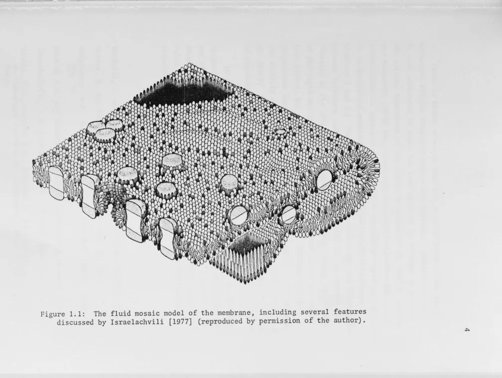
bilayer [reviewed for example by Ti Tien 1974]. Various experimental developments led Singer and Nicholson to propose in 1972 a picture of membranes as a "fluid mosaic" of lipids and proteins, with the lipid molecules more or less oriented normal to the membrane, but free to
representation of such a membrane is shown in Fig. 1.1.
The chemical properties of lipids are sometimes important
(particularly e.g. chlorophyll), but in this thesis I am principally
concerned with their physical properties. Membrane proteins however include most of a cell's enzymes - the organic catalysts by whose
_agencies do biological reactions acquire their remarkable efficiency and specificity. The provision of a membrane environment necessary for the chemical processes of a cell enhances the importance of membranes and allows the physical properties of membrane lipids to affe~t cellular physiological processes.
Lipids are relatively simple biological molecules and their interactions (in particular that interaction which forms bilayers) have been well studied [see review ·by Papahadjopoulos 1973]; however, this is less true of proteins. Proteins are much more complicated molecules whose structures are generally not known. How do they become integral units in a membrane?
A protein is a long and often intricately folded polypeptide
chain. Different peptide groups (some hydrophobic, some hydrophilic) are arranged sequentially to give the primary structure of the protein. This long chain can coil to form local structures - for instance
helices - which are called its secondary structure. The resultant
lumped and coiled chain is then "folded up" (relatively compactly) to form its tertiary structure [see e.g. Levitt 1976]. We must assume
[Israelachvili 1977] that intrinsic membrane-spanning globular
proteins are so constructed in their primary, secondary and tertiary structures that they expose a largely hydrophobic band, about 4 nm
wide, and are elsewhere on the surface largely hydrophilic in order to explain their ready incorporation into the lipid bilayer. (The
tertiary structure may even be "hollow" or toroidal and thus create a
hydrophi 1 ic channel through the membrane.) Alternative tertiary
structures are possible, and it may be that some proteins, when
removed from the membrane into aqueous solution reconform to replace
their hydrophobic surface with more hydrophilic groups, or that
protei~ reconformations are induced by changes in the properties of
~
[image:17.1127.107.1115.13.771.2]There is direct evidence that the physical state of the lipids 1n
membranes affects transport across the membrane - permeability of
E. coli membranes to sugar has been observed to increase at or near
the mel ti_ng transition of the lipids by Linden et al. [ 1973], and Wu
and McConnell [1973] have shown that valinomycin-mediated potassium
conductivity is increased over the phase-separation r.egion of lipids
in an artificial membrane. (Further references and discussion in
Chapters 5 and 8.) There is also direct evidence that the interaction
between intrinsic membrane proteins and membrane lipids affects the
fluidity of the latter [ e .. g. Jost et al. 1973] and alters the nature
of the lipid melting phase transition [Papahadjopoulos et al. 1975].
Less direct, but still I believe convincing, is evidence for the
proposition that the physical state of the surrounding lipids affects
the activity of membrane bound enzymes [Thilo et al. 1977; Overath et
al. 1976]; it is already well established that the presence of lipids
is required .for the activity of many enzymes [e.g. Jacobs et al. 1967].
It will be argued 1n this thesis (Chapters 2, 3 and 8) that the
temperature dependence of the physical properties of membrane lipids
and of their interaction with membrane bound enzymes is in part
responsible for the temperature dependence of biological reaction
rates and therefore physiological processes and even survival of
o_rganisms.
Danielli, J. and Davson, H.A. (1935). J. Cell Comp. Physiol.
l,
495.Gorter, E. and Grendel, F. (1925) . J. Exp. Med. 41, 439.
Hargreaves, W.R., Mulvihill, S.J. and Deamer, D.W. (1977). Nature,
266, 78.
Hinkle, P.C. and McCarty, R.E. (1978). Scientific American 238, 104.
Israelachvili, J.N. (1977). Biochim. Biophys. Acta 469, 221.
Jacobs, E.E., Andrews, E.C., Cohlrab , B. and Cunningham, \II. (1967).
In "Structure Function of Cytochromes", Univ. of Tokyo Press,
Tokyo.
Langmuir, J. (1917). J. Amer. Chem. Soc. 39, 1848. Levitt, M. (1976). J. Mol. Biol. 104, 59.
Linden, C.D., Wright, K., McConnell, H.M. and Fox, C.F. (1973). Proc.
Nat. Acad. Sci. USA 70, 2271.
Mitchell, D. (1966). Biol. Rev. Camb. Phil. Soc.!!_., 445.
Overath, D., Thilo, T. and Trailble, H. (1976). · TIES August, 76, 186. Overton, E. (1899). Vjschr. Naturforsch. Ges. Zurich 44, 88.
Papahadj opoulos, D. (1973). In "Form and function of phospholipids". BBA Library~_. (Ansell, G.B., Hawthorne, J.N., and Dawson, R.M.C., eds.), Elsevier.
Singer, S.J. and Nicholson, G.L. (1972). Science 175, 720.
Thilo, L., Trailble, H. and Overath, P. (1977). Biochemistry 16, 1283.
CHAPTER 2
THE EFFECT OF LOW TEMPERATURE ON PLANTS
IS THE MEMBRANE INVOLVED?
While some aspects of the temperature dependence of biological
processes are applicable to both plants and animals, the dependence on
temperature of the growth rates and ability to survive of plants are
of particular practical interest.* A spectacular example is the cold
resistance of wheat: were the temperature of the northern hemisphere
to fall, then so would the maximum latitude at which this vital crop
can be grown, and the consequent severe reduction in the Canadian and
Russian wheat harvests would have serious global consequences. Even a
modest improvement in low temperature growth or survival rates of this
crop would make a la.rge increase in the world's annual supply of food. Less dramatically this is true of all crops, and temperature remains
one of the major environmental limitations to the world's agricultural
productivity.
There are two basically different ways in which plants are
damaged by low temperature. The first is freezing d(JJTlage: the
formation o·f ice crystals in the tissues may rupture membranes either directly or, more usually, osmotically, by concentrating the unfrozen
aqueous solutions in the cell [Steponkus 1979]. The second is
chilling damage, which cannot be so quickly explained. Many species
of plants die at chilling temperatures - i.e. 0-15 °C - and the only
fundamental explanations offered for this are (a) some vital process
stops at low temperature, or (b) a metabolic imbalance obtains at low
temperature - the rate of use a vital substance exceeds its production,
* With the exception of fish, those animals whose growth and
survival evokes a commercial interest are generally homeotherms, so it is their gross physiological, rather than biochemical, response to low temperatures that is of practical interest. However, the productivity of commercial livestock is in many cases dependent on the cold
or the rate of production of a toxic' substance exceeds its removal. Though the symptoms and causes of chilling damage are various [Lyons 1973; Slack, Roughan and Bassett 1974; and Wr.ight and Simon 1973] the event usually associated with irreversible tissue damage is a
substantial increase in membrane permeability [Murata and Tatsumi 1979; · Christiansen 1979] or, as in freezing injury, lysing of the membrane
[Yoshida et al. 1979]. (Plants may also die or incur inJury at sub-zero temperatures by the mechanism of chilling damage rather than that of freezi.ng darn.age, that is, the membrane might be lysed by the
products of metabolic imbalance rather than by ice formation, but this distinction is rarely made.) Some examples of commercially important chilling-sensitive plants are: corn, sorghum, sugar cane, sweet
potato, capsicum, tomatoes and citrus fruits. Their low survival rate at low temperatures produces such important economic consequences as occasional total crop failure [McWilliam 1979], reduced yield [Downes and Marshall 1971] or damaged and unsaleable products [Lyons 1973]. A further characteristic of such crops which limits their bounty is an unexpectedly low growth rate at low but not fatal temperatures [Evans
et al. 1964; . Ivory and Whiteman 1978a] . In this chapter will be discussed the possible molecular mechanisms which may produce both these characteristics.
A metabolic imbalance can be caused by the differentially temperature-dependent rates of competing reactions. An obviously important example of this is the photo-destruction and production of chlorophyll. The former has a rate which is principally dependent on light intensity only, while the latter has a rate which decreases markedly with temperature. At a given light intensity there is some minimum temperature at which a plant can achieve a net positive
production of chlorophyll, "green up" and grow (discussed more fully in Chapter 4). This is not the whole answer: non-chlorophyllous tissue such as fruit can shO\v chilling damage [Lyons 1973], for example. Further, many other environmental conditions affect the
considerations are discussed 1n depth by Bagnall [1979].
Consequently, there 1s no single temperature above which some
species always lives and below which it always dies. Nevertheless,
for a given set of conditions there is a narrow temperature range over
which growth rates - and chilling dam_age rates - change dramatically. Further, individual biol_ogical reactions, as wel 1 as gross
physiological processes, show this dramatic temperature dependence: a
reaction whose rate may decrease by one order of magnitude from 40 °C
to 10 °C decreases by many orders of magnitude from 10 °C to 5 °C
[e.g. Nolan and Smillie 1977] .clearly something more complicated than can be described by simple reaction kinetics of chemical reactions.
(One of the simplest laws of reaction kinetics is that of Arrhenius.
Since many descriptions of biological activities involve
embellish-ments on Arrhenius' law, a derivation of this law and some of its
consequences is presented in Appendix 2.)
Sharpe and De Michelle [1977] addressed the problem by extending
the model of Johnson
et al.
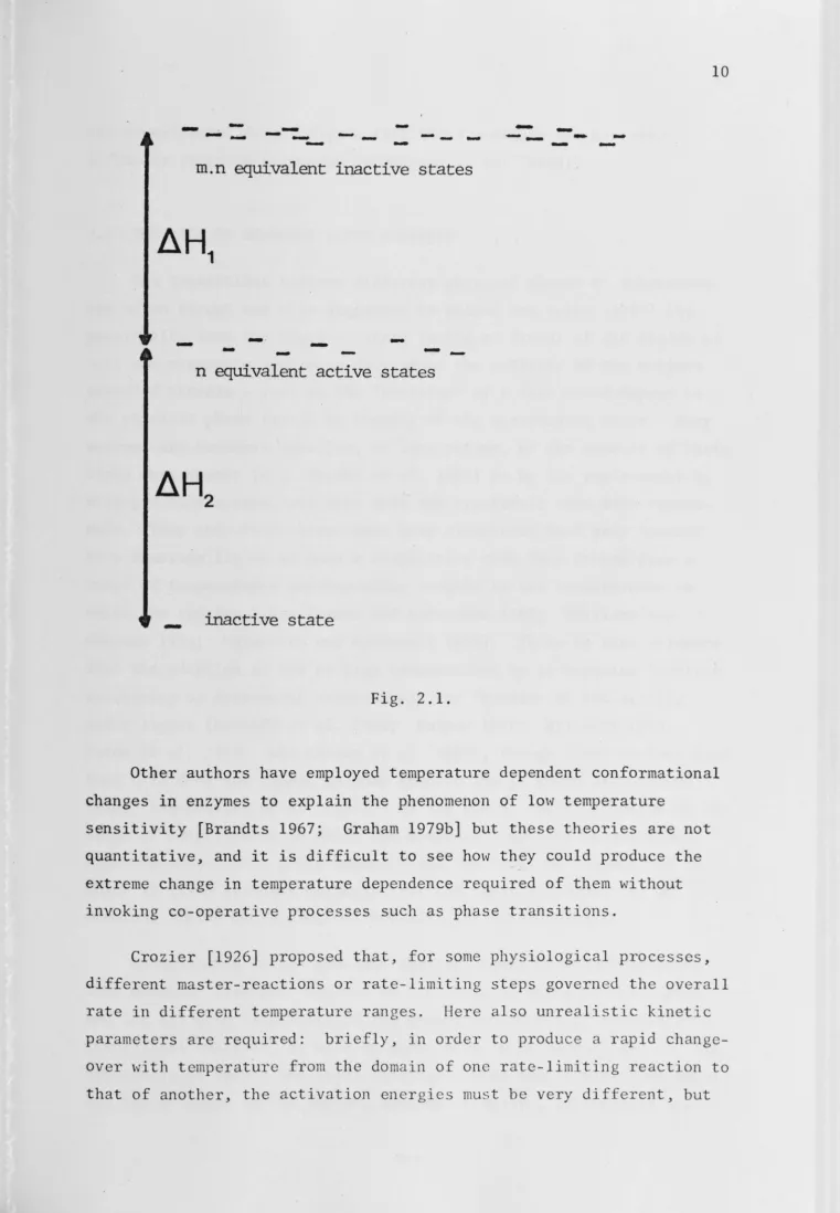
[1942]. They assign to the enzyme low temperature inactive, medium temperature active, and high temperatureinactive states into which enzyme molecules partition according to
Boltzmann statistics. This is pictured in Fig. 2.1, where then and
nm equivalent states at each energy level account for the "entropy of
activation" necessary to produce the large temperature dependence.
Sharpe and De Michelle do not suggest how these states might arise.
In order to produce the rather abrupt temperature dependence in the
biological measurements to which they fitted their model, Sharpe and
De Michelle used extremely large values for the energy of activation
(typically 100 kcal/mole, or 160 times the thermal energy) and entropy
of activation (typically ±200 cal/mole/K, i.e. n and mare of the
order of 1043) . This example is discussed in more detail in Appendix
2. Further, the number of adjustable parameters in this model is at
least six, and so its success in fitting sets of rather undemanding
biological data (most sets fitted have only six to eight points)
can-not be construed as support for this theory. When a many parameter
model requires improbably large values for all of its parameters, it
-
-- -
-
-
-
-
-
-
-
-
--
-
--
-
-
-
-
-
-m.n equivalent inactive states
-
-
-·
-
-
-
--n equivale--nt active states
-
inactive
state
Fig. 2.1.
Other authors have employed temperature dependent conformational
changes in enzymes to explain the phenomenon of low temperature
sensitivity [Brandts 1967; Graham 1979b] but these theories are not
quantitative, and it is difficult to see how they could produce the
extreme change in temperature dependence required of them without invoking co-operative processes such as phase transitions.
Crozier [1926] proposed that, for some physiological processes,
[image:23.788.10.772.25.1124.2]--the experimental data indicate that --these energies are not very different (this is discussed in Johnson et
al.
[1954]).2.1 THE ROLE OF MEMBRANE LIPID FLUIDITY
The transitions between different physical phases of substances are often abrupt and this suggested to Raison and Lyons [1970] the possibility that the physical state (solid or fluid) of the lipids of
cell and organelle membranes determined the activity of the enzymes embedded therein - just as the "activity" of a fish would depend on the physical phase (solid or liquid) of the surrounding water. Many enzymes are rendered inactive, or less active, by the removal of their lipid environment [e.g. Jacobs et
al.
1967] or by its replacement by stronger surfactants, and this made· the hypothesis even morereason-able. They and other researchers have discovered that many species have membrane lipids of such a composition that they freeze over a range of temperatures corresponding roughly to the temperatures in
which the species lives [Lyons and Asmundsen 1965; Williams and
Chapman 1970; Shimshick and McConnell 1973]. There is also evidence
that the adaption to low or high temperatures by an organism involves
increasing or decreasing respectively the fraction -of low melting point lipids [Gerloff et
al.
1966; Kuiper 1970; Willemot 1975;Paton
et al.
1978; and Raisonet al.
1980], though other workers find that this does not happen in some species [De la Roche and Andrews1973; Siminovitch et
al.
1975]. On the whole, the coincidence of the range of temperatures in which the membrane lipids of an organismmight be expected to phase separate and the range of temperature in which it lives strongly suggests that the physical properties of membrane lipids and biological activity are interdependent.
As mentioned in the previous chapter, there is direct evidence
that membrane transport depends on the physical state of the lipids,
and the relative simplicity of the phenomenon admits of immediate
theoretical analysis, as will be undertaken in Chapters 5 and 8. The
hypothesis of Raison and Lyons involves a more complicated phenomenon,
and the evidence in its favour, however extensive, is indirect and
corroborative, and does not exclude the possibility that different
molecular mechanisms are operating. Further, it has been suggested
that some applications or corollaries of this hypothesis are wrong
(see Chapter 3 and Appendix 3 as well as the discussions in Graham
[1979a] and B_agnal 1 [1979]) . The various possible mechanisms for the
effect of lipid condition on enzyme activity had not been listed
before Wolfe [1978] and an examination of these mechanisms must be
undertaken before it can be decided whether the temperature dependent
changes in lipid fluidity and enzymatic activity are related as cause
and effect, or produced by the same cause, or merely coincidence.
2.2
MEMBRANE DEFORMABILITY
A large molecule, such as an enzyme, embedded in a fluid bilayer
can undergo slight changes in shape and thereby deform the adjacent
lipids. The operation of some enzymes involves their changing from
one conformation to another, slightly different, and back again
[Koshland 1973]. Thus the activity of such an enzyme requires that
the adjacent lipids in the membrane be deformable. Consequently, the
value of the activation free energy will depend in part on the lateral
compressibility of the bilayer (this is discussed in Chapter 8).
Further, the lipids, while fluid, can distort to pack around an
irregularly shaped protein (within limits). Once the lipids freeze,
their tails will straighten out and thus tend to impose straight sides
on the hydrophobic region of adjacent proteins. A conformational
change may thus be induced, or the enzyme may be displaced relative to
the bilayer [Borochov and Shinitzky 1976]. Brownian motion of enzymes
has also been implicated in their activity (Junge 1972] - an effect
which is clearly dependent on the surrounding lipid medium.
2.3 AMPHIPHILIC PROPERTIES
The bilayer has a hydrophobic zone sandwiched bet,veen two
hydrophilic zones. A protein which when dissolved in water exposes
its hydrophilic groups, must, to become. an intrinsic protein, lessen
surface to the tails in the bilayer. ' If the protein protrudes on both
sides then it must be circled by a largely hydrophobic band, about
3-5 nm wide. In the frozen bilayer the lipids pack closer together
than when in the fluid state [Marsh 1974]. Thus, in addition to (but
related to) the different "straightening" effect discussed above, the
frozen and fluid phases will have hydrophobic regions of different
thickness. Where both phases exist at the same time (i.e. in a phase
separation) a protruding protein molecule will preferentially dissolve
in one phase or other (depending on the width of its hydrophobic band) [Chen and Hubbell 1973; Overath, Thilo and Trauble 1976]. If forced
to remain in the unfavourable phase the resultant stress may cause the
protein to change shape. This is discussed by Coster [1979].
2.4 SURFACE CHARGES AND DIPOLES ON-MEMBRANES
Many biological lipids dissociate, leaving the head group
negatively charged. This surface charge causes an electric field 1n
the solution near the membrane, and, if the membrane is asymmetric or if there is a trans-membrane potential, in the hydrophobic region also
[McLaughlin, Szabo and Eisenman 1971; Nelson, Colonomis and
McQuarrie 1975].
These fields are very strong (~ 107 Vm- 1 - the same order as the
dielectric breakdown field) and therefore a small fractional change is
still a large absolute change in field strength.
As a result of the negative surface charge, an excess of positive
ions approaches the membrane, and the density of cations increases
from its bulk value to a much higher value in the layer in contact
with the membranes [e.g. Verwey and Overbeek 1948]. Further, any
amphiliphic head group which is not charged must be dipolar, or
zwitterionic, or able to hydrogen-bond with water* (e.g. cholesterol).
It is this that makes the head groups hydrophilic. So there will be a
dipolar field near the membrane surface as well.
*
Non-rando~ alignment of water molecules alone can produce a-In the solid-like phase, the lipids pack more closely together and so the surface density of both charges and dipoles will be greater
than in the fluid-like phase [Trauble et al. 1976]. The larger
negative surface charge density will cause a larger field in the
aqueous solution and a la_rger cation concentration. (This relation-ship is reversible and the temperature at which the phase separation
begins or ends can be altered by changing the pH or ion concentration
[Haller and Freiser 1976].) It will also change the field inside the
membrane and thus cause some counter-ions to traverse the membrane to
maintain the same net membrane potential.
A protein molecule has many charged and dipolar groups and its
conformation will depend upon the interactions between these groups
with each other and with any externally applied fields. The change 1n
surface charge density associated with lipid freezing will change this
applied field. In addition, the local increase in cation
concentration will more effectively shield negative groups and will
change the dissociation equilibria so that there are fewer negative
charges.
The freezing of the membrane lipids can thus change both the
shape and state of ionization of membrane-bound proteins and thus
their activation energies.
2 .. 5 WATER ORDERING
The difference between a solid and a liquid is that the former is
more "ordered" - its molecules vary little in p9sition and orientation
compared with those of the latter. The motional order parameter is a
convenient way of expressing the degree of molecular motion, and takes
on values bet\veen one (for a rigid molecule) and zero (for rapid
isotropic tumbling) .
Water molecules are dipolar and thus in the presence of a strong
electric field will experience different molecular ordering from those
in the bulk state [Hasted 1973]. Thus an ordered state is observed near interfaces several tens of degrees above the bulk freezing
temperature [Clifford 1975]. In this quasi-crystalline state the
water molecules have a different spacing and orientation, and a
mobility, from those in bulk water [Chapman and McLaughlan 1967], and
this may extend a nanometer or more beyond the head groups
[Drost-Hansen 1973].
Many solute molecules in water find themselves trapped within
what is known as a clathrate cage [Davidson 1973] - a complicated
quasi-static framework of water molecules which leaves no hydrogen
bonds exposed. The protruding regions of an intrinsic protein will in
part be surrounded by such a dynamic structure [Brandts 1967] whose
configuration will in part be determined by the exposed hydrophobic
and hydrophilic areas and the placements of sites for hydrogen bonding
between the protein and the water structure [Hagler and Moult 1978].
Highly ordered water molecules also.occur within the tertiary or
quaternary structures of proteins and play an important role in
holding it together [Hagler and Moult 1978].
The clathrate cages at membrane surfaces will be partly
determined by the local electric field and ion concentration, and
therefore may be altered by a change in the surface density of lipid
head groups [Forslind and Kjellander 1975; Drost-Hansen 1973]. Thus
a protein may be stressed into a new configuration by the change in
spacing and orientation of hydrogen bonds with water as a result of
lipid freezing.
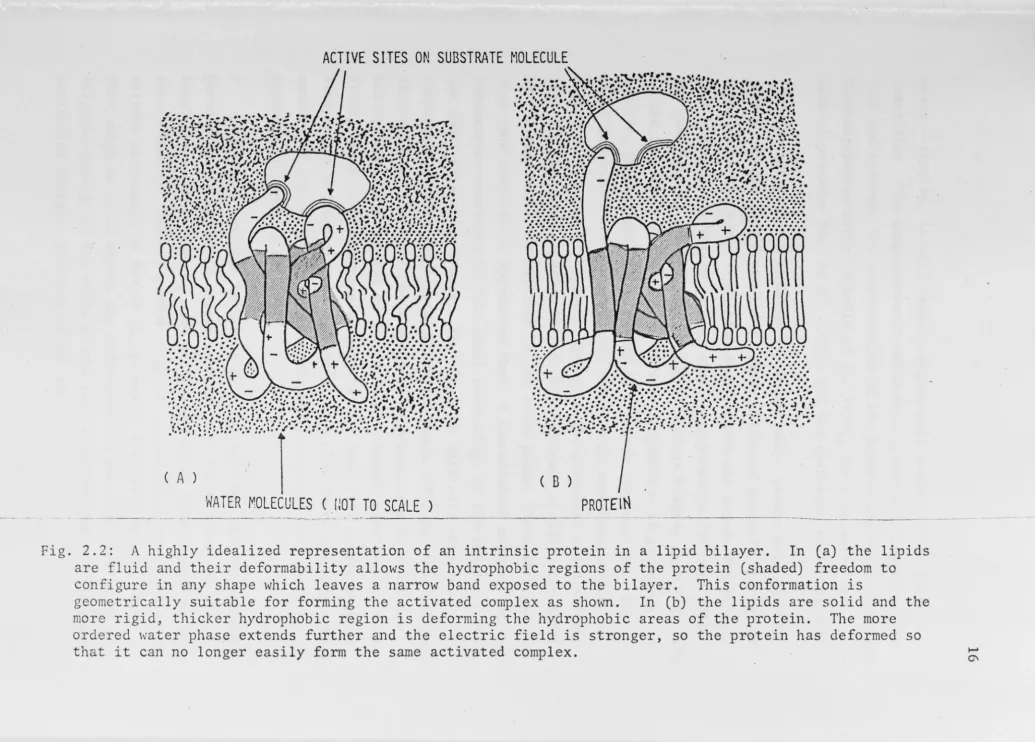
Thus, four of the physical properties of the environment of
membrane proteins (the deformability and geometry of the bilayer, the
electric field and the "structure" of water) are temperature dependent,
and since conformation is affected by these properties, this will be
temperature dependent too (see Fig. 2.2).
2.6 PHASE TRANSITIONS AND SEPARATIONS
A bilayer made of one species of lipid only and containing no
protein, will, over a small range of temperature, change its molecular
Fig.
~
, . . . .... •• · · · ~ · . J :-.. :. • •
1~,.f'..• • f • • "'• ~ .. • ...._._. .a. C f :, ... • • • •••• • • - ~ ., ... . ·,...,:;.,.•~ ... ~ .... _..;-.~· • . , ... ·''•'J•. ~ .. ~··· ,;. . ':•Jj"'l- .... ~ ... <·'·•.I·'·-. ... ( ., .. •,.:, ., .. , .• -\··'· .. ,. & • I• ""' • .I • -..-< ~ • • • ~ • • • T • • •• • • • • : .. 1 • • • • •
=;:,~if/~;:~r~/J:;:r.\,t.:f?':.'
·-;~.':°·:.~1::~!S~·Y:ltn,:1.;:i!:::.;:
'···:~···,:,-; ... ..,..,,~11\r :1-.,., ... ;, ,.:.\•,•,s,
•••• •• • , _ - . ••• I ... .,,
.. , --!·., :11,•,•,..,;,',11.,.,-., . :
l~··
.
!··3:\·
••::--6,,-~ ··-·.} ·l·,···r.··.: ..
~-·,..,,, . ., \: .. J .,~: ,.:,...,;·:,.~ .... ,:, '·
,·.-. ;•_:::: -~~,.; • ;, .,··,'r·'·'
,. {. .. ,,~. . . ... ..
.. .
~.. :r-r · .. ,
"7•• \.' ... ' • :,,., ' ' • >.• ~:. ., •• ,.. ,. ~-.J 'w • ,, !•
.. , , r' J '9; •• ,• • ,/~: 11 '•''• • ~ • :J ••.'•• l •j :: L' •
• ' , • . ..._ .. • • \ • I • • • • ; • • ,J,., ' , • • t , . f
• \ · · · ~ · · . . , . . ••• ••••••• "'(7• • • • • • •
•"J•J•,-.•,.1,.,,: ••••• , \••••··· . ,
'r._,•~) ·1,\, .• ,: ·~
l:,,····"~,,,.1, .. , ,
"!.~:,~:::..:,;;,'!' + ·.-..,: .. 1!., .... :.i.i:·.::!!:~",
•••II\'.~#•'•' ... , •• •• • • •e • • , / , •• •• e
Q'.::RJiQ,
+;Ql~:=~:. ,:-
-~= .
•.•...
.
.
.
. . .
.
...
,
....
.,.,~,.'
-~.
)
; \\~ <
\:,
(~ ~\··~ )' . .
.·
·.· .. ·:·:-,···
... ·
...
·:·:·+ · .. ,;:,:,,.,~,,.,,, •.
·.::.,
.....
·.·.·.·.·.·:.·
:•. :1.
.
.
.
.
.
.
.
,...
,..
.
.
.,
-
. .
.
:-, ~ ·, ,·.• .. •. ·:1!1 ••• '-..!_:,·., ::.- ~-. t • I '\. • • •• + • ,"'I, I",,__ • • •• r •
... , • • ..., r . ,I • - • . • . • ·J'.. ~ , ... :\ • • ... ~· .• ....r-. . ., '.
.r:.:,.~,,,,,:·.·.
.
.
'..
.
.
-.
.. -. ... :.::.·..
. . .
.
.
+ :·· •.•.
·,·\!·,: ... -'•.,,
.
.
,...
.
••• • • • '··· . • ., •. ,,,. \ .•. r '• ' ... • • • • ,·:::.I, .•. ,·:,·~'·· •• • ••• , .... '/. , .. , •• . ~ • .... • • ~#., ..
t •
•9·
I • • c_,••• , ••• ,•. . • ,,, ... , ... , •• , . ,• # ' . <.. .;,, •••••••• ,,, ••• •l'f•-•• • • •• ,.,--,
.. , ..•. ., '# ... ,· .. , ••911\t,. •t.:..-- ,., ... ,,. ,., ,, ... ,·,~
• • , • • , • • , • • • • • • • • • , a 4 f . . . . • • , . . . . .,, • •
'(.
.
~,,.\,,• .. :.·.··.·::.·. ··1
..
'·•·=··..,····
···•,'i•,1'.···-···, ... ,.._. ... , •......
...
,...
.
.
• I • • _,.
( A )
WATER MOLECULES ( lmT TO
SCALE )
( B )
PROTEIN
2.2: A highly fluid and
idealized representation their deformability allows are
configure
of an the
intrinsic protein .... in a lipid bilayer. In (shaded) hydrophobic regions of the protein
(a) the freedom
lipids to in any shape which leaves a narrow to the bilayer. This conformation
complex as shown. In (b) the lipids are the hydrophobic areas of the protein.
field is stronger, so the protein has
band exposed lS
solid and The more deformed geometrically suitable for forming the activated
is deforming the electric more rigid,
ordered water that it can no
thicker hydrophobic region phase extends further and
longer easily form the same activated complex.
the
so
~
[image:29.1133.72.1108.19.761.2]state - that is, it will closely approximate a first-order phase
transition. The temperature at which this occurs will depend on the
salt and hydrogen ion concentration of the aqueous phase [Jacobs-on and
Papahadjopoulos 1975; Tr~uble et al. 1976], the transverse and
lateral pressure [Hui et al. 1975], and the curvature of the bilayer.
The bilayer in a membrane is, regrettably, rather more complex
[Lyons, Wheaton and Pratt 1964]. When different miscible species of
lipids with different transition temperatures are mixed, the bilayer
does not suddenly freeze at a particular temperature [Reisman 1970].
Its behaviour is usually represented on a phase diagram (see Fig. 2.3).
As the temperature of a mixture of some composition is gradually
lowered, it separates into domains of different composition. Thus,
small regions of the frozen state, with a high concentration of the
higher melting point component appear in the otherwise fluid bilayer
[see e.g. Rushbrooke 1949; Shimshick and McConnell 1973; Lee 1977].
If the components are miscible in the solid phase, these increase in
size (and composition approaches that of the original mixture) as the
temperature continues to fall until eventually, on the solidus curve,
the whole bilayer is frozen. The situation with more than two
components is more complicated [Lang and Widom 1975] but qualitatively
similar, i.e. there will be a range of temperatures over which the
bilayer comprises domains of various compositions, some frozen, some
fluid. Another interesting complication is that if a single species
bilayer has a dissociation equilibrium such that there are substantial
amounts of lipids with a net electric charge, it too will phase
separate [Forsyth et al. 1977].
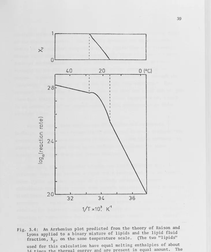
If we were now able to insert proteins into our bilayer without
perturbing it, we should expect to find that, as we lower the
temperature, progressively more protein molecules find themselves in frozen domains, and so, over a range of temperatures, the fraction of enzymes surrounded by frozen lipids varies from zero to one. Over
this range ,ve would expect any membrane-bound reaction rate to be a
Tc1
Tu
T"
TL
Tc
.
2
I
I
I
______
_.__
__,,_______
,
---0
x~
1
I
Fig. 2.3: A simple, two-component phase diagram. Fractional
composition of species· 1 on the x axis, temperature on the y axis. A mixture of X of species 1 and (1 -X) of species 2 will separate
into two phases between TU and TL. At T', one phase will have X'
Cl
of species 1 and the other
x
6
of species 1. TC and TC are the1 2
[image:31.782.15.772.15.937.2]2.7 ·BOUNDARY EFFECTS
To complicate the problem further, however, the interaction
between lipid and protein is different from that between lipid and
lipid, and thus the boundary layer of lipids round a protein is in a
different environment from that of bulk liquids. The next layer is
less affected and so on, but there are several layers of lipids round
a protein molecule whose mobilities will be different from the bulk
values. This boundary layer effect is evident in the experiments of
Jost et al. [1973] and others.
The tails of a lipid in the bulk bilayer can bend and wri.ggle (and the end of the tail thrash about) because the "next door" lipid
can bend and wriggle to accommodate this. If the molecule next to the
lipid is a large and relatively rigid protein molecule it will be
considerably less mobile, and reduce the mobility of the next layer.
Using this concept together with the molecular field approximation,
Marcelja [1976] has calculated the order parameter of lipids as a
function of temperature and distance from a protein. With his
boundary conditions, the order increases (and therefore the mobility
decreases) nearer the protein, and the order of the boundary layers is
a continuous function of temperature. Thus even if the protein
molecules were embedded in a pure, one-species bilayer the lipids
nearby would not undergo an abrupt phase change.
Now if the free energies of interaction between the protein
molecule and each species of lipid molecule were equal, then, in order
to maximize entropy, the lipid composition would be homogeneous up to
and including the boundary layer. If, however, the free energy
interaction is lowest with one species, then that species will be
preferentially adsorbed onto the protein; i.e. that species will form
a larger fraction of the boundary layer than it does of the bulk
[Boggs et al. 1977]. This might be brought about simply by
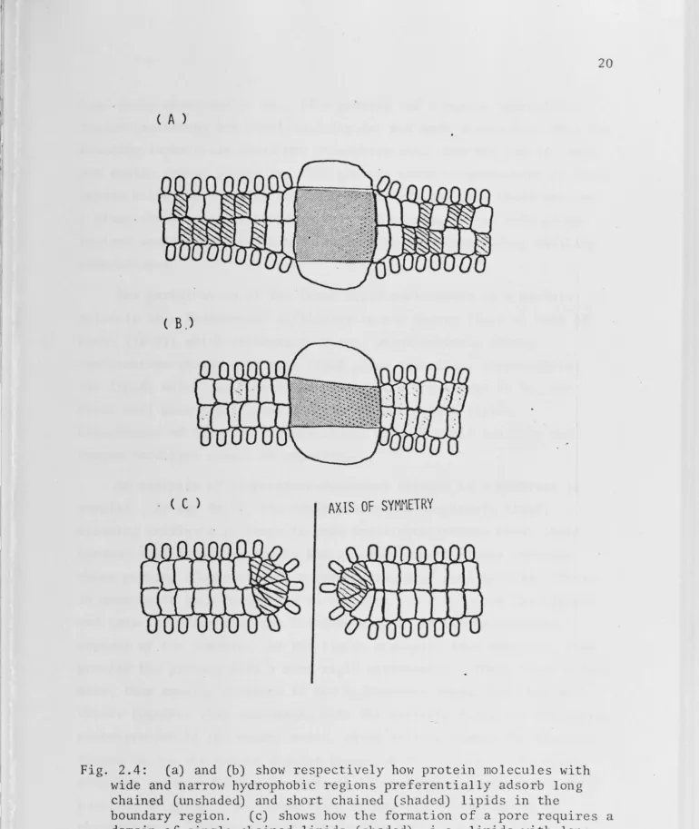
geometrical packing considerations [Israelachvili 1977] and is most
easily shown in Fig. 2.4. A protein with wide hydrophobic band might
preferentially adsorb lipids with long chains; one which bulges in
C A )
(B)
. ( C ) AXIS OF SYMMETRY
Fig. 2.4: (a) and (b) show respectively how protein molecules with wide and narrow hydrophobic regions preferentially adsorb long chained (unshaded) and short chained (shaded) lipids in the
[image:33.797.12.782.17.930.2]head-group areas and so on. If a protein had a narrow hydrophobic
region (selecting fot short-tail lipids) and were shaped such that the
boundary layer tails could not straighten out, then the lipids could
not easily freeze around it. The protein would be surrounded by fluid
lipids below the freezing temperature of even the most fluid species.
A plant whose membrane-bound enzymes had narrow, curved hydrophobic
regions would therefore be better equipped for withstanding chilling
temperatures.
The perturbation of the local lipid environment by a protein
molecule is a fundamental difficulty in any theory (such as that of
Lyons [1973]) which attempts to relate quantitatively enzyme
conformation change with pure lipid phase transition temperatures:
the lipids which can affect the protein are those near to it, and
these will have a different mobility from the bulk lipids.
Coincidence of "critical" temperatures of bulk lipid mobility and
enzyme condition cannot be expected.
An analysis of temperature-dependent changes in a membrane is
complex. At say 30 °Cj the lipid bilayer is completely fluid,
allowing intrinsic proteins to make small oscillations about their
current configuration, and is homogeneous, except around proteins
whose packing shapes require a preponderance of some species. There
is some water structuring due to the electric field near the bilayer
and this is influencing the "clathrate c.age" of the hydrophilic
regions of.the protein. As the lipids gradually lose mobility, they
provide the protein with a more rigid environment. Their tails extend
more, thus causing stresses in the hydrophobic bonds, and they pack
closer together thus increasing both the electric field and the cation
concentration in the nearby water, which in turn change the electric
I .
forces on the charged or dipolar groups on the protein. The
water-ordering extends further, is less fluid, and the hydrogen bonds shift
position sl.ightly. The composition of the lipid environment is
changing and, depending on its shape, the protein may be
preferentially caught in local regions of fluidity or solidity. The
free energy of its activated complex will at least be different - and
thus its activity will change - or the forces may be great enough to
cause a conformational change which precludes the possibility of the
.Though these models are hypothetical - or speculative - they
predict that chilling sensitive and chilling tolerant species will
differ not only in their lipid composition [Lyons and Raison 1979] but
also in the structure of their intrinsic membrane proteins. If the
intrinsic membrane proteins of chilling-sensitive species are
distinguished from those of chilling-resistant species by having rigid
flat sides onto which solid-like lipids can adsorb, then these
proteins will be more prevalent in the solid-like domains of phase
separations. Those of chilling-resistant species, hypothesized to
have irregular hydrophobic regions, would be more prevalent in the
fluid-like regions. This could be determined from freeze fracture
electron micrographs frozen rapidly from phase separation temperatures
[cf. experiments of Chen and Hubbell 1973]. It is not necessary to
postulate that the analogous proteins of chilling-sensitive and
chilling-resistant species are very different - several minor changes
in primary structure could cause a large change in quaternary
structure.
If the activation free energy of an enzyme is changed by changing
electric field and/or changing cation concentration, then its activity
should be affected by change in the concentration in the aqueous
solution of divalent cations. One might therefore look for a greater
dependence on divalent cation concentration in enzymes from
chilling-sensitive species than in those from chilling-resistant species.
Of the various mechanisms and effects suggested and catalogued in
this chapter some are currently under investigation-- Coster [personal
communication 1979] is studying the thickness changes of bilayers in
phase separations and its effect on enzyme activities; Jost [personal
corrununication 1979] is working on preferential adsorption of lipids
onto intrinsic membrane proteins. The two topics considered in detail
in this thesis are membrane compressibility (Chapters 5 and 8) and
preferential solubility of proteins in different phases (Chapter 8).
These theories are developed from well understood physical principles
and will explain the results of those experiments which represent the most direct evidence of the involvement of lipid mobility in the
Bagnall, D.J. (1979). M.Sc. Thesis submitted to the Australian National University, Canberra.
Boggs, J.M., Woods, D.D., Moscarello, M.A. and Papahadjopoulos, D. (1977).
Biochemistry.!.§_,
2325-2329.Borochov, H. and Shinitzky, M. (1976).
Proc.
Nat.
Acad. Sci. USA
73, 4526-4530.Brandts, J.F. (1967). In "Thermo Biology" (Ed. A.H. Rose), Academic Press, 147-218.
Chapman, G. and McLaughlan, K.A. (1967).
Nature
215, 391-392.Chen, Y.S. and Hubbell, W.L. (1973).
Exp.
Eye Res.
l_Z_, 517-532.Christiansen, M.N. (1979). In "Proceedings of the US-Australian-N. Z. Co-operative Science Program Conference on Low Temperature Stress
in Crop Plants: the Role of the Membrane", to be published in Academic Press.
Clifford, J. (1975). In "Water - A Comprehensive Treatise" (Ed. F. Franks), vol. S, p.75. Plenum Press, New York, London.
Coster, H.G.L. (1979).
Crozier, W.J. (1924).
Proc. Aust. Biochem. Soc.
12,Q6.
J.
Gen. Phusiol.
..-
7, 189-216.Davidson, D.W. (1973). In "Water - A Comprehensive Treatise" (Ed. F. Franks), vol. 2, pp.55-234. Plenum Press, New York, London.
De la Roche, I.A. and Andrews, C.J. (1973).
Pl.
Physiol.
51, 468-473.Downes, R.W. and Marshall, D.R. (1971).
Aust.
J.Agric.
Anim.
Hush .
.!_!_, 352-356.
Drost-Hansen, W. (1973).
Ann. N.Y. Acad. Sci.
204, 100.Evans, L.T., Wardlaw, I.F. and Williams, C.N. (1964). In "Grasses and Grasslands" (Ed. C. Barnard), McMillan. pp .102-125.
Forslind, E. and Kjellander, R. (1975). J.
Theor. Biol.
51, 97-109.Forsyth, P.A., Marcelja, S., Mitchell, D.J. and Ninham, B.W. (1977).
Biochem. Biophys. Acta
469, 335-344.Gerloff, E.D., Richardson, T. and Stahmann, M.A. (1966).
Pl. Phys
i
ol.
!!:_, 1280-1284.Graham, D. (1979a). Editor - Proceedings of the US-Australian-N.Z. Co-operative Science Program Conference on Low Temperature Stress
Graham, D. (1979b). In "Proceedings· of the US-Australian-N.Z.
Co-operative Science Program Conference on Low Temperature Stress in Crop Plants: the Role of the Membrane", to be published by Academic Press.
Guinn, G. (1971). Crop Sci. 11, 262-265.
Hagler, A.T. and Moult, J. (1978). Nature 272, 222-226.
Haller, I. and Freiser, M.J. (1976). Biochem. Biophys. Acta 455, 739-748.
Hasted, J.B. (1973). In "Water - A Comprehensive Treatise" (Ed. F. Franks), vol. 2, pp.405-458. Plenum Press, New York, London.
Hui, S.W., Cowden, M., Papahadjopoulos, D. and Parsons, D.F. (1975). Biochem. Biophys. Acta 382, 265-275.
Israelachvili, J. (1977). In "Proceedings of the U.S.-Australian Seminar on the Evolution of Light Trapping Systems, Honolulu", Academic Press.
Ivory, D.A. and Whiteman, P.C. (1978a). Aust. J. Pl. Physiol.
I,
131-148.Ivory, D.A. and Whiteman, P.C. (1978b). Aust. J. Pl. Physiol.
I,
149-157.Jacobs, E.E., Andrews, E.C., Wohlrab, A. and Cunningham, W. (1967). In "Structure and Function of Cytochromes", Univ. of Tokyo Press, Tokyo.
Jacobson, K. and Papahadjopoulos, D. (1975). Biochemistry. 14, 152-161.
Johnson, F.H. , Eyring, H. and Polissar, M.J. (1954). "The Kinetic Basis of Molecular Biologf', Wiley, New York.
Johnson, F.H., Eyring, H. and Williams, R. W. (1942). J. Cell. Comp. Physiol. ~' 247-268.
Jost, P.C., Griffiths, O.H., Capaldi, R.A. and Vanderkooi, G. (1973). Proc. Nat. Acad. Sci. USA 70, 480-484.
Junge, W. (1972). FEES Letts. 25, 109-112.
Koshland, D.E. (1973). Sci. Amer. October, pp.52-64.
Kuiper, P.J.C. (1970). Pl. Physiol. 45, 684-686.
Lang, J.C. and Widom, B. (1975). Physica 81A, 190-213.
Lee, A.G. (1977). Biochem. Biophys. Acta 472, 285.
Lyons, J.M. and Asmundsen, C.M. (1965). J. Amer. Oil Chem. Soc. 42, 1056-1058.
Lyons, J. and Raison, J.K. (1979). In "Proceedings of the
US-Australian-N.Z. Co-operative Science Program Conference on Low Temperature Stress in Crop Plants: the Role of the Membrane", to be published by Academic Press.
Lyons, J.M., Wheaton, T.A. and Pratt, H.K. (1964). Pl. Physiol. 39, 262-268.
Marcelja, S. (1976). Biochem. Biophys. Acta 455, l-7.
Marsh, D. (1974). Biochim. Biophys. Acta 363, 373-386.
McLaughlin, S.G.A., Szabo, G. and Eisenman, G. (1971). J. Gen.
Physiol. 58, 667-687.
McWilliam, J.R. (1979). Co-operative Science in Crop Plants: the Academic Press.
In "Proceedings of the US-Australian-N.Z. Program Conference on Low Temperature Stress Role of the Membrane", to be published by
Murata, N., ·Ono, T. and Sato, N. (1979). In "Proceedings of the US-Australian-N. Z. Co-operative Science Program Conference on Low Temperature Stress in Crop Plants: the Role of the Membrane", to be published by Academic Press.
Nelson, A.P., Colonomos, P. and McQuarrie, D.A. (1975). J. Theor.
Biol. SO, 317-325.
Nolan, W.G. and Smillie, R.M. (1977). Pl. Physiol. 59, 1141-1145.
'
-Overath, P., Thilo, L. and Tr~uble, H. (1976). Tr(J)1s. Biochem. Sci. August, pp.186-189.
Paton, J.C., McMurchie, B.K., May, B.K. and-Elliott, W.H. (1978). J. Bact. 135, 754-759.
Raison, J.K., Berry, J.A., Armond, P.A. and Pike, C.S. (1980). In 11Adaption of Plants to Water and High Temperature Stress", Wiley, New York. In preparation.
Raison, J.K. and Lyons, J.M. (1970). Pl. Physiol. 45, 382-385.
Reisman, A. (1970). "Phase Equilibria: Basic Principles,
Applications, Experimental Techniques", Academic Press, Ne,v York.
Rushbrooke, G.A. (1949). "Introduction to Statistical Mechanics", Oxford University Press, Oxford.