STIM1 mediates multiple signalling pathways
in neuronal growth cones
Camilla B Mitchell
BMedRes (Hons)
Submitted in fulfillment of the requirement for the
Degree of Doctor of Philosophy (Medical Research)
Menzies Research Institute Tasmania
University of Tasmania
Declaration
This thesis contains no material which has been accepted for a degree or diploma by the University of Tasmania or any other institution, except where acknowledged, and by way of background information and duly acknowledged in the thesis, and to the best of my knowledge and belief no material previously published or written by another person except where due acknowledgement is made in the text of the thesis, nor does the thesis contain any material that infringes copyright.
Figure 2.1 A was included in my honours thesis, Mitchell 2009, University of Tasmania
Figure 2.2 B was included in my honours thesis, Mitchell 2009, University of Tasmania
Camilla Mitchell
Authority of Access
This thesis may be made available for loan and limited copying and communication in accordance with the Copyright Act 1968.
Camilla Mitchell
Publications arising from this thesis
Mitchell CB, Gasperini RJ, Small DH, and Foa L. (2012) STIM1 is necessary for store-operated calcium entry in turning growth cones. J Neurochem, 122, 1155-1166.
Statement regarding published work contained in this thesis
Statement of ethical conduct
The research associated with this thesis abides by the international and Australian codes on human and animal experimentation, the guidelines by the Australian Government’s Office of the Gene Technology Regulator and the rulings of the Safety, Ethics and Institutional Biosafety Committees of the University.
Camilla Mitchell
Acknowledgements
Firstly, I’d like to express my sincere gratitude to my primary supervisor, Dr. Lisa Foa. Thank you for supporting me, teaching me and encouraging me when I was down. I am so grateful for your support, both intellectually and personally, and for the guidance you have given me over the past four and a half years. Thank you for believing in me, and only wanting the best for me. I’m extremely grateful that you never minded when I would be in your office every day, and sometimes multiple times a day. You got that demanding student you always wanted! And thanks for all of those Monday morning football debriefs.
To my co-supervisor, Dr Rob Gasperini, thank you for teaching me microscopy. Thank you so much for your help with my calcium imaging, and for taking over when I couldn’t hack it anymore. Thank you for supporting me, helping me and encouraging me when I was having a tough time. I’m extremely grateful for your willingness to answer the late night calls when I was stuck on the scope and didn’t know what I was doing.
To Professor David Small, thank you for your support and encouragement, and for being willing to help when needed. I’m grateful for the constructive criticism in lab meetings and for teaching me how to give a great presentation.
friendship, for your support, for the random lab gifts, and for all of the many, many, many chats, debriefs, and rants late in to the night in the turning room. You have kept me (mostly) sane, and I know I’ve got a forever friend!
To Edgar Dawkins, for your English entertainment and loads of laughs; Claire Hadrill, for the many hot chocolate adventures and always being my friend, even when you disagreed with my point of view; Adrian Thompson, thanks for being a great friend, my football buddy, and for coming back; Emily Ainslie, for being my lab niece, and my first (and best) vegan friend; Dr Dave Klaver, thanks for teaching me the ways of the lab, how to do a western, and always having a chat and a laugh.
To the rest of the Young Small Foians, both past and present, for all of your support, encouragement, and friendship, especially our lab mother Jenny Smith, for all you taught me.
Personally I would like to thank my parents for all of their love and support through my university life, for providing me with so much, so that I could just focus on my studies, and do the very best I could.
Abstract
Calcium is an intracellular second messenger that is vital for normal neuronal function. The maintenance of calcium homeostasis is critical for healthy neuronal function, and disruption in calcium homeostasis has been implicated in diseases such as epilepsy and Alzheimer’s disease. In developing neurons, calcium signalling regulates the precise wiring of neurons, in a process known as axon guidance. Axon guidance is extremely important in the normal healthy development of the nervous system. Aberrant axon guidance is highly associated with several neurodevelopmental disorders including autism and mental retardation syndromes such as fragile-X syndrome. Axons navigate the environment by a dynamic navigational structure located at the distal tip of an extending axon, known as a growth cone. Cytosolic calcium is crucial in mediating growth cone navigation. Correct understanding of the signalling mechanisms that regulate cytosolic calcium is key to understanding normal growth cone function.
The results presented within this thesis demonstrate the presence and function of STIM1-mediated processes within the developing nervous system. This thesis has utilised primary cell culture of embryonic dorsal root ganglia neurons and immunocytochemistry to investigate the presence and localisation of STIM1 within developing growth cones. STIM1, along with its binding partners Orai1 and Orai2 reside in two different localisation patterns within growth cones; active (punctate) and inactive (diffuse). Depletion of calcium stores resulted in the activation of STIM1 within growth cones, increasing the number of growth cones displaying punctate STIM1 protein distribution. Calcium depletion also increased colocalisation between STIM1 and Orai1. Furthermore, STIM1 localisation appeared to be biased towards the turning side of the growth cone, in response to a calcium-dependent guidance cue. These data suggest that STIM1 and the Orai proteins are dynamic proteins that function in the regulation of calcium within growth cones.
While the main function of STIM1 is thought to be the activation of Orai proteins, and subsequent activation of SOCE, STIM1 has been shown to interact with other signalling proteins, including the second messenger cAMP, in a process termed store-operated cAMP signalling. This study utilised cAMP analogues to determine if store-operated cAMP signalling was functional within growth cones. Upon the activation of cAMP, repulsive turning away from Sema-3a was restored in growth cones with reduced levels of STIM1. Sema-3a collapse was also prevented upon addition of cAMP agonists in control growth cones, but not restored in STIM1 morphants. Similar results were achieved with cGMP agonists. These data suggest that STIM1 mediates cyclic nucleotide signalling within growth cones. Furthermore, STIM1 has also recently been implicated in the reciprocal control of type voltage-gated calcium channels (VGCCs) and Orai proteins. While L-type VGCCs are important in mature neurons, there is conflicting data in the literature as to their role in axon guidance. This study investigated whether there was a potential interaction between STIM1 and L-type VGCCS in growth cones, and found that if there is an interaction, it is not essential for growth cone turning, but may be required for axon extension.
Table of Contents
Abbreviations List ...1
Chapter 1. Introduction and literature review ...6
1.1 Axon pathfinding is vital for nervous system formation...6
1.1.1 Pioneer neurons establish a scaffold for axon guidance...6
1.1.2 Growth cones lead the developing axon...7
1.1.2.1 Growth cone structure ...9
1.1.2.2 Growth cone cytoskeleton ...13
1.1.2.3 Directed growth cone motility...15
1.2 Guidance cues mediate axon guidance ...16
1.2.1 Netrins...19
1.2.2 Neurotrophins ...20
1.2.3 Semaphorins ...21
1.3 Two major intracellular second messengers mediate growth cone navigation...23
1.3.1 Cyclic nucleotides as second messengers ...23
1.3.2 Calcium as a second messenger ...26
1.3.3 Second messenger cross talk ...33
1.4 Two major calcium sources in growth cones: extracellular and intracellular...35
1.4.1 Extracellular calcium source: voltage-gated calcium channels ...36
1.4.2 Extracellular calcium source: Transient Receptor Potential Channels...38
1.5.1 Discovery of store-operated calcium entry ...43
1.5.2 STIM1 is the key mediator of store-operated calcium entry ...46
1.5.3 STIM2 ...53
1.5.4 TRPC channels and their role in store-operated calcium entry...54
1.5.5 Orai proteins are essential components of the calcium-release activated calcium channel ...55
1.6 Store-operated calcium entry in neurons ...58
1.6.1 Store-operated calcium entry in growth cones...63
1.6.2 Endoplasmic reticulum in growth cones...63
1.7 Hypothesis ...65
1.7.1 Aims...65
Chapter 2: STIM1 and the Orai proteins are present in dorsal root ganglia growth cones ...68
2.1 Introduction...68
2.2 Materials and Methods...72
2.2.1 Ethical declaration ...72
2.2.2 Materials ...72
2.2.3 SDS-PAGE and western blotting...73
2.2.4 Embryonic dorsal root ganglia (DRG) cell culture...73
2.2.5 Immunocytochemistry...74
2.2.6 Orai1 and Orai2 antibody specificity...74
2.2.7 Analysis and Quantification of STIM1, Orai1 and Orai2 protein distribution ...74
2.2.8 Calcium depletion immunocytochemistry ...75
2.2.10 Asymmetric protein distribution analysis ...77
2.3 Results...78
2.3.1 STIM1, Orai1 and Orai2 proteins are present in DRG sensory neuronal growth cones ...78
2.3.2 Depletion of calcium stores stimulates STIM1, Orai1/2 puncta formation, and STIM1-Orai1 co-localisation in growth cones. ...86
2.3.3 Orai1 is required for STIM1 and Orai2 oligomerisation upon calcium depletion. ...89
2.4 Discussion ...107
Chapter 3: STIM1 mediates neuronal SOCE, growth cone turning and growth cone collapse...115
3.1 Introduction...115
3.2 Materials and Methods...119
3.2.1 Materials ...119
3.2.2 Embryonic DRG cell culture...119
3.2.3 Quantification of STIM1 knockdown: Immunocytochemistry ...119
3.2.4 Verification of STIM1 knockdown: western blotting...121
3.2.5 Calcium imaging in DRG neurons ...121
3.2.6 Growth cone turning assay ...123
3.2.7 Semaphorin-3a collapse assay...124
3.3 Results...125
3.3.1. STIM1 expression is required for SOCE in growth cones...125
3.3.4. STIM1 is required for Sema-3a induced growth cone collapse...139
3.4 Discussion ...143
Chapter 4. L-type voltage-gated calcium channels are not required for STIM1-mediated growth cone navigation...152
4.1 Introduction...152
4.2 Materials and Methods...157
4.2.1 Materials ...157
4.2.2 DRG cell culture...157
4.2.3 Growth cone turning assay ...157
4.3 Results...158
4.4 Discussion ...164
Chapter 5: STIM1 signals via cyclic nucleotides in growth cones ...170
5.1 Introduction...170
5.2 Materials and Methods...174
5.2.1 Materials ...174
5.2.2 Embryonic DRG cell culture...175
5.2.3 Growth cone turning assay ...175
5.2.4 Sema-3a collapse assay ...176
5.3 Results...177
5.3.2. Activation of cGMP restores Sema-3a-induced repulsion after STIM1 knockdown in
growth cones ...180
5.3.3. Activation of cAMP is not sufficient to fully restore BDNF attraction after STIM1
knockdown...183
5.3.4. Activation of cGMP is not sufficient to restore BDNF attraction after STIM1
knockdown...184
5.3.5. Activation of PKA or Epac alone cannot rescue Sema-3a repulsion after STIM1
knockdown...189
5.3.6. Correct cAMP levels are required for Sema-3a-induced growth cone collapse ...193
5.3.7. Optimal levels of cGMP are required for Sema-3a-induced growth cone collapse ...201
5.4 Discussion ...207
Chapter 6. Conclusions and future directions ...219
Abbreviations List
[Ca2+]i Intracellular calcium concentration
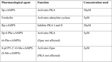
6-Phe-cAMPS Sp-6-Phe-cAMPS
8-Me-cAMPS 8-pCPT-2’-O-Me-cAMPS
AC Adenylate cyclase
Ach Acetylcholine
ATP Adenosine triphosphate
ATPase Adenosine triphosphatase
BDNF Brain derived neurotrophic factor
CAD CRAC activation domain
CaMKII Calcium/calmodulin kinase II
cAMP Cyclic adenosine monophosphate
CaN Calcineurin
cGMP Cyclic guanosine monophosphate
CIF Calcium Influx Factor
CNS Central nervous system
CRAC Calcium-release activated calcium
CICR Calcium-induced calcium release
DCC Deleted in Colon Cancer
DRG Dorsal root ganglia
EGTA Ethylene glycol tetraacetic acid
Epac Exchange protein directly activated by cAMP
ER Endoplasmic reticulum
ER-PM Endoplasmic reticulum-plasma membrane
ERM ezrin/radixin/moesin
f-actin Filamentous actin
FLIP Focal laser-induced photolysis
GTP Guanosine triphosphate
HBSS Hank’s Buffered Salt Solution
ICRAC CRAC current
IP3 Inositol trisphosphate
IP3R IP3 receptor
IR Immunoreactivity
MAG Myelin-associated glycoprotein
MAPK Mitogen-activated protein kinase
mRNA messenger RNA
NaCl Sodium Chloride
NFAT Nuclear Factor of Activated T cells
NGF Nerve Growth Factor
Npn Neuropilin
NT-4/5 Neurotrophin 4/5
P75ntr p75 neurotrophin receptor
PBS Phosphate buffered saline
PDE Phosphodiesterase isozyme
PI3-K Phosphoinositide 3-Kinase
PIP2 Phosphatidylinositol 4,5-bisphosphate
PKA Protein Kinase A
PKG Protein Kinase G
PLC Phospholipase C
PTEN Phosphatase and tensin homolog
PVDF polyvinylidene difluoride
Rac1 Ras-related C3 botulinum toxin substrate 1
RhoA Ras homolog gene family, member A
RIPA Radioimmunoprecipitation assay buffer
RNAi RNA interference
ROI Region of interest
RyR Ryanodine Receptor
SAM Sterile Alpha Motif
SCID Severe Combined Immunodeficiency
SDS Sodium dodecyl sulfate
Sema-3a Semaphorin-3a
SERCA Sarco/endoplasmic reticulum
SNM Sensory Neuron Media
SOC Store- operated calcium
SOCC Store- operated calcium channel
SOCE Store-operated calcium entry
STIM Stromal Interaction Molecule
Tg Thapsigargin
TrK Tyrosine Kinase
TRP Transient receptor potential
TRPC Transient receptor potential canonical
VGCC Voltage-gated calcium channel
w/w weight/weight
Chapter 1:
Chapter 1. Introduction and literature review
1.1 Axon pathfinding is vital for nervous system formation
The nervous system is comprised of millions of neurons that create specific synapses to form a highly ordered neuronal network. The intricacy and precision of neuronal connectivity is the hallmark of neuronal development. One vital process that is required in the formation of this network is axon pathfinding. Axon pathfinding is the process whereby developing neurons send out their axons to navigate through the embryonic environment to find and make connections with their correct targets (reviewed in Tessier-Lavigne and Goodman, 1996). Correct axon pathfinding is crucial to the correct formation of the nervous system. If the process of axon pathfinding goes wrong, aberrant neuronal connections can form. These aberrant connections can lead to neurodevelopmental disorders such as autism spectrum disorder (Geschwind and Levitt, 2007), epilepsy (Yaron and Zheng, 2007), schizophrenia (Bunney et al., 1995, Eastwood et al., 2003, Chen et al., 2011) and mental retardation syndromes including fragile-X syndrome (Waage-Baudet et al., 2005, Bassell and Warren, 2008, Degano et al., 2009). It is extremely important that the process of axon pathfinding is well understood throughout normal development, in order to understand the cellular basis of neuronal disorders and diseases.
1.1.1 Pioneer neurons establish a scaffold for axon guidance
and soluble molecules, acting as guidance cues (Araújo and Tear, 2003). Pioneer axons navigate through this environment establishing a scaffold of axon pathways, or tracks (Bate, 1976, Bentley and Keshishian, 1982, Bastiani et al., 1984, Jacobs and Goodman, 1989, Boyan et al., 1995, Araújo and Tear, 2003, Bak and Fraser, 2003). These early laid axon tracks are important for the guidance of later-developing neuronal axons, referred to as follower axons. Follower axons track along the existing axonal scaffold, fasiculating with individual tracks to reach their final targets (Whitington, 1993, Araújo and Tear, 2003). If pioneer axon pathways are ablated or removed, the guidance of follower axons is perturbed, either by being delayed or misrouted (Raper et al., 1984, Bastiani and Goodman, 1986, Gan and Macagno, 1995, Hidalgo and Brand, 1997). This can result in adverse effects in the development of neuronal connections, however it does not necessarily mean that follower axons will not extend or find their correct targets. Pioneer axons contribute to guidance by providing a suitable substrate for follower axons to extend along. It is rare for pioneer neurons to be essential for follower axons to reach their targets (Pittman et al., 2008)
1.1.2 Growth cones lead the developing axon
complex, larger, but shorter and wider than those of follower axons, which are more elongated in morphology (Bak and Fraser, 2003). The more complex morphology of pioneer growth cones is consistent with their motility as they pause, spread and sample the environment in order to interpret guidance cues to make decisions at choice points such as the optic chiasm or floor plate, as to where to generate axonal scaffolding (Tosney and Landmesser, 1985). Follower axon growth cones have less to interpret, because they are following the pioneer growth cones when they are in the tracks. Therefore, follower growth cones have simpler, narrow, more streamlined growth cone morphologies that grow at a much faster rate (Bovolenta and Dodd, 1990, Wilson and Easter Jr, 1991, Mason and Wang, 1997, Bak and Fraser, 2003, Raper and Mason, 2010), however these can become more complex when follower growth cones reach decision points, such as at the plexus region.
1.1.2.1 Growth cone structure
Neuronal growth cones consist of three domains; the peripheral domain, the central domain and the transitional zone (Fig. 1.1; Goldberg and Burmeister, 1986, Forscher et al., 1987, Forscher and Smith, 1988, Jay, 2000). Each domain is structurally different. The peripheral domain is mostly composed of actin-based structures known as filopodia and lamellipodia, and is thought to be largely devoid of cytoplasmic organelles (Sheetz et al., 1992, Henley and Poo, 2004). The central domain makes up the palm of the growth cone and consists of microtubules, vesicles and organelles. The transitional zone is located between the peripheral and central domains and is characterised by actin-based ruffling activity. Actin filaments and microtubules are domain specific. Actin filaments are found more predominantly in the peripheral domain, while the cytoplasmic central domain contains mostly stable microtubules (Gordon-Weeks, 1987, Forscher and Smith, 1988, Bridgman and Dailey, 1989), with the distal tips of microtubules residing in the transition zone. Dynamic microtubules are also able to extend into the peripheral domain, through the transition zone (Suter and Forscher, 2000).
Figure 1.1. The growth cone structure and cytoskeleton
1.1.2.2 Growth cone cytoskeleton
Growth cone motility is dictated by the motility of filopodia and lamellipodia. Filopodial and lamellipodial motility is based on the dynamics of the underlying cytoskeleton, the actin filaments and microtubules (Bentley and O'Connor, 1994, Letourneau, 1996, Jay, 2000, Suter and Forscher, 2000, Dent et al., 2011).
F-actin operates within the growth cone via several kinetic processes, including: actin filament assembly from G-actin monomers at the leading edge of the growth cone; constant retrograde transport of f-actin networks powered by myosin motors; and proximal recycling of f-actin in the transition zone of the growth cone (Forscher and Smith, 1988, Lin et al., 1996a, Suter and Forscher, 2000, Bard et al., 2008, Chan and Odde, 2008). The extension and retraction of a filopodium reflects the balance between actin polymerization at the barbed ends, and the retrograde flow of entire filaments (Okabe and Hirokawa, 1991, Lin et al., 1996b, Mallavarapu and Mitchison, 1999).
sufficient to induce an attractive or a repulsive growth cone turning response (Buck and Zheng, 2002, Dickson, 2002).
1.1.2.3 Directed growth cone motility
1.2 Guidance cues mediate axon guidance
The formation of ligand-receptor complexes on the growth cone membrane triggers intracellular changes that initiate intracellular signalling cascades, ultimately affecting cytoskeletal dynamics that regulate growth cone motility, resulting in either growth cone attraction or repulsion. Guidance cue receptor activation initiates intracellular signalling through the generation of second messengers, in particular calcium and cyclic nucleotides (Hong et al., 2000, Henley and Poo, 2004, Li et al., 2005, Wang and Poo, 2005, Togashi et al., 2008b, Akiyama et al., 2009). Asymmetrically produced second messengers are thought to coordinate multiple cellular mechanisms, which include membrane trafficking, adhesion dynamics and cytoskeletal reorganisation, all with the purpose of executing bidirectional growth cone turning (Tojima et al., 2011).
their function. Each member of these groups also share distinct cytoplasmic signalling pathways. Calcium-dependent guidance cues also co-activate phosphoinositide 3-kinase (PI3-K) and phospholipase C (PLC)-γ pathways (Ming et al., 1999), and the levels of cyclic adenosine monophosphate (cAMP) and protein kinase A (PKA) activity play a major role in determining the turning response (Tigyi et al., 1996, Cai et al., 1999, Song and Poo, 1999). If PKA is inhibited, the attractive turning responses of netrin-1, BDNF, NGF and Ach are converted to repulsion. If PKA is activated, the repulsive response of MAG is converted to attraction (reviewed in Song and Poo, 1999). In a similar manner, calcium-independent guidance cues are regulated by cyclic guanosine monophosphate (cGMP) and protein kinase G (PKG). If PKG is activated, repulsion in response to Sema-3a is converted to attraction. Inhibition of PKG converts NT3 attraction into repulsion (Song et al., 1997, Ming et al., 1999).
1.2.1 Netrins
vitro (Ming et al., 1997, Wu et al., 2006). Downstream targets of netrin-1 signalling include Rho-GTPases (Causeret et al., 2004, Chang et al., 2004) and Enabled (Gitai et al., 2003) which play roles in cytoskeletal rearrangements within growth cones. Netrin-1 mediated attraction is calcium dependent, and relies upon the downstream signalling of calcium/calmodulin-dependent protein kinase II (CaMKII), mitogen-activated protein kinases (MAPK), calcineurin (CaN), as well as nuclear factor of activated T-cells (NFAT) for gene transcription, which is important for growth cone function (Hong et al., 2000, Graef et al., 2003, Tang and Kalil, 2005).
1.2.2 Neurotrophins
messenger RNA (mRNA) is found predominantly in the central nervous system (CNS) (Leibrock et al., 1989), and its expression is mediated by CREB and CaMKII. BDNF has multiple functions within the nervous system including neuronal survival, morphogenesis, synaptogenesis, and synaptic plasticity, but it also functions as a calcium-dependent chemoattractive guidance cue (Ming et al., 1997, Song et al., 1997). The binding of BDNF to TrKB results in the influx of extracellular calcium into the cytoplasm (Shieh and Ghosh, 1999, Jia et al., 2007), which can activate PLC-γ (Berninger et al., 1993). BDNF has growth promoting actions on filopodia, through signalling via RhoA (Gehler et al., 2004). BDNF can also be chemorepulsive when cAMP levels are low (Song et al., 1998).
1.2.3 Semaphorins
molecules that caused growth cone collapse and were termed ‘collapsins’ (Luo et al., 1993, Luo et al., 1995). It was further found that localised application of these collapsing factors could also result in the turning of a growth cone away from the source of the collapsing factor (Fan and Raper, 1995). Since then, class 3 semaphorins have been shown to elicit repulsive activity on a myriad of neurons, including motor, sensory, olfactory and hippocampal neurons (Luo et al., 1995, Messersmith et al., 1995, Kobayashi et al., 1997, Koppel et al., 1997, Varela-Echavarría et al., 1997, Chedotal et al., 1998)
presence of Sema-3a (Kitsukawa et al., 1997). However, Npn1 does not have any signalling function. Npn1 plays a role in coupling Sema-3a and plexinA to contribute to ligand specificity and enhance the affinity of Sema3a for the receptor complex (Takahashi et al., 1999).
1.3 Two major intracellular second messengers mediate growth cone navigation
Guidance cues elicit their effects on growth cone navigation by activating intracellular second messengers. Two of the most important intracellular second messengers within growth cones are cyclic nucleotides and calcium. While there are other second messengers that are active in growth cones, this thesis will only focus on these two major pathways.
1.3.1 Cyclic nucleotides as second messengers
repulsive, depending on intracellular cAMP levels. Activation of intracellular cAMP on just one side of the growth cone triggers attraction in response to pituitary adenylate cyclase-activating polypeptide, MAG and netrin-1 (Guirland et al., 2003, Murray et al., 2009). Inhibition of PKA, a downstream effector of cAMP, converts both BDNF and netrin-1 turning responses from attraction to repulsion (Ming et al., 1997, Song et al., 1997). One way in which cAMP triggers growth cone attraction is through inhibition of CaN, which is an effector in the growth cone repulsion pathway (Wen et al., 2004). By inhibiting CaN, cAMP is preventing the activation of growth cone repulsion, shifting the growth cone turning response towards attraction.
nucleotides. It is the ratio of cAMP to cGMP that determines growth cone turning (Song et al., 1998, Song and Poo, 1999, McFarlane, 2000, Nishiyama et al., 2003). When the ratio of cAMP:cGMP was systematically varied, with the total concentration remaining at 10µM, turning responses to netrin-1 altered. High ratios of cAMP:cGMP were found to favour growth cone attraction, while low ratios of cAMP:cGMP favoured repulsion (Nishiyama et al., 2003).
1.3.2 Calcium as a second messenger
Calcium signalling does not only occur in combination or cross talk with cyclic nucleotide signalling. Calcium is a major intracellular second messenger in downstream signalling cascades within many cell types (Chilton, 2006). In developing neurons, calcium is particularly important in the transduction of guidance cue signals (Hong et al., 2000, Zheng, 2000), and in the regulation of growth cone motility and turning (Kater and Mills, 1991, Kater et al., 1994, Gomez and Spitzer, 1999, Song and Poo, 1999). Calcium signalling can activate both attractive and repulsive guidance (Fig. 1.2). Higher concentrations of calcium are observed on the side of the growth cone exposed to a source of soluble guidance cue, regardless of whether it is an attractant or a repellent (Zheng, 2000, Tojima et al., 2011). There are two major sources of calcium that are utilised for growth cone function. The first is the extracellular environment, which requires calcium influx across the plasma membrane (Fig. 1.2). The second are internal stores, in particular the ER (Fig. 1.2). Both extracellular and intracellular calcium stores are necessary for the turning of growth cones in response to most guidance cues.
Figure 1.2 Intracellular second messenger signalling within growth cones
of attraction (Zheng, 2000). This work clearly demonstrates that the resting concentration of intracellular calcium, and not only the absolute change in the focal elevation of calcium, is a determining factor in the nature of the growth cone turning response. Although it has been known for a long time that calcium is important in growth cone motility and turning responses (Cohan et al., 1987, Kater and Mills, 1991, Gomez et al., 1995), this study revealed that spatial and temporal characteristics of calcium, in addition to the amount of calcium present, are critical for bidirectional growth cone turning responses.
1988), demonstrating highly spatial, localised calcium elevations are important for growth cone navigation.
Distinct calcium transients and localised signals are activated by specific calcium channels, which are located in both the plasma membrane, and the ER membrane, to gate both major sources of calcium (Bandtlow et al., 1993, Gomez et al., 1995, Tang et al., 2003). Bidirectional growth cone turning most likely relies on the gating of different sets of calcium channels located on the plasma membrane, such as the Transient Receptor Potential Canonical (TRPC) channels, L-type voltage-gated calcium channels (VGCC), and the calcium release-activated calcium (CRAC) channels (Orai proteins). Each of these calcium channels has the ability to trigger select spatial and temporal patterns of calcium signals, and hence growth cone turning (Berridge et al., 2003, Nishiyama et al., 2003, Li et al., 2005, Shim et al., 2005, Wang and Poo, 2005, Tojima et al., 2011, Mitchell et al., 2012).
1.2). The higher amplitude of calcium is sufficient to cause attractive turning on the side of the growth cone with these calcium signals, such as when calcium is released focally on one side of the growth cone (Zheng, 2000). It has also been hypothesised that the calcium effectors are localised to different compartments of the cell. CaMKII is thought to be localised within close proximity of the ER calcium channels, RyRs and IP3Rs (Tojima et al., 2011), which is consistent with the idea that calcium influx across the plasma membrane can be further amplified both spatially and temporally by a secondary release of calcium from the internal stores, resulting in attraction (Takei et al., 1998, Song and Poo, 1999, Tojima et al., 2011). Growth cone repulsion occurs when there are lower amplitude calcium signals, caused only by calcium influx through plasma membrane bound channels, with no additional calcium release from the ER (Henley and Poo, 2004, Ooashi et al., 2005, Tojima et al., 2011). CaN has been hypothesised to exist in a diffuse manner throughout the cytosol, in areas where it is more likely to bind to calcium as it has just entered the cytosol via membrane bound channels (Tojima et al., 2011). These initial and secondary increases in cytoplasmic calcium concentrations are crucial for bidirectional growth cone turning, in response to guidance cues.
1.3.3 Second messenger cross talk
1.4 Two major calcium sources in growth cones: extracellular and intracellular
There are many different calcium channels present within neurons. Some of these calcium channels are important for synaptic transmission, such as multiple types of VGCCs, and the glutamate, NMDA and AMPA receptors. However, this review will only focus on the main calcium channels that have been implicated within growth cones, and in particular growth cone motility and guidance.
on the plasma membrane, activating associated ion channels. How intracellular calcium levels change in growth cones in response to guidance cues is an important question. Guidance cue-induced calcium entry into growth cones can occur via the VGCC, TRP channels and SOC channels. The following section will examine these major calcium sources in more detail.
1.4.1 Extracellular calcium source: voltage-gated calcium channels
upon both calcium influx through the L-type VGCCs on the plasma membrane (Hong et al., 2000). When L-type VGCCs are blocked with nimodipine, netrin-1 induced attraction is converted to repulsion in Xenopus spinal neurons (Hong et al., 2000). Netrin-1 has also been shown to induce an increase in the amplitude of L-type VGCC currents across the plasma membrane in turning growth cones (Nishiyama et al., 2003). Guidance cue receptor activation also stimulates L-type VGCCs on growth cones indirectly, causing calcium influx in to the cytoplasm. Netrin-1 activates TRPC channels on growth cones, through binding of the DCC receptor. Activation of TRPC channels causes calcium influx into the cytoplasm, which in turn depolarises the growth cone membrane. Subsequently, L-type VGCCs are activated which results in a further calcium influx (Shim et al., 2009).
1.4.2 Extracellular calcium source: Transient Receptor Potential Channels
TRP channels are ion channels that are transiently activated in response to temperature, osmolality, mechanical stress and taste (Montell, 2001, Clapham, 2003). Generally, TRP channels are not selective to one ion. Mammalian TRP channel proteins form six-transmembrane cation permeable channels that are further divided into seven sub families. These are TRPC, TRPV, TRPM, TRPA, TRPP, TRPML, and TRPN and they are categorised based on their ligands and modes of activation, and their tissue distribution (Ramsey et al., 2006). TRP channels have been found to be present in neuronal growth cones, with the TRPC family (TRPC1-7) the most extensively studied in axon guidance. In
Xenopus spinal neurons, TRPC1 functions in netrin-1, BDNF, and MAG mediated growth
that members of the TRPC channel family are able to function as potential key mediators for calcium influx and axon guidance regulation during neuronal development. Different TRP channels may serve different distinct regulatory functions within growth cones, potentially by creating different spatio-temporal patterns of calcium influx (Ramsey et al., 2006).
1.4.3 Intracellular calcium source: Calcium-Induced Calcium Release via IP3 receptors and ryanodine receptors
Neuronal cytoplasmic calcium concentrations can increase from two main stores of calcium: the extracellular store and the intracellular store. The intracellular store of calcium resides within the ER lumen.
The process of CICR is initiated by external cues binding to receptors on the neuronal membrane. The activation of two membrane receptor types, G-protein coupled receptors and tyrosine kinase receptors results in the production of IP3 (Berridge, 1993, Berridge, 1998, Berridge et al., 2000). IP3 is highly mobile within the cytoplasm, and diffuses quickly towards the IP3R (Allbritton et al., 1992). IP3 binds to the IP3R on the ER membrane to activate calcium release. When IP3 binds to the IP3R, a conformational change occurs opening an integral calcium channel on the ER membrane (Bootman et al., 2001). This channel opening releases calcium from the high concentration in the ER, to the lower concentration in the cytoplasm (Bootman et al., 2001). The IP3R is not only activated by IP3. There are a variety of factors that the IP3R is sensitive to. One of these is calcium itself. The most effective combination of IP3R activation occurs when both IP3 and calcium act together (Berridge, 1998). The binding of IP3 increases receptor sensitivity to calcium. The receptor has a biphasic action where it activates at lower concentrations of calcium (0.5-1µM), but it inhibits at higher concentrations of calcium (>(0.5-1µM), particularly after calcium releases from the ER, acting as a negative control mechanism (Bootman et al., 2001, Berridge et al., 2003).
for calcium-dependent growth cone attraction (Hong et al., 2000, Jin et al., 2005, Li et al., 2005, Akiyama et al., 2009). Netrin-1-induced growth cone attraction was switched to repulsion upon inhibition of RyRs, and CICR in Xenopus spinal neurons (Hong et al., 2000). A gradient of NGF has been shown to elicit an asymmetric production of IP3 across growth cones, with a higher concentration of IP3 present on the side of the growth cone closest to the NGF source. This asymmetric production of IP3 triggers localised CICR and growth cone attraction in chick DRG neurons (Akiyama et al., 2009). Low concentrations of ryanodine can also increase cytoplasmic calcium concentration through CICR to cause growth cone attraction in Xenopus spinal neurons (Jin et al., 2005). Furthermore, blocking IP3Rs abolishes BDNF-induced attraction in rat cerebellar granule growth cones (Li et al., 2005). In repulsive guidance, RyRs are inhibited by the production of cGMP in response to repulsive guidance cues such as Sema-3a and netrin-1 (Nishiyama et al., 2003, Tojima et al., 2009). Taken together these data demonstrate that CICR is required for the augmentation and amplification of calcium signals in growth cone attraction, but it is not required for repulsion.
future calcium-related events to occur within the cell. ER stores of calcium are replenished by the activity of the calcium adenosine triphosphatase (ATPase) or sarco/endoplasmic reticulum (SERCA) pump, which refills emptied ER stores (Putney Jr, 2007, Smyth et al., 2008, Várnai et al., 2009). However, for the stores to be replenished, there needs to be a mechanism in place for calcium to re-enter the cytoplasm first. In addition, cells require a mechanism to be in place for the augmentation of the initial release of calcium from the ER. Electrophysiologists have been aware of such a mechanism since the 1980’s, where depletion of intracellular calcium stores through IP3Rs and RyRs causes an influx of extracellular calcium into cells (Putney Jr, 1986). The existence of this process, and the channels that allow for this to occur have been confirmed, however, the actual mechanism of action, and the proteins and channels involved have remained a mystery until the last decade.
1.5 Store-operated calcium entry
1.5.1 Discovery of store-operated calcium entry
independently of changes in membrane voltage (Hoth and Penner, 1992). It was postulated that this may be the mechanism by which cells can maintain raised intracellular calcium concentrations as well as replenish empty internal calcium stores (Hoth and Penner, 1992).
communication between calcium stores and the plasma membrane SOC channels, with the use of drugs that promote peripheral actin polymerisation (Patterson et al., 1999, Bakowski et al., 2001), these results led to the idea that a close membrane relationship must exist, potentially through protein-protein interaction. This lead to a search for the proteins involved in the interaction that causes SOCE.
STIM1 is a novel gene that was first cloned from human chromosome region 11p15.5 (Parker et al., 1996). STIM1 was previously designated GOK, and it was originally identified as a growth suppressor gene thought to be associated with tumour development (Parker et al., 1996). Chromosome region 11p15.5 contains genes that have been implicated in Beckwith-Wiedemann syndrome, Wilms tumour (nephroblastoma), rhabdomyosarcoma, rhabdoid tumour development, adrenal carcinoma, hepatoblastoma, bladder, breast, lung, ovarian and testicular cancers (Parker et al., 1996, Sabbioni et al., 1997, Manji et al., 2000). STIM1 protein originally appeared to have no obvious function, but it was found to be highly conserved in mouse and human, suggesting that it may have an important cellular function (Parker et al., 1996). Early studies showed that STIM1 expression was absent from rhabdomyosarcoma, even though it was present in high levels in normal skeletal muscle, where rhadbomyosarcoma originates. This downregulation of STIM1 lead to the hypothesis that STIM1 was involved in tumour development (Sabbioni et al., 1997).
1.5.2 STIM1 is the key mediator of store-operated calcium entry
STIM1 is a type 1 transmembrane protein that resides in the ER membrane (Putney, 2005, Zhang et al., 2005, Wu et al., 2006, Liao et al., 2007). STIM1 is also found in the plasma membrane, although its function there is unclear. STIM1 contains a single transmembrane segment that separates the N terminal (ER luminal) region from the C terminal cytoplasmic region (Williams et al., 2001, Dziadek and Johnstone, 2007). The N-terminus of STIM1 contains a signal peptide, cysteine residues and a single helix-loop-helix region, which conform to an EF-hand motif (Williams et al., 2001). This EF-hand motif binds calcium, allowing STIM1 to function as an ER luminal calcium sensor (Mercer et al., 2006, Stathopulos et al., 2006, Dziadek and Johnstone, 2007). The EF hand has a low affinity for calcium, as well as a 1:1 binding stoichiometry (Stathopulos et al., 2006). STIM1 binds calcium within the ER, when ER stores are full. The ER has a high resting calcium concentration, which is sensed by STIM1 due to its low affinity for calcium. STIM1-triggered calcium release from the ER results in calcium mobilisation. Mutations in the STIM1 EF-hand prevent the binding of calcium, effectively leaving STIM1 constantly active. STIM1 mutants behave as if the ER stores are constantly depleted of calcium (Liou et al., 2005, Spassova et al., 2006, Zhang et al., 2006, Dziadek and Johnstone, 2007). This demonstrates that the binding of calcium to the EF hand in STIM1 is sufficient to prevent the entire sequence of events that lead to SOCE.
2006) (Fig. 1.3). Within this state, STIM1 resides in the ER membrane as a monomer (Ji et al., 2008). The binding of calcium to the EF-hand motif is sufficient to keep CRAC channels in the plasma membrane closed (Zhang et al., 2005, Huang et al., 2006). Depletion of calcium stores within the ER, either through CICR or experimentally by thapsigargin treatment (a pharmacological agent known to deplete calcium stores), causes STIM1 to dimerise and translocate (Fig. 1.3). Redistributed STIM1 oligomerises within seconds into puncta that accumulate within areas of the ER membrane located close to the plasma membrane (Zhang et al., 2005, Baba et al., 2006, Luik et al., 2006, Wu et al., 2006, Liou et al., 2007, Muik et al., 2008). STIM1 oligomerisation is facilitated by the coiled-coil domains, ezrin/radixin/moesin (ERM) domain and sterile alpha motif (SAM) domain located in STIM1 (Williams et al., 2002, Baba et al., 2006, Huang et al., 2006, Luik et al., 2008). The coiled-coil/ERM domain mediates the aggregation of STIM1 dimers into puncta (Williams et al., 2002), and deletion of the SAM domain makes STIM1 incapable of forming puncta in response to ER store depletion (Baba et al., 2006). The coiled-coil domains reside within the ERM domain, which allows for the mediation of protein-protein interactions (Mangeat et al., 1999). STIM1 oligomerisation precedes and triggers the translocation of STIM1 near the plasma membrane in order to activate the CRAC channel (Fig. 1.3) (Liou et al., 2007, Luik et al., 2008, Muik et al., 2008).
Figure 1.3. STIM1 and Orai form the CRAC channel
STIM1 oligomerisation is the primary event in the activation of the CRAC channel and subsequent SOCE. Once STIM1 oligomerisation occurs, all subsequent steps in the SOCE pathway occur (Luik et al., 2008). Although STIM1 activation triggers the SOCE pathway to occur, and it activates the CRAC channel, it does not form the CRAC channel itself. Following the success of RNA interference studies to discover STIM1 and its role in SOCE, similar studies were carried out to discover the molecular makeup of the CRAC channel. Members of two potential protein families have been proposed to interact with STIM1 and play a role in SOCE.
1.5.3 STIM2
being the major STIM homologue within neurons (Berna-Erro et al., 2009). However, recent data from our laboratory disputes this, revealing that although STIM2 is present within the nervous system, it does not localise to neurons (Hadrill, in preparation). Given this, STIM2 will not be discussed in detail within this review.
1.5.4 TRPC channels and their role in store-operated calcium entry
depletion and STIM1 activation, playing some role in SOCE. However, most of the evidence would suggest that TRPC channels are not the classical plasma membrane component that forms the CRAC current.
1.5.5 Orai proteins are essential components of the calcium-release activated calcium channel
al., 2008). Interactions between STIM1 and Orai1 are mediated by the CAD located on the cytosolic C terminus of STIM1, which directly interacts with both the N- and C-termini of Orai1 (Muik et al., 2008, Park et al., 2009). The STIM1 and Orai1 interaction can be reversed upon refilling, suggesting that this interaction only occurs after store-depletion, in order to activate CRAC channels and the CRAC current (Muik et al., 2008).
1.6 Store-operated calcium entry in neurons
The regulation of intracellular calcium concentration is crucial to normal neuronal function. The concentration of intracellular calcium is the dominant regulator of a myriad of neuronal functions, which include differentiation, excitation, synaptic transmission, apoptosis, and gene expression (Ghosh and Greenberg, 1995, Paschen, 2001, Clapham, 2007). Neurons require a low basal concentration of calcium for a strong signal to noise ratio. Neurons also need to be able to sustain an increase in cytoplasmic calcium for the augmentation of calcium signals. It is thought that neurons harness SOCE in order to perform many of these critical calcium-related processes described above (Zufall et al., 2000, Emptage et al., 2001, Venkiteswaran and Hasan, 2009, Gemes et al., 2011).
guidance cues (Gomez and Zheng, 2006). In order to sustain this intracellular calcium level, growth cone stores are emptied. However, these stores are finite and it has been suggested that developing neurons and growth cones utilise the process of SOCE in order to direct and regulate growth cone motility during development (Zufall et al., 2000, Emptage et al., 2001, Li et al., 2005, Wang and Poo, 2005).
well as in a subset of spine synapses (Ng et al., 2011). STIM1 expression has also been shown to increase throughout development, with a stable expression within mature neurons (Keil et al., 2010).
while SOCE has been implicated in the modulation of the activity of neuronal networks, as inhibition of SOCE causes a profound reduction in activity (Steinbeck et al., 2011). SOCE is also present within flight neurons of Drosophila melanogaster (Agrawal et al., 2010). When Orai1 and STIM1 expression are reduced in Drosophila melanogaster, normal flight and flight-associated patterns of rhythmic firing of flight motor neurons is abolished (Venkiteswaran and Hasan, 2009). SOCE also functions in bag cell neurons of Aplysia californica, which are involved in reproductive behaviour (Kachoei et al., 2006). While these studies demonstrate that STIM1, and indeed SOCE, are functional within the nervous system, they do not provide any evidence of the function of SOCE within neuronal growth cones, where the regulation of calcium is crucial for normal motility and guidance throughout development.
1.6.1 Store-operated calcium entry in growth cones
Calcium-dependent guidance cues elicit rises in intracellular calcium within the growth cone (Song et al., 1997, Ming et al., 1999, Hong et al., 2000, Gasperini et al., 2009). Both growth cone turning responses, as well as increases in intracellular calcium continue as long as the guidance cue is present (Gasperini et al., 2009, Mitchell et al., 2012). These results suggest that there is a mechanism within growth cones that refills internal calcium stores to help sustain this rise in intracellular calcium concentration. While SOCE has been proposed to be functional in neuronal growth cones (Li et al., 2005, Wang and Poo, 2005), very little research has been done on the presence and function of SOCE within growth cones. Recently, endogenous STIM1 was demonstrated in DRG neuronal growth cones. STIM1 was shown to be located throughout the central domain of the growth cone, but to also extend in to the periphery and extending filopodial processes (Gasperini et al., 2009). Furthermore, overexpressed STIM1-GFP has also been found to localise and extend into growth cone filopodial extensions. STIM1 puncta also form at the tips of these growth cone extensions (Keil et al., 2010). These studies hint at the presence and localisation of STIM1 within neuronal growth cones.
1.6.2 Endoplasmic reticulum in growth cones
1.7 Hypothesis
While it has been shown that SOCE is necessary for the regulation of calcium within neurons, and indeed throughout the nervous system, there has been little work to examine the presence of STIM and Orai proteins within developing neuronal growth cones. Furthermore, the function of these proteins within growth cone navigation has never been examined. Given that growth cones are highly dynamic structures that rely heavily on temporal and spatially localised calcium signals, it follows that growth cones may utilise the process of SOCE for the regulation of calcium. If this were true, SOCE would play a vital role in growth cone turning, and indeed growth cone navigation throughout the development of the nervous system.
The central hypothesis of this thesis is that:
STIM1 is necessary for store-operated calcium entry in neuronal growth cones, and is required for correct axon guidance
1.7.1 Aims
1. Confirm the presence and localisation of STIM1 and the Orai proteins within embryonic sensory neurons
2. Determine whether STIM1 and Orai1 expression is spatially restricted within navigating growth cones
3. Determine whether SOCE is functional within growth cones, and if it is mediated by STIM1 expression
4. Determine whether STIM1 is required for correct growth cone guidance
5. Determine if there is a functional interaction between STIM1 and L-type voltage-gated calcium channels in navigating growth cones
6. Determine if STIM1 mediates cyclic nucleotide signalling during growth cone navigation
Chapter 2:
STIM1 and the Orai proteins are present in dorsal root
Chapter 2: STIM1 and the Orai proteins are present in dorsal root ganglia growth cones
2.1 Introduction
STIM1 is a crucial mediator of SOCE in many different cell types. Traditionally, much of the work on STIM1 and SOCE has been performed in non-neuronal cells, such as platelets and immune cells, providing most of the knowledge of STIM1 function. Recently, the study of the SOCE machinery, in particular STIM1 function, has begun to expand into the nervous system. It has been suggested that neurons harness SOCE for certain neuronal functions, including gene expression, synaptic transmission, and differentiation (Zufall et al., 2000, Emptage et al., 2001, Clapham, 2007, Venkiteswaran and Hasan, 2009, Gemes et al., 2011). Developing neurons require sustained intracellular calcium levels in order to regulate axonal outgrowth and growth cone turning, in response to calcium-dependent guidance cues (Gomez and Zheng, 2006). This leads to my hypothesis that growth cones require STIM1-mediated SOCE to sustain these rises in intracellular calcium, and replenish stores, in order to undertake these processes.
cone where calcium signals arise (Zheng, 2000). When caged calcium is released on one side of a growth cone, turning can be dictated by this release of calcium (Zheng, 2000). This suggests that calcium signals are spatially distributed throughout growth cones. As such, if SOCE is required at the site of turning within growth cones, STIM1 and Orai proteins would potentially need to be located on the leading edge of growth cones, within both lamellipodia and filopodia.
channel activation. The clustering and redistribution of STIM1 is the signal required for Orai to translocate along the plasma membrane into tetramers, forming the CRAC channel (Liou et al., 2007, Luik et al., 2008, Muik et al., 2008, Clapham, 2009) and activating SOCE, in a highly dynamic process. If STIM1 and Orai proteins are important for growth cone function and navigation, it would be expected that these proteins would display these dynamic distribution patterns as have been described within non-neuronal cells, and that they would be located on the growth cone leading edge.
2.2 Materials and Methods
2.2.1 Ethical declaration
All animal procedures were approved by the Animal Ethics Committee of the University of Tasmania (Ethics number A0012322) and are consistent with the Australian Code of Practice for the Care and Use of animals for Scientific Purposes.
2.2.2 Materials
All materials used throughout this PhD were of analytical reagent grade.
2.2.3 SDS-PAGE and western blotting
Whole dorsal root ganglia (DRG) and cortex were dissected from E16-18 Sprague-Dawley rat embryos, lysed and triturated in radioimmunoprecipitation assay (RIPA) buffer (50mM Tris pH 7.4, 150mM NaCl, 1mM phenylmethylsulphonyl fluoride (Sigma-Aldrich), 1mM ethylenediaminetetraacetic acid (EDTA), 5µg/ml leupeptin (Sigma-Aldrich), 5µg/ml aprotinin Aldrich), 1% NP-40 Aldrich), 1% sodium deoxycholate (Sigma-Aldrich), 0.1% sodium dodecyl sulfate (SDS)). Proteins were separated on 8% polyacrylamide-SDS gels, transferred onto 0.2µm PVDF membranes, and blocked overnight in 5% skim milk. Membranes were incubated with primary antibodies (STIM1, 1:1000; Orai1, 1:1000; and Orai2, 1:200), followed by HRP-conjugated secondary antibodies (1:5000 dilution). Antibody conjugates were detected using chemiluminescent substrate (Millipore) and imaged on a gel documentation system (Chemismart, Vilber-Lourmat).
2.2.4 Embryonic dorsal root ganglia (DRG) cell culture
2.2.5 Immunocytochemistry
Cells were fixed in 4% paraformaldehyde (Sigma-Aldrich) at 4˚C, followed by permeabilisation and blocking with 0.4% Triton X-100 (Sigma-Aldrich) and 5% goat-serum (Sigma-Aldrich). Primary antibodies for STIM1 (1:1000; Sigma-Aldrich), Orai1 (1:400; Cell Signaling Technology) and Orai2 (1:300; Alamone Labs) were detected using Alexa Fluor 488/594 secondary antibodies (Invitrogen). Images were acquired with an Olympus BX50 microscope, cooled CCD camera (Photometrics) and software (NIS Elements, Nikon).
2.2.6 Orai1 and Orai2 antibody specificity
To assess antibody cross reactivity, Orai1 and Orai2 antibodies were preabsorbed with their respective peptides (Alomone) at a ratio of 1:1 (w/w) for 24 hours at 4˚C. The preabsorbed antibody was applied instead of the primary antibody in both immunocytochemistry and western blotting protocols. The same has previously been done for the STIM1 antibody (Gasperini et al., 2009).
2.2.7 Analysis and Quantification of STIM1, Orai1 and Orai2 protein distribution
level to 255. This threshold level was applied to all images across multiple experiments. The resultant images fell in to two main categories; the first, a speckled pattern where isolated pixels were visible with clear background occurring between them (punctate), and the second, a confluent pattern with no pixel isolation or background visible (diffuse). Simple counts were carried out on the resultant images, determining if images were punctate or diffuse. These were counted over 10 experiments, and percentages of diffuse or punctate were determined and analysed using unpaired t-tests (Graph Pad Prism).
2.2.8 Calcium depletion immunocytochemistry
For STIM1 and Orai1 puncta colocalisation experiments, colocalisation was demonstrated using pixel-by-pixel multiplication of STIM1 and Orai1 binary images. Numbers of puncta per growth cone were normalized for growth cone area and compared between control and EGTA/thapsigargin treated cultures. All image analyses were performed using ImageJ, and statistically analysed using unpaired t-tests (Graph Pad Prism).
2.2.9 Knockdown and quantification of endogenous Orai1 and Orai2 protein expression
growth cone and normalised for growth cone area. Statistical analyses performed were unpaired t-tests (Graph Pad Prism).
2.2.10 Asymmetric protein distribution analysis
2.3 Results
2.3.1 STIM1, Orai1 and Orai2 proteins are present in DRG sensory neuronal growth cones
This study sought to determine whether endogenous expression of the SOCE proteins STIM1, Orai1 and Orai2 correlated with the status of internal calcium stores and growth cone motility within embryonic DRG sensory neurons. STIM1 and Orai1 have been demonstrated in DRG neurons previously, although their expression has not been described in detail (Gasperini et al., 2009, Gemes et al., 2011). Western blotting analysis was used to confirm STIM1, Orai1 and Orai2 expression in DRG neurons (Fig. 2.1 A-C). The western blots show staining at the expected bands for STIM1, Orai1 and Orai2. Orai3 was not examined due to unreliable results obtained with several Orai3 antibodies (data not shown). Rat cortex was included as a positive control for both Orai1 and Orai2 staining (Fig. 2.1 B and C). This study confirmed previous work (Gasperini et al., 2009, Gemes et al., 2011) demonstrating the presence and location of STIM1 and Orai1 in DRG, and also suggests that Orai2 is present within DRG of the embryonic peripheral nervous system.
Figure 2.1. STIM1, Orai1 and Orai2 are present in whole DRG
A. Western blot analysis shows STIM1 present in DRG. A band is shown at approximately 90 kDa, the expected size of STIM1. B. Western blot analysis shows Orai1 present in DRG. Rat cortex is shown as a positive control (cortex). Orai1 preabsorbed antibody control is shown (pre). C. Western blot analysis shows Orai2 present in DRG. Rat cortex is shown as a positive control (cortex). Orai2 preabsorbed antibody control is shown (pre). Figure 2.1 A was included in my honours thesis, Mitchell 2009 and was published in Gasperini et al.,
2009. Figure 2.1 B and C were obtained during this PhD, and were published as part of
detected in DRG growth cones, as were Orai1 and Orai2 (Fig. 2.2). Further analysis revealed two distinct STIM1 expression patterns within growth cones. The first was a diffuse pattern, with little apparent discrete puncta. This pattern is similar to previous studies in non-neuronal cells showing STIM1 expression in resting cells with full internal calcium stores (Liou et al., 2005) (Fig. 2.2 A and C). The second pattern was highly punctate, with discrete aggregated puncta and some evident colocalisation. This pattern is similar to activated immune cells where internal calcium stores had been depleted (Liou et al., 2005) (Fig. 2.2 B and D). The pattern of Orai1-IR (Fig. 2.2 A and B) and Orai2-IR (Fig. 2.2 C and D) closely resembled that of STIM1-IR. When the pattern of STIM1-IR was punctate, so was the pattern of Orai1 and Orai2 expression (Figs 2.2 B and D). Similarly, when the expression pattern of STIM1-IR was diffuse, so was the pattern of Orai1 and Orai2 (Figs 2.2 A and C). Preadsorption with Orai antibodies abolished Orai1 and Orai2 immunoreactivity (Fig. 2.2 E and F). No immunoreactivity was observed in the ‘no primary antibody’ controls (Fig. 2.2 G).
Figure 2.2. STIM1, Orai1 and Orai2 are present in growth cones in diffuse or punctate expression patterns.
A-D. Representative growth cones probed for STIM1 (green) and Orai1 or Orai2 (red). Immunoreactivity expression patterns for STIM1, Orai1 and Orai2 were classed as either diffuse, with little apparent discrete puncta (A and C); or punctate, with discrete, aggregated puncta, and some co-localisation evident (B and D). Arrowheads show STIM1-only or Orai1/2-STIM1-only puncta. Arrows show co-localised puncta. Threshold images illustrate the use of ImageJ to classify immunoreactivity patterns as either diffuse or punctate. E. Growth cone stained with preabsorbed Orai1 antibody control, with phalloidin staining (green) to outline growth cone. F. Growth cone stained with preabsorbed Orai2 antibody control, with phalloidin staining (green) to outline growth cone. G. No primary antibody control with both Alexa Fluor-594 and 488 secondary antibodies. Scale bar is 10µm and applies to all images. . Growth cone B was included in my honours thesis, Mitchell 2009. The thresholding analysis was all done as part of this PhD work. Growth cones B, C and D
Figure 2.3. Quantification of STIM1, Orai1 and Orai2 immunoreactivity expression patterns, in randomly growing growth cones
narrow, non-spreading, streamlined growth cones (Fig. 2.2 A, C). These growth cones tended to be smaller in area, ranging from 6 - 78 µm2. Conversely, growth cones displaying punctate STIM1 and Orai1/2 immunoreactivity exhibited spreading lamellipodia (Fig. 2.2 B and D) and tended to be larger in area, ranging between 33 - 247 µm2. The morphology of growth cones displaying a punctate pattern of STIM1/Orai immunoreactivity was reminiscent of growth cones pausing at decision regions (Tosney and Landmesser, 1985). Such growth cones have increased frequency of calcium transients (Gomez and Spitzer, 2000), which may require the activation of SOCE.
2.3.2 Depletion of calcium stores stimulates STIM1, Orai1/2 puncta formation, and STIM1-Orai1 co-localisation in growth cones.
Figure 2.4. STIM1-, Orai1- and Orai2-immunoreactive punctate growth cones increase after calcium store depletion.
expression of protein after calcium depletion, the numbers of STIM1 and Orai1 puncta per growth cone area increased significantly after thapsigargin treatment when compared to vehicle control (Fig. 2.5 C; STIM1 p<0.05, Orai1 p<0.01). Additionally, there was also an increase in the number of co-localised puncta of STIM1 and Orai1 puncta after calcium depletion (Fig. 2.5 C; p<0.05). The y-axis of Fig. 2.5 C represents the number of puncta per growth cone area (µm), so the numbers shown within the graph appear to be small, and are not representative of the whole growth cone. Consistent with these data, an increase in punctate STIM1 and Orai distribution has been previously demonstrated in non-neuronal cells after thapsigargin treatment (Liou et al., 2005, Soboloff et al., 2006b) suggesting that depletion of internal calcium stores activates STIM1- and Orai1/2-mediated SOCE within growth cones.
2.3.3 Orai1 is required for STIM1 and Orai2 oligomerisation upon calcium depletion.