Molecular investigations
on sex determination and
differentiation pathways in the
common carp,
Cyprinus carpio
By Megan L. Barney
B.
Aqua (Hons)
University of Tasmania
Submitted in fulfilment of the
requirements of the degree of
Doctor of Philosophy
Declaration
This thesis contains no material which has been accepted for a degree or diploma by
the University or any other institution. To the best of my knowledge all material
previously published or written by another person is acknowledged in the text of the
thesis.
This thesis contains no material which has been accepted for a degree or diploma by
the University or any other institution. To the best of my knowledge the thesis does
not contain any material previously published or written by another person except
where due acknowledgement is made.
Megan Louise Barney a/r Date 31
N-201 C
Authority of Access
This thesis is not to be made available for loan or copying for three years following
the date this statement was signed. Following that time the thesis may be made
available for loan and limited copying in accordance with the Copyright Act 1968.
Megan Louise Barney Date
6Y°f/24-/Q
Common carp is a highly important species, with significant aquaculture production
and frequent use for fish biology and aquaculture research. In Australia, New
Zealand and the US, common carp are declared as an invasive pest causing damage
to endemic ecosystems. This thesis aims to advance our understanding of the
molecular pathway of sex determination and differentiation in common carp with a
view to facilitate the development of genetic control mechanisms in this species. The
objective was addressed by the molecular cloning of six key genes involved in sexual
development, 3 each critical for female and male development. Spatial expression of
these genes was analysed in adult tissues and the onset and timing of expression was
determined during early ontogeny and through larval development at two
temperatures (20 and 25 °C) allowing investigation of the effect of temperature on
expression and final sex ratios.
The study confirms that there are two isoforms of the cytochrome P450 aromatase
gene in the species, namely the ovarian (cypl9a) and brain (cypl9b). Based on the level of CYP19 expression, the brain was found to be the main site of aromatase
synthesis, contributed predominantly by the brain isoform. Also expressed highly in
the brain were both isoforms of SRY (sex-determining region Y)-box 9 (sox9) and forkhead box L2 (foxL2). Within the gonad, cypl 9a and foxL2 were predominantly expressed in the ovary while doublesex and mab-3 related transcription factor 1
(dmrtl) was primarily expressed in the testis. Ontogenic expression indicated that
cypl9a and sox9a were maternally inherited. Female critical cypl9a showed sexually
dimorphic expression only in fish larger than 20 mm, with warmer conditions (25 °C)
suppressing expression and suggesting a male-skewed final sex ratio. This indicates
sexual differentiation occurs first in the brain, before the gonads are present.
Expression of dmrtl, critical for male development, peaked soon after fertilisation in
the 25 °C treatment indicating a role early in the sex-determining pathway. Peak
expression of sox9 genes and foxL2 occurred prior to hatch, with consequent
expression failing to show any sexually dimorphic expression, suggesting that these
genes play a role in early larval development in the species, but not sex
differentiation.
This thesis found cypl9a and dmrtl to be accurate markers of either ovarian or testis
differentiation respectively. The ability to influence the expression of these genes
may result in manipulation of sex ratios of common carp, and other fish. This would
be of benefit to both pest control, where population sex bias can result in extinction,
and also aquaculture, where monosex populations can improve production efficiency.
By developing a greater understanding of sex determination and differentiation
teleost fish it is possible to gain further insights into the evolution of sex
Declaration I
Authority of Access I
Abstract II
Table of Contents IV
List of Tables XI
List of Figures XII
Acknowledgments XIII
Co-Authorship XV
List of Abbreviations XVII
Chapter 1 - General Introduction
2
1.1 INTRODUCTION TO COMMON CARP, CYPRINUS CARPIO 2 1.1.1 Common carp as an aquaculture species 3
1.1.2 Common carp as a research species 3
1.1.3 Common carp as a pest species 5
1.2
CONSERVED SEX-DETERMINING PATHWAYS ANDMECHANISMS
10
1.3 SEX DETERMINATION AND DIFFERENTIATION IN FISH 13
1.3.1 Types of sex determination 14
1.3.2 Types of gonadal differentiation 15
1.4 MOLECULAR MECHANISMS OF SEX DETERMINATION AND
DIFFERENTIATION IN FISH
17
1.4.1 Molecular mechanisms of sex determination 17 1.4.2 Molecular mechanisms of gonadal sex differentiation 20
1.4.3 Candidate genes 22
1.4.3.1 Aromatase 22
1.4.3.2 FoxL2 25
1.4.3.3 Dmrt1 26
1.6 REFERENCES 33
Chapter 2 - General Materials and Methods
50
2.1 FISH 50
2.1.1 Source 50
2.1.2 Fish breeding and larval rearing 50
2.1.3 Sample collection 51
2.1.3.1 Samples from adult fish 51
2.1.3.2 Sampling of developmental stages 51
2.2 MOLECULAR BIOLOGY 52
2.2.1 Oligonucleotides 52
2.2.2 RNA extraction, reverse transcription and cloning of
cDNA 52
2.2.3 Sequencing and phylogenetic analysis 53
2.2.4 Expression analysis 54
2.2.4.1 Sample preparation and RNA extraction of adult
tissues 54
2.2.4.2 Sample preparation and RNA extraction of
development stages 56
2.2.4.3 Real-time assay 56
2.2.5 Data analysis 57
2.3 REFERENCES 58
Chapter 3 - Distinct cytochrome P450 Aromatase isoforms
in the common carp (Cyprinus carpio): Sexual dimorphism
and onset of ontogenic expression
61
3.1 ABSTRACT 61
3.3.1 Sample collection 64
3.3.2 Molecular biology 64
3.3.2.1 Oligonucleotides 64
3.3.2.2 Real-time assay 66
3.3.3 Data analysis 66
3.4 RESULTS 66
3.4.1 Cloning and Phylogenetic analysis 66
3.4.2 Tissue and sex specific distribution of cyp19a and cyp19b
mRNA in adult common carp 68
3.4.3 Differential expression of cyp19a and cyp19b mRNA
during early development 72
3.5 DISCUSSION 73
3.5.1 Phylogenetics 74
3.5.2 Tissue-specific and sexually dimorphic expression in
adults 75
3.5.2.1 Gonad 75
3.5.2.2 Brain and pituitary 76
3.5.2.3 Liver 79
3.5.2.4 Co-expression of aromatase genes in gonad and brain 80 3.5.2.5 Maternal inheritance and ontogenic expression 81
3.6 CONCLUSION 82
3.7 ACKNOWLEDGEMENTS 83
3.8 REFERENCES 83
Chapter 4 - Expression of Cytochrome P450 Aromatase
through the period of sexual differentiation in the common carp
(Cyprinus carpio)
reared under two temperature regimes 89
4.1
ABSTRACT 894.3.1 Sample collection
93
4.3.2 Molecular biology
93
4.3.3 Data analysis
93
4.3.4 Sex ratio determination
94
4.4 RESULTS 94
4.4.1 Growth
94
4.4.2 Expression of cyp19a and b during larval and gonadal
differentiation
94
4.4.3 Sex ratios from different temperature treatments 99
4.5 DISCUSSION 100
4.6 CONCLUSION 106
4.7 REFERENCES 106
Chapter 5 - Promoter Analysis of Cytochrome P450 Aromatase
genes in the common carp
(Cyprinus carpio).114
5.1
ABSTRACT 1145.2 INTRODUCTION 114
5.3 MATERIAL AND METHODS 116
5.3.1 Sample collection
116
5.3.2 Genomic DNA extraction
117
5.3.3 Construction of DNA libraries
117
5.3.4 Genome walking in libraries
117
5.3.5 Sequencing
119
5.3.6 Promoter analysis
119
5.4 RESULTS 119
5.5 DISCUSSION 124
5.6 CONCLUSION 129
carp
(Cyprinus carpio):
Sexual dimorphism and developmental
expression. 134
6.1 ABSTRACT 134
6.2 INTRODUCTION 134
6.3 MATERIAL AND METHODS 137
6.3.1 Sample collection 137
6.3.2 Molecular biology 137
6.3.2.1 Oligonucleotides 137
6.3.3 Real-time analysis 138
6.3.4 Data analysis 138
6.4 RESULTS 138
6.4.1 Cloning and phylogenetic analysis 138
6.4.2 Tissue and sex-specific expression of foxL2 mRNA in
adult common carp 140
6.4.3 Ontogenic and larval development of common carp 141
6.4.3.1 Growth 141
6.4.3.2 Embryonic and larval foxL2 expression 142
6.5 DISCUSSION 143
6.6 CONCLUSION 147
6.7 ACKNOWLEDGMENTS 148
6.8 REFERENCES 148
Chapter 7 - Cloning and expression of
dmrt1
in the common
carp,
(Cyprinus carpio):
Sexual dimorphism and expression
during development 152
7.1 ABSTRACT 152
7.2 INTRODUCTION 152
7.3.2 Molecular biology 156
7.3.2.1 Oligonucleotides 156
7.3.2.2 Real-time assay 156
7.3.3 Data analysis 157
7.4 RESULTS 157
7.4.1 Cloning and phylogenic analysis 157
7.4.2 Tissue and sex-specific expression of dmrt1 mRNA in
adult common carp 160
7.4.3 Ontogenic and larval development of common carp 161
7.4.3.1 Growth 161
7.4.3.2 Embryonic and larval dmrt1 expression 162
7.5 DISCUSSION 162
7.6 ACKNOWLEDGEMENTS 167
7.7 REFERENCES 167
Chapter 8 -
Sox9genes in the common carp
(Cyprinus carpio):Cloning and expression in adults and during development. 172
8.1 ABSTRACT 172
8.2 INTRODUCTION 172
8.3 MATERIAL AND METHODS 174
8.3.1 Sample collection 174
8.3.2 Molecular biology 174
8.3.2.1 Oligonucleotides 174
8.3.3 Sequencing and phylogenetic analysis 175
8.3.4 Real-time analysis 176
8.3.5 Data analysis 176
8.4 RESULTS 176
8.4.1 Phylogenic analysis 176
8.4.4 Spatial expression of sox9 genes in adult tissues 180 8.5 EMBRYONIC AND LARVAL DEVELOPMENT 183
8.5.1 Growth 183
8.5.2 Embryonic and larval expression of sox9 genes 183
8.6 DISCUSSION 186
8.6.1 Phylogenetics 186
8.6.2 Expression in adults 188
8.6.3 Maternal inheritance and expression during development 190
8.7 CONCLUSIONS 191
8.8 ACKNOWLEDGMENTS 192
8.9 REFERENCES 192
Chapter 9 - General Discussion
197
9.1
GENERAL DISCUSSION 1979.1.1 Interactions of candidate genes in the sex determination
and differentiation pathway 197
9.1.2 Insights into sex determination and differentiation 201
9.1.2.1 Common carp 201
9.1.2.2 Other teleosts 203
9.1.2.3 Higher vertebrates and invertebrates 204 9.1.3 Applied application of outcomes for research, aquaculture
and pest control 206
9.2 FUTURE DIRECTIONS
209
9.3 REFERENCES 211
Table 3.1 Primers for cloning and real-time PCR expression analysis of cypl9a
and cypl9b in common carp 65
Table 5.1 Gene specific primers used for genome walking aromatase
promoters 118
Table 5.2 Selected binding sites / transcription factors predicted from common
carp aromatase promoters. 123
Table 6.1 Primers for cloning and real-time PCR expression analysis of foxL2 in
common carp. 137
Table 7.1 Primers for cloning and real-time PCR expression analysis of dmrtl
in common carp 156
Table 8.1 Primers used for cloning and real-time PCR expression analysis of
sox9 in common carp 175
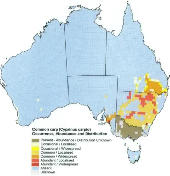
Figure 1.1 Common carp (Cyprinus carpio) occurrence, abundance and
distribution in Australia 6
Figure 1.2 Putative mechanisms of fish sex determination and differentiation. 21 Figure 2.1 Dorsal and lateral view of common carp brain sections used for
RNA extraction 55
Figure 3.1 Phylogenetic tree of vertebrate aromatase proteins. 67 Figure 3.2 Expression of both aromatase genes within tissues showing
differential expression. 69
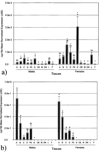
Figure 3.3 Tissue- and sex-specific differences in adult common carp of a)
cypl9a and b) cypl9b expression. 71
Figure 3.4 Temporal expression of cypl9a and b in embryos and larvae reared
at 20 or 25 °C. 72
Figure 4.1 Temporal changes in aromatase expression and TL in larvae reared
at 20 or 25 °C. 96
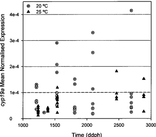
Figure 4.2 Expression of cypl9a in individual larva greater than 20 mm total
length at two temperatures (4) 20°C and • 25 °C) 97
Figure 4.3 Putative females and males as determined by cluster analysis with
size and MNE of cypl9a in larval common carp 98
Figure 4.4 Percentage of male and female common carp 5 cm TL) in groups
reared at 20 °C and 25 °C 99
Figure 5.1 Sequenced 5' flanking region of common carp cypl9a gene. 121 Figure 5.2 Sequenced 5' flanking region of common carp cypl9b gene. 122 Figure 6.1 Phylogenetic tree of available FoxL2 proteins 139 Figure 6.2 Tissue- and sex-specific differences in adult common carp foxL2
expression. 141
Figure 6.3 Temporal changes in length and foxL2 expression of embryos and
larvae reared at 20 or 25 °C 142
Figure 7.1 Gel picture of divrt1 reaction fragments of 5' and 3' nested RACE 158 Figure 7.2 Phylogenetic tree of available DMRT1 proteins 159 Figure 7.3 Tissue- and sex-specific differences in adult common carp churn
expression. 160
Figure 7.4 Temporal changes in TL and churn expression of embryos and
larvae reared at 20 or 25 °C 161
Figure 8.1 Phylogenetic tree of Sox9 proteins. 178
Figure 8.2 Comparative expression of common carp sox9 genes within tissues showing significantly higher expression of sox9a than sox9b. 180 Figure 8.3 Expression of (a) sox9a and (b) sox9b in select adult common carp of
tissues. 182
Figure 8.4 Temporal changes in length and Sox9a and b expression of embryos
I would like to thank my supervisors Dr. Jawahar Patil and Prof. Chris Carter for
giving me the opportunity to undertake this PhD and for their continued guidance,
support and encouragement. Your large efforts in the final stages of thesis
preparation, while contending with personal deadlines and major upheaval, are much
appreciated.
I would especially like to thank Rasanthi for her friendship and assistance in the lab.
Your support and knowledge were essential for the development of my real-time
assays. Your encouragement (especially for knock-off drinks) has helped me through
this PhD.
Thanks also to the CSIRO Carp group for your assistance in the lab and ongoing
support and interest. Thanks especially to Janina for your friendship and enforcing
morning coffee breaks!
Thanks to the team at K&C Fisheries for their assistance collecting broodstock,
maintaining and sampling the larval trials over the many months. Thank you to
Keith, Cate and Piggy for your friendship and for welcoming me into your home for
several weeks at a time, and enduring the .5hourly sampling though the night.
Thanks also to Chris and the boys on the Inland Fisheries Service Carp Team for
suppling fish when needed and for hunting down females when they could. Your
efforts and Christmas parties won't be forgotten. I wish you all the best with the
encouragement. Your ongoing friendship bespite my absence is invaluable.
I wish to thank my family for their ongoing encouragement and support thoughout
my many years of study. Thank you for putting up with my stress and moods. Thanks
also for always welcoming me home with good food, good wine and good company.
Finally I would like to thank Justin for your encouragement and for supporting me,
despite me moving to the opposite end of the state. You have provided a light at the
Chapters 3, 4, 6, 7 and 8 were prepared as scientific manuscripts as identified below
and on the first page of each chapter. In all instances the candidate had the primary
responsibility. The supervisors contributed to the experimental design and
implementation of the research program, data analysis, interpretation of results and
manuscript preparation.
Chapter 3: Barney, M.L. (70%), Patil, J.G., Gunasekera, R.M. and Carter, C.G.
(2008). Distinct cytochrome P450 aromatase isoforms in the common carp
(Cyprinuscarpio):
Sexual dimorphism and onset of ontogenetic expression. Gen. Comp.
Endocrinol., 156 (3), 499-508.
Chapter 4: Barney, M.L (70%), Patil, J.G. and Carter, C.G. Expression of
Cytochrome P450 Aromatase through the period of sexual differentiation in the
common carp
(Cyprinus carpio)reared at two temperature regimes. In preparation.
Chapter 6: Barney, M.L (80%) andand Patil, J.G. Cloning and expression
of foxL2in
the common carp
(Cyprinus carpio):Sexual dimorphism and developmental
expression. In preparation.
Chapter 7: Barney, M.L (80%) and Patil, J. Cloning and expression of
dmrtlin the
common carp
(Cyprinus carpio):Sexual dimorphism and expression during
development. In preparation.
Chapter 8: Barney, M.L (80%) andand Patil, J.G.
Sox9genes in the common carp
(Cyprinus carpio):
Cloning and expression in adults and during development. In
Patil, J.G. (CSIRO) was the primary supervisor and assisted with general
supervision of all aspects of this thesis. This includes expremental design,
interpretation of data and proof reading manuscripts.
Carter, C.G. (UTas) mainly assisted with general supervision and with proof
reading manuscripts.
Gunasekera, R. (CSIRO) assisted with development of the real-time assays and
undertook cloning of the cypl9a promoter.
We the undersigned agree with the above stated 'proportion of work undertaken' for each of the above published (or in preparation) peer-reviewed manuscripts contributing to this thesis.
Co-Supervisor: Co-Supervisor: Dr Jawahar Patil Professor Chris Carter
Head of School: Professor Chad Hewitt
amino acids
arylhydrocarbon receptor Aromatase Inhibitor Analysis of Variance Adapter primer
Androgen response element AhR/nuclear translocation factor benzo(a)pyrene
base pairs
CCAAT enhancer binding protein Cyclic adenosine monophosphate Complementary DNA
cAMP response elements
Commonwealth Scientific and Industrial Research Organisation Cycle Threshold
Cytochrome P450, family 19 (aromatase) Daughterless Carp Project
degree days post fertilisation degree days post hatch Doublesex Mab-3
DM domain ovarian specific
doublesex and Mab-3-related transcription factor DM-related transcription factor Y
Deoxyribonucleic acid days post fertilisation days post hatch Estrogen
Ethylenediaminetetraacetic acid Estrogen response element
Food and Agriculture Organisation of the United Nations Fork head
winged helix/forkhead transcription factor L2 Genetic Sex Determination
Gene Specific Primer
Invasive Animal Co-operative Research Centre kilo base
male aberrant factor 3 Mean Normalised Error NGFI-B/Nur77 binding site nerve growth factor inducible-B Nested Gene Specific Primer Open Reading Frame
RNA Ribonucleic acid
RT-PCR Reverse Transcription PCR SE Standard Error
SNK Student-Newman-Kuels Sox9 SRY-box 9
SRY Sex determing region Y
TCDD 2,3,7,8-tetrachlorodibenzo-p-dioxin TL Total length
TSD Temperature Sex Determination UPM Universal Primer Mix
CHAPTER I
Chapter 1 - General Introduction
1.1 Introduction to common carp, Cyprinus carpio
Common or European carp, Cyprinus carpio, is a teleost (bony) fish belonging to the
order Cypriniformes and family Cyprinidae. The species is recognised by small eyes,
thick lips with two barbels at each corner of the mouth, large scales and strongly
serrated spines in the dorsal and anal fins. Colour can vary, but is often olive green to
silvery grey dorsally, fading to silvery yellow on the belly. Small common carp can
be confused with goldfish, Carassius auratus, however, the latter has no barbels on
the corners of the mouth.
The common carp is native to the Black, Caspian and Aral Sea drainages of western
Asia from where it spread east into Siberia and Asia and west into Europe during the
Pleistocene-Holocene transition. Over the last 200 hundred years it was translocated
or introduced to throughout the world for the purposes of establishing new fisheries
and has consequently now established on all continents except Antarctica (Berg
1964, cited in Balon, 1995).
Kirpitchnikov (1967) recognised four subspecies of carp: C. carpio carpio (Europe),
C. c. aralensis (Central Asia), C. c. haematopterus (East Asia) and C. c.
viridiviolaceus (southeast Asia). However, in the few cases where populations from
different geographical regions were examined two genetically distinct
groups-European and East Asian- could be detected (Brody et al., 1979; Gross et al., 2002;
Kohlmann and Kersten, 1999; Paaver, 1983) supporting the existence of two
subspecies, C. c. carpio and C. c. haematopterus, formerly distinguished on the basis
of differentiation supports the subspecies status of C. c. carpio assigned to European carp and C. c. haematopterus assigned to Asian carp but does not justify a separate subspecies status (C. c. aralensis) for the Central Asian carp. European and Central Asian carp are closely related and there is evidence for a single origin of present day
European domesticated and wild/feral carp from a common ancestor with Central
Asian carp (Kohlmann et al., 2003). The species investigated in the current study are
of the European strain, C. c. carpio and will be hereafter referred to as common carp.
1.1.1 Common carp as an aquaculture species
Common carp is an important freshwater aquaculture finfish species, probably with
the earliest domestication records among fishes, going back approximately 4000
years with a book called Kwai Sin Chak Shik written during the Sung Dynasty in A.D. 1243, describing the transportation and trading of 'carp' fry in bamboo baskets
(Balon, 1995, 2004). According to the Food and Agriculture Organisation (FAO)
(2007) common carp had the 4th highest production of all aquaculture species
(including fish, crustaceans and molluscs) with over 3.1 million tonnes produced in
2006. The majority of commercial common carp production is from polyculture in
China, with additional intensive pond and tank culture in Europe and Israel and
semi-intensive pond polyculture in India (Carter, 2006).
1.1.2 Common carp as a research species
As well as a food source, the common carp has been used extensively in fish biology
and aquaculture research (for review see Horvath and Orban, 1995; Hulata, 1995).
Common carp is believed to have evolved from allotetraploidization (species
hybridization) and many loci are still expressed in duplicate (David et al., 2003;
Larhammar and Risinger, 1994). Its chromosome number (2n=100) is twice that of
al., 1967). Additional evidence for carp tetraploidy is that many enzymes are
expressed in duplicate (Engel et al., 1971; Ferris and Whitt, 1977). Enzymes which
appear to be expressed as one variant only are assumed to have another that has
become a pseudogene. Using the proportion of lost duplicate loci to estimate the time
elapsed since the tetraploidization event, it is suggested that tetraploidy in carp is
more ancient than in salmonids with carps found to express fewer duplicate loci than
tetraploid salmoid fishes (Engel et al., 1971). Using c-myc genes the tetraploidization
event of common carp is estimated to have taken place 58 MYA (Zhang et al., 1995).
Other duplicated common carp genes suggest a more recent divergence time of less
than 16 MYA (Larhammar and Risinger, 1994). Microsatellite analysis found two
distinct modes in distribution of differences between paralogs, suggesting one
whole-genome duplication (about 12 MYA) and a more recent wave of segmental
duplications (between 2.3 and 6.8 MYA)(David et al., 2003). It is therefore likely
that common carp and goldfish derived from a shared ancestor after the
tetraploidisation event as goldfish have the same number of chromosomes (Ojima et
al., 1966) and can form hybrids with common carp (Hubbs, 1955; Hume et
al., 1983).
Larhammar and Risinger (1994) state that the absence of multiple forms of some of
the proteins could be due to complete absence of electrophoretically detectable
differences between duplicate loci because of a much shorter time since
tetraploidization. The coding capacity at both common carp loci seems to be
maintained and both loci give rise to somatotropin mRNA as cDNA clones which
have been isolated from both loci (Chao et al, 1989, Koren et al., 1989).
Furthermore, it was found that numerous insertions/deletions have occurred in the
3'UT but not in the coding region, indicating that there is still a selection against
species an interesting evolutionary model for examining tetraploidisation, with a role
for polyploidisation in speciation and diversification.
As well as being important in aquaculture, common carp are often used as a sentinel
species in studies investigating potential alterations to the function of the endocrine
system of humans and wildlife as a result of chemical exposure (Lavado et al., 2004;
Moens et al., 2007; Moens et al., 2006).
Segregation and sex-reversal studies also widely use common carp, however
genomic information is limited; currently only 2262 nucleotide and 2325 protein
sequences are available for common carp in GenBank, compared to 171,119
nucleotides and 71,687 proteins for zebrafish, Danio rerio (14/07/09
http://www.ncbi.nlm.nih.gov/) . As a popular aquaculture species, with established
methods of artificial spawning and rearing, common carp is a traditional subject for
chromosome set manipulation studies since it has high fecundity and unique
morphological genetic markers such as genes for scale cover types and colour
mutations (Gomelsky, 2003; Horvath and Orban, 1995).
1.1.3 Common carp as a pest species
In parts of Australasia and America common carp are an introduced species widely
regarded as a pest and a threat to endemic ecology (Koehn, 2004; Zambrano et al.,
2006). In Australia, they were first introduced over 100 years ago (Brown, 1996) and
there have been at least three separate introduction of distinct strains or genetic-types
(Kailola et al., 1993) including Koi, a colourful variety of the same species (Davis et
al., 1999). It was not until the early 1960's that the `Boolarra' strain, that was
IN Present - Abundance / Distribution Unknown Occasional / Localised
Occasional / Widespread Common / Localised Common / Widespread Abundant / Localised Abundant / Widespread Absent
Unknown
Common carp (Cyprinus carpio) Occurrence, Abundance and Distribution
into the Murray-Darling basin, is believed to have caused an upsurge in common
carp abundance becoming very widely distributed in little more than 10 years
(Shearer and Mulley, 1978). Common carp are now widely distributed throughout
[image:25.547.129.464.182.528.2]south-eastern Australia including Tasmania (IFS, 2004) (Figure 1.1).
Figure 1.1 Common carp (Cyprinus carpio) occurrence, abundance and distribution in Australia (Courtesy IA CRC, 2008)
Their adaptability and tolerance to poor water quality allow common carp to inhabit
a wide range of conditions and dominate the fish communities of the southern
Murray-Darling Basin (Gehrke et al., 1995; Harris and Gehrke, 1997). The
abundance of native fish populations and species diversity in the Murray-Darling
Basin has reportedly declined over the last century (Bomford and Tilzey, 1997;
parts of their distribution (Cadwallader and Gooley, 1984). In contrast, common carp
have undergone a rapid expansion in range and abundance throughout this region
since their introduction in the 1960's (Koehn et al., 2000). The life-history of
common carp is one characterised by flexibility, with long breeding seasons (up to 9
months), the ability to spawn multiple times each year (Smith and Walker, 2004) and
with high fecundity (Sivakumaran et al., 2003). Floodplains and slow flowing
pool-type habitats appear to be the preferred spawning habitat (Sivakumaran et al., 2003;
Stuart and Jones, 2006). Male and female common carp are long-lived (up to 28
years) and mature relatively early compared with similar-sized native fish (Brown et
al., 2005).
Common carp also have significant impacts on aquatic systems, causing extensive
habitat and water quality degradation and altering composition, abundance and
diversity of macrophytes as well as diversity of macroinvertebrates (Fletcher et al.,
1985; Miller and Crow!, 2006; Roberts et al., 1995) as well as increasing water
turbidity and nutrient concentrations (Driver et al., 2005; King, 1997). There is still
some debate however as to what extent common carp are the cause of major
disturbances in freshwater systems and to what extent they are a response to
disturbance, after which they may gain opportunistic dominance (Harris and Gehrke,
1997). Common carp also pose an economic threat to industries that depend on
pristine water quality and aquatic habitats, including domestic and irrigation water,
agriculture, tourism and commercial and recreational fisheries (Adams, 2008).
A suite of techniques has been implemented to control the abundance and
environmental impacts of common carp. These include commercial and recreational
spawning, biomanipulation (stocking with predatory fish), exclusion with screens or
barriers, poisoning and biological control, bioacoustics and bubble barriers, and
genetic manipulation (Berg et al., 1997; Gervai et al., 1980; Koehn et al., 2000;
Propst and Gido, 2004; Shields, 1958; Stuart and Jones, 2006; Stuart et al., 2006;
Taylor et al., 2005).
Several methods of common carp management have been discussed in Australia
(Roberts and Tilzey, 1996) including employment of a daughterless trait, a heritable
sex ratio manipulation mechanism predicted to cause localised extinction of
populations (Hamilton, 1967; Werren et al., 1981). The Murray-Darling Basin
Commission is supporting the Austral-Asian Invasive Animal Co-operative Research
Centre and its research partner CSIRO Marine Research to undertake the
Daughterless Carp Project (DCP) as a tool to control common carp in the basin. The
DCP focuses on genetically manipulating the population sex ratio of common carp
by introducing exclusively male-bearing fish. Through the use of gene technology, it
is hoped to introduce multiple copies of a `daughterless' gene into common carp
which would be periodically released into the wild. Copies of the gene are carried by
the males, and introgessed though the pest population, resulting in male bias of
population sex ratios and ultimately resulting in drastic reduction of female offspring
in the population and potentially leading to localised extinction of pest populations.
The research described in the current thesis is part of this project as it explores
potential candidate genes (Section 1.4.3) in the sex determination pathway of
common carp that may be targeted to manipulate sex ratios. At the start of this
The primary gene target for the `daughterless' approach is the ovarian isoform of
P450 aromatase gene (cypl9a) due to its essential role in estrogen synthesis and
ovarian development (see section 1.4.3.1). Estrogen is required for many functions
and in many tissues, it is therefore important to know where cypl9a is locally
expressed, and co-expressed with the brain isoform, to determine potential undesired
or compensatory effects of 'switching off this gene. However, this information was
not available in the literature and the current thesis not only identifies both ovarian
and brain aromatase isoforms but also demonstrates tissue- and sex-specific
expression in adults and onset of timing. As aromatase is the primary target for the
DCP, addition effort was made to clone and identify the promoter region of both
aromatase isoforms to determine potential regulatory element involved in
transcription. FoxL2 is one such regulatory element of aromatase (section 1.4.3.2 and
Chapter 6). This thesis describes, for the first time, the cloning and expression of this
gene in adult and developing common carp. In addition, cloning and the expression
profiles of dmrtl (section 1.4.3.3 and Chapter 7) and sox9 (section 1.4.3.4 and
Chapter 8) are reported; both factors involved in male development.
Exploring the molecular pathways that lead to ovary and testis development in
teleost fish will result in a greater understanding of sex determination and
differentiation in all vertebrate species. More specifically this allows greater
understanding of a potential 'Achilles heel' or vulnerable aspects of sexual
development that may be used to control sex ratio and in turn, pest species. Examples
of this would be controlling phenotypic sex by inducing or repressing expression of
critical genes involved in development of either females (cypl9a -see Chapter 4) or
males (dmrtl -see Chapter 7). More generally, the ability to manipulate sex ratios of
bias can result in extinction, and also aquaculture, where monosex populations can
improve production efficiency.
This introductory chapter will summarise what is understood of sex determination
and differentiation mechanisms in fish, with particular focus on gonochoristic species
including the common carp. Also reviewed are candidate genes of interest that have
also been explored in this thesis to elucidate their role in sex determination and their
potential as candidates for developing daughterless technology.
1.2 Conserved sex-determining pathways and mechanisms
In mammals, there is greater knowledge of the molecular mechanisms of sex
determination and differentiation (Brennan and Capel, 2004) allowing investigation
into other vertebrate systems through comparative studies. Comparative studies
between whole genomes of mammals and fish have shown significant similarities
(Aparicio et al., 1995; Postlethwait et al., 2000) suggesting that some sex
differentiation genes of the regulatory hierarchy may be conserved (foxL2 for
example Baron et al., 2005a).
In contrast to many developmental processes, sex-determining mechanisms show no
clear evolutionary conservation between phyla. For example the mammalian
male-inducing master gene, SRY (sex-determining region Y), seems to be specific to
mammals (Capel, 2000). As there is no sex-specific SRY homologue in birds or any
reptile, SRY is unlikely to have been the original sex-determining gene in the
However, it has been shown that sex chromosomes of even widely divergent groups
now appear to have changed very little over the last 300+ million years. Even
independently derived sex chromosomes seem to have followed the same set of
evolutionary rules; male heterogamety (XX female:XY male system-typified by
mammals), female heterogamety (ZW female:ZZ male system- in birds and snakes),
as well as a variety of genetic and environmental sex determining systems (found in
teleosts and reptiles) (Graves and Shetty, 2001). The sex-determining pathway seems
to be extremely conserved, although the control of the genes in this pathway is vested
in different elements. Furthermore it has been suggested that sex-determining genes
can be made redundant, and replaced by control at another step of a conserved
sex-determining pathway. The possibility of several genes acting as a sex switch may
have led to the evolution of new sex-determining/chromosome systems.
Sex-determining systems in fish appear to be at a primitive stage of evolution
(Charlesworth, 2004; Nanda et al., 1992; Solari, 1994; Thorgaard, 1977), but
collectively represent a wide variety of systems ranging from monogenic to
polygenic systems. Moreover the phenotypic sex is known to be influenced or
manipulated by environmental factors such as temperature and exogenous treatment
of sex steroids. Therefore, knowledge of the relationships between sex-determining
genes, sex steroids and environmental factors will provide critical information to
understand animal sex determination and sex differentiation in general and
particularly to elucidate the conserved mechanisms that operate behind sex
determination in vertebrates.
Genetic mechanisms which trigger sex determination appear to be diverse in non-
mammalian vertebrates, especially in teleost species that exhibit male heterogamety,
Nagahama, 2002; Godwin et al., 2003; Matsuda, 2003; Nakamura et al., 1998).
However, studies indicate that many downstream gene products of sex determination
genes are functionally similar in diverse species. Many steroidogenic pathways and
genes that are important in sex determination in mammals are also conserved in other
vertebrates and show gonad-specific expression during the period of sex
determination (Matsuda, 2003).
Despite a range of different mechanisms, morphological development of the gonads
in all vertebrate groups appears to have been conserved through evolution. The basic
structure of the testis is highly conserved among vertebrates, and strong parallels in
invertebrates such as the fly, Drosophila melanogaster (Drosophila) suggest that the
mechanistic pathways that control the architecture and cellular development of the
organ are also conserved (Capel, 2000). For these reasons, information about
molecular pathways of gonadogenesis in other animals may provide valuable insights
into the mammalian system.
Investigating the coordinated interaction between the transcription and hormonal
factors of sex determination may be important to understand the mechanisms that
lead to testicular and ovarian differentiation. Among the vertebrates, fish exhibiting a
wide range of gonadal differentiation types including gonochorism and
hermaphroditism will continue to provide an excellent model for the comparative
study of sex determination and gonadal sex differentiation. Greater understanding of
these processes in fish will broaden our understanding of vertebrate sex
determination and differentiation, and will have important applications for fisheries
and aquaculture (Devlin and Nagahama, 2002). For example, elucidation of a
sex-determining gene would contribute toward reliable production of monosex fish for
1.3 Sex determination and differentiation in fish
The expression of sex is governed by two processes: sex determination (for the
genetic or genotypic sex) and sex differentiation (for the gonadal or phenotypic sex).
Sex determination refers to the mechanisms that direct sex differentiation, whereas
sex differentiation refers to the development of testis or ovaries from
undifferentiated or bipotential gonad (Hayes, 1998). Differentiation also includes
sexually influenced development of morphology, behaviour and biochemical
secondary sex characteristics.
Sexual dimorphism is a common trait among vertebrates (Morrish and Sinclair,
2002; Sinclair et al., 2002; Zarkower, 2001) and is achieved by complex regulatory
cascades that direct an early bipotential embryo down either a male or female path of
sexual development (Devlin and Nagahama, 2002; Swain and Lovell-Badge, 1999).
Although detailed morphological information on sexual dimorphism has been well
documented in many species of fish, the molecular pathways of sex determination
and differentiation are relatively unknown (Devlin and Nagahama, 2002; Penman
and Piferrer, 2008; Piferrer and Guiguen, 2008).
It appears that mechanisms of primary sex determination evolve rapidly 'with initial
sex-determining cues at the top of the regulatory hierarchy differing greatly between
species (Zarkower, 2001). This is particularly evident in fish, which due to the great
number of species (c. 31,100) and the large variety of aquatic environments
inhabited, display extreme diversity in modes of reproduction and sex-determining
systems, even among closely related species (Bernard, 2005; Devlin and Nagahama,
1.3.1 Types of sex determination
In teleost fish, sex determination is influenced by a balance of genetic, steroidogenic
and/or environmental processes (Devlin and Nagahama, 2002). The interactions
between these factors are the primary control directing the course of sex
differentiation and the associated physiological processes supporting gonadal
development and function. Many fish have genetic systems that direct gonadal
differentiation during early development and maintain the differentiated state
throughout adulthood, or in the case of sequential hermaphrodites, until the pattern of
gene expression changes and sex reversal occurs in response to age or environmental
cues (Sadovy and Shapiro, 1987). Known types of genetic sex determination in fish
are: male (XX/XY) and female (ZZ/ZW) heterogamy, such as in medaka, Oryzias
latipes (Matsuda, 2005) and the lizardfish, Trachinocephalus myops, (Ueno et al.,
2001) respectively; multiple sex chromosomes (X, Y, Z, W) as in platyfish,
Xiphophorus maculatus (Volff and Schartl, 2001); polygenic sex determination
(postulated for zebrafish (Lawrence et al., 2008) and pufferfish; fugu, Takifugu
rubripes and Tetraodon, Tetraodon nigroviridis (Li et al., 2002)); and autosomal
genes which can override the sex-determining activity of the primary sex
chromosomes, as seen in swordtails, Xiphophorus Poeciliidae (Devlin and
Nagahama, 2002). Although morphologically distinct sex chromosomes cannot be
identified in common carp (Kirpichnikov, 1981), sex determination is thought to be
of the XX/XY system with conventional diploid offspring yielding 1:1 sex ratios
(Cherfas et al., 1994a; Komen et al., 1992; Manzoor Ali and Satyanarayana Rao,
1989).
Environmental influences have an impact on sex determination. Temperature is a
Ictalurus punctatus (Patino et al., 1996), pejeffey Odontesthes bonariensis
(Striissmann et al., 1996a; Striissmann et al., 1996b), with a specific temperature
range inducing sex-specific development. Dissolved oxygen levels have also been
suggested to alter sex ratio, with hypoxia leading to male-biased populations of
zebrafish (Shang et al., 2006). Another factor found to influence phenotypic sex in
some species is pH, where high and low pH usually result in female and male-biased
offspring, respectively (Baroiller et al., 1999; Rubin, 1985).
In highly evolved heterogametic systems, the mode of gonadal differentiation is
expected to be gonochoristic, however sex chromosomes have also been reported in a
few hermaphroditic species (Devlin and Nagahama, 2002). Environmental factors
causing sex inversion in hermaphroditic fish have been extensively reviewed
(e.g. Baroiller et al., 1999; Devlin and Nagahama, 2002; Godwin et al., 2003; Ross,
1990). However, there is limited knowledge on the genetic mechanisms that regulate
hermaphrodism, and it is unknown if primary sex determination is indeed regulated
by genetic or environmental factors. A study on mangrove rivulus, Rivulus
marmoratus indicates that temperature can influence sex determination as well as
sex-inversion in hermaphroditic fish, with low temperature increasing the occurrence
of primary males (5-35%) and increased temperatures induced sex-inversion
(ie. secondary male production) (Harrington, 1967, 1968).
1.3.2 Types of gonadal differentiation
Teleostean fish demonstrate diversity in reproduction, with different modes of
gonadal differentiation including gonochorism, hermaphroditism, and unisexuality
and other factors are capable of redirecting the gonadal development (Devlin and
Nagahama, 2002; Nakamura et al., 1998).
Gonochoristic fish develop as either males or females and retain the same sex
throughout adulthood (Devlin and Nagahama, 2002; Yamamoto, 1969). There are
two types of gonochorism, differentiated and undifferentiated. In differentiated
gonochorists, ovarian and testicular differentiation proceeds directly from the
undifferentiated gonad (e.g. common carp, Cyprinus carpi° (Komen et al., 1992) and
European seabass, Dicentrarchus labrax (Blazquez et al., 1998)), while
undifferentiated gonochorists pass during development through a non-functional
female phase when the gonads of all individuals, irrespective of the genetic sex,
display primary stage oocytes (e.g. zebrafish, Danio rerio (Takahashi, 1977) and
Sumatra barb, Barbus tetrazona). Later on, the population differentiates into males
and females in approximately equal proportions. However, social factors such as
density and relative size of juveniles in a cohort has been found to influence
sex-determination in some species (Francis and Barlow, 1993; Francis, 1984).
Hermaphrodite fish function as both sexes, either simultaneously/synchronous or
sequentially, at some time during their life. Synchronous hermaphrodites have the
ability to produce male and female gametes simultaneously (e.g. Serranus sp.
(Oliver, 1991, 1997; Petersen, 1990; Pressley, 1981) or Gobiidae (Cole, 1990; St.
Mary, 1998), while sequential hermaphrodites undergo sex-change from
male-female (protoandry) (e.g. Gilthead seabream, Sparus aurata (Brusle Sicard and
Fourcault, 1997) or female-male (protogyny) (e.g. wrasse, Thalassoma bifasciatum
2002). There is yet another set of species that exhibit bidirectional sex reversal
(Munday et al., 1998).
1.4 Molecular mechanisms of sex determination and differentiation in fish
1.4.1 Molecular mechanisms of sex determination
A complex series of interacting biochemical processes are involved in the sex
determination pathway. At some point early in the cascade a primary
sex-determining gene in activated, resulting in a commitment to the production of either
male or female gametes (Devlin and Nagahama, 2002; Wilkins, 1995). In genetic
systems, mechanisms range from purely polygenic controls, to those with dominant
sex-determining factors mixed with autosomal controls, to highly evolved sex
chromosomes with heterogametic (XY) males or heterogametic (ZW) females (Bull,
1983). In worms and flies, sex is determined by the ratio of X chromosomes to
autosomes (Cline and Meyer, 1996; Parkhurst and Meneely, 1994). In mammals, sex
is determined genetically by the presence or absence of a Y chromosome (Capel,
2000; Swain and Lovell-Badge, 1999).
Fishes provide a rich material for studying the evolution of sex chromosomes.
Although sex determination in most fishes is likely regulated by genetic factors,
relatively few teleosts have karyotypically distinct sex chromosomes (Arkhipchuk,
1995). In most species, the sex chromosomes are still in early stages of
differentiation, and do not show distinct differences in length or gene content (Lee et
al., 2004). Both XY and WZ gonosomal systems have evolved repeatedly in various
groups of fishes (Devlin and Nagahama, 2002). However, additional autosomal loci
also contribute to sex determination in many species (Kosswig, 1964). Master genes
Sinclair et al., 1990). However, identifying sex-determining loci in fish has been less
successful, with no apparent sex chromosomes and no sex-linked loci or molecular
markers discovered in popular fish models, such as zebrafish, fugu and Tetraodon
(Li et al., 2002; Schartl, 2004), suggesting polygenic sex determination.
Within the pathway of genetic systems, certain components, or combinations of
components, may become dominant in influencing the direction of sex determination
such that environmental factors have little influence. In polygenic systems, the sex of
a particular individual will be determined by the strength of the genetic factors it
receives from its parents with some genes directing ovarian and others testicular
development. Over time, one component may gain such influence over the direction
of the pathway that other genetic loci do not override its effects. If this occurs, sex
will be determined by a simple, single locus genetic system, and sex chromosomes
may develop (Devlin and Nagahama, 2002).
Species such as medaka (Matsuda et al., 2002) and platyfish, Xiphophorus
(Schultheis et al., 2006) have been shown to possess sex chromosomes and have a
well defined sex-determining region on the Y chromosome. However, only in
medaka has the primary sex-determining gene been revealed in the sex-determining
region. This gene encodes a conserved DM-domain protein, DMY (Matsuda et al.,
2002; Nanda et al., 2002). DMY is a new doublesex/mab-3 gene most closely related
to DMRT1 (Kondo et al., 2003). DMY expressed specifically in the somatic (sertoli)
cells surrounding the primordial germ cells prior to morphological sex differentiation
(Nagahama, 2005). Additionally, the DMY protein is localised to the sertoli cell
nuclei continuously from embryonic stages to adulthood, suggesting that it is an
the differentiated state of the testis (Nagahama, 2005). Loss/gain function studies
provide strong evidence that DMY is the master sex-determining gene in this
species; with naturally sex reversed XY females showing mutations affecting DMY,
and transgenic studies with a genomic DMY fragment showed that transgenic XX
fish develop into males, in some cases fully fertile (Nagahama, 2005).
Sequence comparison showed that DMY had greatest homology with DMRT1,
which in vertebrates has a conserved role in male gonadal development (Lutfalla et
al., 2003; Nanda et al., 2002). It is suggested that DMY originated from a duplicate
copy of autosomal dmrtl, that in a new genetic environment acquired a determining function (Matsuda, 2005). However it may not represent a general
sex-determining gene in fishes as it has not been found in a distant species of medaka or
any other fish species studied (Kondo et al., 2003). Some strains of medaka with
uncompromised sex-reversed XX males, indicate that autosomal factors can
over-ride the role of DMY as a male determinant (Nanda et al., 2003). This highlights that
the genes at the top of the sex-determining cascade leading to males and females are
highly variable in vertebrates and particularly fish.
The platyfish is another interesting model for sex determination study, as it belongs
to the Poeciliidae family which displays diverse genetic sex-determining systems
ranging from simple heterogametic, to polyfactoral sex determination (Volff and
Schartl, 2001). With three different but homologous sex chromosomes (X, Y, Z),
platyfish can be XY or YY males and XX, XW or YVV females (Volff and Schartl,
2001). These sex chromosomes appear to be an early stage of differentiation as there
are no karyomorphological differences and viable YY males. This multiple sex
gonosomes but only the Y-linked allele/s are expressed, with X and W alleles
repressed by autosomal factors or b) a dosage dependant mechanism, with a varying
copy numbers of the sex-determining genes on each sex chromosome (Volff and
Schartl, 2001). While the sex-determining loci in platyfish remain unknown, recent
molecular analysis of the Y chromosome's sex-determining region has revealed a
number of potential candidate genes for this species, however all gene candidates
identified so far have been detected on both the X and the Y, reflecting the poor
degree of differentiation of the gonosomes and their probable recent origin (Bane et
al., 2008; Schultheis et al., 2006; Volff et al., 2003). Even if it can not be excluded
that some genes are functional only on one type of sex chromosome, this contrasts
with the situation observed for the master sex-determining genes in medaka (DMY)
and mammals (SRY), which are specific to one sex chromosomes, the Y. This further
emphasises the recent evolution of primary sex-determining genes in fishes. None of
the genes identified so far in the Y sex-determining region of the platyfish are similar
to known sex-determining genes such as dmrt (DMY or doublesex) or HMG-domain
genes (SRY or sox9) described in other organisms (Schultheis et al., 2006).
Although a greater understanding of sex-determining loci is being developed as more
species of fish are being examined, the timing and influence of these sex-determining
mechanisms on sex steroid production and consequent gonadal differentiation remain
unclear.
1.4.2 Molecular mechanisms of gonadal sex differentiation
Although much is known about the process of sex differentiation in fish, the precise
mechanisms underpinning sex differentiation as well as those involved in primary
sex-determination remain undefined (Devlin and Nagahama, 2002). Fish gonadal sex
SF 1 FoxL2 -
\
...., ..41P,.... ." • '..
, —
0
0 z I -- • u ■
-4
CROSS TALK> SF-1 i
1 g
P. r> ...
—
-- Daxl Wnt47 14' — — —• ,
°
Z oDMY? i
cd 0
0
1 1
Chapter 1. General Introduction
found in mammals (Baron and Guiguen, 2003). The differentiation of the bipotential
gonad to either testis or ovaries relies on the regulation of the steroidogenic pathway.
In lower vertebrates, sex steroid (estrogens and androgens) play a critical role in
gonadal differentiation (Devlin and Nagahama, 2002; Striissmann and Nakamura,
2002). This is evident from functional sex reversal reported in a number of species
following exogenous administration of sex steroid hormones (for review see
Cheshenko et al., 2008; Piferrer, 2001; Tzchori et al., 2004). In common carp it is
possible to produce meiotic gynogens (Komen et al., 1988), mitotic gynogens
(Cherfas et al., 1993b), hybrid gynogens (Cherfas et al., 1994b), induced triploids
(Cherfas et al., 1993a), hybrid triploids (Wu et al., 1993), mitotic androgens
(Bongers et al., 1994), hybrid androgens (Cherfas et al., 1994b), mitotic tetraploids
(Cherfas et al., 1993b), bred tetraploids (Cherfas et al., 1994b), and hybrid tetraploids
(Cherfas et al., 1994b). However, the mechanism of action of these steroids on gonad
sex differentiation is not well understood.
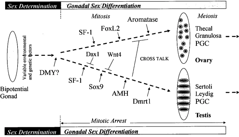
Sex Determination Gonadal Sex'Differentiation
Bipotential
Gonad
Mitosis
Aromatase
Sox9
AMH
Dmrtl
Meiosis
cl* 4 0
® (D
®
<001.44
<=>
Thecal
Granulosa — —.
PGC
Ovary
Sertoli — LeydigPGC
V
Testis Mitotic Arrest4
[image:40.546.88.502.450.687.2]IWININIMMI
Gonadal Sex DifferentiationFigure 1.2 Putative mechanisms of fish sex determination and differentiation.
Unlike sex-determining systems, the genes involved in gonadal differentiation appear
to be relatively conserved (Figure 1.2). Genes such as steroidogenic factor-1 (SF-1 or
Ad4BP), cyp19, foxL2, AMH, sox9 and dmrtl that have been identified as factors in
mammalian sex differentiation are largely conserved across fish and other vertebrates
studied, albeit some species specific differences are exhibited. The large majority of
these molecules had not been isolated, let alone characterised in the common carp.
Therefore, with the view to understand these mechanisms, as well as facilitate the
manipulation of sex in the species, the isolation, characterisation, molecular analysis
and potential interaction of four key genes (inclusive of isoforms), and their response
to temperature cues was analysed in this study. This represents two genes that may
have a role in ovarian (aromatase and foxL2) and two in testicular (dmrtl and sox9)
differentiation.
1.4.3 Candidate genes
1.4.3.1 Aromatase
The hormonal balance between estrogens and androgens appears crucial in the
process of sex differentiation in developing teleost fish. This balance relies on the
availability and activity of steroid synthesizing enzymes, and in particular
cytochrome P450 aromatase complex expressed by the cyp19 gene (Fenske and
Segner, 2004). In the majority of mammals, there is expression of only a single
functional cyp19 gene. Conversely, two genetically distinct cyp19 isozymes with
different catalytic activity and with distinct or partially overlapping expression
patterns have been identified in many teleost species including goldfish (Callard and
Tchoudakova, 1997), zebrafish (Chiang et al., 2001a; Kishida and Callard, 2001;
Trant et al., 2001); mosquitofish, Gambusia holbrooki (Patil et al., 2004); Atlantic
al., 2003). These isozymes are termed cypl 9a and cypl9b for ovarian and brain P450
aromatase, respectively, based on their exclusive or predominant expression in these
tissues. The existence of two paralogous genes is suggested to be due to the
occurrence of genome duplication early in the teleost linage (Taylor et al., 2003).
Commonly, cyp19 is considered to be closely involved in the process of sexual
differentiation in fish, based both on the profiles of the enzyme expression and
activity around the reproductive cycle and on the observations that treatment with
aromatase activators or inhibitors can lead to skewed sex ratios (Devlin and
Nagahama, 2002; von Hofsten and Olsson, 2005). Female-male sex reversal in many
fish after blocking of aromatase with inhibitors or treatment with estrogen receptor
antagonists, before or during sex differentiation, indicates estrogen signaling is
required for early ovarian differentiation (Baroiller et al., 1999; Guiguen et al., 1999;
Nakamura et al., 1998; Piferrer et al., 1994). In male tilapia, steroidogenic enzymes
were not detected until after gonadal differentiation, unlike in female fry (Nakamura
et al., 2000) indicating a greater role of steroid hormones in ovarian differentiation
than testis.
The expression of cyp19 genes is regulated by transcription factors and response
elements located in the promoter region. Analysis of the promoter region of
aromatase genes in fish shows the presence of binding sites for the piscine equivalent
to many mammalian transcription factors implicated in the cascade of sex
determination and differentiation. While transcription factors appear to be conserved
among vertebrates, response elements appear to have adapted to facilitate localized
expression of paralogous genes. For example, in fish there is a unique presence of an
brain aromatase is mainly involved in estrogen-mediated neural estrogen synthesis.
Because neural aromatase activity and cypl9b mRNA are upregulated by estrogen in
the adult goldfish brain and zebrafish embryo, it is tempting to speculate that control
is exerted directly on these EREs. No EREs are found within a region 356 bp
upstream of the predominant brain exon la of the zebra finch aromatase
(Ramachandran et al., 1999), nor has an ERE been recognized in the human or
mouse brain-specific promoter region (Honda et al., 1999; Honda et al., 1994). Thus,
estrogen effects on neural aromatase expression in avian and mammalian brain must
take place through other regulators or transcription factors.
Although the two aromatase isoforms have similar gene structures in the coding
region, the binding regions of SF-1 and SRY Isox9, which are sex-determining factors
in mammals, are present in the 5' flank region of cypl9a but not cypl9b of tilapia
(Chang et al., 2005), zebrafish (Kazeto et al., 2001; Tong and Chung, 2003) and
goldfish (Tchoudakova et al., 2001). This data indicates that cypl 9a plays a decisive
role in sex differentiation of these species. However, in medaka the SF-1 region has
been identified in both isoforms (Kuhl et al., 2005; Tanaka et al., 1995). SF-1 is an
important cis-element that regulates expression of several steroidogenic enzymes in
gonads and adrenals (Hatano et al., 1994) and has shown expression dependant on
cyp19 in diverse vertebrates (Crews et al., 2001; Lynch et al., 1993). During gonadal
differentiation in mammals (Ikeda et al., 1994), turtle, Trachemys scripta (Fleming et
al., 1999), and rainbow trout, Oncorhynchus mykiss (Baron and Guiguen, 2003),
SF-1 is up-regulated in males and down-regulated in females. However, in alligator,
Alligator mississippiensis (Western et al., 2000), American bullfrog, Rana
catesbeiana (Mayer et al., 2002), chicken (Smith et al., 1999b) and tilapia
the pattern of expression is highly variable between species, it is likely that the
interaction between SF-1 and other co-factors may cause sex-specific regulation of
aromatase, as is implicated by the paradoxical actions of gonadotropins on cypl9a
gene (Yoshiura et al. (2003).
1.4.3.2 FoxL2
Another transcription factor that shows sexually dimorphic expression corresponding
to ovarian aromatase is foxL2, a member of the winged helix/forkhead family of
transcription factors known to be involved in ovarian development granulosa cell
differentiation, and thus the proper maintenance of ovarian function (Cocquet et al.,
2003; Loftier et al., 2003; Ottolenghi et al., 2005; Pisarska et al., 2004; Schmidt et
al., 2004; Uda et al., 2004; Yao, 2005). It is the earliest known sexually dimorphic
marker, expressed in the somatic cells during early development and later in
granulosa cells surrounding the oocytes (Cocquet et al., 2002). FoxL2 has been found
to correlate with the level of cyp19 expression in a diverse range of species,
including medaka (Nakamoto et al., 2006), tilapia (Yoshiura et al., 2003), rainbow
trout (Baron et al., 2005b), Japanese flounder, Paralichthys olivaceus (Yamaguchi et
al., 2007), wrinkled frog, Rana rugosa (Oshima et al., 2008), chicken (Govoroun et
al., 2004), turtle (Ramsey et al., 2007) and mammals (Pannetier et al., 2006).
Interestingly, rainbow trout has two genetically independent paralogue genes, foxL2a
and b (Baron et al., 2004; Vizziano-Cantonnet et al., 2008; Vizziano et al., 2007).
FoxL2a expression patterns in rainbow trout are similar to that observed for the
mammalian foxL2 gene. However, foxL2b is specific to somatic cells of the ovary,
and is expressed later than foxL2a. This is perhaps an example of specialised
function of duplicated genes where other species have only maintained one
which whole genome sequences are available, it appears that foxl2 is duplicated in
many more teleosts (Baron et al., 2004; Vizziano-Cantonnet et al., 2008; Vizziano et
al., 2007).
It appears that both foxL2 and SF-1 activate cyp19 expression by binding directly to
the promoter, with foxL2 enhancing SF-1 activated transcription by forming a
heterodimer (Nagahama, 2005). Sex reversal studies show that in vivo expression in
female tilapia of a transgenic dominant-negative-mutant-foxL2 construct reduced
aromatase expression causing ovaries to degenerate and testicular tissue to develop
with complete sex-reversal resulting in viable males (Nagahama, 2005). Sex reversal
experiments in rainbow trout resulted in foxL2 expression in the ovaries of
sex-reversed XY genetic males following estrogen treatment, while female-male sex
reversal with an aromatase inhibitor resulted in greatly reduced foxL2 expression
(Baron et al., 2004). This suggests either a positive feedback loop, possibly via
estrogen, or male-promoting factors in the masculinised gonad such as dmrtl, may
influencefoxL2 transcription.
1.4.3.3 Dmrtl
In higher vertebrates, the female pathway is generally considered the default in the
absence of any male inducing factor (e.g., SRY). In humans, a sex reversing
syndrome associated with deletions of chromosome 9p is pinpointed to a critical
region at the end of the short arm, and the dmrtl was cloned from this interval
(Raymond et al., 1998). Since this human chromosome is equivalent to the bird Z
chromosome, it is not surprising to find that dmrtl maps to the chicken Z (Nanda et
al., 1999). With no copy on the W, there is a dosage difference of two copies (male)
to one (female). The gene is expressed specifically in the testis at about the same