0022-538X/80/04-0067/06$02.00/0
Temperature-Sensitive Mutants of Foot-and-Mouth Disease
Virus
with Altered
Structural Polypeptides
II.
Comparison of Recombination and Biochemical Maps
ANDREW M. Q. KING,* WILLIAM R. SLADE, JOHN W. I. NEWMAN,AND DAVID McCAHON GeneticsDepartment, Animal Virus Research Institute, Pirbright, Woking, GU24 ONF, England
The structuralpolypeptides offoot-and-mouthdisease virus were digested with
Staphylococcus aureus V8 protease in the presence of sodium dodecyl sulfate.
Theprotease-resistant peptidesderived from temperature-sensitive mutants were
compared with those ofthe wild typeby electrofocusing in a polyacrylamide gel.
Covariation between the charge shifts of different peptides indicated that they
shared common sequences: onlyfive independent peptides in all were derived
fromVP1, VP2, and VP3, accounting forapproximately 50% of the polypeptide
sequences. In twoinstances,aminoacid substitutions that caused similar shifts in
theisoelectricpointwerefound to belocatedin different peptides. However, 15
mutants that possessed identical
shifts
in VP2 could not be distinguished bypeptide
analysis.
Thepolypeptides ofrevertantsableto grow atthenonpermissivetemperature werecompared withthose of the parentalmutants. By this test, 6 of
the12distinguishable classesof coat protein mutations were found to covary with
temperaturesensitivity.Inadditionto true revertants,several phenotypic
revert-ants which possessed a second charge change, either in a
different
structuralpolypeptide orina
different
region of the samepolypeptide,were isolated. Theorientation of the recombination map was deduced from the loci of the coat
protein mutations.
Picornaviruses provide a unique example of genetic recombination between RNA molecules. Much workwasdevotedtoquantitating
recom-bination frequencies measured in crosses
be-tween markers for temperature sensitivity or
inhibitor resistance. This work culminated in recombinationmapsforpoliovirus and foot-and-mouth disease virus (FMDV or aphthovirus) which were
approximately
additive and linear (4,12, 15).The problem of
relating
agenefunctionto amap locus is
that,
thusfar,
nogenetic
markerineither the
poliovirus
ortheFMDVmaphaseverbeen correlated witha
physicochemical
charac-ter in the viral RNA or in a virus-coded
poly-peptide. In the caseofpoliovirus, considerable
progress was made
by
observing physiological
defects ofmutantsunderrestrictive conditions
(5). Most of the
map's
mutationswereassigned
tothatregionof thegenome,
approximately
40%of the
total,
that codes for structuralprotein.
However,many membersofthisclassofmutant
were also defective for other
functions,
forex-ample, RNA
synthesis.
The FMDV map
appeared
to be similar tothat of
poliovirus
inbothrespects. Aselectionof18of thetotal70
temperature-sensitive (ts)
mu-tants atvarious map lociwerefoundtobeeither
acid labileor
cystine
dependent (13;
McCahon,
unpublished observations). Since both
charac-tersindicate defects in structural protein (14),
this result implied either that the entire map
represents only the coat protein region of the
genome or that many of the ts mutants have
second mutations affecting the coat proteins.
Twenty-nine mutants were examined for viral
RNA and
antigen
syntheses at the restrictivetemperature. Differences were demonstrated
among mutants,but therewas noclear
correla-tion with the map locus (R. A. J. Priston,
un-published observations). These results expose two limitations of the physiological approach: (i) it isimpreciseinthat, for example, individual structural
polypeptides
cannotbedistinguished; (ii)pleiotropic
effects make it difficultto mapnonstructuralfunctions.
Electrofocusing
themutantgeneproducts
of-fers asolutiontotheproblem
ofphysically
lo-cating missense mutations. The accompanyingpaper (9) describes the
application
ofelectrofo-cusingtothestructural
polypeptides
of FMDVand itsmutants. In thispaper we describe the
propertiesofrevertantsandthe
electrofocusing
ofpeptidefragmentstodifferentiate among
mu-tations that cause similar
charge
changes.
Twentytsmarkersof the FMDV recombination
mapareidentifiedasmissense mutations
affect-ing structural
polypeptides.
Since the poly-67on November 10, 2019 by guest
http://jvi.asm.org/
68 KING ET AL.
peptide-coding positions are known (6, 17), we
have therebydetermined the orientation of the
map.
MATERIALS AND METHODS
The origin of the FMDV mutants was described
previously (11-13).tsmutantsnumberedless than 100
wereinduced, and those numberedmorethan 100and
ts 03 were spontaneous. Growth, purification, and
trypsintreatmentof FMDV were asdescribed inthe
precedingpaper(9).
Peptide fingerprints. Virus pellets containing 50
jigofproteinweredisrupted by heatingto 100°C for
1min in50[L of 1% sodiumdodecyl sulfate (SDS)-2%
(vol/vol) 2-mercaptoethanol-0.01 M Tris-10% (vol/
vol)glycerol. Ten microliters of 1-mg/ml
Staphylococ-cusaureus V8protease (Miles Laboratories, Inc.) in
0.01 MTris-hydrochloride (pH 7.4)-i mM EDTAwas
added, andthemixturewasincubated for1h at37°C
before the reactionwasstopped byheatingto 100°C
for1 min. Half the preparationwasloadedpergel.
Electrofocusing. Electrofocusingwas asdescribed
for method (ii) of reference 9, exceptthe
electropho-resis was continued for the times indicated in the
figurelegends.
Determination of molecular weight. Molecular
weights ofpeptides were determined from a plot of
electrophoretic mobility in SDS-polyacrylamide gel
versus thelogarithm ofmolecularweight. Standards
usedwerethe intact formsofVP1,VP2 andVP3(each
with a molecular weight of 30,000), soybean trypsin
inhibitor (molecular weight, 22,500), myoglobin
(mo-lecular weight, 16,400), cytochrome c (molecular
weight, 12,400), and insulin (molecular weights, 2,600
and3,200). Amixtureof[35S]methionine-labeled and
unlabeled FMDV polypeptides was fingerprinted as
described above, and the gel was stained. Peptide
bands were cut out of the gel and washed twice in
waterfor10min and then in 0.125 M
Tris-hydrochlo-ride (pH 7.4)-2% 2-mercaptoethanol-0.1% SDS-10%
glycerol for 30 min. Each slice was loaded, with
markers in thesamesolvent,ontoan
SDS-polyacryl-amidegel (85 by6mm) containing 17.15%acrylamide
and0.35% N,N'-methylenebisacrylamide and
electro-phoresed with the Laemmli (10) discontinuous buffer
systemfor 6.5hat40V.TheSDS-polyacrylamide gels
werestained, slicedlongitudinally, dried, and
autora-diographed todetermine thepositions of the labeled
peptides.
Isolation ofrevertants. Two methodsfor isolat-ingrevertantswereused. In the first,whichwasused
withts 12, 16, 58, 107, and 109, testtube cultures of
BHKcellswereinfectedat37°C for30minandthen
washed with acid buffer, 0.1 M NaPO4 (pH6.2), to
destroy inoculum virus. After fresh medium was
added, the cultures were incubated at 37°C for a
further 75 min, followed by 4 h at 410C. The virus
yields were plaque assayed at 41°C. Plaques were
pickedaspresumptiverevertantsand clonedat410C.
Nomore thanonerevertantwasisolatedperculture.
The second method, which was used for the other
mutants listed in Table 1, except for ts 3, was a
modification ofthe procedureof Lake and Mackenzie
(11) for isolatingmutants.Each mutant wasassayed
by the agar-cell suspension technique(3), and plaques
were allowed to form at37°C for 16 to 20 h. An agar
layercontainingtetrazolium stainwasthen added to
theplates, and the incubationwascontinued for 20 to
24 h at41°C. Plaqueswithclearlydefined halos were
picked and cloned at 41°C. Seed stocks were prepared
in small monolayer cultures at 41°C. Working stocks
were prepared by a single passage of the seed stocks inmonolayercultures at37°C.
RESULTS
Location of amino acid substitutions. Ta-ble 1 lists the 24 ts mutants of FMDV (strain 0
Pacheco) thatpossessedvariant structural
poly-peptides. All70 ts mutants of the recombination
map were screenedbyelectrofocusing forshifts
in the isoelectric points ofVP2, VP3, VP4 and
themajor fragmentofVP1, VP1L,producedby
trypsintreatmentof the virus(1).For 29 of these
mutants, thesearch was extended to intactVP1,
but no additional variants were found. Also
screened werethreets mutants not onthemap,
oneofwhich, ts3, was found to bevariant; its
properties arealso included in Table 1. Both of
the mutants that carried the guanidine
resist-ance (gs+) marker, tsgs+ 03and tsgs+ E18, had
normal structural polypeptides. The
accompa-nying paper (9) describes all of the classes of
charge shifts that could bedistinguishedonthe
TABLE 1. Summaryofelectrofocusingmutations
observedinFMDVstructuralpolypeptides
Score of revert-Location ants
Class of amino with
of mu- Mutant acid with
tation substitu-
wild-tion' type
poly-peptide
a ts 12 VPl/b 0/3
b ts 16 VP1l3b 0/3
c ts 40 VP1a 2/4
d ts 58 VP1a 0/3
e ts103 VP1a 1/4
f ts 109 VP1a 0/3
g ts 3 VP2-yb ND'
h ts65, 63, 33, 58, 62, VP2,8 11/16d
47, 59, 37, 42, 23,
61, 66, 44, 41, 67
ts 22 VP2,8 0/8
ts 109 VP2a 3/3
k ts 28 VP3a 1/7'
1 ts 107 VP3a 2/3
aa,
/,
and-y
specifypeptides producedby the actionofS. aureus V8protease in the presence of SDS.
bThesesubstitutions arelocated in polypeptide
seg-mentsthat areabsent from peptidefingerprints.
'ND, Not determined.
dTotal for this class of mutation. Inaddition, four
suppression reversions were found that affected the
samepolypeptide.
'Also foursuppression reversionsaffecting VP 1.
J. VIROL.
on November 10, 2019 by guest
http://jvi.asm.org/
FMDV RECOMBINATION MAP: COAT PROTEIN MUTATIONS 69
basis of magnitudeordirection. In several cases,
different mutants exhibited identical charge
shifts. Thus, the variant VP2 of ts 109 was
indistinguishable fromthatof ts 22, the
differ-encebetween the VP1Lof ts 16 and that of ts
109wasuncertain,and,most notably, there were 15 identical cases of altered VP2, designated
"class h" in Table1.
To discriminate among as many different
classes of mutationalevents aspossible, the
var-iant polypeptides were subjected to a simple form ofpeptide fingerprinting,adapted from the
method of Cleveland et al. (2). Virus samples were denatured with SDS and, before
electro-focusing, were
digested
withS. aureus V8pro-tease. The resulting fingerprints are shown in
Fig.1.
Atotal ofseven prominentand reproducible
peptideswerederived from VP1,VP2, and VP3.
Theorigin ofeach peptide could be inferredby aninspectionofmutantfingerprints. For
exam-ple, only one peptide, VPla, is derived from VP1, and it is clear which peptide when the fingerprints ofts 103 (gel
f)
and wild-type ts+(gel
b)arecompared.
Thistechnique enabledus to distinguish those VP1 mutants that are lo-catedwithin this peptide (ts40, 58, 103,and109)from those thatare not(ts 12and 16). Figure 1
also shows that the
6,000-dalton
VPla iscon-tainedentirely within VP1L.
Three
peptides
are derived from VP2: theapeptide
isaltered in thecaseofts' 109(Fig.
lm),whereas the other two bands are shifted
coor-dinately ints 22(gel k) and classh mutants (gel
i). Thesetwohomologouspeptides are therefore
designated VP2,B. Thus, there are three
distin-guishable locations foraminoacidsubstitutions
in VP2: the a and,B peptides and segments of
VP2 that are not represented at all in the
fin-gerprintsof Fig. 1. ts 3 (gelh) and a suppressor
ofts 37 (seebelow)arein the third category.
ts 28
(gel
n) and 107(gel o), thetwomutantsaltered inVP3, have amino acid substitutionsin
the same (a) pair of homologous peptides,
al-though the shifts were of differing magnitude.
The moreacidic(lower) of the pair co-runs with
the lower of thetwo
VP2f8
bands. Aprominentpeptide toward the low-pH end of thegradient was also assigned to VP3: in Fig. 1 it is
desig-nated
VP3,8.
None of the 0 Pacheco mutantsexhibited an alteredVP3,B; however, VP3
mu-tantsoftwootherserotype0-strains of FMDV
were found to have a variant peptide at this
isoelectricpoint (datanotshown).
Molecular weights were determined for the
peptides derived from VP1, VP2, and VP3 by electrophoresis on an SDS-polyacrylamide gel (Table 2). The seven peptides leave much of VP2,VP3,and,especially,VP1unaccounted for.
Electrofocusing the SDS-peptide complexes
to-ward
the
cathode afterpreparing the sampleasdescribed by O'Farrell et al. (16) resulted in fingerprints thatweresimilartothose of Fig.2,
but inverted (data not shown). However, no
I-
*.4,2-' 1 a Jr doa * . .j3
*
|!_
_ *m _- _ dW*3.6- -w .T.
*1 -a
. ,., I r w A .
a b c de hg h
j
* / m n oFIG. 1. Peptide fingerprints ofFMDV mutantswith altered structuralpolypeptides. Virus polypeptides
weredisruptedin SDS andelectrofocusedtoward theanodeforIh at 200V, followedby 6.5 h at400V, after
either(a)notreatmentor(btoo)digestionwith S.aureusV8protease.(aand b)ts+;(c)
ts'
previously treatedwithtrypsinto cleave VP1 toVP1L;(d)ts12;(e)ts16;(f)ts103; (g) ts 40; (h) ts 3; (i) ts 37;(j)ts37R50; (k) ts
22; (1)ts58; (m)ts109; (n)ts107;(o)ts 28.
A.
3p-
4-* - -9 VOL. 34,1980
4
on November 10, 2019 by guest
http://jvi.asm.org/
[image:3.508.105.400.410.617.2]70 KING ET AL.
TABLE 2. Molecularweights
of
peptides generatedbyproteolysis in SDS solution
Peptide Mol wt
VP1a 6,000
VP2a 10,000
VP2,83 (upper)' 10,000
VP2,8 (lower) 10,000
VP33a (upper) 11,000
VP3a (lower) 16,000
VP3,B 6,000
Upper (mostbasic)of thetwoVP2,Bbands inFig.
1.
additionalpeptideswerefound.Furthermore,no
diffusible peptides were revealed by rapidly
staining gels immediately after electrofocusing.
The smallpolypeptide, VP4,wascomparatively
resistant to proteolysis under the conditions
used forfingerprinting.
Peptide fingerprints show that the variant
formsof VP2 in ts22and109,although
indistin-guishable in the intact form, have mutations
affectingdifferentpeptides.Hence, the two
mu-tations cannot be sistersand arelistedseparately
inTable 1.Similarly,the VP1 mutations ofts 16
and 109 wereshowntoaffect differentregionsof
VP1. However, fingerprints failed to
discrimi-nate among any of the class h mutations of
Table 1: all 15 exhibited identical shiftsin the
same 10,000-dalton peptides derived from VP2. Twoexamples,ts 37and58, are shown inFig. 1.
Thus, the 26 coat protein mutations were
re-solved into 12 classes (Table 1, a through 1),
distinguishable by electrofocusing the variant
polypeptides and theirfragments.
Revertants. Revertants were selected for
their abilitytogrow at thenonpermissive
tem-perature.Electrofocusingwasthenperformedto
determine whether the variantpolypeptidehad
alsoreverted.Table 1shows that for thedouble
mutants ts 58 and 109 it is the VP2 alteration
that covaries with temperature sensitivity and
not the alteration in VP1. No covariation was
observedfor three tsmutants, ts12, 16,and 22,
althoughaminimum of three revertants of each
was studied. The mutation within ts 3 had not
beenmapped and was not tested. Theremaining
classes oftsmutation inTable 1 each scored at
least one instance ofreversion in their variant
polypeptide. Five examples only from among the
class h mutationswere tested for covariant
re-version. They all scored positive, and it is
as-sumed that all 15members of this class covary
with temperature sensitivity. On the basis of
theseresults, 20 of the 26 electrofocusing
muta-tionslisted in Table 1 were assigned to the coat
protein region of the genome. These 20
muta-tions fell into six physically distinguishable
classes (Table 1, c, e, h, j, k, and 1).
This test does not distinguish between a true
reversion and a suppression reversion that
hap-pens to restore thewild-type chargeby a
substi-tution elsewhere in thepolypeptide.The
distinc-tion is not important since bothprovide positive
evidence for covariation. Such compensating
charge shifts might be revealedbyfingerprinting
the revertant polypeptide. However, when this
was done (e.g., revertants of class h and k
mu-tations), no hidden suppressors were found (data
notshown).
It is of interest that several suppression
re-vertants were found among those rere-vertants in
which the wild-type charge on the polypeptide
was not fully restored. The suppressors were of
twotypes, examplesof which areshown inFig.
2. ts37R50possessedamutation thatsuppressed
ts 37and that compensated only partlyfor the
chargeshift in VP2.Fingerprintsof ts 37 and ts
37R50 were identical (Fig. 1), showingthat the
ts mutation and its suppressor affect different
regions of thepolypeptide. For the VP3variant,
ts 28, suppression revertants that possessed a
charge shiftin another polypeptide, VP1, were
isolated (Fig. 2).
DISCUSSION
Anovel method ofpeptidefingerprintingwas
used to distinguishamong similar
electrofocus-ing mutations. The technique was also useful for
identifying suppression reversions and for
set-tingalimit of 0.3kilobase on thephysical
sepa-ration of the 15 class h mutations. A
determi-
4.-FIG. 2. Suppressionreversions. Virus was treated
withtrypsin, and after disruption with SDS the
poly-peptides were electrofocused toward the anode for 1
hat200V,followed by 5 h at 400 V. (a) ts+;(b)ts28;
(c)ts28and ts+,co-electrofocused; (d) ts28R30;(e)ts
28R30 and ts+;
(t)
ts37; (g) ts 37 and ts+;(h) ts 37R50;(i)ts37R50 and ts+.
J. VIROL.
on November 10, 2019 by guest
http://jvi.asm.org/
[image:4.508.286.423.421.600.2]FMDV RECOMBINATION MAP: COAT PROTEIN MUTATIONS
nation of the position of each peptide in the
polypeptide sequence would open the way to
"proteasecleavagemapping" of missense
muta-tions by electrofocusing. However, it should be
noted that approximatelyhalf of the
informa-tionalcontent was lost by digestion tofragments
thatwere too small to detect or too acidic or
basictofocus.
It might be expected that phenotypic
rever-sions, inwhich a second mutationcompensates for thets defect, would predominate over true
reversions (8). Hence, a lack ofcovariation
be-tween tsandelectrofocusingmutations does not
prove that the mutations are independent,
whereas a single instance of covariation was
taken as sufficient evidence that the two
char-acters werecaused by a single mutation.
For several phenotypic revertants,
suppres-sion mutationswererevealedas asecond charge
change in one of the structural polypeptides. One class ofsuppressors that altered the a
pep-tide of VP1 suppressed the ts defect of ts 28,
located in VP3a. This suggeststhat these two
regions of the polypeptide arefunctionally
as-sociated.
Fivecharge changemutationslisted in Table
1 failed to covary with temperature sensitivity in reversion studies and are therefore omitted
from the discussion of the recombinationmap.
Infour of these cases, thereare additional rea-sons for believing that their alterations are
causedby second mutations, independentof the
tscharacter. ts 58and109each havetwovariant
polypeptides, only one of which covaried with
the tslesion. Preliminary recombinationstudies
show thattsandcharge change markers ofts16
and 22 canbe separated. For example, ts+
re-combinants, produced by crossing ts 22 with
viruses having mutations that map to its left,
invariablypossessthe alteredVP2ofts 22 (data notshown). The statusofonemutant, ts 12, is
stilluncertain, but its inclusion wouldnotaffect theinterpretationwhichfollows.
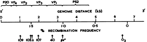
Comparison
of biochemical andrecom-binationmaps. FMDV RNA hasatotallength of8.0kilobases. All of theknown geneproducts
P20VP VP2
VPt3
VP1 __are encoded along a 7.5-kilobase sequence
bounded
on onesideby
apolycytidylic
acidtract near the 5' end of the virus RNA and on theother side by the 3'-terminal polyadenylic acid
tract (6, 7, 17). Reversion studiesidentified20 ts
mutations that covariedwithalterationsinVP1,
VP2, or VP3(Table1). These threepolypeptides
are encoded together near the 5' end of the
genome, as shown in Fig. 3. Within this set of
mutations, there is noapparentcorrelation
be-tweenindividualmaploci and
polypeptide-cod-ingregions. However,thedistribution ofthe 20
chargechange lociis
significantly
nonrandominthat they all lieat, or tothe left of,a recombi-nationfrequencyof1.25%,nearthecenterof the
map. It follows that the recombination map
representsthe genome oriented with its 5'
ter-minusontheleft.
The
recombination
data from which themapsofboth FMDV and
poliovirus
wereconstructedarenotoriously subjecttovariation. Ofthe 70ts
mutations of the FMDV map, five loci at the
extreme left, includingtwo charge change
mu-tants, ts 28and65, were
singled
outby
McCahonet al. (15) as being
particularly speculative.
If theseareeliminated,
arelationsbip
betweentherecombination frequency and the
genomic
dis-tance becomes clear.
Figure
3 shows the maplociof the five different classes ofchargechange mutation. The h
class,
represented by
mutantswithidentical substitutions in the
fB
peptide of VP2, isdesignated
hinFig.
3.gs+ and ts03
arestandard reference
points
of the map (15). h specifiesaposition
between0.8and1.6kilobases on the biochemical map, and since it wasmapped
independently
14times, itslocus isac-curately
known. The standard deviation ofthe hloci,
+0.2% recombinationfrequency,
is ameasureof themapping
precision
forindividualmarkersinthecoat
protein region.
Figure 3 shows a
possible
alignment of thebiochemical
and recombination maps. Recom-bination frequency values coincide withpoly-peptide-coding regions
for all of the mutantsexcept ts103, whichlies
approximately
onestan-darddeviation from the
boundary
oftheVP1-P52
S GENOME DISTANCE (kb)
0 1 2 3 4 5 6 7
I I 1 I
'.5 I I
1.0 0.5
% RECOMBINATION FREQUENCY
3'
I
t
03
FIG. 3. Tentativealignmentoftherecombinationmap(lower scale)with the biochemicalmap(upper scale) oftheFMDV genome. Genetic markersareplottedatthelocipreviouslydeterminedbyMcCahon et al. (15).
h isplottedatthemeanlocusoftheclass hmutations(seetext).kb,Kilobase.
71
VOL. 34, 1980
IL
KI'A .7
1
Ile
on November 10, 2019 by guest
http://jvi.asm.org/
[image:5.508.121.407.553.636.2]72 KING ET AL.
coding region. Since ourinformation islimited toagroup of proteins encoded at one end of the genome, it is notpossible toestimate precisely
howmuchofthe genomeisrepresentedby the
recombination map.
Ourphysical interpretation of the FMDV re-combination map shown in Fig. 3 must be
re-gardedastentative. However,itprovides a use-ful theoretical basis for future recombination studies, which we hope will lead to a better
description of genetic recombination in RNAat
themolecular level.
LITERATURE CITED
1. Burroughs,J.N.,D. J.Rowlands,D. V.Sangar, P.
Talbot, and F. Brown. 1971. Further evidence for
multiple proteins in the foot-and-mouth disease virus
particle.J. Gen. Virol. 13:73-84.
2. Cleveland, D.W., S. G.Fischer, M. W. Kirschner,
and U. K. Laemmli.1977.Peptide mapping bylimited proteolysis in sodiumdodecylsulfate andanalysis by
gelelectrophoresis.J. Biol. Chem.252:1102-1106.
3. Cooper, P. D. 1961. Animproved agar-cell suspension
plaque assay forpoliovirus:somefactorsaffecting effi-ciencyofplating. Virology13:153-157.
4. Cooper, P. D. 1968. Ageneticmap ofpoliovirus
temper-ature-sensitivemutants.Virology35:584-596.
5. Copper,P. D. 1977. Genetics ofpicomaviruses,p.
133-207.In H. Fraenkel-Conratand R. R.Wagner (ed.),
Comprehensive virology, vol. 9. Plenum Publishing
Corp., NewYork.New York: Plenum Press.
6. Doel, T. R.,D. V. Sangar,D. J. Rowlands, and F.
Brown. 1978.Areappraisalof thebiochemical map of
foot-and-mouthdisease virus RNA. J. Gen. Virol.41:
395-404.
7. Harris,T.J.R.,and F. Brown.1976.Thelocationof
thepoly(C) tract in the RNA offoot-and-mouth disease virus. J. Gen. Virol.33:493-501.
8.Jarvik, J., and D. Botstein. 1975. Conditional-lethal mutations that suppress genetic defects in morphogen-esis byaltering structural proteins. Proc. Natl. Acad. Sci. U.S.A. 72:2738-2742.
9. King, A.M.Q.,and J. W. I. Newman. 1980. Tempera-ture-sensitive mutants offoot-and-mouth disease virus withaltered structural polypeptides.I.Identification by electrofocusing. J. Virol. 34:59-66.
10. Laemmli, U. K. 1970. Cleavage of structural proteins
during theassemblyofthehead of bacteriophage T4. Nature(London)227:680-685.
11. Lake,J., and J. S. Mackenzie. 1973. Improved
tech-niquesfor the isolation of temperature-sensitive mu-tantsoffoot-and-mouth disease virus. J. Gen. Virol. 12: 665-668.
12. Lake,J.R.,R. A. J.Priston,and W. R.Slade.1975. A geneticrecombination map of foot-and-mouth disease virus. J. Gen. Virol.27:355-367.
13. Mackenzie, J. S., W. R. Slade, J. Lake, R. A. J.
Priston, J. Bisby,S. Laing, and J. Newman. 1975. Temperature-sensitive mutants of foot-and-mouth dis-easevirus: theisolation of mutants and observations on their properties and genetic recombination. J. Gen. Virol.27:61-70.
14.McCahon, D.,and P. D.Cooper.1970.Identification of
poliovirus temperature-sensitive mutants having
de-fects in virusstructural protein. J. Gen. Virol. 6:51-62.
15.McCahon, D.,W. R.Slade,R. A. J.Priston,and J. R.
Lake. 1977. Anextendedgenetic recombination map
forfoot-and-mouth disease virus. J. Gen. Virol. 35:555-565.
16. O'Farrell,P.Z.,H. M.Goodman,and P. H. O'Farrell.
1977. Highresolutiontwo-dimensionalelectrophoresis ofbasic aswell asacidic proteins.Cell 12:1133-1142.
17.Sangar,D.V.,D. N.Black,D.J.Rowlands, andF.
Brown. 1977. Biochemical mapping of the foot-and-mouth disease virus genome. J.Gen. Virol. 35:281-297. J. VIROL.