R E S E A R C H
Open Access
Structural divergence of chromosomes
between malaria vectors
Anopheles lesteri
and
Anopheles sinensis
Jiangtao Liang
1, Biao Cheng
1, Guoding Zhu
2, Yun Wei
1, Jianxia Tang
2, Jun Cao
2, Yajun Ma
3, Maria V. Sharakhova
4,5,
Ai Xia
1*and Igor V. Sharakhov
4,5*Abstract
Background:Anopheles lesteriandAnopheles sinensisare two major malaria vectors in China and Southeast Asia. They are dramatically different in terms of geographical distribution, host preference, resting habitats, and other traits associated with ecological adaptation and malaria transmission. Both species belong to theAnopheles hyrcanus group, but the extent of genetic differences between them is not well understood. To provide an effective way to differentiate between species and to find useful markers for population genetics studies, we performed a comparative cytogenetic analysis of these two malaria vectors.
Results:Presented here is a standard cytogenetic map forAn. lesteri, and a comparative analysis of chromosome structure and gene order betweenAn. lesteriandAn. sinensis. Our results demonstrate that much of the gene order on chromosomes X and 2 was reshuffled between the two species. However, the banding pattern and the gene order on chromosome 3 appeared to be conserved. We also found two new polymorphic inversions, 2Lc and 3Rb, inAn. lesteri, and we mapped the breakpoints of these two inversions on polytene chromosomes.
Conclusions:Our results demonstrate the extent of structural divergence of chromosomes betweenAn. lesteriand An. sinensis, and provide a new taxonomic cytogenetic tool to distinguish between these two species. Polymorphic inversions ofAn. lestericould serve as markers for studies of the population structure and ecological adaptations of this major malaria vector.
Keywords:Cytogenetic map, Chromosomal inversions,Anopheles lesteri,Anopheles sinensis, Gene order, Arm homology, Polytene chromosomes
Background
Anopheles sinensis Wiedemann and Anopheles lesteri
Baisas & Hu, 1936 (syn.An. anthropophagus) are
mem-bers of theAnopheles hyrcanusgroup. Due to their
iden-tical morphological characteristics, they have been considered cryptic species [1]. The earliest studies in the
1960s identified two egg types of An. sinensis in China
[2]. Since then, the egg type with a wide deck has been
assigned toAn. sinensis, while the taxonomic status and
specific name for the narrow egg type remains question-able. Xu & Feng [3] initially proposed that the narrow
egg type of An. sinensis represents a subspecies of An.
lesteri, and named it An. lesteri anthropophagus. Later,
Ma [4] elevatedAn. anthropophagusto the species level.
However, recent studies of the internal transcribed spacer 2 (ITS2) of the ribosomal DNA (rDNA) have
pro-vided evidence thatAn. anthropophagusis a junior
syno-nym of An. lesteri, leading to a resurrection of the
species nameAn. lesteriBaisas & Hu, 1936 [4–6].
There-fore, the previously recognized An. anthropophagusand
An. lesteriin China are considered the same species. Despite being morphologically indistinguishable,
mem-bers of the An. hyrcanus group, An. lesteri and An.
sinensis differ in their geographical distribution, host preference, resting habitats, and other features important
for malaria transmission [7]. Anopheles sinensis is the
* Correspondence:xiaai@njau.edu.cn;igor@vt.edu
1Department of Entomology, Nanjing Agricultural University, Nanjing, China 4Department of Entomology, Fralin Life Science Institute, Virginia Tech, Blacksburg, VA, USA
Full list of author information is available at the end of the article
most widespread species, with a range that is continuous
throughout 29 provinces and regions in China.
Anoph-eles lesteri is mostly sympatric with An. sinensis but is
limited to central China, south of 33°N [7]. Anopheles
lesteri has been established as the primary vector of Plasmodium vivax malaria in central China because of
the higher human blood index ofAn. lesteriand its close
association with malaria outbreaks. In central China,
most malaria outbreaks appeared in areas whereAn.
les-teriwas present. The regions containingAn. lesterihave
suffered more serious malaria epidemics than those
areas where the only vector was An. sinensis [8]. In
addition, An. lesteri naturally transmits P. falciparum
[8, 9] and also is a major vector of a filarial worm, Brugia malayi, in China [10, 11]. An. sinensis is more zoophilic and has significantly lower human-biting rate, human blood index, infection rate, and entomological
inoculation rate with P. vivax than An. lesteri [9]. For
decades it has been considered a secondary malaria vector.
However, the importance of An. sinensis as the P. vivax
malaria vector in areas cultivated for rice (Oryza sativaL.)
cannot be ignored. Recent evidence indicates that An.
sinensis was responsible for the outbreak of the vivax malaria in central China in 2006 [12, 13].
The availability of well-developed polytene
chromo-somes in the salivary glands of An. lesteri and An.
sinensis makes these species suitable for cytogenetic studies [14, 15]. Before the high-resolution cytogenetic
photomap of An. sinensis was published in 2014 [14],
several chromosomal maps were developed for this cies [15–19] and used to distinguish among sibling
spe-cies within the An. hyrcanus group. However, because
most of the published maps are drawn manually, their resolution is low, which has introduced subjectivity into the interpretation of chromosome banding patterns. In contrast to the previously drawn maps, the standard
photomap for An. sinensis, constructed from
high-resolution chromosome images [14], provides more de-tailed banding patterns, making it more suitable for genome mapping and other studies.
In this study, a detailed cytogenetic photomap was
developed for the Asian malaria vector An. lesteri. A
comparative analysis of the chromosome banding
pat-terns betweenAn. lesteri and An. sinensis demonstrated
a limited pattern identity between chromosomes X and 2.
Gene mapping, using fluorescent in situ hybridization
(FISH), further established whole-arm homology between the two species. A gene order comparison determined re-arrangements of the X, 2R, and 2L chromosomes, while arms 3R and 3L were more conserved between the two species. We also mapped two new polymorphic
inver-sions: 2Lc in field-collected An. lesteri specimens from
Hainan, and 3Rb in the Wuxi laboratory strain of An.
lesteri. These could serve as markers for distinguishing
between the two species and for population genetics and ecological studies ofAn. lesteri.
Methods
Mosquito strains and chromosome preparation
The Wuxi laboratory strain (Jiangsu Institute of Parasitic Diseases, Wuxi, China) and a field strain ofAn. lesteri col-lected from Hainan were used in this study. The Wuxi strain ofAn. lesterihas been maintained in the insectary of the Key Technical Laboratory for Prevention and Control of Parasitic Diseases of the Ministry of Health (MOH) in the Jiangsu Institute of Parasitic Diseases (JIPD), Wuxi, China, for over 30 years. The salivary glands were dissected from early fourth-instar larvae ofAn. lesteriand were used for obtaining polytene chromosome preparations as previ-ously described [14]. The quality of polytene chromosomes was examined with an Olympus BX43 phase-contrast microscope (Olympus Corp., Tokyo, Japan). Chromosome preparations with clear banding patterns were placed in li-quid nitrogen for several minutes, and then cover slips were removed and slides were dehydrated in 50%, 70%,
90% and 100% ethanol for imaging and in situ
hybridization. Chromosomes were imaged with an Olym-pus BX43 microscope with a DP72 digital camera and Cell-Sens imaging software (Olympus Corp.). The best 50 of approximately 100 images were used to construct a
cytogenetic map forAn. lesteri, using Adobe Photoshop
software as previously described [20].
Fluorescencein situhybridization
The Genome sequences were acquired from theAn.
gam-biae PEST strain (https://www.vectorbase.org/organisms/
anopheles-gambiae/pest) andAn. sinensis(https://www.vec
torbase.org/organisms/anopheles-sinensis/sinensis). The PCR primers were designed using the Primer3 Program
[21], based on the above genome sequences fromAn.
gam-biaeandAn. sinensis. Genomic DNA ofAn. lesteriwas ex-tracted from live fourth-instar larvae, using a DNeasy Blood & Tissue Kit (Qiagen GmbH, Hilden, Germany), and utilized as a template for PCR.
After the standard PCR procedures, amplified PCR products were loaded on agarose gel and cut for purifi-cation using a QIAquick Gel Extraction Kit (Qiagen). The DNA fragments were labeled with either Cy3.5-AP3-dUTP or Cy5.5-Cy3.5-AP3-dUTP (GE Healthcare UK Ltd, Chalfont StGiles, UK), using a Random Primed DNA Labelling Kit (Roche Applied Science, Penzberg,
Germany). In situ hybridization was performed using a
previously described method [20]. Fluorescent signals were detected and recorded with a Zeiss LSM 710 laser scanning microscope (Carl Zeiss Microimaging GmbH, Oberkochen, Germany), and placed on the polytene
Results and discussion
A polytene chromosome photomap forAn. lesteri
The polytene chromosome complement in salivary glands ofAn. lestericonsists of three chromosomes (X, 2, and 3). The X chromosome is represented by one arm, while chromosomes 2 and 3 have two arms. Chromosomal arms usually form the chromocenter, by joining their pericen-tromeric regions (Fig. 1). Polytene chromosomes with clear banding patterns from the salivary glands ofAn.
les-teri were analyzed and utilized to develop a standard
photomap. The chromosomal map contains five elements: the shortest X chromosome, the longest 3R arm, and then similarly sized 2R, 2L, and 3L arms. Chromosome
ele-ments of An. lesteri were subdivided into 39 numbered
divisions and 116 lettered subdivisions, by analogy with the chromosome map ofAn. sinensis[14] (Fig. 2).
The landmarks which could be used for arm recogni-tion were as follows. In addirecogni-tion to its short length, the
X chromosome of An. lesteri could be recognized by
telomeric region 1A, the end of which has a“flared fan”
shape with two thin granulated bands (Fig. 2). The centromeric end of the X chromosome has a bulbous, granulated region, 5B, with an unclear banding pattern. Autosome 2 consists of two arms of almost equal length (Fig. 2). The telomeric end of 2R has three narrow bands at the tip, followed by a large puffy area in subdivision 6B. A wide dark band and a bright inter-band in region 9A, and a puffy region 9B with three double bands, could also be used as landmarks for the 2R arm. A dark double band in region 11B and two dark bands followed by four thin bands in region 11C provide additional landmarks for the middle part of the 2R arm. The centromeric end of the 2R arm has a small puffy region,
14D, with a dark band on the very end. In contrast to the bulb-shaped and striped telomeric end of the 2R arm, the 2L arm has a flared, bright, granulated telo-meric end, followed by four dark bands in region 23A. A large, bright, puffy region, 18C, with six dark bands, pro-vides supporting information that this is the 2L arm. The pericentromeric area is easily recognized by a dark band with a low level of polyteny in region 15A, and two bands in region 15B.
Chromosome 3 has arms that are unequal in length. The 3R arm is the longest among them (Fig. 2). The telomeric area of 3R has a large region with a flared fan shape and a dark double band inside followed by three thin bands at the beginning of region 24B. Another con-sistent feature of 3R is the presence of two large puffy areas in regions 25D–26B; one contains three dark bands and the other has one dark double band in the middle. The presence of one dark band in regions 31A, sur-rounded by bright granulated areas on both sides, could be considered an additional landmark for this arm. Close to the centromere, the 3R arm is composed of a series of continuous dark bands in regions 31B–32B, which makes this region look striped.
The 3L arm is slightly shorter than the chromosome 2 arms. The telomeric end of 3L is bright and flared, as the 2L arm, but can be easily distinguished by the pres-ence of three dark bands in the 39B-C region. Two puffy areas in the 36B-C region, one with close three dark bands and the other with two dark bands in the middle, could also be considered an important landmark of the 3L arm. Closer to the centromere, this arm contains an apparent constriction in the 33B-C region, with a dark band in the middle. The centromeric end of the 3L arm consists of a large dark band, which is absent in the centromeric areas of the other chromosomal arms.
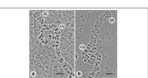
Polymorphic inversions inAn. lesteri
The results of our study of field-collected and laboratory strains demonstrated the presence of two polymorphic
inversions in chromosome arms 2L and 3R ofAn. lesteri.
A polymorphic inversion in a heterozygote state on the 2L (2Lc) arm was found in 20 of 76 samples from the
field strain of An. lesteri collected in Hainan (Fig. 3).
This inversion was not present in the Wuxi laboratory strain. The distal and proximal breakpoints of the 2Lc inversion were mapped to the regions 22B and 17C of 2L (Fig. 2). Our results revealed a landmark for recog-nizing this inversion. In the case of the standard
ar-rangement of chromosome arm 2L (+c/+c), the banding
pattern was arranged as one puff with four dark bands followed by three thin bands (18C-B) near the proximal part of chromosome. In the inverted chromosome (2Lc/ c), the bands appeared as three thin bands, plus one puff
[image:3.595.57.291.500.693.2]Fig. 2A standard cytogenetic map forAnopheles lesteri.Numbered divisions and lettered subdivisions are shown below the chromosomes. Brackets indicate the positions of the polymorphic inversions 2Lc and 3Rb
[image:4.595.60.539.88.420.2] [image:4.595.62.538.453.703.2]containing four dark bands near the distal part of the chromosome.
We also discovered another polymorphic inversion on 3R (3Rb), but only in the Wuxi laboratory strains of An. lesteri, with a frequency of heterozygote arrange-ment of about 17%. We localized the breakpoints to regions 27C and 31A on 3R (Fig. 2). The landmark of
the standard 3R arrangement (+b/+b) was four closely
located bands followed by a dark triple band located in the proximal position (30D). In an inverted chromo-some (3Rb/b), this specific banding pattern is at a distal position.
Inversion polymorphisms are commonly used as tools in mosquito taxonomy, ecology, and population genetics. Data from a previous study indicated that adaptations of mosquitoes to various environments are often associated with the composition and frequency of polymorphic
in-versions [22, 23]. Despite the importance of An. lesteri
for malaria transmission in Asia, inversion polymor-phisms have not been investigated.
In this study, we identified the polymorphic inversion 2Lc (17C-22B) from a field population in Hainan, and
3Rb (27C-31A) from Wuxi laboratory strains ofAn.
les-teri. We previously published one polymorphic inversion
(3Ra) on chromosome arm 3R of An. sinensis, and
mapped the breakpoints to regions 28A-31A [14]. How-ever, without genome sequencing data, we could not analyze the molecular structures of inversion breakpoints and could not determine if the 3Rb inversion inAn. lesteri and 3Ra inAn. sinensisare the same. Nevertheless, the
in-version polymorphism reported here forAn. lesteriwould
help our understanding of epidemiological and ecological heterogeneities in this malaria vector.
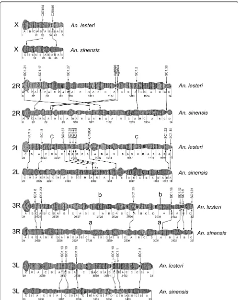
Structural divergence of chromosomes and gene order comparison betweenAn. lesteriandAn. sinensis
Banding pattern comparisons based on cytogenetic maps ofAn. lesteriandAn. sinensis[14], and gene order analysis using FISH of 31 PCR-amplified DNA probes on chromo-somes of both species, enabled us to perform a compara-tive cytogenetic study (Fig. 4). To amplify DNA probes for FISH, two pairs of PCR primers (Ag8026 and Ag9894)
were designed, based on the An. gambiae PEST strain
DNA sequences available from the Vector Base (https://www.vectorbase.org/organisms/anopheles-gam biae). Primers for 27 probes were inferred from the
genome sequence of An. sinensis (https://www.vectorba
se.org/organisms/anopheles-sinensis/sinensis). An add-itional three pairs of PCR primers (C20446, C07454, and C15834) were developed based on our sequence database (Additional file 1: Table S1). All DNA probes yielded
unique signals on the polytene chromosomes ofAn. lesteri
and An. sinensis (Table 1). Two examples of FISH, with one clear signal on each, are shown in Fig. 5.
By comparing the physical locations of the probes in An. lesteri and An. sinensis (Table 1), we demonstrated that all five arms are homologous between the two spe-cies (X = X; 2R = 2R; 2L = 2L; 3R = 3R; 3L = 3L). When we further analyzed the physical locations of DNA probes and compared the banding patterns on five chromosomal arms (Fig. 4), we found that gene locations on the sex chromosome X and autosome 2R were reshuffled. The chromosomal structure showed limited similarity between the two species. Chromosome arm 2L, and particularly arms 3R and 3L, demonstrated rela-tively consistent gene orders and had some similarities in their banding patterns. Cytogenetic analyses identified specific landmarks on each chromosomal arm that could be used for species recognition.
We hybridized only two probes on the X chromosomes of the two species. These probes, C07454 and C20446, were mapped to different X chromosome regions of the
two species: subdivisions 3A and 4A inAn. lesteriand 5A
and 1A inAn. sinensis(Fig. 4), suggesting the existence of rearrangements between the species.
The banding patterns of X chromosomes had no recognizable similarity between the two species, imply-ing the presence of multiple chromosomal inversions that reshuffled the order of the bands. As a result of significant order reshuffling, the boundaries of the in-versions remained uncertain, based on the cytogenetic analysis. Telomeric and centromeric areas of the X chromosomes in the two species could be easily used
for species diagnostics. The telomeric end inAn.
sinen-sisstarts with two dark, granulated bands in region 1A,
while, inAn. lesteri, it begins with a bright, granulated, flared end. Secondly, the pericentromeric region 5B ends with a clear dark band inAn. sinensis, but a bright granulated bulb-shaped area inAn. lesteri.
Analysis of the positions of seven markers mapped on chromosome arm 2R also identified multiple rearrange-ments (Fig. 4) between the two species. Using these markers and the Genome Rearrangements in Man and Mouse (GRIMM) program [24], we calculated the
mini-mum number of fixed inversions on arm 2R betweenAn.
lesteriandAn. sinensis. The orders of the markers were 1, 2, 3, 4, 5, 6 and 7 inAn. lesteriand 2, 5, 4, 1, 3, 6 and 7 in An. sinensis. When we used the GRIMM program, without assuming directionality of the markers (unsigned option), there were three calculated inversions. When we used the signed option to perform a pairwise analysis, the number of rearrangements increased to five. The banding patterns of these arms significantly differed between the two species.
The most reliable landmarks to distinguish between these arms in the two species are the following. First, six large bands in the middle of arm 2R in regions
arm in An. sinensis. The puffy region, 9B, with three
double bands in An. lesteri(the chromosome location of
marker SC1.27), changed its position on theAn. sinensis
chromosome, where it was located in the inverted pos-ition in the middle of the arm in region 11B. Two dark
bands in region 12A inAn. lesteri(the chromosome
lo-cations of markers Ag8026 and Ag9894) could be traced
in region 8A of An. sinensis, also an inverted order.
Telomeric areas of An. lesteri and An. sinensis were
similar to each other, which might suggest that marker SC1.21 was transferred to a new position, not by an in-version, but by a transposition.
The analysis of the order of nine probes on arm 2L for the two species revealed no order reshuffling. However, the distance between the markers SC1.5 and SC1.37 in An. sinensiswas twice that of An. lesteri, suggesting the presence of rearrangements in 2L. These rearrange-ments moved four areas with puffs from the middle of
arm 2L (region 18C-20B) in An. lesteri to the
centro-meric regions 17C-18C in An. sinensis. This difference
could be used to distinguish between the 2L arms of the two species.
The positions of seven and six DNA probes in arms 3R and 3L, respectively, demonstrated no gene order
reshuffling between An. lesteri and An. sinensis.
Al-though the banding pattern of chromosome 3 was
simi-lar between the two species, An. lesteri could be
distinguished by a flared 3L telomere, and An. sinensis
could be identified based on a narrow, dark telomeric
band. Another specific feature of the 3L arm in An.
les-teriwas constriction in region 33B_C, which was absent
in An. sinensis. The landmark in the An. lesteri3R arm was four thin dark bands followed by a dark triple band in the region 30D, which was absent inAn. sinensis.
To investigate the pattern of chromosome rearrange-ments over larger evolutionary distances, we conducted
BLAST searches of homologous sequences in the An.
atroparvus[25] andAn. gambiae[26] genomes.Anopheles atroparvus is the only member of the An. maculipennis complex with a sequenced genome. Some of the genomic
scaffolds of An. atroparvus were recently mapped to
chromosomes of this species [25, 27]. These data enabled
us to assign the An. lesteriprobes to chromosomal arms
ofAn. atroparvus(Table 2).
There was good correspondence in arm assignment
betweenAn. lesteriandAn. atroparvus. The close
asso-ciation of arm assignments inAn. lesteri, An. atroparvus
[image:7.595.307.539.87.245.2]Fig. 5Fluorescencein situhybridization results of DNA probes SC1.35 and SC1.1. Arrows show the positions of signals produced by probes (a) and SC1.1 (b). Blue signal were labeled with Cy5.5 and red were labeled with Cy3.5 dyes.Scale-bars: 10 µm
Table 1DNA probes used forin situhybridization with theAn. lesteriandAn. sinensispolytene chromosomes
Clone name
Source Location in
An. lesteri
Location in An. sinensis
1 C20446 Additional file1: Table S1 X:4A X:1A
2 C07454 Additional file1: Table S1 X:3A X:5A
3 SC1.21 supercont1.21 2R:6A 2R:10C
4 SC1.17 supercont1.17 2R:7B 2R:7A
5 SC1.27 supercont1.27 2R:9B 2R:11B
6 Ag8026 AGAP008026 2R:12A 2R:8B
7 Ag9894 AGAP009894 2R:12A 2R:8A
8 SC1.2 supercont1.2 2R:13A 2R:13A
9 SC1.30 supercont1.30 2R:14C 2R:14C
10 SC1.9 supercont1.9 2L:23C 2L:23C
11 SC1.5 supercont1.5 2L:23A 2L:23A
12 SC1.37 supercont1.37 2L:21C 2L:20B
13 SC1.35 supercont1.35 2L:21B 2L:20A
14 SC1.23 supercont1.23 2L:21A 2L:19B
15 SC1.48 supercont1.48 2L:20D 2L:19B
16 C15834 Additional file1: Table S1 2L:20B 2L:18C
17 SC1.22 supercont1.22 2L:15B 2L:15B
18 SC1.15 supercont1.15 2L:15A 2L:15A
19 SC1.8 supercont1.8 3R:24B 3R: 24B
20 SC1.20 supercont1.20 3R:25A 3R:25A
21 SC1.33 supercont1.33 3R:30A 3R:30A
22 SC1.18 supercont1.18 3R:31B 3R:31A
23 SC1.12 supercont1.12 3R:32A 3R:32A
24 SC1.6 supercont1.6 3R:32B 3R:32B
25 SC1.31 supercont1.31 3R:32C 3R:32C
26 SC1.13 supercont1.13 3L:38A 3L:38A
27 SC1.19 supercont1.19 3L:37C 3L:37C
28 SC1.39 supercont1.39 3L:37A 3L:37A
29 SC1.10 supercont1.10 3L:34C 3L:34C
30 SC1.1 supercont1.1 3L:34B 3L:34B
[image:7.595.57.287.108.564.2]andAn. sinensissupport their close relationships, as they
belong to the subgenus Anopheles. In contrast, the 2R
arm of An. lesteri corresponded to the 3R arm in An.
gambiae, and vice versa, indicating a whole-arm trans-location between the species. Thus, our results provide further support for evidence of a whole-arm
transloca-tion between subgenera Anopheles and Cellia [28], to
whichAn. gambiaebelongs.
Conclusions
In this study, we constructed a standard cytogenetic map for the Asian malaria vectorAn. lesteri. In addition, we identified polymorphic inversions: 2Lc, from a field population in Hainan, and 3Rb on the 3R chromosome
from the Wuxi laboratory strain of An. lesteri. The
[image:8.595.58.533.118.609.2]in-version breakpoints were localized to subdivisions 22B and 17C of 2L, and 27C and 31A of 3R. Polymorphic
Table 2Genomic locations of the DNA probes inAn. atroparvus,An. sinensisandAn. gambiae. The locations were acquired using https://www.vectorbase.org/blast against genomic scaffold sequences ofAnopheles atroparvusEBRO strain,Anopheles gambiaePEST strain, andAnopheles sinensisChina strain. The e-values are indicated in parentheses after the locations
Clone name
Source Location inAn. atroparvus Location inAn. sinensis Location inAn. gambiae
1 C20446 Additional file1: Table S1 KI421895: 3842010–3842293(3e-128);
KI421886: 1702493–1702773(8e-98)
AS2_scf7180000695491: 102496–102779(2e-144) X:533418–533760(6e-107)
2 C07454 Additional file1: Table S1 KI421888: 9432816–9432478(1e-76) AS2_scf7180000695709:907649–907989(2e-175) 2L:47798210–47798610(1e-96)
3 SC1.21 supercont1.21 2R:KI421882: 9912851–10045608(0) AS2_scf7180000696054: 1195628–1428863 (0) 3R:10,114,393–10,249,464 (0) 4 SC1.17 supercont1.17 KI421890: 1035344–1389232(0) AS2_scf7180000695920: 44079–328299 (0) 3R:38705671–38,709,129(0) 5 SC1.27 supercont1.27 KI421890: 4179223–4195140(0) AS2_scf718000069604:488088–637274(0) 3R:26871040–26944051(0) 6 Ag8026 AGAP008026 KI421890: 6995505–6995742(5e-91) AS2_scf7180000696017: 292678–292915(3e-81) 3R:4,499,175–4,507,999(0)
7 Ag9894 AGAP009894 2R:KI421882: 15579762–15579934(2e-52) AS2_scf7180000696027: 184208–184042(2e-53) 3R:44,903,740–44,912,595(0)
8 SC1.2 supercont1.2 KI421900: 2361929–2838297(0) AS2_scf7180000696013: 1377801–1404489(0) AS2_scf7180000695708: 122951–293931(0)
3R:49911437–50,354,787(0)
9 SC1.30 supercont1.30 KI421900: 1492320–1595685(0) AS2_scf7180000696012: 752618–1009731(0) 2L:5,452,203–5,453,482(0) 10 SC1.9 supercont1.9 2L:KI421884: 10941708–11186226(0) AS2_scf7180000695483: 403208–410683(0) 2L:47,782,863–48,943221(0) 11 SC1.5 supercont1.5 2L:KI421884: 77975–136.076(0)
KI421916: 285574–290011(0)
AS2_scf7180000696060: 1083588–1123553(0) 2L:33212653–33,787,298(0)
12 SC1.37 supercont1.37 KI421886: 11137039–11199731(0) AS2_scf7180000696049: 2284815–2306994(0) AS2_scf7180000696129: 257010–238213(0)
2L:36,690,171–36,694,229(0)
13 SC1.35 supercont1.35 2L:KI421886: 9346759–9417257(0) AS2_scf7180000696058: 981634–993925(0) 2L:27,916,638–27,918,874(0) 14 SC1.23 supercont1.23 KI421891: 7010523–7018018(0) AS2_scf7180000695983: 404330–428660(0) 2L:11,396,721–11,400,344(0) 15 SC1.48 supercont1.48 KI421891: 6344135–6346162(0) AS2_scf7180000695983: 1052702–1083490(0) 2L:10,641,721–10,643,463(0) 16 C15834 Additional file1: Table S1 2L:KI421886: 1283313–1283772(1e-167) AS2_scf718000069587: 127420–127901(0) 2L:26,996,740–26,997,260(9e-133)
17 SC1.22 supercont1.22 2L:KI421886: 998049–1001402(0) AS2_scf7180000690255: 166481–193533(0) 2L:14,179,779–14,182,651(0) 18 SC1.15 supercont1.15 KI421924: 86788–88570(0) AS2_scf7180000695974: 485746–503624(0) 2L:2,429,065–2,431,713(0) 19 SC1.8 supercont1.8 KI421888: 4420609–4428976(0) AS2_scf7180000695742: 846781–876896(0) 2R:11,955,880–11,963,441(0) 20 SC1.20 supercont1.20 KI421897: 3217947–3221214(0) AS2_scf7180000695544: 217804–260601(0) 2R:40,388,954–40,391,204(0) 21 SC1.33 supercont1.33 KI41906: 789097–795883(0) AS2_scf7180000695987: 203015–213211(0) 2R:9,258,761–9,262,721(0) 22 SC1.18 supercont1.18 KI421885: 5623993–5627430(0) AS2_scf7180000691904: 29065–60946(0) 2R:53,122,916–53,125,899(0) 23 SC1.12 supercont1.12 KI421883: 793616–798090(0) AS2_scf7180000696020: 346403–391124(0) 2R:25,078,045–25,082,179(0) 24 SC1.6 supercont1.6 KI421885: 9679627–9684068(0) AS2_scf7180000696131: 1492568–1550122(0) 2R:51,006,383–51,010,426(0) 25 SC1.31 supercont1.31 KI421908: 618377–622446(0) AS2_scf7180000695941: 606638–632554(0) 2R:59,756,833–59,757,717(1e-156)
26 SC1.13 supercont1.13 KI421893: 6014521–6018625(0) AS2_scf7180000696026: 562865–534327(0) 3L:8,096,738–8,100,402(0) 27 SC1.19 supercont1.19 KI421899: 300613–303079(0) AS2_scf7180000695747: 106108–123837(0) 3L:26,049,483–26,051,315(0)
X:15,344,337–15,345,182(2e-162)
inversions are useful markers for studying the popula-tion genetics of mosquitoes. These results provide the foundation for a better understanding of the epidemio-logical and ecoepidemio-logical roles of polymorphic inversions in An. lesteri.
By comparing the physical locations of 31 probes of An. lesteriandAn. sinensis, and the banding patterns of polytene chromosomes, we found that all five
chromo-some arms were homologous for An. lesteri and An.
sinensis. We demonstrated that the chromosome arms X, 2R, and 2L were rearranged between the two species, because of the presence of fixed inversions. Chromo-some structures of the 3R and 3L arms were more
simi-lar, and gene orders were conserved, betweenAn. lesteri
andAn. sinensis.
Our results provide reliable cytogenetic information
for discriminating An. lesteri and An. sinensis, and a
foundation for understanding the genetic content associ-ated with species-specific ecological adaptation and
vec-torial capacity. Cytogenetic and physical maps for An.
lesteri and An. sinensis would serve as convenient out-groups for phylogenetic reconstruction, based on fixed
inversions in other mosquitoes of the subgenus
Anoph-eles, such as theAn. maculipenniscomplex.
Additional file
Additional file 1: Table S1.The sequences of DNA probes from An. sinensisShanghai Chinese strain. (DOCX 15 kb)
Acknowledgments
We thank an anonymous reviewer for their helpful comments.
Funding
The work was supported by a National Natural Science Foundation of China (31301877) and the Fundamental Research Funds for the Central Universities (KJQN201431) to AX. The comparative study withAnopheles atroparvuswas supported by the grant from the Russian Science Foundation№15-14-20011 (to IVS). The funding agencies had no role in the design of the study and collection, analysis, and interpretation of data and in writing the manuscript.
Availability of data and materials
All data are presented in the main paper and Additional file 1: Table S1.
Authors’contributions
AX conceived and designed the experiments, jointly performed data analysis and drafted the manuscript. JL, BC and YW performed the experiments. GZ, JT, JC and YM contributed to mosquito strains collecting. IVS and MVS jointly contributed to data analysis and writing the manuscript. All authors read and approved the final manuscript.
Competing interests
The authors declare that they have no competing interests.
Consent for publication Not applicable.
Ethics approval and consent to participate Not applicable.
Author details
1Department of Entomology, Nanjing Agricultural University, Nanjing, China. 2Key Laboratory of National Health and Family Planning Commission on Parasitic Disease Control and Prevention, Jiangsu Provincial Key Laboratory on Parasite and Vector Control Technology, Jiangsu Institute of Parasitic Diseases, Wuxi, Jiangsu Province, China.3Department of Tropical Infectious Diseases, Faculty of Tropical Medicine and Public Health, Second Military Medical University, Shanghai 200433, China.4Department of Entomology, Fralin Life Science Institute, Virginia Tech, Blacksburg, VA, USA.5Laboratory for Ecology, Genetics and Environmental Protection, Tomsk State University, Tomsk, Russia.
Received: 15 August 2016 Accepted: 23 October 2016
References
1. Rueda LM, Wilkerson RC, Li C.Anopheles(Anopheles)lesteriBaisas and Hu (Diptera: Culicidae): neotype designation and description. Proc Entomol Soc Wash. 2005;107(3):604–22.
2. Ho C, Chou TC, Ch’En TH, Hsueh AT. TheAnopheles hyrcanusgroup and its relation to malaria in east China. Chin Med J. 1962;81:71–8.
3. Xu JJ, Feng LZ. Classification of chineseAnopheles hyrcanussubspecies. J Entomol. 1975;18:77–89.
4. Ma YJ, Xu J. The Hyrcanus group ofAnopheles(Anopheles) in China (Diptera: Culicidae): species discrimination and phylogenetic relationships inferred by ribosomal DNA internal transcribed spacer 2 sequences. J Med Entomol. 2005;42(4):610–9.
5. Qu FY. Historical review on the classification and rectification ofAnopheles anthropophagustoAn. lesteriin China. Chin J Parasitol Parasit Dis. 2008; 26(3):234–5.
6. Hwang UW. Revisited ITS2 phylogeny ofAnopheles(Anopheles) Hyrcanus group mosquitoes: reexamination of unidentified and misidentified ITS2 sequences. Parasitol Res. 2007;101(4):885–94.
7. Rueda LM, Zhao T, Ma YJ, Gao Q, Zhu GD, Khuntirat B, Sattabongkot J, Wilkerson RC. Updated distribution records of theAnopheles(Anopheles) hyrcanusspecies-group (Diptera: Culicidae) in China. Zootaxa. 2007;1407:43–55. 8. Gu ZC, Shang LY, Chen JS, Zheng X, Su YJ, Li AM, Liu H, Luo MZ, Qian HL,
Tang LH. The role ofAnopheles anthropophagusin malaria transmission in in Xinyang City of Henan Province. Chin J Parasitol Parasit Dis. 2001;19(4):221–4. 9. Liu C. Comparative studies on the role ofAnopheles anthropophagusand
Anopheles sinensisin malaria transmission in China. Zhonghua liu xing bing xue za zhi. 1990;11(6):360–3.
10. Saeung A, Hempolchom C, Baimai V, Thongsahuan S, Taai K, Jariyapan N, et al. Susceptibility of eight species members in theAnopheles hyrcanus group to nocturnally subperiodic Brugia malayi. Parasit Vectors. 2012;6(1):1–8. 11. Zhang XM, Bai LY, Zhang FN, Wen CL, Wang MS, Lu SH, et al. Vectorial
capacity ofAnopheles lesteri anthropophagusin transmitting filariasis malayi in Leshan Prefecture, Sichuan Province. Chin J Parasitol Parasit Dis. 1987;5(3):191–3.
12. Zhang HW, Liu Y, Zhang SS, Xu BL, Li WD, Tang JH, et al. Preparation of malaria resurgence in China: case study of vivax malaria re-emergence and outbreak in Huang-Huai Plain in 2006. Adv Parasitol. 2014;86:205–30. 13. Zhu G, Xia H, Zhou H, Li J, Lu F, Liu Y, et al. Susceptibility ofAnopheles
sinensistoPlasmodium vivaxin malarial outbreak areas of central China. Parasit Vectors. 2013;6(1):1051–60.
14. Liang J, Sharakhova MV, Lan Q, Zhu H, Sharakhov IV, Xia A. A standard cytogenetic map forAnopheles sinensisand chromosome arm homology between the subgeneraAnophelesandCellia. Med Vet Entomol. 2014;28 Suppl 1:26–32.
15. Miao J. The salivary gland chromosomes ofAnopheles anthropophagusXu et Feng. Acta Entomol Sin. 1988;31(2):176–83.
16. Ye B, Huang P, Jiang Z, Geng E, Liu P. The salivary gland chromosomes of Anopheles sinensis. Acta Genet Sin. 1981;111:616–8.
17. Ye B, Huang P, Li B. Further studies on the salivary gland chromosomes of Anopheles sinensis. Acta Genet Sin. 1983;10(6):489–92.
18. Li B, Liu D, Xie C, Ye B. Identification of the short arm of the sex chromosome of the polytene chromosomes of the salivary gland of Anopheles sinensis. Chin J Parasitol Parasit Dis. 1988;6(2):118–20. 19. Li B, Liu D, Xie C. Polytene chromosomes of the salivary gland ofAnopheles
20. Sharakhova MV, Xia A, McAlister SI, Sharakhov IV. A standard cytogenetic photomap for the mosquitoAnopheles stephensi(Diptera: Culicidae): application for physical mapping. J Med Entomol. 2006;43(5):861–6. 21. Rozen S, Skaletsky H. Primer3 on the WWW for general users and for
biologist programmers. Methods Mol Biol. 2000;132:365–86.
22. Xia A, Sharakhova MV, Leman SC, Tu Z, Bailey JA, Smith CD, Sharakhov IV. Genome landscape and evolutionary plasticity of chromosomes in malaria mosquitoes. PLoS One. 2010;5(5):e10592.
23. Coluzzi M, Sabatini A, Della Torre A, Di Deco MA, Petrarca V. A polytene chromosome analysis of theAnopheles gambiaespecies complex. Science. 2002;298(5597):1415–8.
24. Tesler G. GRIMM: genome rearrangements web server. Bioinformatics. 2002;18(3):492–3.
25. Neafsey DE, Waterhouse RM, Abai MR, Aganezov SS, Alekseyev MA, Allen JE, et al. Highly evolvable malaria vectors: the genomes of 16Anopheles mosquitoes. Science. 2015;347(6217):1258522. doi:10.1126/science.1258522. 26. Holt RA, Subramanian GM, Halpern A, Sutton GG, Charlab R, Nusskern DR,
et al. The genome sequence of the malaria mosquitoAnopheles gambiae. Science. 2002;298(5591):129–49.
27. Artemov GN, Sharakhova MV, Naumenko AN, Karagodin DA, Baricheva EM, Stegniy VN, Sharakhov IV. A standard photomap of ovarian nurse cell chromosomes in the European malaria vectorAnopheles atroparvus. Med Vet Entomol. 2015;29(3):230–7.
28. Sharakhov IV, Artemov GN, Sharakhova MV. Chromosome evolution in malaria mosquitoes inferred from physically mapped genome assemblies. J Bioinform Comput Biol. 2016;14(2):1630003. doi:10.1142/
S0219720016300033.
• We accept pre-submission inquiries
• Our selector tool helps you to find the most relevant journal
• We provide round the clock customer support
• Convenient online submission
• Thorough peer review
• Inclusion in PubMed and all major indexing services
• Maximum visibility for your research
Submit your manuscript at www.biomedcentral.com/submit