R E S E A R C H
Open Access
Renal function after out-of-hospital cardiac
arrest; the influence of temperature
management and coronary angiography, a
post hoc study of the target temperature
management trial
Malin Rundgren
1,13*, Susann Ullén
2, Matt P. G. Morgan
3, Guy Glover
4, Julius Cranshaw
5, Nawaf Al-Subaie
6,
Andrew Walden
7, Michael Joannidis
8, Marlies Ostermann
9, Josef Dankiewicz
10, Niklas Nielsen
11and Matthew P. Wise
12Abstract
Background:To elucidate the incidence of acute kidney injury (AKI) after out-of-hospital cardiac arrest (OHCA) and to examine the impact of target temperature management (TTM) and early coronary angiography on renal function. Methods:Post hoc analysis of the TTM trial, a multinational randomised controlled trial comparing target temperature of 33 °C versus 36 °C in patients with return of spontaneous circulation after OHCA. The impact of TTM and early angiography (within 6 h of OHCA) versus late or no angiography on the development of AKI during the 7-day period after OHCA was analysed. AKI was defined according to modified KDIGO criteria in patients surviving beyond day 2 after OHCA.
Results:Following exclusions, 853 of 939 patients enrolled in the main trial were analysed. Unadjusted analysis showed that significantly more patients in the 33 °C group had AKI compared to the 36 °C group [211/431 (49%) versus 170/422 (40%) p= 0.01], with a worse severity (p= 0.018). After multivariable adjustment, the difference was not significant (odds ratio 0.75, 95% confidence interval 0.54–1.06,p= 0.10].
Five hundred seventeen patients underwent early coronary angiography. Although the unadjusted analysis showed less AKI and less severe AKI in patients who underwent early angiography compared to patients with late or no angiography, in adjusted analyses, early angiography was not an independent risk factor for AKI (odds ratio 0.73, 95% confidence interval 0.50–1.05,p= 0.09).
Conclusions:In OHCA survivors, TTM at 33 °C compared to management at 36 °C did not show different rates of AKI and early angiography was not associated with an increased risk of AKI.
Trial registration:NCT01020916. Registered on www.ClinicalTrials.gov 26 November 2009 (main trial).
Keywords:Out-of-hospital cardiac arrest, Acute kidney injury, Angiography, Induced hypothermia (target temperature management), Contrast
© The Author(s). 2019Open AccessThis article is distributed under the terms of the Creative Commons Attribution 4.0 International License (http://creativecommons.org/licenses/by/4.0/), which permits unrestricted use, distribution, and reproduction in any medium, provided you give appropriate credit to the original author(s) and the source, provide a link to the Creative Commons license, and indicate if changes were made. The Creative Commons Public Domain Dedication waiver (http://creativecommons.org/publicdomain/zero/1.0/) applies to the data made available in this article, unless otherwise stated.
* Correspondence:malinrundgren@skane.se;malin.rundgren@skane.se
1
Department of Clinical Sciences, Anaesthesia and Intensive Care, Skane University Hospital, Lund University, 221 85 Lund, Sweden
13Department of Intensive and Perioperative Care, Skane University Hospital,
Lund University, 221 85 Lund, Sweden
Background
Cardiac arrest (CA) with a return of spontaneous circu-lation (ROSC) represents a multisystem insult. Although neurological injury accounts for the majority of deaths, refractory cardiovascular failure is common. The post cardiac arrest syndrome is characterised by multi-organ dysfunction as a result of an acute inflammatory re-sponse and persistent haemodynamic instability. Ischaemia-reperfusion injury and on-going shock may aggravate renal injury [1–3]. Acute kidney injury (AKI) is common in these circumstances, affecting 12–81% of patients depending on definition and patient selection [1, 2, 4, 5]. It is possible that routine therapeutic inter-ventions and diagnostic procedures, such as target temperature management (TTM) and scans with con-trast, impact the development of organ dysfunction, in-cluding the development of AKI.
There are limited data on the renal effect of induced hypothermia and TTM after CA with ROSC. In the hypothermia after cardiac arrest study [6], patients rando-mised to treatment at 33 °C had a lower calculated glom-erular filtration rate compared to controls [7]. In other clinical circumstances, hypothermia and rewarming had mixed effects [8,9]. Prolonged hypothermia after coronary artery bypass surgery did not confer renoprotection, and relatively rapid rewarming to normothermia during car-diopulmonary bypass was associated with more renal dys-function than less aggressive rewarming [10].
The acute coronary syndrome is a common cause of CA. Current recommendations for ST-elevation myocar-dial infarction (STEMI) after CA with ROSC include emergent coronary angiography, and if necessary, percu-taneous coronary intervention (PCI) within 120 min [11]. However, in comatose CA patients after non-STEMI, the decision for angiography and the optimal timing remains controversial [11, 12]. Trials are addressing the risk-benefit ratio of emergent coronary angiography in this patient group [13,14]. Whether early angiography impacts the risk of AKI after CA is unknown.
The aims of this post hoc analysis were to describe the incidence and outcome of AKI in survivors of out-of-hospital CA (OHCA) of presumed cardiac origin and to investigate the impact of TTM and early coronary angiography.
Methods
We performed a post hoc analysis of the target temperature management trial, a parallel group rando-mised clinical trial in 36 intensive care units (ICUs) in Europe and Australia between 2010 and 2013 which compared the effect of treatment at 33 °C or 36 °C fol-lowing ROSC on all-cause mortality (trial registration number NCT01020916, registered on www.Clinical-Trials.gov 26 November 2009). The rationale and main
results have already been reported [15]. Ethics commit-tees in all participating countries approved the protocol. Waived, delayed and/or consent from legal surrogates and delayed written informed consent from patients regaining mental capacity were obtained according to decisions by ethical boards in respective countries.
Patient population
Comatose adult patients with ROSC after OHCA of the presumed cardiac cause were eligible for inclusion. Pa-tients were excluded if the time between ROSC and screening exceeded 4 h, CA was unwitnessed with asys-tole as first recorded rhythm, intracranial bleeding or is-chaemic stroke was suspected or initial temperature on admission was less than 30 °C. Sustained ROSC was de-fined as no requirement for chest compressions for 20 consecutive minutes and persisting signs of circulation, including systolic blood pressure (BP) > 80 mmHg with or without vasoactive drugs. The time of ROSC was de-fined as the start of sustained ROSC [16]. Shock on ad-mission was defined as need for vasopressors, intra-aortic balloon pump counterpulsation (IABP) or other mechanical support to maintain a systolic BP > 80 mmHg for > 30 min after ROSC.
Randomisation, intervention and post cardiac arrest care
Randomisation was stratified by the centre in a 1:1 ratio be-tween TTM at 33 °C (TTM-33) and 36 °C (TTM-36) and temperature controlled using commercially available cool-ing devices. Centres were encouraged to facilitate coolcool-ing by using 30 mL/kg of crystalloid fluid at cold or room temperature, depending on temperature allocation. Target temperature was induced and maintained for 28 h after ROSC, followed by controlled rewarming at a maximum rate of 0.5 °C/h. In the absence of pre-specified criteria, withdrawal of life-supporting treatment was not allowed prior to 108 h after randomisation [17]. Other management decisions were at the discretion of the clinicians responsible for the patient.
General data collection
The following data were collected from the TTM data-base: demographics, pre-hospital factors, concurrent dis-eases, status on arrival in hospital, Sequential Organ Failure Assessment (SOFA) score, serum creatinine, urine output, fluid balance, level of inotropic/vasoactive support and use of renal replacement therapy (RRT) on a daily basis until day 7 or discharge from ICU (which-ever occurred first), ICU and hospital length of stay and survival status at discharge and 6 months after CA.
Selection of patients for analysis of AKI after CA
end-stage renal failure on chronic dialysis before CA were excluded. In the first analysis, the development of AKI was stratified by temperature allocation. In a second analysis, patients undergoing early coronary angiography within 6 h of CA were compared with patients who had delayed or no angiography. The 6-h time limit defining early angiography in the study was chosen to allow time for transport and primary resuscitation in case of haemodynamic instability. If the time of angiography was not available, patients were excluded from the angi-ography analysis.
Renal function assessment
AKI was defined according to the KDIGO criteria [18] with modifications. The analysis included the ICU length of stay up to a maximum of 7 days. For the creatinine-based criteria, we used the first recorded cre-atinine after arrival in the hospital as the baseline value. Daily serum creatinine was measured locally in hospital la-boratories or using point-of-care devices. As only total daily urine output data on individual days were recorded in the case report forms, a mean hourly urine rate per day was calculated and adjusted for the recorded body weight. For this reason, we applied the urine criteria of the KDIGO classification as follows: AKI stage 2 was defined by a mean urine production < 0.5 mL/kg/h for the whole 24-h period, and AKI stage 3 was defined by a mean urine production < 0.3 mL/kg/h for the whole 24-h period. The urine criterion was not used to define AKI stage 1. For a summary of definitions, see Additional file1: Table S1.
Patients were included in the study at any time of day. As such, the 24-h urine output and fluid balance data were incomplete on the day of CA (day 1) and also po-tentially on the last day in ICU. To avoid misdiagnosing patients as having AKI based on a potentially erroneous low urine output, data from incomplete days were ex-cluded from the analysis. The urine output on the last day of ICU stay was disregarded if the patient stayed less than 7 whole ICU days.
The worst stage of AKI based on serum creatinine, urine output or initiation of RRT was determined on a daily basis.
Statistical analysis
Data are presented as counts and percentages or, for con-tinuous data, as mean (standard deviation) or median [interquartile range (IQR)] depending on distribution. Between-group comparisons were made using chi-squared or Mann-WhitneyUtests. Logistic regression was used to assess the impact of TTM and early coronary angiography on AKI. The model was adjusted for patient factors and previous comorbidities [age, sex, hypertension, diabetes, New York Heart Association (NYHA) heart fail-ure class 3 or 4 and serum creatinine on admission],
cardiac arrest circumstances [applications of bystander cardiopulmonary resuscitation (CPR), shockable rhythm, time from CA to ROSC] and post cardiac arrest factors (lactate clearance defined as a reduction of lactate > 50% during the first 12 h or lactate < 2 mmol/L at 12 h after CA, use of vasopressors and/or inotropes, IABP insertion, highest blood glucose during the first 24 h, temperature allocation and angiography within 6 h of CA). As a sec-ondary analysis, a logistic regression was performed to ad-dress the impact of angiography within 36 h after the CA on AKI. P < 0.05 was considered significant. No correc-tions for multiple comparisons were made. The IBM SPSS Statistics for Windows, Version 22 (IBM Corp. Armonk, NY) was used for the required analyses.
Results
Nine hundred thirty-nine patients were included in the original TTM study. Following the exclusion of 86 pa-tients, 853 patients were included in the analyses (Add-itional file 1: Figure S1). The patients excluded due to early death survived 15 (IQR 10–21) hours after CA. Seven hundred six patients (82%) were males, and the median age was 65 (56–73) years. An initial shockable rhythm was present in 80% with bystander CPR per-formed in 625/853 patients (73%). The median time from CA to ROSC was 25 (16–38) minutes. More than 80% of the patients required vasoactive drugs during the first 3 days of intensive care and 112/859 (13%) were in shock at the time of inclusion in the study. More than half of the patients had a maximum circulatory SOFA score of 4 during the first 3 days after CA (Table1).
During days 2–7, 138 patients (16%) had maximum AKI stage 1, 116 (14%) had AKI stage 2 and 127 (15%) had AKI stage 3 of whom 74 received RRT. There were 472 patients (55%) who did not fulfil the AKI criteria. The daily prevalence of AKI during the first week is shown in Additional file1: Figure S2. The number of patients diag-nosed with AKI based on creatinine and urinary output criteria is shown in Fig. 1. In 541 patients (264 TTM-33 and 277 TTM-36), their last day of the ICU stay was in-complete because the patient left the ICU either due to improvement or death/withdrawal of intensive care. Urine output analysis was therefore excluded for that day.
Patients who developed AKI were characterised by an older age, a greater proportion suffering from NYHA heart failure stages 3 and 4, a lower incidence of a shockable rhythm, a longer duration from CA to ROSC, a higher lac-tate and serum creatinine on admission and more frequent treatment with an IABP, while early angiography and TTM-36 were associated with less AKI (Table1).
Impact of TTM
versus 40%,p= 0.01). The severity of AKI was also worse in the TTM-33 group (p= 0.018, Fig.2), but there was no difference in the proportion of patients who received RRT. The TTM-33 group had a significantly more positive fluid balance, and more patients required vasoactive drugs dur-ing days 2–3 (Additional file1: Table S2). Fifty-six percent of patients in the TTM-33 group and 47% in the TTM-36 group had a cardiovascular SOFA score of 4 on at least 1 day during days 1–3 (p= 0.45).
Logistic regression analysis with correction for co-morbidities and factors related to CA and critical care demonstrated that TTM-36 was not an independent risk factor for AKI (p= 0.10, odds ratio (OR) 0.75, 95% confi-dence interval (CI) 0.54–1.06) (Table2).
Impact of early coronary angiography
[image:4.595.59.538.101.563.2]Early angiography within 6 h of CA was performed in 516/ 836 patients (62%) (Table3). The median time between CA
Table 1Patient characteristics split into no AKI or any AKI
Baseline variables No AKI (n= 472) Any AKI (n= 381) pvalue Age (years) 63 (54–72) 66 (59–74) 0.0001 Sex (male) 374 (79%) 321/381 (84%) 0.06 Body mass index 25.3 (23.3–27.8) 26.3 (24.2–29.7) < 0.0001 Hypertension 179/471 (38%) 162/379 (43%) 0.18 CHF (NYHA 3–4) 23/472 (5%) 33/379 (9%) 0.03 Any IHD, PCI, AMI, CABG 137/472 (29%) 129/377 (32%) 0.12 Diabetes 63/469 (13%) 58/379 (15%) 0.49 Cardiac arrest variables
Shockable rhythm 403/472 (85%) 283/381 (74%) < 0.0001 Bystander CPR 357/472 (76%) 268/381 (70%) 0.09 Time CA-ROSC (min) 23 (15–33) 29 (19–41) < 0.0001 Shock on admission 41/472 (9%) 71/381 (19%) 0.0001 Lactate on admission (mmol/L) 5.3 (2.7–8.4) (n= 437) 6.2 (3.0–9.8) (n= 362) 0.004 IABP 52/472 (11%) 84/381 (21%) < 0.0001 TTM 33 °C 220/472 (47%) 211/381 (55%) 0.01 Early angiography 301/459 (66%) 215/376 (57%) 0.01 Any vasoactive drug day 1 357/467 (76%) 292/378 (77%) 0.81 Day 2 384/466 (82%) 321/377 (85%) 0.30 Day 3 311/466 (67%) 301/377 (80%) < 0.0001 Noradrenaline or epinephrine > 0.1μg/kg/min days 1–3 207/472 (44%) 233/381 (61%) < 0.0001 Renal variables/outcomes
Baseline creatinine (μmol/L) 100 (80–115) (n= 455) 110 (90–135) (n= 372) < 0.0001 Creatinine > 130μmol/L 51/455 (11%) 99/372 (27%) < 0.0001 Worst AKI stage in the first week
Stage 1 NA 138/381 (36%) NA
Stage 2 NA 116/381 (30%) NA
Stage 3 NA 127/381 (33%) NA
Daily fluid balance
Day 2 1300 (400–2300) (n= 467) 1800 (800–3100) (n= 381) < 0.0001 Day 3 400 (−700 to + 1100) (n= 454) 700 (−100 to + 2000) (n= 376) < 0.0001 RRT during the first week NA 74/381 (20%) NA Survival at 6 months 317/472 (67%) 159/381 (42%) < 0.0001
Patient characteristics, cardiac arrest-related factors, treatment and renal outcome data split into AKI or no AKI during days 2–7 of ICU stay
Abbreviations: CHFchronic heart failure,IHDischaemic heart disease,PCIpercutaneous coronary intervention,AMIacute myocardial infarction,CABGcoronary artery bypass grafting,CPRcardiopulmonary resuscitation,CA-ROSCtime from cardiac arrest to return of spontaneous circulation,IABPintra-aortic balloon pump,
and angiography was 2 (1–3) hours (Fig.3). Two hundred sixteen over five hundred seventeen (42%) of patients who underwent early angiography developed AKI compared to 161/319 (50%) of patients who had an angiogram later than 6 h following OHCA or not at all (p= 0.014). The severity of AKI was worse in patients without early angiography (p= 0.007). This difference was driven by patients not re-ceiving any angiography. Logistic regression analysis dem-onstrated that increasing time to ROSC, a higher serum creatinine on admission and treatment with IABP were in-dependently associated with a higher risk of AKI whereas an initial shockable rhythm was associated with less AKI. Timing of angiography was not an independent risk factor (p= 0.09) (Table3). There were 128 patients in the late/no angiography group who underwent angiography more than 6 h after the CA (range 7 h–68 days after CA, Add-itional file 1, Fig. 3). A secondary analysis of angiography
prior to 36 h did not alter the results. Mortality
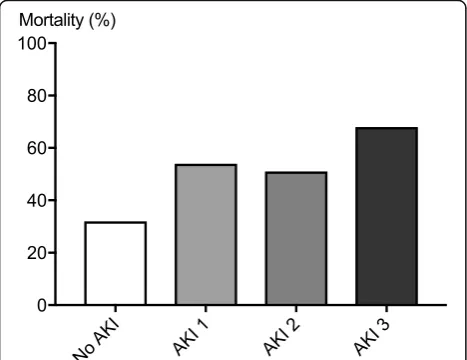
Since only patients surviving beyond day 2 were included in the study, overall 6-month mortality was relatively low at 44%. In patients with AKI stage 3, 6-month mortality was significantly higher at 68% compared to 33% in pa-tients without AKI (Fig.4). Nine of 17 patients who were still RRT-dependent 7 days after the CA were alive at 6 months with none of them requiring long-term dialysis.
Discussion
In survivors of OHCA from the presumed cardiac cause, temperature management at 33 °C did not show different rates of AKI from management at 36 °C. In addition, emergency contrast angiography within 6 h of OHCA was not associated with a higher risk of AKI compared to later or no angiography. This finding is important and should be evaluated in properly designed trials given the possibility that the number of patients managed with early angiography may increase if the recommended clinical strategy for STEMI is extended to non-STEMI. Fig. 1The frequency of criteria used to determine the worst stage
of AKI days 2–7 after CA. AKI stages 2 and 3 are split into the different diagnostic criteria urine output, S-creatinine concentration or a combination thereof. For stage 1, AKI urine output criterion was by study definition not applicable and as a consequence neither was the combination of urine output and creatinine level
[image:5.595.57.290.88.201.2]Fig. 2The maximum stage of AKI during days 2–7 after cardiac arrest stratified by temperature allocation. The category of AKI was worse in the TTM-33 group (p= 0.018, Mann-WhitneyUtest)
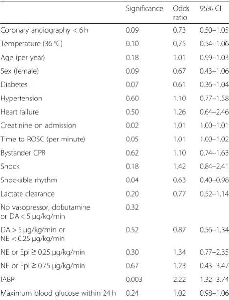
Table 2Results of logistic regression
Significance Odds ratio
95% CI Coronary angiography < 6 h 0.09 0.73 0.50–1.05 Temperature (36 °C) 0.10 0,75 0.54–1.06 Age (per year) 0.18 1.01 0.99–1.03 Sex (female) 0.09 0.67 0.43–1.06 Diabetes 0.07 0.61 0.36–1.04 Hypertension 0.60 1.10 0.77–1.58 Heart failure 0.50 1.26 0.64–2.46 Creatinine on admission 0.02 1.01 1.00–1.01 Time to ROSC (per minute) 0.05 1.01 1.00–1.02 Bystander CPR 0.62 1.10 0.74–1.63 Shock 0.18 1.42 0.84–2.41 Shockable rhythm 0.04 0.63 0.40–0.98 Lactate clearance 0.20 0.77 0.52–1.14 No vasopressor, dobutamine
or DA < 5μg/kg/min
0.32 DA > 5μg/kg/min or
NE < 0.25μg/kg/min
0.52 0.87 0.56–1.34 NE or Epi≥0.25μg/kg/min 0.30 1.34 0.77–2.35 NE or Epi≥0.75μg/kg/min 0.67 1.23 0.43–3.47 IABP 0.003 2.22 1.32–3.74 Maximum blood glucose within 24 h 0.24 1.02 0.98–1.06
Results of logistic regression, angiography within 6 h of cardiac arrest vs. no early angiography. Increasing time to ROSC, a higher serum creatinine on admission and treatment with IABP were independently associated with a higher risk of AKI whereas an initial shockable rhythm was associated with less AKI. Normal lactate clearance was defined as lactate < 2.0 mmol/L alt 12 h or a decrease in lactate > 50% within 12 h of cardiac arrest
Abbreviations: ROSCreturn of spontaneous circulation,CPRcardiopulmonary resuscitation,DAdopamine,NEnorepinephrine,Epiepinephrine,IABP
[image:5.595.304.539.97.405.2] [image:5.595.57.290.522.692.2]Reported incidences of AKI after CA range from 12 to 81% depending on the definition used [1,4]. Our finding of an incidence of AKI of 45% is in line with previous work [5, 19]. We also show an association between AKI after OHCA and increased mortality, which again is in agreement with previous reports [1,2,5,19–21].
[image:6.595.59.539.100.599.2]The TTM-33 group contained more patients with AKI and more severe AKI than the TTM-36 group. The temperature groups were reasonably well balanced with regard to patient and cardiac arrest factors. We believe the exclusion of patients who died on days 1 and 2 after OHCA from our analysis did not affect our conclusions
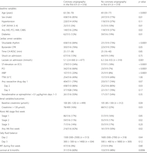
Table 3Patient characteristics split into angiography within 6 h of CA or later/no angiography
Coronary angiography in the first 6 h (n= 516)
No coronary angiography
in first 6 h (n= 319) p value Baseline variables
Age (years) 63 (56–70) 69 (59–77) < 0.0001 Sex (male) 438/516 (85%) 247/319 (77%) 0.01 Hypertension 220/514 (43%) 118/319 (37%) 0.11 CHF (NYHA 3–4) 25/515 (5%) 31/319 (10%) 0.01 Any IHD, PCI, AMI, CABG 149/516 (29%) 118/319 (37%) 0.02 Diabetes 62/515 (12%) 59/316 (19%) 0.01 Cardiac arrest variables
Shockable rhythm 438/516 (84%) 231/319 (73%) < 0.0001 Bystander CPR 393/516 (76%) 223/319 (70%) 0.05 Time CA-ROSC (min) 25 (17–38) 25 (16–40) 0.85 Shock on admission 67/516 (13%) 44/319 (14%) 0.75 Lactate on admission (mmol/L) 5.1 (2.4–8.8) (n= 477) 6.2 (3.6–9.5) (n= 310) 0.001 ST-elevation on ECG 279/513 (54%) 57/315 (18%) < 0.0001 PCI 342/516 (66%) 23/319 (7%) < 0.0001 IABP 107/515 (20%) 25/319 (8%) < 0.0001 TTM 33 °C 254/516 (49%) 157/319 (49%) 1.00 Any vasoactive drug day 1 404/511 (79%) 233/317 (74%) 0.07 Day 2 438/510 (86%) 252/316 (80%) 0.03 Day 3 377/508 (74%) 221/317 (70%) 0.17 Noradrenaline or epinephrine > 0.1μg/kg/min days 1–3 261/516 (50%) 171/317 (54%) 0.35 Renal variables/outcomes
Baseline creatinine (μmol/L) 100 (85–120) (n= 499) 105 (85–130) (n= 312) 0.04 Creatinine > 130μmol/L 78/499 (16%) 68/312 (22%) 0.03 Worst AKI stage first week
Stage 1 86/516 (17%) 51/319 (16%) 0.85 Stage 2 59/516 (11%) 55/319 (17%) 0.02 Stage 3 71/516 (14%) 55/319 (17%) 0.20 Any AKI first week 216/516 (42%) 161/319 (50%) 0.02 Daily fluid balance
Day 2 1500 (500–2500) (n= 513) 1600 (500–2700) (n= 318) 0.64 Day 3 300 (−500 to + 1400) (n= 504) 500 (−400 to + 1800) (n= 309) 0.12 RRT during first week 47/516 (9%) 27/319 (9%) 0.80 Survival at 6 months 311/516 (60%) 153/319 (48%) 0.0006
Patient characteristics, cardiac arrest-related factors, treatment and renal outcome data split into patients exposed to angiography within the first 6 h of cardiac arrest or not. Dichotomous results are presented as numbers and percentages; continuous data are presented as median and interquartile range. Thepvalues were calculated using Fisher’s exact test and Mann-Whitney test respectively
Abbreviations: CHFchronic heart failure,IHDischaemic heart disease,PCIpercutaneous coronary intervention,AMIacute myocardial infarction,CABGcoronary artery bypass grafting,CPRcardiopulmonary resuscitation,CA-ROSCtime from cardiac arrest to return of spontaneous circulation,IABPintra-aortic balloon pump,
since death in the first 2 days was evenly distributed be-tween temperature allocations. The patient characteristics were also balanced with respect to the patient and cardiac arrest factors. During the ICU stay, vasoactive drugs were used more frequently in the TTM-33 group compared to the TTM-36 group. This may reflect differences in cardio-vascular performance between the groups. A previous sub-study of the TTM trial including 171 patients showed that patients randomised to TTM-33 had a reduced car-diac index and increased systemic vascular resistance compared to TTM-36 [22]. The optimal blood pressure/ perfusion parameters for renal recovery after cardiac ar-rest are not known. In well-characterised patients with
vasoplegia, an increase in MAP from 60 to 75 mmHg led to an increase in glomerular filtration rate (GFR) and Cr-EDTA clearance without further improvement with an increase to 90 mmHg [23,24]. In OHCA patients, there is an association not only between increasing MAP and im-proved renal function indices, but also between increasing level of vasoactive support and decreased GFR/increased need for RRT [25]. Considering CA patients and renal function, the optimal balance between MAP and vaso-active support is not known. Patients in the TTM-33 group also received more intravenous fluids than the TTM-36 group. Fluid overload is a risk factor for develop-ment and progression of AKI [26,27]. This aspect of clin-ical management after CA deserves further attention in future studies. After correction for important risk factors, temperature allocation was no longer an independent risk factor for AKI.
After correction for important risk factors, early angi-ography was not independently associated with an in-creased risk of AKI. This is encouraging, as fear of contrast-induced nephrotoxicity may lead clinical teams to withhold potentially life-saving emergency coronary angiography unnecessarily. Similar results were noted by Petek et al. [28]. Whether the TTM protocol recommen-dation to administer intravenous fluids on induction of TTM (30 ml/kg) modified the development of AKI is unclear.
Our principal reason for focusing on the effects of early angiography (within 6 h of CA) was to investigate a clinical scenario in which the renal system is exposed to several potentially harmful insults simultaneously. In the post CA setting, where cardiac output may be reduced, administration of contrast during emergency angiog-raphy might potentially compromise the chances of renal recovery further [29]. The lack of an increased in-cidence of AKI in this setting is reassuring especially in light of current guidelines [12] and consensus statements [30,31]. Early angiography may result in faster recovery of cardiac function and better renal perfusion, which may outweigh the potential risks associated with contrast media. Our results are supported by a recent study showing no significant difference in the release of early AKI biomarkers in critically ill patients after contrast exposure [32].
Both patient characteristics and OHCA factors were associated with development of AKI. An increasing time from CA to ROSC, an indicator of ischaemic burden, was independently associated with a higher risk of AKI, as was a higher serum creatinine on admission. The as-sociation of the intensity of ischaemia/reperfusion injury after CA (whether assessed as amount of epinephrine administered, time to ROSC, lactate concentration or the early presence of shock) and AKI has been noted in several studies [3–5, 19, 21]. Increased serum creatinine Fig. 3The time from cardiac arrest to angiography. The majority of
patients had an early angiography (within 6 h of cardiac arrest). Note varied time intervals
[image:7.595.57.290.87.265.2] [image:7.595.57.291.501.681.2]concentrations on admission and risk of AKI are also well described [4,5,19]. Thus, creatinine concentrations have been incorporated in most AKI risk-scoring sys-tems [33]. The reason for the increased baseline creatin-ine concentration in our patient group developing AKI is not known. Patient factors (age, pre-existing cardiac failure, higher body mass index with associated muscle mass and unrecognised CKD (chronic kidney disease) and peri-arrest factors (longer time to ROSC and more pronounced post ROSC haemodynamic instability) may have contributed [34].
The presence of an IABP was strongly associated with the development of AKI, but shock and poor lactate clearance were not. Since the use of IABPs was unevenly distributed between centres, availability and indications for its applica-tion may have differed. It is possible that the use of IABP is a surrogate marker of continued haemodynamic instability and continued shock, but it is also possible that using an IABP after OHCA increases the risk of AKI per se.
Early angiography was not independently associated with a lower incidence of AKI. When extending the ana-lysis to all patients with angiography during the ICU stay, angiography was independently associated with a reduction of AKI but the selection and survivor bias contributes to this finding.
Limitations
It is important to acknowledge limitations to this post hoc analysis based on a randomised controlled trial designed primarily to address the postulated protective effect of temperature control after OHCA, and therefore, the results should be regarded as hypothesis generating. Data on pre-existing renal function were not available. We elected not to use the MDRD formula to assess baseline creatinine since race (black/non-black) was unknown. The use of the first recorded serum creatinine concentration on arrival to the hospital might have underestimated baseline renal func-tion resulting in a reduced incidence of AKI [1, 7]. How-ever, we note that it takes 24–36 h for serum creatinine changes to occur after a definite renal insult [35]. Therefore, it is likely that the creatinine values within the immediate period after OHCA are reasonably representative of pre-existing values, but it is conceivable that we missed a certain degree of unrecognised CKD. Since only daily urine output data were available, the urinary criteria of the KDIGO classification of AKI had to be modified, an ap-proach commonly taken in large cohort studies [36]. The consequence of this modification was likely to be a reduced recorded incidence of AKI. Urine output may have been modified by for instance initial volume loading, cold-induced diuresis or use of diuretics; however, on day 2, the first complete day included in the study, there was no difference in urine output between TTM-33 and TTM-36.
Since the TTM-study was not randomised by timing of angiography, there may be a selection bias towards early angiography in relation to the first analysed rhythm (ven-tricular fibrillation/tachycardia) and ECG findings (ST-ele-vation) reflecting resuscitation guidelines. Haemodynamic instability and cardiogenic shock may have played a less prominent role in a similar selection bias because early angiography was performed with similar frequency in those patients excluded due to early death and those in-cluded. Early angiography seems to have been employed equally despite more pronounced lactate elevation and cardiogenic shock in the excluded group.
We cannot exclude that the use of early angiography was influenced by the initial creatinine concentration as the initial creatinine was lower in the early angiography group. How-ever, the patients excluded due to early death had higher baseline creatinine than the included patients. Despite this, early angiography was applied with similar frequency. The necessary exclusion of 73 patients (8%) dying before the end of day 2 after CA may have produced an unknown bias.
We cannot exclude that some patients in our study may have been exposed to other early contrast examina-tions, for instance to exclude a pulmonary embolism. Fi-nally, the TTM trial data did not include the type or volume of contrast administered including during coron-ary angiography.
Conclusions
In survivors of OHCA from the presumed cardiac cause, temperature management at 33 °C did not show different rates of AKI from management at 36 °C. Coronary angi-ography within 6 h of CA was not associated with an in-creased incidence of acute kidney injury compared to later or no coronary angiography. The additional effect of coronary angiography on kidney function after cardiac arrest was negligible compared to other factors causing acute kidney injury after cardiac arrest.
The present results suggest that coronary angiography should not be deferred in a patient after cardiac arrest due to concerns over the risk of acute kidney injury.
Additional file
Additional file 1:Table S1.Definitions of AKI stage as used in the study.
Abbreviations
AKI:Acute kidney injury; BP: Blood pressure; CA: Cardiac arrest; CABG: Coronary artery bypass grafting; CHF : Chronic heart failure; CKD : Chronic kidney disease; CPR: Cardiopulmonary resuscitation; DA: Dopamine; Epi: Adrenaline/ epinephrine; GFR: Glomerular filtration rate; IABP: Intra-aortic balloon pump; ICU: Intensive care unit; IHD: Ischaemic heart disease; IQR: Interquartile range; KDIGO: Kidney disease improving global outcomes; MAP: Mean arterial pressure; NA: Noradrenaline/norepinephrine; NYHA: New York Heart Association; OHCA: Out-of-hospital cardiac arrest; PCI: Percutaneous coronary intervention; ROSC : Return of spontaneous circulation; RRT: Renal replacement therapy; SOFA: Sequential Organ Failure Assessment; STEMI: ST-Elevation myocardial infarction; TTM: Target temperature management
Funding
The TTM trial was funded by independent research grants from the Swedish Heart-Lung Foundation; Arbetsmarknadens försäkringsaktiebolag (AFA) Insurance Foundation; The Swedish Research Council; regional research support, Region Skåne; government funding of clinical research within the Swedish National Health Services; Thelma Zoega Foundation; Krapperup Foundation; Thure Carlsson Foundation; Hans-Gabriel and Alice Trolle-Wachtmeister Founda-tion for Medical Research; Skåne University Hospital, Sweden; TrygFonden, Denmark; the European Clinical Research Infrastructures Network; the European Critical Care Research Network; the Ministry of Higher Education and Research of Luxembourg; and the National Research Fund, Luxembourg. No commercial funding was received. The funding organizations did not have any access to the data, nor did they have any influence on data analysis or interpretation.
Availability of data and materials
The datasets used and/or analysed during the current study are available from the corresponding author on reasonable request.
Authors’contributions
MR, SU, MW, and MO planned the post hoc study. SU and MR provided the statistical analysis of the study. MR, SU, MW, SO, and MJ interpreted the data. All authors contributed in writing the manuscript. All authors read and approved the final manuscript.
Ethics approval and consent to participate
Ethics committees in all participating countries approved the protocol. Waived, delayed and/or consent from legal surrogates and delayed written informed consent from patients regaining mental capacity were obtained according to decisions by ethical boards in respective countries.
Consent for publication
Not applicable
Competing interests
MR, SU, MM, JC, NA, MJ, MO, JD and MW have no competing interest to declare.
AW has given consultancy advice to BARD Healthcare 2012 and Baxter Healthcare 2016. GG has received consultation fees from Bard Medical. NN has received a speaker fee from Bard Medical and has been a scientific advisor to BrainCool.
Publisher’s Note
Springer Nature remains neutral with regard to jurisdictional claims in published maps and institutional affiliations.
Author details
1
Department of Clinical Sciences, Anaesthesia and Intensive Care, Skane University Hospital, Lund University, 221 85 Lund, Sweden.2Foprum South,
Skane University Hospital, Lund, Sweden.3Honorary Research Fellow, Cardiff
University School of Medicine, Cardiff, UK.4Department of Intensive Care,
Guys and St Thomas’Hospital, Kings College London, London, UK.
5Department of Anaesthetics and Intensive Care Medicine, Royal
Bournemouth Hospital, Bournemouth, UK.6Adult Intensive Care Directorate,
St George’s Hospital London, London, UK.7Department of Intensive Care
Medicine, Royal Berkshire Hospital, Reading, UK.8Division of Intensive Care and Emergency Medicine, Department of Internal Medicine, Medical University Innsbruck, Innsbruck, Austria.9Department of Critical Care and
Nephrology, Guy’s and St Thomas’Hospital, King’s College London, London,
UK.10Department of Cardiology, Skane University Hospital, Lund University,
Lund, Sweden.11Department of Anaesthesia and Intensive Care, Helsingborg
Hospital, Helsingborg, Sweden.12Adult Critical Care, University Hospital of
Wales, Cardiff, UK.13Department of Intensive and Perioperative Care, Skane University Hospital, Lund University, 221 85 Lund, Sweden.
Received: 17 November 2018 Accepted: 11 March 2019
References
1. Domanovits H, Schillinger M, Müllner M, Thoennissen J, Sterz F, Zeiner A, Druml W. Acute renal failure after successful cardiopulmonary resuscitation. Intensive Care Med. 2001.
2. Yanta J, Guyette F, Doshi A, Callaway C, Rittenberger J. Renal dysfunction is common following resuscitation from out-of-hospital cardiac arrest. 2013;84: 1371–4.
3. Roman-Pognuz E, Elmer J, Rittenberger J, Guyette F, Berlot G, Rosa SD, Peratoner A, Bd A, Lucangelo U, Callaway C. Markers of cardiogenic shock predict persistent acute kidney injury after out of hospital cardiac arrest. Heart Lung. 2019;48(2):126–30.
4. Kim Y, Cha K, Cha Y, Kim O, Jung W, Kim T, Han B, Kim H, Lee K, Choi E, et al. Shock duration after resuscitation is associated with occurrence of post-cardiac arrest acute kidney injury. J Korean Med Sci. 2015;30:802–7. 5. Geri G, Guillemet L, Dumas F, Charpentier J, Antona M, Lemiale V, Bougouin
W, Lamhaut L, Mira J, Vinsonneau C, et al. Acute kidney injury after out-of-hospital cardiac arrest: risk factors and prognosis in a large cohort. Intensive Care Med. 2015;41:1273–80.
6. Hypothermia after Cardiac Arrest Study Group. Mild therapeutic hypothermia to improve the neurologic outcome after cardiac arrest. N Engl J Med. 2002;346:549–56.
7. Zeiner A, Sunder-Plassmann G, Sterz F, Holzer M, Losert H, Laggner A, Müllner M. The effect of mild therapeutic hypothermia on renal function after cardiopulmonary resuscitation in men. Resuscitation. 2004;(3):253–61. 8. DeRosa S, Cal MD, Joannidis M, Villa G, Pacheco J, Virzì G, Samoni S,
D'ippoliti F, Marcante S, Visconti F, et al. The effect of whole-body cooling on renal function in post-cardiac arrest patients. BMC Nephrol. 2017;18:376. 9. DeRosa S, Antonelli M, Ronco C. Hypothermia and kidney: a focus on
ischemia-reperfusion injury. Nephrol Dial Transplant. 2017;32:241–7. 10. Boodhwani M, Rubens F, Wozny D, Nathan H. Effects of mild hypothermia
and rewarming on renal function after coronary artery bypass grafting. Ann Thorac Surg. 2009;87:489–95.
11. Windecker S, Kolh P, Alfonso F, Collet J, Cremer J, Falk V, Filippatos G, Hamm C, Head S, Jüni P, et al. 2014 ESC/EACTS Guidelines on myocardial revascularization: the Task Force on Myocardial Revascularization of the European Society of Cardiology (ESC) and the European Association for Cardio-Thoracic Surgery (EACTS) developed with the special contribution of the European Association of Percutaneous Cardiovascular Interventions (EAPCI). Eur Heart J. 2014;35:2541–619.
12. Callaway CW, Donnino MW, Fink EL, Geocadin RG, Golan E, Kern KB, Leary M, Meurer WJ, Peberdy MA, Thompson TM, et al. Part 8: post-cardiac arrest care: 2015 American Heart Association guidelines update for
cardiopulmonary resuscitation and emergency cardiovascular care. Circulation. 2015;132:S465–82.
13. Kern K: Early coronary angiography versus delayed coronary angiography (PEARL). ClinicalTrials.gov Identifier: NCT02387398 Available online: https:// clinicaltrialsgov/ct2/show/NCT02387398.
14. Spaulding C: EMERGEncy versus delayed coronary angiogram in survivors of out-of-hospital cardiac arrest (EMERGE). ClinicalTrials.gov identifier: NCT02876458.https://clinicaltrials.gov/ct2/show/NCT02876458?term= NCT02876458&rank=1.
15. Nielsen N, Wetterslev J, Cronberg T, Erlinge D, Gasche Y, Hassager C, Horn J, Hovdenes J, Kjaergaard J, Kuiper M, et al. Targeted temperature
management at 33 °C versus 36 °C after cardiac arrest. N Engl J Med. 2013; 369:2197–206.
of Canada, InterAmerican Heart Foundation, Resuscitation Councils of Southern Africa). Circulation. 2004;110:3385–97.
17. Nielsen N, Wetterslev J, al-Subaie N, Andersson B, Bro-Jeppesen J, Bishop G, Brunetti I, Cranshaw J, Cronberg T, Edqvist K, et al. Target temperature management after out-of-hospital cardiac arrest--a randomized, parallel-group, assessor-blinded clinical trial--rationale and design. Am Heart J. 2012;163:541–8. 18. Khwaja A. KDIGO clinical practice guidelines for acute kidney injury.
Nephron Clin Pract. 2012;120:c179–84.
19. Tujjar O, Mineo G, Dell’Anna A, Poyatos-Robles B, Donadello K, Scolletta S, Vincent J-L, Taccone F. Acute kidney injury after cardiac arrest. Crit Care. 2015;19:169.
20. Hasper D, Sv H, Storm C, Jorres A, Schefold J. Changes in serum creatinine in the first 24 hours after cardiac arrest indicate prognosis: an observational cohort study. Crit Care. 2009;13(5):R168.
21. Chua H, Glassford N, Bellomo R. Acute kidney injury after cardiac arrest. Resuscitation. 2012;6:721–7.
22. Bro-Jeppesen J, Hassager C, Wanscher M, Østergaard M, Nielsen N, Erlinge D, Friberg H, Køber L, Kjaergaard J. Targeted temperature management at 33 °C versus 36 °C and impact on systemic vascular resistance and myocardial function after out-of-hospital cardiac arrest: a sub-study of the target temperature management trial. Circ Cardiovasc Interv. 2014;7:663–72. 23. Larsson JS, Bragadottir G, Redfors B, Ricksten S. Renal effects of
norepinephrine-induced variations in mean arterial pressure after liver transplantation: a randomized cross-over trial. Acta Anaesthesiol Scand. 2018;62(9):1229–36.
24. Redfors B, Bragadottir G, Sellgren J, Swärd K, Ricksten S. Effects of norepinephrine on renal perfusion, filtration and oxygenation in vasodilatory shock and acute kidney injury. Intensive Care Med. 2011;37:60–7. 25. Grand J, Hassager C, Winther-Jensen M, Rundgren M, Friberg H, Horn H,
Wise M, Nielsen N, Kuiper M, Wiberg S, et al. Mean arterial pressure during targeted temperature management and renal function after out-of-hospital cardiac arrest. J Crit Care. 2018;50:234–41.
26. Perner A, Prowle J, Joannidis M, Young P, Hjortrup P, Pettilä V. Fluid management in acute kidney injury. Intensive Care Med. 2017;43:807–15. 27. Raimundo M, Crichton S, Martin J, Syed Y, Varrier M, Wyncoll D, Ostermann
M. Increased fluid administration after early acute kidney injury is associated with less renal recovery. Shock. 2015;44:431–7.
28. Petek B, Bravo P, Kim F, Id B, Kudenchuk P, Shuman W, Gunn M, Carlbom D, Gill E, Maynard C, et al. Incidence and risk factors for postcontrast acute kidney injury in survivors of sudden cardiac arrest. Ann Emerg Med. 2016;67:469–76. 29. Laurent I, Monchi M, Chiche J, Joly L, Spaulding C, Bourgeois B, Cariou A,
Rozenberg A, Carli P, Weber S, et al. Reversible myocardial dysfunction in survivors of out-of-hospital cardiac arrest. J Am Coll Cardiol. 2002;40:2110–6. 30. Noc M, Fajadet J, Lassen JF, Kala P, MacCarthy P, Olivecrona GK, Windecker
S, Spaulding C, European Association for Percutaneous Cardiovascular I, Stent for Life G. Invasive coronary treatment strategies for out-of-hospital cardiac arrest: a consensus statement from the European association for percutaneous cardiovascular interventions (EAPCI)/stent for life (SFL) groups. EuroIntervention. 2014;10:31–7.
31. Joannidis M, Druml W, Forni L, Groeneveld A, Honore P, Hoste E, Ostermann M, Straaten HO-V, Schetz M. Prevention of acute kidney injury and protection of renal function in the intensive care unit: update 2017 : expert opinion of the Working Group on Prevention, AKI section, European Society of Intensive Care Medicine. Intensive Care Med. 2017;43:730–49.
32. Ostermann M, McCullough PA, Forni L, Bagshaw SM, Joannidis M, Shi J, Kashani K, Honore P, Chawla L, Investigators JKoboaS. Kinetics of urinary cell cycle arrest markers for acute kidney injury following exposure to potential renal insults. Crit Care Med. 2018;46:375–83.
33. Lamiere N, Kellum J, Group ftKAGW. Contrast-induced acute kidney injury and renal support for acute kidney injury: a KDIGO summary (part 2). Crit Care. 2013;2013:205.
34. Forni L, Darmon M, Ostermann M, Straaten HO-V, Pettilä V, Prowle J, Schetz M, Joannidis M. Renal recovery after acute kidney injury. Intensive Care Med. 2017;43:855–66.
35. Ostermann M, Joannidis M. Acute kidney injury 2016: diagnosis and diagnostic workup. Crit Care. 2016;20:299.