R E S E A R C H
Open Access
Dynamic changes of CX3CL1/CX3CR1 axis
during microglial activation and motor
neuron loss in the spinal cord of ALS
mouse model
Jingjing Zhang
1,2,3, Yufei Liu
1,2, Xinyao Liu
1,2, Song Li
1,2, Cheng Cheng
1,2, Sheng Chen
4*and Weidong Le
1,2*Abstract
Background:Neuron-microglia communication plays a crucial role in the motor neurons (MNs) death in amyotrophic lateral sclerosis (ALS). Neurons can express chemokine (C-X3-C motif) ligand 1 (CX3CL1), which mediates microglial activation via interacting with its sole receptor CX3CR1 in microglia. In the present study, we aimed to investigate the dynamic changes of CX3CL1/CX3CR1 axis during microglial activation and MNs loss in
SOD1G93Amouse model of ALS.
Methods:qPCR, western blot and immunofluorescent staining were used to examine the mRNA and protein levels and localization of CX3CL1/CX3CR1 in the anterior horn region of spinal cord in both SOD1G93Amice and their age-matched wild type (WT) littermates at 40, 60, 90 and 120 days of age. The M1/M2 microglial activation in the spinal
cord tissues of SOD1G93Amice and WT mice were evaluated by immunofluorescent staining of M1/M2 markers and
further confirmed by qPCR analysis of M1/M2-related cytokines.
Results:The immunofluorescent staining revealed that CX3CL1 was predominately expressed in MNs, while CX3CR1 was highly expressed in microglia in the anterior horn region of spinal cord. Compared with age-matched WT mice, CX3CL1 mRNA level was elevated at 40 days but decreased at 90 and 120 days in the anterior horn region of spinal cords in ALS mice. Consistently, CX3CR1 mRNA level was increased at 90 and 120 days. Western blot assay further confirmed the dynamic changes of CX3CL1/CX3CR1 axis in ALS mice. Additionally, the levels of M1/M2 markers of microglia and their related cytokines in the anterior horn region of spinal cord in ALS mice were increased at 90 and 120 days. Moreover, while M1-related cytokines in ALS mice were persistently increased at 120 days, the upregulated M2-related cytokines started to decline at 120 days, suggesting an altered microglial activation.
Conclusions:Our data revealed the dynamic changes of CX3CL1/CX3CR1 axis and an imbalanced M1/M2 microglial activation during ALS pathological progression. These findings may help identify potential molecular targets for ALS therapy.
Keywords:ALS, CX3CL1/CX3CR1 axis, Microglial activation, SOD1G93Amice
* Correspondence:[email protected];[email protected]
1
Center for Clinical Research on Neurological Diseases, the First Affiliated Hospital, Dalian Medical University, Dalian 116021, People’s Republic of China
4Department of Neurology, Ruijin Hospital, Shanghai Jiao Tong University
School of Medicine, Shanghai 200025, China
Full list of author information is available at the end of the article
Introduction
Amyotrophic lateral sclerosis (ALS) is a chronic neuro-degenerative disease characterized by the loss of both lower and upper motor neurons (MNs). Most ALS pa-tients present progressive muscle weakness and atrophy, and eventually died within 5 years of the disease onset. At present, no therapy could delay the disease progres-sion except riluzole and edaravone, which can extend some ALS patients’lifespan for only a few months. The etiology and pathogenesis of ALS are believed to be multifactorial, and the exact mechanisms have not been fully understood. Most ALS cases are sporadic, while around 10% of patients are familial. Identification of pathogenic genes for familial ALS has facilitated the re-search for key pathogenic mechanisms of this disease [1]. Cu/Zn superoxide dismutase 1 (SOD1) was the first gene identified to be associated with ALS, accounting for about 10–20% of familial ALS. SOD1G93A mice car-rying mutant human SOD1 gene develop similar symp-toms and pathological features as ALS [2], and this mouse model has been widely used in the field of ALS research.
Microglia play a dual role in the pathogenesis of ALS. On one hand, it has been reported that activated micro-glia contributed to MNs death in ALS mice via induc-tion of inflammatory responses [3–6]. Inhibition of microglial activation could ameliorate disease
patholo-gies through the blockage of NF-κB signaling in
SOD1G93A mice [7]. On the other hand, one recent
study has found that microglia can promote the recovery of MNs function in a mouse model of TDP-43 proteino-pathy [8]. The cytotoxic or neuroprotective roles of microglia may be dependent on their phenotypes, M1 and M2 [9]. M1 microglia is characterized by the ele-vated expression of inducible nitric oxide synthase (iNOS) and cluster of differentiation 86 (CD86), and can usually reduce neuronal survival through the
overpro-duction of reactive oxygen species (ROS) and
pro-inflammatory cytokines such as tumor necrosis fac-torα (TNF-α) and Interleukin 1β(IL-1β). M2 microglia is characterized by the expression of arginase 1 (Arg1) and cluster of differentiation 206 (CD206), which can enhance neuronal survival through the release of trophic and anti-inflammatory factors such as insulin-like growth factor 1(IGF-1) [10]. It has been speculated that M2 microglia may protect MNs through preventing neu-roinflammation at an early stage, whereas M1 microglia induce MNs loss by promoting neuroinflammation dur-ing the late phase of disease [11].
Communications between MNs and microglia are thought to play an important role in the pathogenesis of ALS [12, 13]. While microglia regulate MNs fates, MNs are also able to influence microglial activity through various mechanisms [14, 15]. For example, extracellular
mutant SOD1 protein, predominately secreted from MNs, directly triggers microglial activation [14, 16]. Chemokine (C-X3-C motif ) ligand 1 (CX3CL1), also named fractalkine, is the only representative of the CX3C chemokine, being constitutively expressed by neu-rons. Strong neuronal excitation can cause fractalkine to be cleaved from the neuronal surface. This soluble form of fractalkine then interacts with nearby microglia which express low levels of CX3CL1 receptor CX3CR1 [17]. The dynamic changes in the expression of CX3CL1 in MNs and its receptor CX3CR1 in microglia may be in-volved in fundamental processes of neurons/microglia communication [18]. Disruption of CX3CL1/CX3CR1 axis could result in microglial activation and overexpres-sion of IL-1β, TNFα [19]. Moreover, polymorphism of the CX3CR1 gene can influence the fate of ALS [20]. Until now, the dynamic changes of CX3CL1/CX3CR1 axis in the spinal cord anterior horn of ALS are not well-documented during disease progression. Therefore, in the present study, we aimed to examine the dynamic changes of CX3CL1/CX3CR1 axis and detected the microglial activation status and the related cytokines production in the anterior horn region of spinal cord in SOD1G93Amouse model of ALS at the age of 40, 60, 90 and 120 days. The study outcome may help us under-stand the roles of CX3CL1/CX3CR1 axis in the disease progression of ALS and may provide new clues for ALS therapy.
Materials and methods
SOD1G93Atransgenic mice and their treatments
Male SOD1G93Atransgenic (TG) mice overexpressing 20 copies of mutant human SOD1 with a Gly93Ala substi-tution (SOD1G93A) were obtained from Jackson
Labora-tories (B6SJL-Tg(SOD1*G93A)1Gur/J, stock number
002726). The hemizygous TG mice start to show a de-crease of max running speed and abnormal gait from 50 days of age. Around 90–100 days of age, visible symp-toms of hind limb paralysis could be found, followed by a complete hind limb paralysis around 125 days of age [21]. All animals were housed under standard conditions of constant temperature and controlled lighting (12/12 h light/dark cycle). TG mice were maintained as heterozy-gote by crossing TG males with wild-type (WT) females of the same C57BL/6 J genetic background. At 30 days, genotypes of TG mice were identified by PCR according to our previously reported protocol [22]. In order to ex-clude the possible impacts of gender difference on dis-ease onset and lifespan [23], only male TG and WT mice were used in the current study.
sacrificed at the age of 40 days (asymptomatic stage), 60 days (pre-symptomatic stage), 90 days (symptomatic stage) and 120 days (terminal stage). Then the mice were perfused through heart with ice-cold 0.1 M PBS. The lumbar spinal cord was separated on ice. The anterior horn region was prepared by cutting ventral half of the lumbar spinal cord and removing the tissues surround-ing anterior horn that marked with the black line (as shown in Additional file1: Figure S1) under the dissect microscope. The tissues were collected and stored at− 80 °C. Total RNA was extracted from the anterior horns and lysed in RNAiso plus reagent according to manufac-turer’s instructions (Takara Bio, Japan). RNA concentra-tion and purity were measured by UV spectroscopy.
Complementary DNA (cDNA) synthesis and quantitative real-time PCR (qPCR)
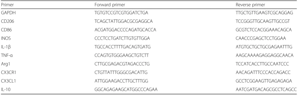
cDNA was obtained by reverse transcription using the primescript RT reagent kit with gDNA eraser (Takara Bio, Japan) by two-step method. qPCR was performed with TransStart TOP green qPCR supermix (Transgen Biotech, China) using the Applied Biosystems 7500 Real-Time PCR System (Thermo Fisher Scientific, USA). GAPDH was selected as an internal control for gene level. The primers sequences for qPCR were summarized in Table 1. The relative level was calculated as 2–ΔΔCt normalizing to GAPDH and relative to age-matched WT mice.
Frozen sections for immunofluorescent staining
TG mice at 40, 90 and 120 days and age-matched WT litter-mates were sacrificed by perfusion through heart with ice-cold 0.1 M PBS and 4% paraformaldehyde (PFA). The lumbar spinal cords were removed and immersed in 4% PFA. After 12 h, the tissues were gradually dehydrated in 15 and 30% sucrose solution for 24 h, then embedded in Opti-mal Cutting Temperature Compound and cut into serial sec-tions of 10μm thickness in freezing microtome at −25 °C. Sections were attached to glass slides for immunofluorescent
staining as previously described [22]. Spinal cord sections were baked for 30 min at 55 °C and washed by 0.1 M PBS for three times, then blocked with 5% bovine serum and 0.3% triton in PBS for 1 h at room temperature. Sections were incubated with following primary antibodies overnight at 4 °C: anti-NeuN (MAB377, 1:500, Millipore), anti-Iba1 (019–19,741, 1:1000, WAKO), anti-CX3CL1 (AF472, 1: 200, R&D Systems), anti-CX3CR1 (AF5825, 1: 50, R&D Systems), anti-CD86 (553,689, 1:1000, BD Biosciences), anti-Arg1 (sc-271,430, 1:50, Santa Cruz), anti-GFAP (Z0334, 1:2000, Dako). After washing in PBS three times, secondary donkey anti-rabbit IgG conjugated Alexa 555 (A0453, 1:2000, Beyo-time), IFKine™Green donkey anti-goat IgG (A24231, 1:500, Abbkine), goat anti-rabbit IgG conjugated Alexa 594 (8889S, 1:2000, Cell Signaling Technology), goat anti-mouse IgG conjugated Alexa 488 (4408S, 1:2000, Cell Signaling Tech-nology), Cy3 goat anti-rat IgG (A0507, 1:2000, Beyotime, China), goat anti-mouse IgG conjugated Alexa 594 (8890S, 1:2000, Cell Signaling Technology) were applied respectively at room temperature for 2 h. Hoechst was used for nuclei staining. After washing, sections were covered with mount-ing medium and analyzed by fluorescent microscope.
Cell counting
[image:3.595.55.544.98.254.2]For CX3CR1+/Iba1+, CD86+ /Iba1+ or Arg1+/Iba1+ cell counting, five sections spaced 100μm apart in each mouse were used and both sides of the anterior horn re-gion (two views in one slide and at least five slides per animal) were captured at 200 × magnification. Then the total number of cells in both sides of anterior horn re-gion was manually counted by a person who was blind to experimental design. Counts from both sides of anter-ior horn region were averaged and divided by the area in square millimeters. The average count from five sections was used as a single measurement per mouse for statis-tical analysis. The integrated density of CX3CL1 staining was measured by Image J software on all five sections per animal [25].
Table 1Primers for qPCR
Primer Forward primer Reverse primer
GAPDH TGTGTCCGTCGTGGATCTGA TTGCTGTTGAAGTCGCAGGAG
CD206 TCAGCTATTGGACGCGAGGCA TCCGGGTTGCAAGTTGCCGT
CD86 ACGATGGACCCCAGATGCACCA GCGTCTCCACGGAAACAGCA
INOS CCCTCCTGATCTTGTGTTGGA CAACCCGAGCTCCTGGAA
IL-1β TGCCACCTTTTGACAGTGATG ATGTGCTGCTGCGAGAATTTG
TNF-α CCAGTGTGGGAAGCTGTCTT AAGCAAAAGAGGAGGCAACA
Arg1 CTTGCGAGACGTAGACCCTG TCCATCACCTTGCCAATCCC
CX3CR1 CTGTTATTTGGGCGACATTG AACAGATTTCCCACCAGACC
CX3CL1 ATTGGAAGACCTTGCTTTGG GCCTCGGAAGTTGAGAGAGA
Western blotting and qualification
Anterior horns in lumbar spinal cords from both TG mice and WT littermates were dissected and sonicated in ice cold lysis buffer (P1003B, Beyotime, China). The total protein was extracted and separated by 10% SDS-polyacrylamide gel elec-trophoresis and then transferred onto nitrocellulose mem-branes (Merck Millipore, Germany). After blocking with 5% skim milk prepared in tris-buffered saline-Tween 20 (TBST) (20 mM Tris, pH 7.2, 150 mM NaCl, 0.1% Tween 20) for 1 h at room temperature, membranes were incubated for 14 h at 4 °C with primary antibodies against CX3CL1 (bs-0811R, 1: 1000, Bioss), CX3CR1 (AF5825, 1: 100, R&D Systems),
β-tubulin (2128S, 1:2000, Cell Signaling Technology). After washing, membranes were incubated with HRP-conjugated goat-anti-rabbit IgG (7074S, 1:2000, Cell Signaling Technol-ogy) or HRP-conjugated donkey-anti-goat IgG antibody (A0181, 1:2000, Beyotime) for 1 h at room temperature. The target protein bands were quantified by using FluorChem Q system (ProteinSimple, San Jose, CA, USA).
Statistical analysis
Data were expressed as Mean ± Standard deviation (SD). Statistical analysis was performed using statistical program for social sciences (SPSS) 22.0. Two-way ANOVA with Tukey post-hoc for multiple comparisons was used to de-termine whether significant differences existed between TG and WT mice at different time points. Ap value of less than 0.05 indicates statistical significance.
Results
CX3CL1 was highly expressed in MNs and decreased during disease progression in TG mice
Dramatic MNs loss in the spinal cord anterior horn re-gion is an important feature of ALS mice during disease progression [24]. The immunoflurescent staining showed
that CX3CL1 was mainly expressed in MNs (Fig. 1a),
and no obvious CX3CL1 staining was observed on microglia or astrocytes of ALS mice at the age of 90 days (Additional file2: Figure S2). Interestingly, the integrated density of CX3CL1 staining in MNs was elevated in the spinal cord anterior horn region of TG mice at 40 days but decreased accompanied with the MNs loss in the an-terior horn region at the age of 90 and 120 days (Fig.1b).
We further measured the mRNA level of CX3CL1. At 40 days of age, the mRNA level of CX3CL1 in the spinal cord anterior horn region of TG mice was 1.7 fold higher than that of WT mice; however, at 90 and 120 days, the mRNA level of CX3CL1 in the spinal cord an-terior horn region of TG mice was significantly lower than that of WT mice (Fig. 2a). Western blot also re-vealed similar dynamic change of CX3CL1 protein, as shown by the elevated level of CX3CL1 in TG mice at 40 days but declined at 90 and 120 days, as compared
with age-matched WT mice (Fig. 2b, c). All these data may predict a loss of CX3CL1 signaling in MNs during the pathogenesis and progression of ALS.
CX3CR1 was mostly located in microglia and consistently increased in TG mice at 90 and 120 days
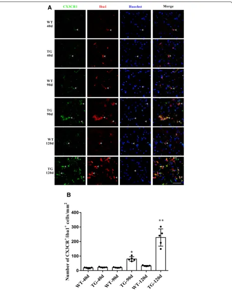
As the disease progression, significant microgliosis and microglial activation could be found in the spinal cord anterior horn region of TG mice [26]. We found that the CX3CL1 receptor CX3CR1 was dominantly located in microglia and its expression was consistently increased to-gether with microglial proliferation (Fig.3a, b). Moreover, compared with WT mice, CX3CR1 mRNA level did not increase as CX3CL1 did in the spinal cord anterior horn region of TG mice at 40 days, but increased significantly at 90 and 120 days (Fig.4a). Western blot also revealed a consistent elevation of CX3CR1 protein in the spinal cord anterior horn region of TG mice at 90 and 120 days com-pared with their age-matched WT mice (Fig. 4b, c). The dynamic changes of CX3CR1 might be a consequence of direct microgliosis or a compensational response to CX3CL1 downregulation.
Imbalanced M1 and M2 microglial activation in the spinal cord anterior horn region of TG mice at 120 days
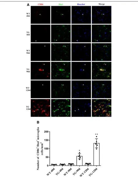
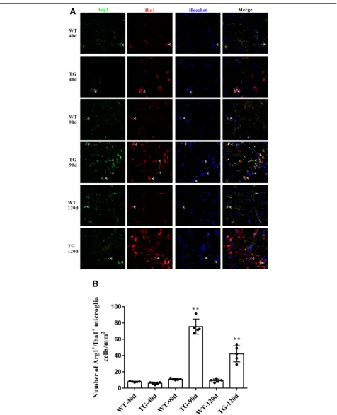
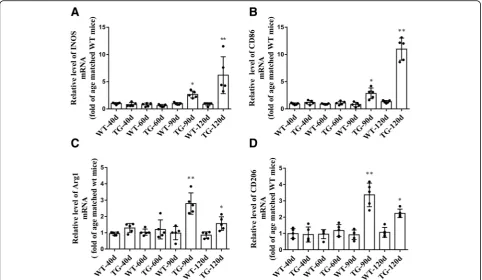
Accompanied by the impaired CX3CL1/CX3CR1 axis, the number of activated microglia was dramatically increased in the spinal cord anterior horn region of TG mice, evi-denced by the immunoflurescent changes of M1/M2 microglia markers. The number of CD86+/Iba1+M1 micro-glia was increased at 90 days and further increased at 120 days (Fig. 5a and b), whereas the number of Arg1+/Iba1+ M2 microglia was significantly increased at 90 days but de-clined in TG mice at 120 days (Fig.6a and b). These results were further confirmed by qPCR, as shown by the in-creased mRNA levels (about 2.7–3.4 fold) of both M1 phenotype (CD86, iNOS) and M2 phenotype (CD206, Arg1) microglial markers in the spinal cord anterior horn region of TG mice at 90 days. Moreover, consistent with the microglial quantification analysis, at the age of 120 days, while M1 microglial markers (CD86, INOS) were persist-ently increased (about 6.2–11.0 fold higher than those in age-matched WT mice), mRNA levels of M2 microglial markers (CD206, Arg1) were reduced compared with those at 90 days of age (still significantly higher than those in WT mice) (Fig.7). All these data may predict an altered M1/M2 polarization during ALS progression, as indicated by an ac-tivated M1/M2 microglia at 90 days and subsequently a relatively suppressed M2 polarization at 120 days.
Microglia play important roles in modulating
neuroin-flammation through secreting proinflammatory or
in most neurodegenerative disease. In contrast, as a neuro-protective cytokine associated with M2 phenotype
microglia, interleukin-10 (IL-10) can inhibit the
production of neuro-toxic factors by microglia to pro-mote cell survival. Here, we found that the mRNA levels of IL-1β and TNF-α were increased by about 5.1–5.4 Fig. 1CX3CL1 were mainly located in neurons and decreased during MNs loss in TG mice.aDouble immunofluorescent staining of CX3CL1
(green) and NeuN (red) in the anterior horn of lumber spinal cord of WT and TG mice at the age of 40, 90 and 120 days. Scale bar = 50μm.b
Integrated density of CX3CL1 staining. The values are expressed as mean ± SD, *p< 0.05 versus age-matched WT mice, **p< 0.001 versus
[image:5.595.57.542.84.625.2]Fig. 2The mRNA and protein levels of CX3CL1 in the spinal cord anterior horns.aqPCR analysis of CX3CL1 mRNA level.bWestern blot analysis
of CX3CL1 protein level.cQuantitative analysis the CX3CL1 Protein level. The values are expressed as mean ± SD, *p< 0.05 versus age-matched
[image:6.595.58.539.83.697.2]Fig. 3CX3CR1 was mainly located in microglia and was increased in the TG mice at the age of 90 and 120 days.aDouble immunofluorescent staining of CX3CR1 (green) and Iba1(red) in the anterior horn of lumber spinal cord of WT and TG mice at the age of 40, 90 and 120 days. Scale
bar = 50μm.bNumber of CX3CR1+/Iba1+microglia per mm2. The values are expressed as mean ± SD, *p< 0.05 versus age-matched WT mice, **
[image:7.595.61.538.85.680.2]Fig. 4The mRNA and protein levels of CX3CR1 in the spinal cord anterior horns.aqPCR analysis of CX3CR1 mRNA level.bWestern blot analysis
of CX3CR1 protein level.cQuantitative analysis the CX3CR1 Protein level. The values are expressed as mean ± SD, *p< 0.05 versus age-matched
[image:8.595.58.538.83.698.2]Fig. 5The changes of M1 microglia in the TG mice at different time points.aDouble immunofluorescent staining of CD86 (red) and Iba1 (green)
at different time points, Sale bar = 50μm.bQuantitative analysis of the M1 microglia per mm2at different time points. The values are expressed
[image:9.595.62.536.84.692.2]Fig. 6The changes of M2 microglial phenotypes in TG mice at different time points.aDouble immunofluorescent staining of Arg1 (green) and
Iba1 (red) at different time points, Sale bar = 50μm.bQuantitative analysis of the M2 microglia per mm2at different time points. The values are
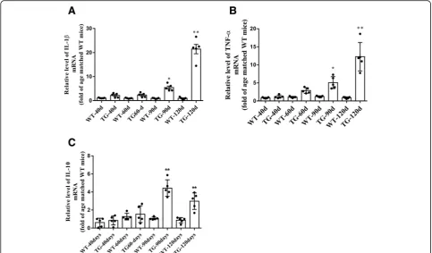
[image:10.595.60.537.84.672.2]fold in spinal cord of TG mice at 90 days, and persist-ently increased by about 12.3–21.5 fold at 120 days,
compared with age-matched WT mice (Fig. 8a, b).
Moreover, the IL-10 mRNA increased by about 4.4 fold at 90 days but reduced to about 3.0 fold at 120 days in TG mice compared with age matched WT mice (Fig.8c). These results were consistent with the data of microglial quantification and microglial markers determination and further indicate the imbalanced activation of M1/M2 microglia in the spinal cord anterior horn region of TG mice at 120 days.
Discussion
Microglia have been studied extensively for their roles in various neurodegenerative diseases including ALS. Their ac-tivation has even been considered as the“hallmark”of many neurodegenerative conditions. However, the exact roles of microglia in ALS are still under debate. Previous study reported that lower mutant SOD1 expression within micro-glia significantly extended the survival of LoxSOD1G37Rmice [27], whereas a later study showed that the elimination of proliferating microglia couldn’t prevent MNs loss in SOD1G93Amice [28]. A latest research revealed that micro-glia could selectively clear neuronal hTDP-43, which was ne-cessary for recovery of MNs function in a mouse model of TDP-43 proteinopathy [8]. Therefore, manipulating the
function of microglia has the potential to improve ALS prognosis.
As the only member of the CXC subfamily of chemo-kines, CX3CL1 exists in two distinct forms: a full-length membrane-bound form and a shed form containing the N-terminal chemokine domain. The shed chemokine do-main of CX3CL1 acts, when cleaved, as a signaling mol-ecule and binds to microglial-expressed CX3CR1 receptor to modulate microglial activation [18]. Here, we found CX3CL1 was up-regulated in the MNs at early stage of ALS (40 days) but decreased at the later phase of disease (90 and 120 days) in ALS model. Simultaneously, the ex-pression level of CX3CR1, the microglia-located sole re-ceptor for CX3CL1, was increased at the age of 90 and 120 days, consistent with the alteration of CX3CL1. In addition, accompanied with this impaired CX3CL1/ CX3CR1 axis, microglia were consistently activated in the spinal cord anterior horn of ALS mice in a M1/M2 imbal-anced manner.
Previous studies have shown that the impaired
CX3CL1–CX3CR1 axis was usually accompanied by
ab-normal microglial activation in various animal models of neurological diseases [29–31]. It has been reported that CX3CL1 could attenuate LPS-induced microglial activa-tion by inducing phosphatidylinositol-3 kinase pathway and reducing the production of nitric oxide (NO), IL-6, Fig. 7The mRNA levels of M1/M2 microglial markers in the TG mice at different time points.a,bThe mRNA levels of M1 markers (INOS, CD86).
c,dThe mRNA levels of M2 markers (Arg1, CD206). The values are expressed as mean ± SD, *p< 0.05 versus age-matched WT mice, **p< 0.001
[image:11.595.57.540.88.368.2]and TNF-α [32]. Microglial activation usually occurred
when CX3CL1 was decreased [32, 33]. Moreover,
CX3CL1 protein could be up-regulated in injured neu-rons to protect neuneu-rons against glutamate excitotoxicity and cell death [34, 35]. The deficiency of its receptor CX3CR1 could promote microglial activation and ex-acerbate MNs loss in ALS mice [36]. Here, we found a significant increase of CX3CL1 level in MNs of the an-terior horn of ALS mice at the age of 40 days, while the TG mice showed neither obvious clinical symptoms nor MNs loss. We assumed that the increased CX3CL1 level might be the early sign of MNs degeneration, which may in turn affect the microglial function at the early stage of disease. After disease onset, there was a significant
de-crease of CX3CL1 immunoflurescent staining on
remaining MNs in TG mice. It is likely that the CX3CL1 reduction after disease onset may reflect the abnormally reduced expression on MNs and MNs loss as well.
Microglial proliferation may result in CX3CR1 eleva-tion at 90 and 120 days. Up-regulaeleva-tion of CX3CR1 may be also a compensation for CX3CL1 down-regulation in the spinal cord of ALS model. The function of CX3CL1/
CX3CR1 axis requires the coordination between
CX3CL1 and CX3CR1 level, and the change of CX3CR1 alone may not be able to compensate for the function of CX3CL1/CX3CR1 axis. The impaired CX3CL1/CX3CR1
axis seems to be associated with microglial activation at ALS disease onset and might contribute to the disease progression. Further study on the molecular mechanisms of CX3CL1/CX3CR1 axis in regulating microglial activa-tion might provide useful informaactiva-tion to determine the value of CX3CL1/CX3CR1 axis as a target for ALS therapy.
Moreover, in the present study, we also found an al-tered microglial polarization during disease progression of ALS. Microglia are the resident immune cells of the central nervous system (CNS) and are the primary medi-ators of neuroinflammation. The activated microglia have been categorized into the classical M1 and alterna-tive M2 phenotype based on their polarization status [9]. Activation of microglia is regulated by multiple extracel-lular and intracelextracel-lular factors, cross talk between neu-rons and microglia could modulate M1 or M2 microglial activation during progression of ALS [37, 38]. It is be-lieved that M2 polarization may be triggered as a pro-tective compensation during the early stage of ALS, while a strong M1 polarization may be dominant during the later phases of the disease in ALS [38]. Adult micro-glia isolated from ALS mice at disease onset showed an M2 phenotype and protected MNs, whereas microglia isolated from ALS mice in end-stage of disease adopted a neurotoxic M1 phenotype [39]. Our present data Fig. 8The changes of M1/M2 microglia related cytokines mRNA levels at different time points.a,bmRNA levels of two cytokines related to M1
microglia (IL-1βand TNF-α).cmRNA levels of IL-10 related to M2 microglia. The values are expressed as mean ± SD, *p< 0.05 versus age-matched
[image:12.595.56.540.88.371.2]revealed that the dynamic changes of CX3CL1/CX3CR1 axis might be associated with alteration of microglial phenotype as ALS progression in the TG mice. CX3CR1
could regulate microglial phenotypes in a time
dependent manner. During the early disease stage CX3CR1 may be a protective molecule. However, as dis-ease progresses, it becomes harmful to neurons [40]. Al-though M1 microglia and pro-inflammatory cytokines were significantly increased at the age of 90 days, the in-creased M2 microglia and anti-inflammatory cytokines were paralleled with elevated M1 microglia. Thus, this balance between M1 and M2 microglial polarization may be result in stabilization of ALS at initial disease stage. Following the disease progression, the role of CX3CR1 is changed, and the balance between M1 and M2 microglia might be disrupted, as shown by a con-tinuous increase in M1 microglia polarization and
over-production of pro-inflammatory cytokines, but a
declined M2 microglia activation and decreased
anti-inflammatory cytokines production. These results indicate that an altered M1/M2 microglial activation may contribute to the ALS progression, and reversing such imbalance between M1 and M2 microglia may delay the progression of ALS.
Conclusions
In conclusion, impaired CX3CL1/CX3CR1 axis and balanced M1/M2 microglial activation may play an im-portant role in the pathogenesis and progression of MNs degeneration in ALS. Regulating CX3CL1/CX3CR1 axis and microglial activation may be a feasible option for fu-ture ALS therapy.
Additional files
Additional file 1:Figure S1.Roadmap for the dissection of anterior horn region in lumber spinal cord. (TIF 1640 kb)
Additional file 2:Figure S2.The immunofluorescent staining of CX3CL1 on microglia or astrocytes. (A) Double immunofluorescent staining of CX3CL1 (green) and GFAP (red) positive astrocytes. (B) Double immunofluorescent staining of CX3CL1 (green) and Iba1 (red) positive
microglia. Scale bar = 50μm. (TIF 1941 kb)
Abbreviations
ALS:Amyotrophic lateral sclerosis; Arg1: Arginase 1 (Arg1); CCL: CC
chemokine ligands; CD206: Cluster of differentiation 206; CD86: Cluster of differentiation 86; CNS: Central nervous system; CX3CL1: Chemokine (C-X3-C motif) ligand 1; CX3CR1: Chemokine (C-X3-C motif) ligand 1 receptor;
IL-10: Interleukin-10; IL-1β: Including interleukin-1β; IL-4: Interleukin-4;
iNOS: Inducible nitric oxide synthase.; MNs: Motor neurons; SOD1: Cu/Zn
superoxide dismutase 1; TGF-β: Transforming growth factor-β; TNF-α: Tumor
necrosis factor-α
Acknowledgements Not applicable.
Funding
This work was supported by funding from the National Natural Sciences Foundation of China (NSFC 81430021 and 81370470), the Program for
Liaoning Innovative Research Team in University (LT2015009), and Liaoning Science and Technology Project (2015225008).
Availability of data and materials
All data generated or analyzed during this study are included in this published article.
Authors’contributions
WL and SC contributed to the conception and design of this research. JZ, YL and XL were responsible for experimental procedures and data acquisition. JZ, CC and SL were involved in statistical analyses and data interpretation. All authors contributed to drafting the manuscript and approve the final version. They also agree to the accuracy and integrity of the manuscript.
Ethics approval
All experimental procedures on animals in this study were conducted in accordance with the laboratory animals care guidelines and approved by institutional animal care committee at Dalian Medical University.
Consent for publication Not applicable.
Competing interests
The authors declare that they have no competing interests.
Author details
1Center for Clinical Research on Neurological Diseases, the First Affiliated
Hospital, Dalian Medical University, Dalian 116021, People’s Republic of China.2Liaoning Provincial Key Laboratory for Research on the Pathogenic Mechanisms of Neurological Diseases, the First Affiliated Hospital, Dalian Medical University, Dalian 116021, People’s Republic of China.3Chifeng
Municipal Hospital, Chifeng 024000, People’s Republic of China.4Department
of Neurology, Ruijin Hospital, Shanghai Jiao Tong University School of Medicine, Shanghai 200025, China.
Received: 23 June 2018 Accepted: 21 November 2018
References
1. Ferraiuolo L, Kirby J, Grierson AJ, Sendtner M, Shaw PJ. Molecular pathways
of motor neuron injury in amyotrophic lateral sclerosis. Nat Rev Neurol.
2011;7(11):616–30.
2. Gurney ME, Pu H, Chiu AY, Dal Canto MC, Polchow CY, Alexander DD, et al.
Motor neuron degeneration in mice that express a human Cu, Zn
superoxide dismutase mutation. Science. 1994;264(5166):1772–5.
3. Clement AM, Nguyen MD, Roberts EA, Garcia ML, Boillee S, Rule M, et al.
Wild-type nonneuronal cells extend survival of SOD1 mutant motor
neurons in ALS mice. Science. 2003;302(5642):113–7.
4. Pramatarova A, Laganière J, Roussel J, Brisebois K, Rouleau GA.
Neuron-specific expression of mutant superoxide dismutase 1 in transgenic mice
does not lead to motor impairment. J Neurosci. 2001;21(10):3369–74.
5. Lino MM, Schneider C, Caroni P. Accumulation of SOD1 mutants in
postnatal motoneurons does not cause motoneuron pathology or
motoneuron disease. J Neurosci. 2002;22(12):4825–32.
6. Puentes F, Malaspina A, van Noort JM, Amor S. Non-neuronal cells in ALS:
role of glial, immune cells and blood-CNS barriers. Brain Pathol. 2016;26(2):
248–57.
7. Frakes AE, Ferraiuolo L, Haidet-Phillips AM, Schmelzer L, Braun L, Miranda CJ,
et al. Microglia induce motor neuron death via the classical NF-kappaB
pathway in amyotrophic lateral sclerosis. Neuron. 2014;81(5):1009–23.
8. Spiller KJ, Restrepo CR, Khan T, Dominique MA, Fang TC, Canter RG,
et al. Microglia-mediated recovery from ALS-relevant motor neuron degeneration in a mouse model of TDP-43 proteinopathy. Nat Neurosci.
2018;21(3):329–40.
9. Tang Y, Le W. Differential roles of M1 and M2 microglia in
neurodegenerative diseases. Mol Neurobiol. 2016;53(2):1181–94.
10. Henkel JS, Beers DR, Zhao W, Appel SH. Microglia in ALS: the good, the bad,
and the resting. J Neuroimmune Pharmacol. 2009;4(4):389–98.
11. Zhao W, Beers DR, Appel SH. Immune-mediated mechanisms in the
pathoprogression of amyotrophic lateral sclerosis. J NeuroImmune
12. Appel SH, Zhao W, Beers D, Henkel J. The microglial-motoneuron dialogue
in ALS. Acta Myol. 2011;30(1):4–8.
13. Brites D, Vaz AR. Microglia centered pathogenesis in ALS: insights in cell
interconnectivity. Front Cell Neurosci. 2014;8:117.
14. Urushitani M, Sik A, Sakurai T, Nukina N, Takahashi R, Julien JP.
Chromogranin-mediated secretion of mutant superoxide dismutase proteins
linked to amyotrophic lateral sclerosis. Nat Neurosci. 2006;9(1):108–18.
15. Biber K, Neumann H, Inoue K, Boddeke HW. Neuronal‘On’and‘Off’signals
control microglia. Trends Neurosci. 2007;30(11):596–602.
16. Hernangomez M, Carrillo-Salinas FJ, Mecha M, Correa F, Mestre L, Loria F,
et al. Brain innate immunity in the regulation of neuroinflammation: therapeutic strategies by modulating CD200-CD200R interaction involve the
cannabinoid system. Curr Pharm Des. 2014;20(29):4707–22.
17. Chapman GA, Moores KE, Gohil J, Berkhout TA, Patel L, Green P, et al. The
role of fractalkine in the recruitment of monocytes to the endothelium. Eur
J Pharmacol. 2000;392(3):189–95.
18. Harrison JK, Jiang Y, Chen S, Xia Y, Maciejewski D, McNamara RK, et al. Role
for neuronally derived fractalkine in mediating interactions between neurons and CX3CR1-expressing microglia. Proc Natl Acad Sci U S A. 1998;
95(18):10896–901.
19. Rogers JT, Morganti JM, Bachstetter AD, Hudson CE, Peters MM, Grimmig
BA, et al. CX3CR1 deficiency leads to impairment of hippocampal cognitive
function and synaptic plasticity. J Neurosci. 2011;31(45):16241–50.
20. Calvo A, Moglia C, Canosa A, Cammarosano S, Ilardi A, Bertuzzo D, et al.
Commonpolymorphisms of chemokine (C-X3-C motif) receptor 1 gene modify amyotrophic lateral sclerosis outcome: A population-based study.
Muscle Nerve. 2018;57(2):212–6.
21. Kanning KC, Kaplan A, Henderson CE. Motor neuron diversity in
development and disease. Annu Rev Neurosci. 2010;33:409–40.
22. Zhang X, Chen S, Song L, Tang Y, Shen Y, Jia L, et al. MTOR-independent,
autophagic enhancer trehalose prolongs motor neuron survival and ameliorates the autophagic flux defect in a mouse model of amyotrophic
lateral sclerosis. Autophagy. 2014;10(4):588–602.
23. Choi CI, Lee YD, Gwag BJ, Cho SI, Kim SS, Suh-Kim H. Effects of estrogen on
lifespan and motor functions in female hSOD1 G93A transgenic mice.
J Neurol Sci. 2008;268(1–2):40–7.
24. Zhang X, Chen S, Li L, Wang Q, Le W. Decreased level of
5-methyltetrahydrofolate: a potential biomarker for pre-symptomatic
amyotrophic lateral sclerosis. J Neurol Sci. 2010;293(1):102–5.
25. Schneider CA, Rasband WS, Eliceiri KW. NIH Image to ImageJ: 25 years of
image analysis. Nat Methods. 2012;9(7):671–5.
26. Hall ED, Oostveen JA, Gurney ME. Relationship of microglial and astrocytic
activation to disease onset and progression in a transgenic model of
familial ALS. Glia. 1998;23(3):249–56.
27. Boillée S, Yamanaka K, Lobsiger CS, Copeland NG, Jenkins NA, Kassiotis G,
et al. Onset and progression in inherited ALS determined by motor neurons
and microglia. Science. 2006;312(5778):1389–92.
28. Gowing G, Philips T, Van Wijmeersch B, Audet JN, Dewil M, Van Den Bosch
L, et al. Ablation of proliferating microglia does not affect motor neuron degeneration in amyotrophic lateral sclerosis caused by mutant superoxide
dismutase. J Neurosci. 2008;28(41):10234–44.
29. Castro-Sanchez S, Garcia-Yague AJ, Lopez-Royo T, Casarejos M, Lanciego JL,
Lastres-Becker I. Cx3cr1-deficiency exacerbates alpha-synuclein-A53T induced neuroinflammation and neurodegeneration in a mouse model of
Parkinson’s disease. Glia. 2018; 66(8):1752–62
30. Tang Z, Gan Y, Liu Q, Yin JX, Liu Q, Shi J, et al. CX3CR1 deficiency
suppresses activation and neurotoxicity of microglia/macrophage in experimental ischemic stroke. J Neuroinflammation. 2014;11:26.
31. Zhang L, Xu J, Gao J, Wu Y, Yin M, Zhao W. CD200-, CX3CL1-, and
TREM2-mediated neuron-microglia interactions and their involvements in
Alzheimer’s disease. Rev Neurosci. 2018; 29(8):837–48
32. Lyons A, Lynch AM, Downer EJ, Hanley R, O’Sullivan JB, Smith A, et al.
Fractalkine-induced activation of the phosphatidylinositol-3 kinase pathway attentuates microglial activation in vivo and in vitro. J Neurochem. 2009;
110(5):1547–56.
33. Mecca C, Giambanco I, Donato R, Arcuri C. Microglia and aging: the role of
the TREM2-DAP12 and CX3CL1-CX3CR1 axes. Int J Mol Sci. 2018;19(1):e318.
34. Chapman GA, Moores K, Harrison D, Campbell CA, Stewart BR, Strijbos PJ.
Fractalkine cleavage from neuronal membranes represents an acute event in the inflammatory response to excitotoxic brain damage. J Neurosci. 2000; 20(15):RC87.
35. Cerbai F, Lana D, Nosi D, Petkova-Kirova P, Zecchi S, Brothers HM, et al. The
neuron-astrocyte-microglia triad in normal brain ageing and in a model of neuroinflammation in the rat hippocampus. PLoS One. 2012;7(9):e45250.
36. Cardona AE, Pioro EP, Sasse ME, Kostenko V, Cardona SM, Dijkstra IM, et al.
Control of microglial neurotoxicity by the fractalkine receptor. Nat Neurosci.
2006;9(7):917–24.
37. Geloso MC, Corvino V, Marchese E, Serrano A, Michetti F, D’Ambrosi N. The
dual role of microglia in ALS: mechanisms and therapeutic approaches. Front Aging Neurosci. 2017;9:242.
38. Hooten KG, Beers DR, Zhao W, Appel SH. Protective and toxic
neuroinflammation in amyotrophic lateral sclerosis. Neurotherapeutics. 2015;
12(2):364–75.
39. Liao B, Zhao W, Beers DR, Henkel JS, Appel SH. Transformation from a
neuroprotective to a neurotoxic microglial phenotype in a mouse model of
ALS. Exp Neurol. 2012;237(1):147–52.
40. Febinger HY, Thomasy HE, Pavlova MN, Ringgold KM, Barf PR, George AM,