Identification of the Lytic Origin of DNA Replication
in
Human
Cytomegalovirus by
a
Novel
Approach
Utilizing
Ganciclovir-Induced
Chain Termination
FAYEZ M. HAMZEH,"2'3 PAUL S. LIETMAN,12'3 WADE GIBSON,3'4AND GARYS. HAYWARD34*
TheDivision of Clinical Pharmacology1 and The Virology Laboratories,4 Department of Medicine,2 and Departmentof
Pharmacology
andMolecular Sciences,3 The Johns Hopkins School of Medicine,Baltimore, Maryland 21205
Received 3July 1990/Accepted 3 September 1990
Infection withhuman cytomegalovirus in thepresenceof the antiviral nucleotide analog ganciclovir results in continuing low-level viral DNA synthesis and the accumulation of relatively small fragments of double-strandedprogenyDNA. These fragments consistently provedtorepresentamplification ofsequencesfromonly
one small section of the viral genome (EcoRI-V) lying near the center of the unique L segment. Further mapping revealed that the viral sequencesrepresented in these fragments occurredingradients of abundance thatdecreased inboth directions fromapointnear0.35 to 0.4mapunit. The proportion of amplifiedsequences increased with both time after infection and dosage of ganciclovir used. We conclude that theprimary lytic cycle replication origin of human cytomegalovirus lies withina3-to4-kbregion immediately upstream and to
the right of the promoter for the single-stranded DNA-binding protein (DB140). The amplified origin-containing DNA molecules appeared to arise by continuing rounds of bidirectional initiation on truncated fragments of the genome that were generated as a result of chain termination effects induced by the incorporation of ganciclovir into the viral DNA. Inspection of the DNA sequence in the vicinity of ori-Lyt revealed a large complex upstream region that may be a noncoding intergenic domain and that bears no
homology to anypreviously describedherpesvirus origin. This 2.5-kb region includes manyduplicated and
inverted sequences, together with consensus CRE/ATF and other transcription factor-binding sites, and an
interesting set of 23 copies of an interspersed decamer consensus element AAAACACCGT that is also
conserved at theequivalent locus in simian cytomegalovirus. This work represents the first identification ofan
origin domain inacytomegalovirusgenomeand is the first demonstration ofabidirectional mechanism forany
herpesvirus lytic cycle origin.
Bothprimary and reactivated infections with human
cyto-megalovirus (HCMV) can lead to serious clinical conse-quences inacquired immunodeficiency syndrome or
other-wise immunocompromised patients. In recent years, the
nucleotide analog ganciclovir [9-(1,3-dihydroxy-2-propoxy-methyl) guanine (DHPG)] has received considerable atten-tion as the first antiviral agent that shows some efficacy
against HCMV (2). While establishingaDNAhybridization assayforstudying the effects of DHPGoncytomegalovirus
(CMV) replicationincellculturesystems,weobservedthat,
contrary to expectations, newly synthesized viral DNA continuedtoaccumulate in the nucleus atDHPGdoses that totally inhibited virionproduction (F. M. Hamzehand P. S. Lietman, submitted for publication). Further analysis, as
described here, has revealed that this DNA is ofrelatively low molecular weightandrepresents amplification of onlya
limitedportion of the viral genome.
The locations ofindividual defined replication origins in mammalian genomes have proved extremely difficult to
define and characterize (18). Therefore, the origins for initiation ofDNAsynthesis in DNAviruseshave attracted much attention as model systems. A great deal is known about the detailed mechanism of these events in the
rela-tively small and specialized papovavirus and adenovirus
systems and also in the highly lytic herpes simplex virus (HSV). The situation prevailing in some of the more
cell-* Corresponding author.
associated herpesviruses and papillomaviruses, which ap-pearcapable ofreplicating ina latent state in close coordi-nation with cellular replication controls, is expected to
provideadditionalinsights.
In Simian Virus 40 (SV40), adenovirus, and HSV,
rela-tively short minimal origin DNA sequences are involved, although in HSVessentially the same sequences are
dupli-cated at three distinctloci, ori-L, ori-Si, and ori-S2 (6, 16, 30, 41, 42, 45). Each of these three viruses also encodesan
origin-specific DNA-binding initiator protein (e.g., SV40 large T antigen, adenovirus terminal protein, and HSV
UL9). UL9 binds to consensus GTTCGCAC sequences located on both arms of 47-bp (ori-S) or 142-bp (ori-L)
palindromic sequences, which also have an A+T-rich cen-tralcore(9,25, 33, 44). HSVreplicatesinanextremelywide range of host cell types and encodes six additional viral
polypeptidesnecessaryforreplication, includingadeltalike
DNApolymeraseandpolymerase-associated protein,a
sin-gle-strand DNA-binding protein, and primase and helicase activities (3, 7, 32, 46). Large T antigen also possesses ATPase and unwindase activities, and adenovirus encodes itsown DNApolymeraseandsingle-stranded DNA-binding
protein,butotherwisereplicationin vitro of bothSV40and adenovirus DNA depends upon several additional and
es-sential host cellproteins (4).
In the case of Epstein-Barr virus (EBV), two distinct
replicationoriginshave been identifiedbyDNA transfection
procedures. Oneoperates duringlatencyand is responsible
for maintenance of circular plasmids in B lymphocytes
6184
0022-538X/90/126184-12$02.00/0
Copyright C 1990,AmericanSociety for Microbiology
on November 10, 2019 by guest
http://jvi.asm.org/
IDENTIFICATION OF ori-Lyt IN HCMV 6185
(ori-P). Plasmid replication is coupled to the cell cycle and utilizes the cellular DNA polymerase and a single origin-specific viral DNA-binding protein, EBNA-1 (36, 48, 49). There are 24 binding sites for EBNA-1 in the relatively large (1.5-kb) ori-P region: 4 in an initiator domain with dyad symmetry and 20 arranged as 30-bp tandem repeats in a distinct terminator domain (5, 14, 34, 47). In contrast, uncoupled lytic-cycle replication in EBV requires a dupli-cated 1,000-bp region referred to previously as DSL and DSR (17). These two ori-Lyt sites contain many repetitive and
palindromic
features and include an enhancer domain with binding sites for several cellular proteins and for the EBV Zta transcriptional trans activator (10, 19, 28; Y.-N. Chang, G. S. Hayward, and S. D. Hayward, unpublished data).Each of the known viral lytic DNA replication origins appears to contain at least one recognizable A+T-rich initial melting site and lies within a divergent intergenic noncoding region adjacent to or intermingled with transcriptional con-trol elements for key immediate-early promoters or replica-tion-related genes. The physical structures of replicating SV40, adenovirus, and HSV DNAs are dramatically dif-ferent from one another. SV40 proceeds primarily through a circular "Cairns" or "theta" replicative intermediate, whereas adenovirus utilizes a single-strand displacement mechanism proceeding from either end of the linear genome. Herpesviruses all produce fast-sedimenting multigenomic concatemeric intermediates and have been proposed to first circularize their input linear genomes and then replicate by a
"rolling-circle"
process (13, 20, 21, 37). However, the only direct evidence for this model comes from the well-defined tandem-repeat structure of defective HSV DNA species that accumulate at high multiplicities of infection (11). These defective DNA populations retain only short segments of the viral genome from adjacent to one or the other of the origins and include terminal packaging sequences. Their structure provided the initial evidence for the map location of theori-S and ori-L origins in HSV (6, 12, 22, 29).No information is available as yet about the molecular events in DNA replication of the 240-kb genome of HCMV, which produces efficient permissive lytic cycle infection in cell culture only in diploid human fibroblasts. However, it is known that newly synthesized intracellular replicating HCMV DNA, like that of HSV and EBV, contains joined termini (27), and there is some evidence in murine CMV for circularization of input parental genomes (31). Because of previously unsuccessful attempts to identify the origin of HCMV replication by the traditional DNA cotransfection and superinfection assays pioneered by Stow (44) and by Challberg (3), the alternative approach described here of chain termination mapping induced by DHPG has proved valuable.
(This study represents work submitted by F.M.H. in partial fulfillment of requirements for his Ph.D. degree in Pharmacology and Molecular Sciences from The Johns Hopkins University.)
MATERIALS AND METHODS
Cells and virus. Human embryonic lung fibroblast (MRC-5) cells were obtained from the American Type Cul-ture Collection (Rockville, Md.). Thecells were maintained and passaged in Dulbecco minimal essential medium (DMEM) containing 10% fetal bovine serum (GIBCO). The cells were plated in either six-well plates or individual 100-mm dishes. HCMV (Towne) was plaque purified and
maintained by passaging at a low multiplicity ofinfection (0.01 to 0.1PFU/cell) in the above cells (27). The virus was
stored in DMEM at-80°C. Cell cultures wereinfectedwhen they reachedconfluence. The virus was added at the desig-nated multiplicity of infection in DMEM containing 3% heat-inactivated fetal bovineserum. Thecultures were incu-bated for 90min at37°C in a 5% CO2 incubatorfor adsorp-tion of the virus. The adsorpadsorp-tionmedium was thenaspirated, and the monolayer was washedtwice with phosphate-buff-ered saline (0.1 ml/cm2 of growth surface). Fresh medium was added with orwithout DHPG at thedesignated concen-trations. DHPG was obtainedoriginally fromtheBurroughs Wellcome Co.,ResearchTriangle Park, N.C., andcan now
be obtained from Syntex, SanFrancisco, Calif.
Virion DNA preparation. MRC-5 cells were plated in a
75-cm2 flask. The cells were infected with HCMV at alow multiplicity of infection as described above. On day 5after infection, themedium was replaced withfreshmediumfor 3 days. On day 8 afterinfection, themediumwastransferred toa sterile tube and thencentrifuged at 5,000 rpmfor 15 min to remove cellular debris. The supernatant fluid was col-lected and transferred to a new sterile tube. Thevirus was
pelleted by centrifuging the above supernate fluid in an
RC-2B Sorvall centrifuge at 18,000 rpmfor 2 h at4°C. The virus pellet was digested with proteinase K, andthe virion DNA was extracted with phenol as describedbelow.
Intracellular DNA extraction. At specific time
points,
infected monolayers were washed twice with phosphate-buffered saline and theneitherscrapeddirectlyinto 2.5 ml of phosphate-buffered saline or collected aftertrypsinization.
After cells were pelleted by centrifugation at 2,000 rpm at4°C for 5 min, they were suspended in phosphate-buffered saline and counted and then stored at -80°C for dot blot hybridization without nucleic acid extraction. For
larger-scale DNA extraction, the cells were suspended inlysis
buffer (1.0% Sarkosyl, 0.01 M EDTA, 0.01 M Tris hydro-chloride [pH7.6]).Proteinase K(preincubated for30 min at37°C) was added to a final concentration of200
,g/ml.
The lysate was thenincubated at 37°C for30 min. Proteins wereextracted by successive cycles of phenol,
phenol-chloro-form-isoamyl alcohol (50:48:2), andchloroform-isoamyl
al-cohol (48:2).Nucleic acids wereprecipitated with ethanolat-20°C and then suspended in TE buffer (0.01 M Tris hydrochloride 0.01 M EDTA [pH7.4]). Total DNA
concen-tration was determined, and the samples were stored at
-20°C forfurther analysis.
Viral DNA synthesis as measured by dot blot DNA-DNA
hybridization. Cells (2 x 10⁵/ml) were prepared fordot blot hybridization without DNA extraction by
adding
sodium hydroxide to a final concentration of 0.5 M andincubating
for 1 h at 37°C. At the end of the incubationperiod,
the samples were neutralized with an equal volume of 3 Msodium acetate (pH 5.2). The samples were
applied
tonylon
membranes (Biodyn A; Pall Corp.) under vacuum with a 96-well apparatus (Schleicher &
Schuell, Inc.).
The mem-branes weredried at 80°C for3 handthen storedinaclosed plastic bag until hybridization. Prehybridization was carried out in asealed bag in asolutioncontaining
6x SSC(lx
SSC is 0.15 M NaCl plus 0.015 M sodium citrate[pH
7.4]),
5x Denhardt solution (0.1%Ficoll, 0.1%polyvinyl
pyrollidone,
0.1% bovine serum albumin), 0.2% sodiumdodecyl
sulfate (SDS), and 100,ugof salmon sperm DNA per ml(all
reagents from Sigma). After prehybridization for 2 h at67°C,
the membranes were hybridized for24 h at67°C inamixture of the same composition as prehybridizationbuffer,
but with theaddition of 107 cpm ofaradioactive HCMV DNAprobe.
VOL.64, 1990
on November 10, 2019 by guest
http://jvi.asm.org/
The probe was labeled with [32P]dCTP by nick translation with a commercial nick translation kit (Bethesda Research Laboratories) or by randomprimingasrecommendedbythe manufacturer of the Oligolabeling Kit (Pharmacia). At the end ofthehybridization period, eachmembranewaswashed in at least 250 ml of each ofthe following solutions: 0.5% SDS-2xSSC for 5 min,0.1%SDS-2xSSC for 15 min, 0.5% SDS-0.lx SSC at 650C for 2 h with gentle agitation, and finally 0.5% SDS-0.lx SSC for 30 min at65°C. The mem-branesweredriedat80°Cfor2h and thenexposedtoX-ray film(Kodak X-Omat). Finally, individual dotswere cutout, andtheradioactivity associated withtheprobewas quanti-fiedby scintillation counting inFluorosol(National Diagnos-tics) with a Beckman LS-233 scintillation spectrometer.
Sucrose gradient centrifugation of DNA. Total cellular DNA samples extracted from DHPG-treated and untreated HCMV-infected cultures were layered atthetopoflinear 10 to 30% (wt/vol) sucrose gradients prepared in TBS buffer (0.15MNaCl, 0.05 M Trishydrochloride,0.01MEDTA[pH 7.4]). The gradients were centrifuged in a Beckman SW41 rotorin an OTD-50 Sorvallultracentrifugeat35,000 rpmfor 3 h at18°C. Fractions of 0.5 ml eachwerecollectedfromthe topofthegradient. Theviral DNA contentofeachfraction was determined by dot blot DNA-DNA hybridization with thedesignated probe.
Southern blot analysis. The fractions from the sucrose
gradientwere combined as indicated belowforeach exper-iment. The combined fractions were dialyzed against TBS buffer (250 ml) with three changes ofthe dialysis buffer. Finally, the DNA was precipitated with ethanol at -20°C andsuspended in TEbuffer forstorage at -20°C.The DNA samples were cleaved withthe designated restriction endo-nuclease and then electrophoresed in agarose gels and transferred to a nylon membrane (39). The DNA from the top of the gradient, representing the slowly sedimenting molecules, was 32P labeled as above and hybridized to the Southernblot membranes by using the procedure and wash-ing conditionsdescribed above.
Cloned DNA probes. The individual BamHI and Hindlll fragments from HCMV (Towne) virion DNA described by LaFemina and Hayward (26), Thomsen andStinski (43), and D'Aquila et al. (8) were cloned in plasmid pBR322 in an Escherichia coli HB101 (rec) host. The resulting plasmids used in this work were pRL3 (BamHI-C, 16.2 kb), pRL11 (BamHI-K, 8.5 kb), pRL16 (BamHI-Q, 6.1 kb), pRL104 (HindIII-D, 20.3 kb),and pRL113 (HindIII-M, 8.9 kb).The map locations of these fragments are shown in Fig. lb and 3b. Plasmid DNA was purified by CsCl-ethidium bromide density gradient centrifugation followed by ethanol precipi-tation and dialysis.
Mitochondrial DNA probe.Themitochondrial DNA probe
was prepared from human blood platelets as described by Schuster et al. (38). In summary, the blood platelets were centrifuged at200 x g for 10 min at room temperature to
remove contaminating cells. The platelets were pelleted at 750 x gfor 20 min and digested with proteinase K; then the nucleic acids were extracted with phenol. RNA was re-moved by digestion with DNase-free RNase (0.1 mg/ml). The remaining DNA was digested with BamHI and sub-jected to electrophoresis in 0.8% low-melting-temperature agarose. The single 16.4-kb mitochondrial fragment was recovered and then labeled with
[32P]dCTP
by the random primingprocedure described above.0 3 10 13 20 25
Fraction number
FIG. 1. Reduced size and limited sequence complexity of HCMVDNAin DHPG-treated cultures: sucrosegradient sedimen-tationanalysis of total intracellularorvirion DNAextracted 96 h after infectioninthe presenceorabsence of DHPG.Thedistance sedimentedincreases from lefttoright. Theamountofviral DNAin each fraction was detected by dot blot filter hybridization with cloned HCMVfragmentDNAprobes. (A) ControlDNAextracted frompurified virions; BamHI-C probe. (B) IntracellularDNAfrom cultures treated with DHPG at 0 (0), 1 (O), or 10 (A) p.g/ml;
BamHI-Cprobe. (C) IntracellularDNAfrom HCMV-infected cells analyzed with probes from different regions across the genome: BamHI-K(0), HindIII-M(0),andBamHI-Q(A).Eachfraction is plotted as a ratio (percentage) of the radioactivityhybridizing to
DNAfromaculture treatedwith DHPGat10,ug/mlrelativetothat obtainedwith DNAfromaparalleluntreated culture.
RESULTS
Short DNAfragments that accumulate in the presence of DHPG represent selectivelyamplified sequences.Treatmentof HCMV-infectedcells with DHPG inhibits theproductionof infectiousprogenyvirusasmeasured byaplaquereduction assaybutpermits continued synthesisof viral DNA detect-able by dot blot hybridization with a cloned HCMV BamHI-C DNA probe (Hamzeh and Lietman, submitted). TheHCMVDNAthataccumulates consists of short incom-pleteviralDNAfragments rather thancompletegenomesas
judged by sucrose gradient sedimentation properties. We hypothesized that these short DNA fragments could have resulted from chain termination, which might result in the selective accumulationofviral DNA sequencesfromregions of the genome that are close to the origin or origins of replication. To test this hypothesis, we designed several
on November 10, 2019 by guest
http://jvi.asm.org/
[image:3.612.336.551.71.419.2]IDENTIFICATION OF ori-Lyt IN HCMV 6187
HCMV
UL
a,. ,,,, K O. M.U L H YX J D , A I co N B Q C ZbW PTR S F V
,Z S G i,jop A md F E IRK1 C p PWeXfX O D MohT J USZ:ZL
U
Oji
F h G CYS Nba D K OV E WF^X Jiln It,,I,
A U 8 P of I
RijOU;
:d.I .,@.
FIG. 2. Physical maplocations inthe intact HCMV(Towne)genomeof the four DNA probes used in these experiments (solid bars).
Complete restrictionenzymecleavage mapsfor HindlIl,BamHI, andEcoRIare modifiedfrom the data of LaFemina and Hayward (26),
Kembleetal. (23), and D'Aquilaetal. (18). Open barsindicate theinvertedrepeats encompassingthe UL andUssegments.
experiments to ask whether specific regions ofthe HCMV
genomeareselectively represented in these short fragments.
In the first experiment, intracellular DNA samples from HCMV (Towne)-infected MRC-5 cells grown either in the
absenceof DHPGorthepresenceof 1or10 ,ug of DHPGper
ml were extracted and fractionated by sedimentation
through sucrose density gradients. The collected fractions were analyzed for viral DNAcontentby dot blot hybridiza-tionwith thesameBamHI-C probe (pRL3) used previously.
Thisanalysis revealed bothareduction in total yield of viral
DNA andashift fromrapidly sedimenting virion-sized DNA
moleculesto slowlysedimenting short DNA fragments (Fig. 1A and B). However, differentresults wereobtained when
probesfromother sections of the HCMVgenome wereused.
Forexample,hybridizationwith aBamHI-K probetoDNA samplesfrom acrossthe sedimentation profile of
intracellu-lar DNAgrownincellsinfected in the presenceof 10 pLgof DHPGpermlgave asimilar resulttothatwith theBamHI-C probe, but neither the HindIII-M norBamHI-Q probe
de-tected any significant amounts of homologous viral se-quencesinanyportion ofthegradient (Fig. 1C). The BamHI C and K fragments map adjacent to one another near the
center ofthe unique L segment ofHCMV (Towne) DNA, whereas BamHI-Q lies in the unique S segment and
HindIII-Mcomesfromneartheleft-hand end of theunique
Lsegment(Fig. 2). Therefore, although alternative
explana-tions werepossible, it seemed likelythatthe
DHPG-gener-ated fragments represented selective amplification of se-quencesfrom only alimitedportion ofthe viralgenome.
Ina second set ofexperiments, enrichmentofBamHI-K
sequencescomparedwithBamHI-Q sequenceswas studied
as afunction ofboth the concentration ofDHPGused and
the length of time of the treatment. Both dot blot and
Southern blot experiments were carried out with total
un-fractionated intracellular DNA. Measurementofthe ratioof remaining sequences that were complementary to the
BamHI-KorBamHI-Qprobesat1 and25 ,ug/mlofDHPG,
relative to untreated controls, showed that the fraction of BamHI-Qsequences presentfelltoonly 35 and2%,
respec-tively, of that in the untreated controlswithin 2 days after
infection (Fig. 3A). However, the relative levels of BamHI-K sequences neverfellbeyond 60and 8%,
respec-tively, at the two doses and recovered to 90 and 23%, respectively, at 4 days. Since there was a considerable increase intotal HCMVDNAat4dayscomparedwith that at 2 days in the untreated control samples, these data
indicated that there was a net gain in total BamHI-K
sequences and that this part of the genome is selectively
replicatedatincreasingly higherrates thanaretheBamHI-Q sequences in the presence of greater concentrations of DHPG.
Southern blothybridization toBamHI andEcoRIdigests
of total intracellular DNApreparedat 4daysafter infection in thepresenceofincreasing doses of DHPGconfirmedthat theseprobes were, indeed, hybridizing tothe correct viral DNA fragments and also that the phenomenon can be demonstratedatthelevel of the abundance ofspecificintact
viralDNAfragments (Fig. 3B). Moreover, sinceBamHI-K isa largerfragment thanBamHI-Q, the relative increase in
BamHI-Ksequences clearly involvesanamplificationevent and isnot simplyamatterofrandomdegradation.
Identification of the viral DNA locus thatisamplifiedinthe
presenceofDHPG.Thepeakfractionscontainingmostof the
slowly sedimenting short DNA fragments from a
DHPG-treatedculture(Fig. 1B,fractions3to8)werecombined and
digested with either BamHI or EcoRI restriction enzyme. The cleavage products were fractionated by agarose gel
electrophoresis and then transferred to a nylon membrane and processed for Southern blot hybridization. Another
sampleof the shortfragments of infected-cell DNA fromthe
sucrose gradient was 32p labeled by random priming and used as a probe. The hybridization results revealed only
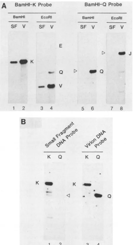
three homologousbands in theBamHI digest of theslowly sedimenting DNA (Fig. 4A, lane 1) and four bands in the EcoRIdigests (Fig. 4A,lane2).Thebands thathybridizedto theprobe in theBamHI digests corresponded tothe virion DNA fragments BamHI-K (8.7 kb)and BamHI-R(6.1 kb),
whereasthethirdpredominantbandprovednot tobeof viral
origin (see below). Similarly, two of the four bands that
hybridizedtothe short-fragment probein the EcoRIdigests correspondedtoHCMV virionDNAfragmentsrepresenting
EcoRI-V(4.2 kb)andEcoRI-Q(5.8 kb).Thesame Southern blot thatwasused for thehybridizationtotheshort-fragment
DNAprobewasthenstrippedof thatprobeandrehybridized
to amitochondrial DNA probe (Fig. 4A, lanes 5 to 8). The mitochondrial probe hybridized to the largest BamHI
frag-ment (16.8 kb; Fig. 4A, lane 6) and to two of the EcoRI
fragments (9.0 and 8.2 kb; lane 8). The expected 1.2-kb EcoRIfragmentofmitochondrial DNAwasalsopresentbut
ranoff thegelinthisexperiment (datanotshown). The viral band thatgave the strongestautoradiographic imageonthe X-ray film was EcoRI-V, followed in order by BamHI-K, EcoRI-Q, and BamHI-R, which all lie adjacent to one another in a single block located near the middle of the
Hind III
RnmH
IIaS
.0-
NC J TZ
I 6 --I- I I 0
%
VOL. 64,1990
4
on November 10, 2019 by guest
http://jvi.asm.org/
[image:4.612.96.509.80.211.2]lo..
'O' 0ta
'4
B6mHI-0 jg/m1
CS BamHl-K 25 j&g/ml BomHiI-Q25jJml
0 2
6
2L-1 2 3 4
B
V
tion,
days
DHPG (ug/mi)
0.1 1.0 10 25 50
_K ___ _p
Q
E
wJ o
BamHl
EcoRI
FIG. 3. Selective amplification of BamHI-K compared with
BamHI-Q DNA sequences with increasing time and increasing
doses ofDHPG. (A)Total intracellular DNA preparedatdifferent
timesafterHCMVinfectionin thepresenceofeither1 or25 ,ugof
DHPG per ml was applied to filters and analyzed by dot blot
hybridizationwith eitheraBamHI-Kor aBamHI-Q plasmidDNA
probe. The results are plotted as percentages of the level of
hybridization to untreated infected cell DNA samples. (B) Total
intracellularDNApreparedat 4daysafterHCMVinfection inthe
presence of different concentrations ofDHPG was cleaved with
eitherBamHIorEcoRI andanalyzedbySouthernblothybridization
with a 32P-labeledprobe containinga 1:2 mixture oftheBamHI-K
andBamHI-QplasmidDNAs.
unique L region in the physical map of the HCMV genome (Fig. 4B).
To confirm our interpretation that the amplified HCMV DNAsequences obtained in the presence of DHPG
(referred
to asDHPGDNA) containedpredominantlyBamHI-K and EcoRI-V sequences, we isolated slowly sedimenting DNA fragments from the top of anothersucrosegradient
(pooled
fractions 1 to 8) and used them as both the probe and the target forhybridization with selected cloned HCMV DNA fragments. A comparison between the DHPG DNA short fragments and intact total virion DNA afterhybridization with plasmid DNA containing the BamHI K fragment is shown in Fig. 5A. The single band obtained in the lane representing the DHPG DNA fragments (Fig. 5A, lane 1) matches thecorresponding band in the virion DNA(lane2). Similarly, the EcoRI digests of the DHPG DNA(lane 3) and the intact virion DNA(lane 4) show three comigrating bands representing the expected homology between BamHI-K sequences and the EcoRI-V, EcoRI-Q, and EcoRI-E spe-cies. The same Southern blot was then stripped of the BamHI-Kprobe and rehybridized toaBamHI-Q probe. The BamHI Q fragment, which is derived from the right-hand endof the viral genome, detectedthe appropriatefragments inboth theBamHI and EcoRI digests of virion DNA(lanes 6 and 8) but did not yield any signal with the small DHPG fragment DNA sample after digestion with either BamHIorEcoRI (lanes 5 and7).
The reciprocal experiment with cloned DNA from the BamHI-K andBamHI-Q plasmid DNA samples placed on
theSouthern blot was also carried out (Fig.5B). Hybridiza-tionwith the short DHPG DNAprobe revealed the presence of BamHI-K sequencesonly (lane 1) and not anyBamHI-Q sequences (lane 2). To confirm that the input BamHI-Q plasmid DNA sequences were present on the blot in the appropriate ratios, the same Southern blot membrane used in lanes 1 and 2 was stripped and hybridized to a probe preparedbylabelingtotal HCMVvirion DNA. Thiscontrol experimentshowed that thelanerepresentingthe BamHI Q fragment (lane 4) actually gave a more intense autoradio-graphic imagethan thatof BamHI-K (lane 3), suggesting that there was in fact more BamHI-Q DNA than BamHI-K DNA
on theblot. Therefore, we conclude that there is selective amplification of sequences complementary to BamHI-K in the presence ofDHPG and that the BamHI-Q fragment from theextremeright-hand end of the genome in the short unique segment is not represented in the DHPG-truncated small DNAfragments.
Since theHindIll Mfragment from the extreme left-hand end of the long unique regionwas also totally absent from short-fragment DNA samples (Fig. 1C) and because hybrid-ization experiments with the short DHPG fragment probes produced no evidence for the presence of any other viral sequences exceptfor thosewithinoradjacent to the BamHI
K and EcoRI V fragments, we also conclude that this procedureresultsinamplificationof DNAfrom justasingle localized domain within the HCMV genome. Logically, this site must represent a lytic origin of replication, which continually reinitiates in the presence of DHPG, despite a reduction in size of the replicating DNA molecules caused bythechain-terminationeffectsinduced by incorporation of DHPGnearthe end of thegrowing primer (35).
Direction ofreplication. The DHPG-induced chain-termi-nation mapping approach also was used to study the direc-tion of HCMV DNA replicadirec-tion. We reasoned that there should be a decreasing gradient of enrichment of the
se-quences on one or both sides ofthe origin and that larger
A
ui
81
C,) 0
L-4-0
0 U)
-a-02
4-,
C,Z
131
on November 10, 2019 by guest
http://jvi.asm.org/
[image:5.612.77.296.100.583.2]IDENTIFICATION OF ori-Lyt IN HCMV 6189
DHPG DNAProbe
BamHI EcoRi
V D V D
32P 32p
Mit DNAProbe
BamHI EcoRi
V D V D
A
BamHI-K
ProbeBamHI EcoRI
SF V SF V
BamHI-Q Probe
BamHJ EcoRI
SF V SF V
rR |
Ot
_e
. V_
E
> _ J
_ K
Q > _ Q
-ow v
1 2 3 4 5 6 7 8 1 2 3 4
D A
203 229
D K EOV
5?t 869 5 6 4.2 12 5 9
S---+- -- -s -
---E R K C
14.3 5- 9.5 162
-;~~~---t~~ +- -- -
-t-+-FIG. 4. Identification of themosthighly amplifiedsequences.(A) Pooled DNAfrom fractions 3to8inasucrosegradient
sedimenta-tion profile of DHPG-treated intracellular DNA was analyzed by
Southern blot analysis with 32P-labeled probes representing either the small DNA fragments themselves (lanes 1 to 4) or platelet
mitochondrial DNA(lanes5to8). The size marker reference lanes
containBamHI-digested (lane 1) orEcoRI-digested (lane 3), 32p_
labeled HCMV virion DNA, whereas lanes 5 and 7 contain
unla-beled HCMV virion DNA. DNA fragments representing human mitochondrial DNAareindicatedby the solid arrowheads (4). (B)
Physicalmapsof the relevantportion of the central ULsegmentof theHCMV(Towne)genomeshowing relative positions, sizes, and overlaps ofHindIll, BamHI, and EcoRI restriction fragments and the location of the abundant EcoRI-V species found in the short DHPG-treated HCMV DNA molecules (open bar). Cloned
frag-mentsusedasprobes in Fig. 5, 6, and7areshownassolid bars.The
nomenclature used and estimated fragment sizes in kilobases are
derived from LaFemina and Hayward (26; unpublished data). Ge-nomic sequence coordinates given for the HindIII fragments are
thosefor theequivalent species in the HCMV (AD169)genome(4a).
DHPG fragments should contain more of the flanking se-quences than smaller DHPG fragments. Fractions 1 to 4
(small fragments) and 5 to 8 (intermediate-sized fragments)
fromasucrosegradient separation of DHPG DNA fragments
were pooled; the DNA was digested with EcoRI and then electrophoresed in a 1% agarose gel and hybridized with a
probecontaining the HindlIl D HCMV DNA fragment from the left-hand side of BamHI-K (Fig. 6B). Because the fragments in the intact virion DNA lane mustbepresent at
equimolar concentrations, theintensity of each band should therefore be proportional to the size of the segment of the fragment thatis complementary to the HindlIl probe (Fig. 4B). Accordingly, the probe detected the corresponding
bands in theintactvirionDNAcontroldigest (lane 6)inthe following orderofdecreasingintensity: EcoRI-D=EcoRI-K
>EcoRI-Q. However, the lane representing the DNAfrom fractions 1 to 4 (lane 7) shows a faint EcoRI-Q band only, whereas all three fragments were displayed in the lane
B
K Q
K _
5 6 7 8
'0
K Q
K _
< . _ Q
1 2 3 4
FIG. 5. Confirmation that the slowly sedimentingDHPG DNA containsamplifiedHCMV BamHI-Ksequences.(A) Southern blot
analysis of small fragment DNAfrom DHPG-treated cultures with cloned viral DNA probes. SF,BamHI orEcoRI digestsofpooled DNAfrom isolated smallfragments obtained fromfractions 1to8of
asucrosegradient sedimentation profileofDHPG-treatedDNA.V,
BamHIorEcoRIdigestsof cleaved unlabeled DNA extracted from
HCMV virions. Lanes: 1 to 4, hybridization with 32P-labeled BamHI-Kplasmid DNA probe;5to8,32P-labeledBamHI-Q probe.
(B) Southern blot analysis with small DHPG DNA fragments as
probes. Lanes: 1and3, BamHI-cleavedBamHI-Kplasmid DNA;2 and4, BamHI-cleaved BamHI-Q plasmid DNA;1 and2, hybridiza-tion with 32P-labeled small DHPG DNAfragment probe; 3 and4, hybridizationwith control 32P-labeled total virion DNAprobe.
representing the DNA from fractions 5 to 8 (lane 9) with
relative intensities that decreased in the following order:
EcoRI-Q = EcoRI-K > EcoRI-D. Thus, the reversed rela-tiveintensityof the bands in intermediate-sized DHPGDNA
compared with that in virion DNA strongly suggests that
EcoRI-Qis representedathighermolar concentration in all DHPG fragments than are EcoRI-K and EcoRI-D. This
finding, inconjunctionwith the otherdata describedabove, strongly suggests that HCMV DNA replication starts in
A
B
Hindill H/E EcoRi B/E BamHl B/H
VOL.64, 1990
_m 4
on November 10, 2019 by guest
http://jvi.asm.org/
[image:6.612.56.297.78.356.2] [image:6.612.321.547.80.495.2]A DHPG Probe SF IF V D M DM
D -so
E
K --a
315
B
HindHil-D
ProbeSF F
V D M D M
CMitDNA Probe
SF IF
V DMD M
D
-K _
Q -_
[image:7.612.68.302.79.232.2]6 7 8 9 10 11 12 13 14 15
FIG. 6. Detection ofagradient of amplifiedsequencesadjacent
to BamHI-K in DHPG-treated intracellularDNA. Comparison by
Southern blotanalysis of slow- andintermediate-sedimenting
frac-tionsof intracellular DNA from cultures infected with HCMVinthe
presenceof DHPG. All DNAsamplesonthe blotwerecleavedwith
EcoRI. (A) 32P-labeled intracellular DHPGDNAprobe (fractions1 to8); (B) cloned 32P-labeled HindIII-D probe; (C) 32P-labeledhuman
platelet mitochondrial DNA probe. Lanes: V, unlabeled HCMV virion DNA; SF, small DNA fragments from sucrose gradient
fractions1to4; IF, intermediate-sized DNA fragments fromsucrose
gradient fractions5 to 8; D, total intracellular DNA from
DHPG-treated HCMV infected cells; M, total intracellular DNA from
mock-infected cells. Bandscorresponding toindividual viral DNA
fragmentsareidentifiedbythestandard nomenclature (seemapin
Fig. 4B), and mitochondrialDNAfragmentsaredenotedby
arrow-heads(4).
BamHI-K or EcoRI-V and proceeds leftward toward EcoRI-Q,then toEcoRI-K, and latertoEcoRI-D.
The same Southern blot after hybridization to a probe
made from labeled DNA preparedfrom fractions 1 to 8 (a
mixture ofboth slow- andintermediate-sedimenting DHPG-DNA fragments) detected five major bands in the intact
virion DNA control sample (Fig. 6A, lane 1).These bands
correspond to EcoRI-V and EcoRI-E, in addition to the
EcoRI-D, -K, and -Q species detected above. Since our results with the smallest DHPG fragments imply that the
origin itself is located within BamHI-K and EcoRI-V, the appearanceof EcoRI-E(lane 4),which isjusttotherightof EcoRI-V on the physical map (Fig. 4B), suggests that the
replication alsoproceedstotheright from EcoRI-V toward
EcoRI-E, as well as to the left. This possibility is further
supported by theappearance of EcoRI-V (andsomeEcoRI
Q fragments) in the smallest DHPG DNA sample (fractions
1to4;lane2)and theaddition ofEcoRI-E, -D,and -K in the intermediate-sized DNA from fractions 5 to 8 (lane 4). Again, some of the additional bands thatwere detected by
the probe from fractions 1 to 8 in the intermediate-sized
fragment position were foundto be of mitochondrialorigin (Fig. 6C,lane14),andthey correspondtotheonlytwobands detectedatthis sizepositioninmock-infected cells(lane 15).
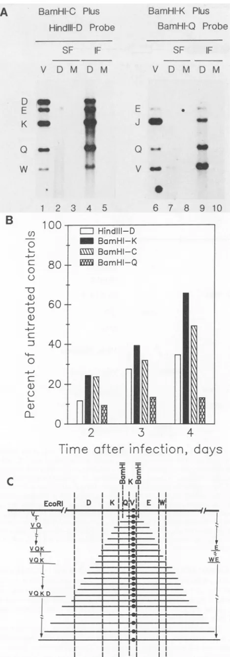
For further analysis of the direction of replication, we
usedtwo probes in combination, onefromjustto theright
andonefromjusttotheleft of theputative origin.
Simulta-neous hybridization with the HindIII-D (left of BamHI-K)
and BamHI-C (right ofBamHI-K) probes gave a different relativeintensityfor EcoRI-EcomparedwithEcoRI-D inthe
intermediate-sized DNA from fractions 5to8(Fig. 7A, lane
4) compared with their relative intensity within the virion DNA sample (lane 1). This result suggeststhat EcoRI-E is
present at a severalfold higher molar concentration than
EcoRI-D.
Similarly, EcoRI-Q
ismoreabundant thanEcoRI-W, -K,
or-D,
andEcoRI-Kismoreabundant thanEcoRI-D(Fig. 4B).
Thepresence ofa5-to10-fold overabundance of sequencescomplementary
to BamHI-Kcompared
withBamHI-Q
in thissamesample
of intermediate-sizedDHPGfragments
wasalsoconfirmedby
simultaneoushybridization
with BamHI-KandBamHI-Q probes (Fig. 7A,
lanes 6to10).
The results from theseexperiments,
when considered inconjunction
with the other evidence describedabove,
strongly
suggest that thereplication
of HCMV DNA isbidirectional, proceeding
both leftward andrightward
from EcoRI-V.For
quantitation
of this type ofanalysis,
we studied the total intracellularviral DNA from DHPG-treated culturesby
using
dot blothybridization,
andwecompared
the levels of accumulation ofsequencescomplementary
toprobes
attheputative origin
ofreplication
relativetothoseadjacent
to ordistant from the
origin (Fig.
7B).
Again,
there was greateramplification
of sequences that arecomplementary
to the BamHI Kfragment
ascompared
with the sequences eithertotheleft ofthe
putative origin (HindIII-D)
ortotheright
of theputative origin (BamHI-C). Furthermore,
thesequencescomplementary
tothe HindIII-D and BamHI-Cprobes
were alsoamplified
severalfold relative toBamHI-Q
sequences fromthe shortunique region
attheextremeright-hand
end of the genome.Therefore,
thisanalysis
(even
with unfraction-atedDNA)
again
suggests that theorigin
of HCMV DNAreplication
lies withinBamHI-K,
and thatviral DNArepli-cation
proceeds
both leftwardtoward HindIII-D andright-ward toright-wardBamHI-C before
reaching BamHI-Q.
Amodelillustrating
therelative sizesandpredicted
maplocations of the truncatedreplicated
DNAfragments
present in the DHPG-treated intracellular DNApool
isgiven
inFig.
7C. Note thatwe do not know whether individual DNA mole-culesproceed
inbothdirectionsorwhether thepopulation
is
a mixture ofsome molecules
proceeding
leftwardonly
and othersproceeding rightward only.
Inaddition, although
they
are
depicted
as linear structures, we cannot at present exclude thepossibility
of branchedor circularstructures.DISCUSSION
The
experiments
described in this report represent afollow-up
to ourunexpected
observation thatHCMVDNAsynthesis appeared
tocontinue
in the presence ofDHPGeven at inhibitor concentrations that resulted in
complete
cessationofinfectious virion
production (Hamzeh
and Liet-man,submitted). However,
we now show that this resultonly
applies
to aspecific
segmentoftheviral genome, which becomesselectively
amplified
andprogressively
smallerin size. Weimply
that theamplified
sequences result from continuous reinitiationonincompletely replicated
viralDNA molecules that have become truncatedby
theincorporation
ofDHPG nearthe terminus of the
growing
primer strand,
preventing
furtherelongation
(35). By
measuring
therelative abundanceof viral DNA sequences inadjacent
regions
ofthe genome from sucrosegradient
fractionscontaining
DNA molecules of differentsizes,
wedetermined that the smallest molecules have thehighest
concentrations oforigin
se-quences and that
larger
molecules containincreasing
abun-dancesofadjacent
sequences that maptoboth the left andright
of theorigin
sequences.Only
asingle
origin
locus appearstobepresent in theamplified
DNAsamples,
and the sequences present atthehighest
concentration werefoundto map within the 4.2-kb EcoRI V and 8.5-kb BamHI K
fragments
of HCMV(Towne).
Infact,
in the mosthighly
on November 10, 2019 by guest
http://jvi.asm.org/
A
BamHFC Plus
HindIll-D Probe
SF IF
V D M D M
D _
E
-K
Q
_
I
*..
LI-J
0
V w
B
0
-o
4-j 0 -o
G)
C)
ci)
cL-1 2 3 4 5
100
-
80-
60-
40-
20-0
2
3
BamHI-K Plus
BamHF-Q
Probeamplified
samples
these twospecies
appearto be theonly
viral DNA fragments that remain in an intact form at all, although many DNA molecules are present that contain SF IF these sequences and are even smaller than the 4.2-kb EcoRI-Vspecies. TheEcoRI V fragment of HCMV (Towne) V D M D M maps between nucleotide positions 88020 and 92210 within the unique L segment of the HCMV viral genome (map coordinates 0.367 through 0.388). These two fragments are equivalent to theBamHIMandEcoRIVfragments of strain * HCMV (AD169), which has been completely sequenced by Chee and colleagues in Cambridge (4a). Inspection of the DNA sequenceof this region of the HCMV genome reveals that it includes the UL57 gene that gives rise to a 4.2-kb-*
" leftward transcript encoding the 140-kDa early single-stranded DNA-binding protein (DB140, equivalent to the - " UL29 or ICP8major DNA-binding protein of HSV [1, 23]). However, upstreamand to the right of UL57 there is a large * 2.5-kb block of DNA sequences that has only small, poorly defined potential open reading frames (UL58 to UL60) and 6 7 8 9 10 containsmany interesting sequence elements and repetitive motifs. Thisregion, which spans nucleotide positions 90340 through 92966, occupies the entire right-hand half of EcoRI-V and proceeds beyond it toward the right-hand boundary of BamHI-K. An analysis of sequence features in this area, which we propose represents the lytic origin of HCMV, is illustrated in Fig. 8.The HCMV DNA replication origin that we haveidentified heremustfunction in the capacity of a lytic cycle origin that is uncoupled from cell cycle controls, but it lacks any recognizableDNA sequence orevolutionary homology with any previously identified viral DNA replication origin
re-gions, including
those in HSV and EBV.Therefore,
the HCMVlytic origin
represents
a newfourth class ofherpes-virus DNA replication origin that is distinctly different
structurally
fromthe HSVtype
oforigin
and from the EBV ori-P and ori-Lyt regions. Nevertheless, the genomic loca-tionadjacenttothe 5'endofthe genefor the single-strandedDNA-binding
protein
of HCMVparallels
that ofori-L in HSV, and the entire 2.5-kb repetitive region may (like the HSV and EBVlytic origins)
represent
anoncoding
inter-genic domain. The presence of multiple ATF and core or
4
Time
after
infection,
day,
D K
I I ji iiE E I I
,'Vl
E
IW!
I t!
I !!I
r
17-1I
ZJi
I
I
S FIG. 7. Selective amplification of adjacent fragments on both sidesoftheputative lyticorigin site. (A) Southern blot analysis of intermediate-sizedDNAmoleculesfrom DHPG-treated intracellular DNA. Lane designations and arrangements as described in the legendto Fig. 6. All DNAsamples onthe blotwere cleaved with EcoRI. Lanes: 1 to 5, mixed probe of BamHI-C plus HindIII-D plasmidDNAs; 6 to 10, mixedprobe of BamHI-K plusBamHI-Q plasmidDNAs. Relevant overlapmaps are shown in Fig.4B. (B) Quantitative dot blothybridization analysis oftherelative propor-tionsofadjacentsequencesderivedfromtheright (BamHI-C)orleft (HindIII-D)of BamHI-K in total intracellularDNA from HCMV-infectedcells treated with DHPGat10,ug/ml. Again,theresultsare
plottedas the percentageofradioactivity in thetreated culturesat
different times after infection compared with that in parallel
un-treated cultures. (C)Model for the derivation and structureofthe small truncated DNA moleculescontaining ori-Lytsequencesfound in intracellularHCMV DNA from DHPG-treated infected cells. In the smallest amplified molecules only the EcoRI V or BamHI K fragments remain intact. Larger truncated molecules contain de-creasing gradientsofadjacentsequencesonboth sidesof EcoRI-V. Therefore,asinglepredominantori-Lyt site isproposedtolie within EcoRI-V. Although all of the slowly sedimentingDNA molecules
areshown tobederived bybidirectionalreplication, wecannot at
present exclude the possibility that some individual molecules proceed rightwardonlyandothersproceed leftwardonly.
E WE
6191
m Hindlil-D _ BamHI-K
EXX5
BamHI-C- BamHI-Q
11
R
C
EcoRl
va
-I-VOK VOK
I
VT
VQ K D
I-
r-I
I
on November 10, 2019 by guest
http://jvi.asm.org/
[image:8.612.62.296.57.726.2]HCMV
- L --- ----
S--(N
v
44
0 OaD 0 Oc mD
a
4 44 44 44 4 44 4 4a
10, 1011
*o
b.pl4 3 10-bpRepeats
Fsp 1/ Sph I
a
2Obp
so 38bp 28bp
A A
5x l9bp
2x28bp
Palindromes
Direct
JRepeats
FIG. 8. Structuralorganization of the predicted ori-Lyt locus. (A) Map location of the ori-Lyt locus (0) adjacenttothe 5' endoftheDB140 single-stranded DNA-binding proteingene(UL57, dbp, ssb) in the HCMVgenome.Theinverted repeatsencompassing the uniqueL andS segmentsof thegenome areshownasopenbars. Major blocks of primordial herpesvirusgenesthatareevolutionarily conserved between
EBV, HSV, and CMV are denoted by solid bars. pol, DNA polymerase; hel, helicase subunit. (B) Locations ofrepeated sequencesand putative transcriptional control elementsorfactor binding sites withina3.7-kb regionontheright-hand side of the EcoRI V and BamHI K
fragments, i.e., HCMV (AD169) sequence coordinates 89700 to 93360. The coding region for the single-stranded DNA-binding protein (DB140) occupies the entire left-hand half of EcoRI-V.Symbols: V (MTLF),consensusadenovirusmajor late transcription factor binding site (GGTCAGCTGACC); <I , consensus TATAAA and AATAAA elements; 0, consensus CRE- or ATF-binding sites (TGACGTCA,
TGACGACA, TGACGCA, TGACGTA, TGACGT); O,consensusSP-1 factor-binding sites (CCCGCCC); O,consensusCAAT transcription
factor-binding site (TACCAAT);4*, family of decamer dispersedrepeatelements(at least 7outof 10 matches with theconsensussequence
AAAACACCGT);0,family ofsevenFspI-SphI 11-bprepeats(TGCGCATGCG); A,two13-bp directrepeats(GAAAACCTATATA); *,five
19-bp directrepeats;GCCGGTAAAAAAATTTTTCCACT;V, two28-bpdirectrepeats.
palindromic CREsequences,plusa consensusMLTFsite,is
consistent with an association with transcriptional control
elements and the presence ofnumerous other tandem and
invertedrepeat structuresis typical ofthe local architecture within the DSL-DSR region encompassing the EBV lytic origin. Either of the repeated sequence elements GAAAA
CCTATATA(two copies)orGCCGGTAAATTCCACT (five
copies)withinthetandemly repeated region mightrepresent theanticipated A+T-rich initiation site(s). In addition, there
are23 interspersed copies ofadecamer consensus element
AAAACACCGT betweenpositions-250 and -1250 relative totheDB140genecapsite(Table 1)and sixclusteredcopies
of the 11-bp TCGGCATGCGC FspI-SphI element(Fig. 8),
whichbothseemlikelytobebindingsites foreither viralor
cellular factors involved in regulation oftranscription and DNA replicationevents.
The highly restricted host range and slow virus growth cycle, together with an unfortunate location of restriction enzyme cleavage sites, appearsto havefrustratedprevious
attempts toidentifythe HCMVorigin bycotransfectionplus
A
IzI
0
s9
li
MLTF
D. TATAA
Poly-A
CRE/ATF
mm sP- 1
CTF
I
prrl..M
..I
i
-4---.4 ---- - --- ---- ---- ----
-I
e
'46,
1
AIO
IT, 40 '4
0
.\ q
f
on November 10, 2019 by guest
http://jvi.asm.org/
[image:9.612.113.503.78.500.2]IDENTIFICATION OF ori-Lyt IN HCMV 6193
TABLE 1. Family of interspersed decamer repeat elements upstreamfrom the promoters for the single-stranded DNA-binding
protein genes in HCMV and SCMVI
Position andsequenceofrepeatelementb
HCMV(AD169) SCMV (Colbum)
-224AAAATA£CGT -233 -204 AAAATAGCGT -213
-293 TAAACGACGT -284 -254AAAACAACGT -263 -299AGCACACCGT -308 -296AAAACAACGC -287 -354 AAAACCCCAC -363 -327 AAAACGGTGT -336 -376AAAACACCGT -385 -339AAAACACCGT -330 -392 AAAACGGCGT -401 -343AAAaCAACGT -352 -404 AAAACGCCGT -395 -370AAAACACCG, -361
-455AAAACACCGT -464 -408 QAAACACCGT -417 -474_AAACACCGT -465 -436AAAGCAzCGT -445 -498AAaAAACCGT -507 -454AAAACACCGA -445 -538AAAACACCGT -529 -474AAAACGACGa -465 -547 AAAGAACCGC -556 -573 AGAACAGCGT -564 -574 GAAACACCGC -583 -593AAAACGACGT -602 -668 AAAACGCCGT -677 -651AAAACGGTGT -660 -694AAAAACCCGC -703 -663 CAAACACCGT -654 -754CAAACCGCGT -763 -679AAAACACCGT -688 -774£AAACCACGT -765
-906AAAACACATT -915 -953 GAAACACCGT -962 -980 ACAACACCGT -971 -1047AAAACACCGG -1038 -1096AAACCGCCGT -1087 -1139 AAAACCCCGT -1130 -1221AACACACTGT -1230 -1232AAACCCGCGT -1241
aConsensussequences areAAAACACCGT for both HCMVandSCMV. Deviations from the 10-bpconsensus sequence areunderlined.
bPositionsgivenare calculatedrelative totheconsensusTATAAA-box elements(assignedtopositions -28 to -23) intheHCMV(AD169) DB140
andSCMV (Colburn)DB129 genes.Sequencedata wereobtained fromMark
Chee (4a)andDavidAnders(in press).
superinfection procedures (R. LaFemina and G. S. Hay-ward, unpublished data; E. Mocarski, personal communica-tion).However, inparallel studies,D. G. Anders and S. M. Punturieri(submitted forpublication)havesuccessfullyused DNA transfection procedures with the Colburn strain of African green monkey simian CMV (SCMV) and defined a
lytic cycle origin to within a 2.2-kb fragment located in a
positionthatisexactly analogousto ours(i.e.,justupstream from the promoter for the DB129 single-stranded DNA-bindingprotein gene [1]). DNA sequence analysis bythose authors has also revealed 16 copiesof the conserved inter-spersed decamer AAAACACCGT consensus element
be-tween positions -250 and -800 upstream from the DB129 gene mRNA start site in SCMV DNA (Table 1), plus two
copies of the 11-bp FspI-SphI element, togetherwith multi-ple ATFandCRE elements.
Asaconfirmation of thegeneral validityand complimen-tarity of thesetwomethods, we havealso carriedoutchain termination plus hybridization with SCMV (Colburn)-in-fected cells. Our results indicated that after48 hof infection inthepresenceof DHPG therewasselectiveamplificationof the EcoRI-D, SalI-G and Sall-H, andXbaI-J, -M, and -U, -V,or-Yfragments.These DNAfragmentsdefineanexactly equivalent positionatmapunit 0.35to0.4 nearthecenterof theuniqueLsegmentofthe 220-kb SCMV genome(29)and fulfill precisely the predictions from the DNA transfection-superinfection studies (Anders and Punturieri, submitted). In the reciprocal experiments with HCMV (Towne) DNA, Anders and Punturieri (submitted) obtained positive evi-dence for replication of the 22-kbHindlIl A fragment but
foundno
activity
with theBamHI KorCfragment,
suggest-ing
that akey
element of the HCMVorigin
may be evenfarther than 2.5 kb upstreamfromthe promoterfor the
major
DNA-binding protein.
In viral genomes like those of HCMV and SCMV that involve
giant
220- to 240-kbreplicons
withrelatively large
origin
domains,
thechain-terminationprocedure
appears tobe a very useful
complement
to the DNA transfection approach. The transfectionprocedure
cannot work with target DNAfragments
that donot containthe entireorigin
domainin cisorifdeletions orrearrangements of
palindro-micfeaturesoccurwithinbacterialvectorsystems(asis the
case with HSV ori-L). In contrast, the chain
termination-hybridization procedure
overcomestheseobstaclesby
being
applied directly
within virus-infected cells and has the ad-vantage ofyielding
information about directionality. How-ever, the chain terminationapproach
suffers at presentby
not
permitting
outside boundariesof theorigin
tobedefined and therefore does notprovide
a great deal ofprecision
about the exactlocation ofkey
elements. Furtherpotential
development
of this latterprocedure
foranalysis
of thestructure of the
replicating
DNAfragments by
electronmicroscopy
and two-dimensionalgel
electrophoresis,
etc., will undoubtedly beuseful,
althoughsubject
to the usual limitations ofinterpreting potentially
aberranteffects causedby
theuse of inhibitors.Despite plausible
models forherpes
simplex
virus DNAreplication
occurring by
arolling-circle
replication
mecha-nism,
thatis notnecessarily
the caseforCMV,
and wedonotconsiderit
likely
that the small HCMV DNAfragments
accumulating
in the presence ofDHPG areproduced
by
arolling-circle
mechanism.Although
it has yet tobeproved,
the most
likely
scenario is that thesefragments
representproducts
resulting
frommultiple
rounds of bidirectional initiation events that occuron shortened lineartemplates.
Conceivably,
there could be twopopulations
withsingle
growth
forksproceeding
inopposite directions,
orindividual molecules could containmultiple growth
forks. It should be cautionedthat,
becausethey
weregenerated
inthe presence ofDHPG,
the structure of theorigin-enriched
fragments
doesnot
necessarily
make anypredictions
aboutthenormal mode of HCMVreplication. Nevertheless,
it isinteresting
that
tandem-repeat-defective
CMV DNA ofthe type seenroutinely
in HSV has notyet beenobserved,
despite
delib-erateattemptsto generate it
by
passaging
of SCMV athigh
multiplicities
ofinfection forover20successivepassages(D.
Ciufo and G. S.
Hayward,
unpublished data). Instead,
shortened,
circularly
permuted
linear genomefragments
ofapproximately
150 kb arerapidly
accumulatedduring
high-multiplicity
passaging
in HCMV infections(15,
24, 40).
Furthermore,
these defective DNApopulations
exhibit ab-errant-sizedforms ofthe EcoRI E andBamHI Cfragments
that could represent rearrangements across part of the
ori-Lyt
region (R.
L.LaFemina,
R.Pritchett,
and G. S.Hayward,
unpublished data).
The results described in this work should stimulate
rapid
future progress in
identifying
theproteins
andspecific
cis-acting
sequences involved inreplication
of thelarge
andcomplex
genomesofCMVs,
andsimilarprocedures
have thepotential
for usefulapplication
tootherherpesvirus
systemsas well.
ACKNOWLEDGMENTS
F.M.H. was supported byascholarship from the University of
Jordan, Amman, andbythe Division of ClinicalPharmacology of the The JohnsHopkins UniversitySchoolofMedicine. The study
VOL.64, 1990
on November 10, 2019 by guest
http://jvi.asm.org/
[image:10.612.60.300.106.366.2]was supported by private grants to P.S.L. andby Public Health ServicegrantRO1-AI24576toG.S.H. from the NationalInstitutes of Health.
We thankNadia Badiee and Mabel Chiuforexcellent technical assistance, Pamela Wright for photography,andSarahHeaggansfor help with preparation of the manuscript. We areespecially grateful
to MarkChee and Bart Barrell (Medical ResearchCouncil, Cam-bridge, England) for providing HCMV DNA sequencedata andto
David Anders (Wadsworth Center, Albany, N.Y.) forexchanging information about the ori-Lytregions of HCMV and SCMV before publication.
LITERATURE CITED
1. Anders, D. G., and D. W. Gibson. 1988. Location, transcript
analysis, and partial nucleotide sequenceofthe
cytomegalovi-rusgene encodinganearly DNA-binding proteinwith similar-itiestoICP8 ofherpes simplex virustype 1.J. Virol. 62:1364-1372.
2. Biron,K.K., S. C.Stanat, J.B.Sorrell,J. A.Fyfe,P. M.Keller, C. U.Lambe,and D.J.Nelson.1985. Metabolic activationof the nucleoside analog 9-{[2-hydroxy-1-(hydroxymethyl)ethoxy]
methyl} guanine in human diploid fibroblasts infected with humancytomegalovirus. Proc. Natl. Acad. Sci. USA 82:2473-2477.
3. Challberg,M. D. 1986. Amethod foridentifyingtheviralgenes required for herpesvirus DNA replication. Proc. Natl. Acad. Sci. USA 83:9094-9098.
4. Challberg, M. D., and T. J. Kelly. 1989. Animal virus DNA
replication. Annu. Rev. Biochem. 58:671-717.
4a.Chee,M.S., A. T.Bankier,S.Beck, R. Bohni, C. M. Brown, R. Cerny,T. Horsnell, C. A. Hutchison III, T. Kouzarides, J.A. Martignetti, E. Preddie, S. C. Satchwell, P. Tomlinson, K. M. Weston,and B. G. Barrell.1990.Analysisof theprotein-coding content of the sequence of human cytomegalovirus strain AD169. Curr.Top. Microbiol. Immunol. 154:126-169. 5. Chittenden, T.,S. Lupton, and A. J.Levine. 1989. Functional
limits ofori-P,theEpsteinBarrvirusplasmidorigin of replica-tion. J.Virol. 63:3016-3025.
6. Ciufo,D.M.,andG. S.Hayward. 1981.Tandem repeat defec-tiveDNAfrom theL segmentoftheHSVgenome.Dev. Mol. Virol. 1:271-306.
7. Crute, J. J., T. Tsurumi, L. Zhu, S. K. Weller, P. D. Olivo, M. D. Challberg, E. S. Mocarski, and I. R. Lehman. 1989. Herpes simplex virus 1 helicase-primase: a complex of three
herpes-encoded gene products. Proc. Natl. Acad. Sci. USA 86:2186-2189.
8. D'Aquila, R. T., G. Hayward, and W. C. Summers. 1989. Physical mapping of the human cytomegalovirus (HCMV) (Towne)DNA polymerase gene: DNA-mediated transfer ofa
genetic marker foranHCMV gene. Virology171:312-316. 9. Elias, P.,andI. R.Lehman. 1988. Interaction oforiginbinding
proteinwith anoriginofreplicationofherpes simplexvirus 1. Proc. Natl. Acad. Sci. USA85:2959-2963.
10. Freeze,U. K., G. Laux, J. Hudewentz, E.Schwarz,andG. W. Bornkamm. 1983. Two distantclusters ofpartially homologous smallrepeatsofEpstein-Barrvirusaretranscribedupon induc-tion ofanabortiveorlytic cycle ofthe virus. J. Virol. 48:731-743.
11. Frenkel, N., R. J. Jacob, R. W. Honess, G. S. Hayward, H. Locker,and B. Roizman.1975.Anatomy ofherpessimplexvirus DNA. III. Characterization of defective DNA molecules and
biological properties ofvirus populations containing them. J. Virol.16:163-167.
12. Frenkel, N., H. Locker, W. Batterson, G. Hayward, and B. Roizman. 1976. Anatomy of herpes simplex virus DNA. VI. Defective DNA originatesfromthe "S" component. J. Virol. 20:527-531.
13. Friedman, A.,andY. Becker. 1977.Circular andcircular-linear DNAmoleculesofherpes simplexvirus. J. Gen.Virol. 37:205-208.
14. Gahn, T. A., and C. L. Schildkraut. 1989. The Epstein-Barr virus origin of plasmid replication, ori-P, contains both the
initiationand termination sites of DNAreplication.Cell 58:527-535.
15. Geelen, J. L. M. C., C. Walig, P. Wertheim, andJ. VanDer Noordaa. 1978. Human cytomegalovirus DNA. I. Molecular weight andinfectivity. J. Virol. 26:813-816.
16. Gray, C.P., and H. C. Kaerner. 1984.Sequenceof theputative originofreplication in the UL regionofherpes simplex virus type1 ANG DNA. J. Gen.Virol. 65:2109-2119.
17. Hammerschmidt, W., and B. Sugden. 1988. Identification and characterization of ori-Lyt, a lytic origin of DNA replication of Epstein-Barrvirus. Cell 55:427-433.
18. Handeli,S., A. Klar, M. Meuth, and H.Cedar. 1989. Mapping replication units in animal cells. Cell 57:909-920.
19. Hudewentz, J., H. Delius, U. K. Freese, U.Zimber, and G. W. Bornkamm.1982. Two distant regions of the Epstein-Barr virus genome with sequence homologies have the same orientation and involve small tandem repeats. EMBO J. 1:21-26.
20. Jacob, R. J., L. S. Morse, and B. Roizman. 1979. Anatomyof herpes simplex virus DNA. XII. Accumulation of head-to-tail concatemers in nuclei of infected cells and their role in the generation of the four isomeric arrangements of viral DNA. J. Virol. 29:448-457.
21. Jean, J.-H., M. L.Blankenship, and T. Ben-Porat. 1977. Repli-cation of herpesvirus DNA. I. Electron microscopic analysis of replicative structures.Virology 79:281-291.
22. Kaerner, H. C., A.Ott-Hartmann, R. Schatten, C. H. Schroder, and C. P. Gray. 1981. Amplification of a short nucleotide sequencein the repeat units of defective herpes simplex virus type 1Angelotti DNA. J. Virol. 39:75-81.
23. Kemble, G. W., A. L. McCormick, L. Pereira, and E. S. Mocarski. 1987. A cytomegalovirus protein with properties of herpes simplex virus ICP8: partial purification of the polypep-tide and map position of the gene. J. Virol. 61:3143-3151. 24. Kilpatrick, B. A., E.-S. Huang, and J. S. Pagano. 1976. Analysis
of cytomegalovirus genomes with restriction endonucleases HindIll and EcoRI. J. Virol. 18:1095-1105.
25. Koff, A., and P. Tegtmeyer. 1988. Characterization of major recognition sequences for a herpes simplex virus 1 origin-binding protein. J.Virol. 62:4096-4103.
26. LaFemina, R. L., and G. S. Hayward. 1980. Structural organi-zation of the DNA molecules from human cytomegaloviruses. ICN-UCLASymp. Mol. Biol. 18:39-55.
27. LaFemina, R. L., and G. S. Hayward. 1983.Replicative formsof humancytomegalovirus DNA withjoined terminiarefound in permissively infected human cells but not in non-permissive Balb/c-3T3 mouse cells. J. Gen. Virol. 64:373-389.
28. Lieberman, P. M., J. M. Hardwick, J.Sample,G.S.Hayward, and S. D. Hayward. 1990. The Zta transactivator involved in induction of lytic cyclegeneexpression in Epstein-Barr virus-infectedlymphocytes bindstoboth AP-1andZREsites intarget promoterandenhancerregions. J.Virol. 64:1143-1155. 29. Locker, H., N. Frenkel, and I. Halliburton. 1982. Structure and
expression of class Il defective herpes simplex virus genomes encoding infected cell polypeptide number 8. J. Virol. 43:574-593.
30. Lockshon, D., and D. A. Galloway. 1986. Cloning and charac-terization oforiL2,alargepalindromic DNAreplicationoriginof herpes simplex type 2. J. Virol. 58:513-521.
31. Marks, J. R., and D. H. Spector. 1984. Fusion ofthe termini of the murine cytomegalovirus genome after infection. J. Virol. 52:24-28.
32. McGeoch, D. J., M. A. Dalrymple, A. Dolan, D. McNab,L. J. Perry, P. Taylor, and M. D. Challberg. 1988. Structures of
herpes simplex virus type 1 genes required for replication of virus DNA. J. Virol. 62:444-453.
33. Olivo, P. D., N.J. Nelson, andM. D. Challberg. 1988. Herpes simplex virus DNA replication: the UL9 gene encodes an
origin-binding protein. Proc. Natl. Acad. Sci. USA 85:5414-5418.
34. Rawlins, D. R., G. Milman, S. D. Hayward, and G. S. Hayward. 1985. SequencespecificDNAbindingof the Epstein-Barrvirus nuclear antigen (EBNA-1) to clustered sites in the plasmid maintenanceregion. Cell 42:859-868.
on November 10, 2019 by guest
http://jvi.asm.org/
IDENTIFICATION OF ori-Lyt IN HCMV 6195 35. Reardon, J. E. 1989. Herpes simplex virustype 1 and human
DNA polymerase interactions with 2'-deoxyguanosine 5'-triphosphate: kinetics of incorporationintoDNA and induction of inhibition. J. Biol. Chem.264:19039-19044.
36. Reisman, D., J. Yates, and B. Sugden. 1985. A putative origin of replication of plasmids derived from Epstein-Barr virus is composed of two cis-acting components. Mol. Cell. Biol. 5:1822-1832.
37. Roizman, B., G. S. Hayward, R. Jacob, S. Wadsworth, and R.W.Honess. 1974. Human herpesvirus.I.Amodelfor molec-ularorganization of herpesvirus virions and their DNA. Ex-cerptaMed. Int.Congr. Ser. 2:188-198.
38. Schuster, R. C., A. J. Rubenstein, and D. C. Wallace. 1988. MitochondrialDNAin enucleated human blood cells. Biochem. Biophys. Res. Commun. 155:1360-1365.
39. Southern, E.1975.Detection of specificsequences among DNA fragments separated by gel electrophoresis.J.Mol.Biol.9:503. 40. Stinski, M. F., E. S. Mocarski, and D. R.Thomsen. 1979. The DNAofhumancytomegalovirus: sizeheterogeneity and defec-tiveness resulting from serial undiluted passage. J. Virol. 31: 231-239.
41. Stow, N.D. 1982. Localization of an origin of DNA replication withintheTRs/TRsrepeated region of the herpessimplexvirus type 1genome. EMBO J. 1:863-867.
42. Stow, N. D., and E. C. McMonagle. 1983. Characterization of theTRS/IRS origin ofDNAreplication of herpessimplex virus
type 1.Virology 130:427-438.
43. Thomsen, D. R., and M. F. Stinski. 1981. Cloningof thehuman cytomegalovirus genome as endonuclease XbaI fragments. Gene 16:207-216.
44. Weir, H. M.,J. M. Calder, and N. D. Stow. 1989.Binding of the herpes simplexvirus type 1 UL9gene product toan originof viral DNAreplication. Nucleic AcidsRes. 17:1409-1425. 45. Weller, S. K., A. Spadaro, J. E. Schaffer, A. W.Murray, A. M.
Maxam, and P. A. Schaffer. 1985. Cloning, sequencing, and functionalanalysis oforiL, aherpes simplex virustype 1origin ofDNAsynthesis. Mol. Cell. Biol. 5:930-942.
46. Wu, C. A., N.J. Nelson, D. J. McGeoch, and M. D. Challberg. 1988. Identification ofherpes simplex virus type 1 genes re-quired for origin-dependent DNA synthesis. J. Virol. 62:435-443.
47. Wysokenski, D. A., and J. L. Yates. 1989. Multiple EBNA-1 binding sites are required to form an EBNA-1-dependent en-hancer andtoactivateaminimalreplicative origin within ori-P ofEpstein-Barr virus. J.Virol. 63:2657-2666.
48. Yates, J., N. Warren, D. Reisman, and B. Sugden. 1984. A cis-acting element from the Epstein-Barr viral genome that permits stablereplication of recombinant plasmids inlatently infected cells. Proc. Natl.Acad. Sci. USA 81:3806-3810. 49. Yates, J. L., N. Warren, and B.Sugden.1985. Stable replication
ofplasmids derived fromEpstein-Barrvirus in various mamma-lian cells. Nature(London) 313:812-815.
VOL.64, 1990