0022-538X/84/010132-10$02.00/0
Copyright C)1984, AmericanSociety forMicrobiology
Analysis
of
Adenovirus
Transforming Proteins
from
Early
Regions
1A
and
1B
with Antisera
to
Inducible Fusion
Antigens Produced in
Escherichia
coli
KATHERINER. SPINDLER, DEBRA S. E. ROSSER, AND ARNOLDJ. BERK*
Department ofMicrobiology andMolecularBiology Institute, University of California, Los Angeles, Los Angeles,
California 90024
Received18July 1983/Accepted 19September 1983
Plasmidvectors wereconstructed which expressedthreeadenovirus tumorantigens fused to aportion of thetrpEprotein of Escherichia coli. Insertion ofadenovirus type 2 DNA from early region 1A (E1A) into such aplasmidled to afusionprotein which containedtheC-terminal 266 amino acids of the 289-aminoacid protein encoded by the viral 13SmRNA. Similarly,insertion of adenovirus type 5 DNAcorrespondingto theE1B55-and21-kilodaltonproteins led toproduction offusionproteins containingamino acidsequences
from these proteins. Afterinduction with indoleacrylic acid, fusion proteins accumulated stably in the E.
coli cells. Byusingasimple extraction of insoluble protein, 1 to 10 mg of fusionproteinperliter of culture wasobtained.Thefusionproteins were purified on preparative polyacrylamidegels and used toimmunize rabbits. Specificantisera for the ElA 289- andclosely related243-amino acid proteinsand the E1B 55-and 21-kilodaltonproteinswereobtained. These sera were used toimmunoprecipitate the tumor antigens incells infected with wild-type and various mutants of adenovirus or to analyze them by an immunoblotting procedure.MutantElA
proteins
in which theC-terminal70aminoacids are deleted werephosphorylatedtomuchlower extents than thewild-typeElAproteins. Thisindicatesthatthe deletedregionisimportantfor
the process of phosphorylation. The ElA proteins were extracted, sedimented in glycerol gradients, analyzed byimmunoprecipitation, and found to sedimentprimarily asmonomers.
The portion oftheadenovirusgenomeresponsiblefor the
transforming activity of the virus is subdivided into two
regions,earlyregions1A(ElA) and 1B
(EIB)
(Fig. 1). These regions are defined by two transcriptional units (2, 8) and twogeneticcomplementationgroups (20, 25). Twooverlap-ping mRNAs transcribed at early times of infection from
ElA, 13S and 12S, encode two closely related proteins of 289 and243 amino acids (1, 45). The primary sequences of
theseproteins differonly by 46internal aminoacidsunique
to the larger protein (45). During infection two mRNAs
accumulate fromElB, a22Sand a 13SmRNA, fromwhich
the two major early products of ElB, a 55-kilodalton (kd) and a 21-kdprotein,aretranslated (4, 46,60).Transcription and translation ofthesemajorproteins ofElAandElB(and
otherswhichmay be encodedbyEl mRNAs; see references 11,18, 26) areimportant foravarietyofreasons.(i)Thegene products ofElAandElBarerequired for cellular
transfor-mation of rodent cells in vitro (14, 16, 22, 25, 58). (ii) The
ElA 289-amino acid protein induces the expression of all
other early viral genes (3, 24, 42, 48). (iii) Transcription of ElAisregulatedby bothupstream sequencesandsequences
withinthestructuralportion ofthe gene(43,44; T. Osborne
and A.Berk,unpublisheddata). (iv)ElAsequences or gene
products or both may enhance transcription of nonviral
genes introduced into cells by infection ortransfection (R.
Gaynor,D. Hillman,T.Osborne,and A. Berk,unpublished data). (v) In adenovirus-transformed cells the ElB 55-kd
protein is associated with a cellular 54-kd protein; this cellular 54-kdproteinisalsofoundinassociationwithsimian virus40 large Tantigen insimian virus40-transformedcells
(51). Thus, theregulation of transcriptionby El and its role in oncogenic transformation have focused considerable in-terestin the proteins encoded by ElAand ElB.
TheproteinsofElAarepresent at very low levels in both
*Corresponding author.
transformed andproductively infectedcells(21). Theyhave been visualized on two-dimensional gels of total protein isolated from cellsin whichElmRNAand protein syntheses were increased due toprolonged inhibition of DNA synthe-sis (11). The
El
proteins have also been observed by immunoprecipitation of productively infected andtrans-formedcells,usingserafromanimals eitherbearing adenovi-rus-induced tumors or immunized with early infected cell
extracts (5, 12, 23, 30, 32, 50, 53). Monoclonal antibodies directed against adenovirus-transformed mouse cells have
been used to identify the 55-kd protein of the
E1B
region (52). Proteins identified with these various antisera have beencorrelated withspecific ElmessagesbymRNAhybrid selection and invitro translationstudies (17-19, 33,35, 55). Recentlysynthetic peptides correspondingtouniqueregionsor carboxy termini of
El
proteins have been used to raise antisera to the El proteins(10, 62; K. R. Spindlerand A. J.Berk,unpublished data).
We describe here the production of high-titer rabbit anti-sera directed against specific adenovirus El proteins. By using an approach similar toone used to make a foot-and-mouth disease virus vaccine(28),wehavemadeuseofDNA sequence information to produce large, in-frame fusion
proteins of the Escherichia coli trpE gene product with
adenovirusElAandElB
proteins.
UponinductionofE.colicontainingthese
plasmids,
largeamountsof thetrpE-adeno-virusfusion proteins accumulated stablyand wererecovered byarelatively simpleextraction of insoluble
proteins.
Theseproteins have been usedto immunize
rabbits,
and antiseraspecific for the
ElA,
ElB 21-kd, and ElB 55-kd proteinshavebeen obtained and used toexaminethe tumor
antigens
in wild-type and mutantsofadenovirus.
MATERIALS ANDMETHODS
Cellsandviruses.Adenovirus type
2/type
5(Ad2/5)
pm975has a point mutation mapping at nucleotide 975 (42). AdS
132
on November 10, 2019 by guest
http://jvi.asm.org/
d1312 isadeletionmutantfrom nucleotides 448to1,349(54): AdS d1313 is a deletion mutant from nucleotides 1,275 to
3,625 (6, 54); and AdS hrl is a single base deletion of
nucleotide 1,055 (48). Wild-type AdS and mutant Ad2/5 pm975 were grown in HeLa cell suspension cultures, and titers were determined by plaque formation on HeLa cell
monolayers. Mutants Ad5 d1312, Ad5 d1313. and Ad5 hrl
were grown and titrated onthecomplementing 293cell line
(15, 20).
Construction of recombinant plasmids. The construction and structure of the trpE-containing vectors pKRS101 and pKJB231.1 are described in Fig. 2a. Plasmids were kindly
supplied as follows: pPS21 by R. Gunsalus, University of
California, LosAngeles,pKJB1, pKJB231.1, and pUC8CM by K. Buckley, University of California, San Diego; and C131 by U. Petterson, University of Uppsala, Uppsala, Sweden. Enzymes were obtained from New England
Bio-labs orBethesda Research Laboratories andusedas
recom-mended. BamnHI linkers were obtained from Collaborative
Research and treated with kinase before use. Restriction
fragments were purified from 7% polyacrylamide gels (40).
Preparative quantities of plasmids were obtained, and liga-tions and transformation of E. coli C600 or HB101 were
performed accordingtothemethods of Maniatisetal.(37).A 20-pL.g/ml portion of tryptophanwas added toallmedia used in transformations using plasmids containing trp operon sequences. HB101 was initially transformed with
recombi-nantplasmids; after screening, desired plasmids were used
to transform C600.
Induction ofexpression plasmids. C600 strains containing recombinant plasmids were grown overnight at 37°C in M9 (37) plus 1% Casamino Acids, 20 ,ug oftryptophan per ml,
and 100Fgofampicillin perml. Thesecultureswere diluted 1:100 in M9 plus Casamino Acids and ampicillin. At an
optical densityat600nmof0.2,a1:1,000dilutionofa10-mg/
ml stock ofindoleacrylic acid(SigmaChemicalCo.)in100% ethanol wasadded tothe culturestobeinduced(9). Growth
was continued to saturation (8 to 16 h) and the cells were
harvested. Total cell lysates were prepared by boiling in
Laemmli gel sample buffer (29). Alternatively, cells were
lysed and treated with DNase I inhigh saltfor 1 h,and the insoluble protein pellet was recovered exactly as described
previously (28). This insoluble pellet was dissolved by
EIA E1B
243aa 21kd
12 S - ^ * - - * 13 S
55 kd
289 aa 21 kd
Ad5
Ad2 T T T
625 1007 1339 Pvull SmaI XbaI
13S ^
SstI KpnI Pvull 1770 2049 2487
BgIll SstI
3329 3642
1833 PstI
FIG. 1. EIA and E1BDNA sequences andgene products. The
DNAsequencesof AdSandAd2 fromtheleftendof thegenome are
shownatthe bottom,withrestriction sitesused incloning marked.
Nucleotidesequencenumbersaregiven forAdSand Ad2 above and belowthe line, respectively (57). Above this the heavy horizontal
arrowsjoined bycarets indicatethe exons ofmRNAs: the
arrow-headsindicate their 3' ends. Boxes abovethe exons represent the translatedregions;notethat theE1B55-kdproteinistranslatedfrom adifferent reading frame than the 21-kd protein(indicated by the shading) (4).
boiling in 1x to 2x Laemmli gel sample buffer for 5 to 30 min.
Gel electrophoresis. Proteins were electrophoresed on gels (1.5 mm by 12 cm by 18 cm) of 8, 10, or12%polyacrylamide with a5% stacking gel at 20to 30 mA (constant current) until the bromophenol blue reached the bottom of the gel (29). Proteins were visualized by staining with Coomassie brilliant blue (induction experiments). Radiolabeled samples were fixed and dried (32PO4-labeled samples) or fixed and treated with 1 M sodium salicylate for 1 h, dried (35S-labeled samples), and exposed to X-ray film, using intensifying screens. Molecular weight markers for staining were ob-tained from Sigma; '4C-labeled protein standards were from Amersham Corp.
Immunization. Fusion proteins were prepared by electro-phoresis in preparative 8% polyacrylamide gels. Protein position and concentrations were estimated by staining side marker lanes. Gel strips were excised and emulsified in complete Freund adjuvant; initial injection of 2-kg New Zealand female rabbits contained 200 to 500 pLg of protein. Rabbits were boosted after 3 weeks with 100 to 200
jg
of protein in incomplete Freund adjuvant. and serum was collected 7 to 10 days later. Subsequent boosts and serum collection were approximately 2 weeks apart.Labeling, infection, and lysis of cells. A total of 2 x 10⁷ monolayer HeLa cells were infected at the multiplicity of infection (MOI) indicated in the presence of 20
,utg
of cytosine arabinoside (araC) per ml (11), which was replen-ished every 12 h. At the indicated times (36 to 45 h after infection) the cells were washed three times with phosphate-buffered saline (for [35S]methioninelabeling) or 0.15 M NaCl (for 32P04 labeling). Dulbecco minimum essential medium minus methionine or P04, respectively, with 25jiCi
of[35S]methionine
or 300 iLCi of 32P04 per ml was added in a volume of 7 ml. After 2 h the cells were washed three times, divided into aliquots, and frozen at -20°C. Cell lysates were prepared in two ways. (i) Cells were lysed and digested with micrococcal nuclease, RNase A, and DNase I as described before (11) except that the clarified supernatant was not lyophilized but diluted directly into RIPA buffer (0.15 M NaCI, 0.01 M NaPO4[pH 7.0],
1% Nonidet P-40, 1% sodium deoxycholate, 0.1% sodium dodecyl sulfate, 0.02%NaN3,
100 U ofTrasylol [Mobay Biochemical] per ml). (ii) Alterna-tively, cells were lysed by the procedure of Manley et al. (38) except that only 0.7 packed-cell volume of saturated (NH4)2SO4 was added. After incubation for 60 min on ice, the lysates were centrifuged at 40,000 rpm for 60 min in a Beckman 5OTi rotor. The supernatant was removed and dialyzed against phosphate-buffered saline or diluted into RIPA buffer or both.
Immune precipitation and glycerol gradients. Precipitations were performed in 0.5 ml of RIPA buffer with 5 il of antiserum at0°C for 12 to 16hand then incubated with 50
LIl
of Staphylococcus
aiurelus
Cowan I (27) for 30 min. The immunoprecipitates werepelleted,washed twice with 0.5 ml of RIPA and once with 1 ml of 50 mM Tris-hydrochloride (pH 6.8), and eluted byresuspending in 25 LIofLaemmli gel sample buffer (29) and boiling for 3 min. For sedimentation analysis, lysates were prepared by method (ii), dialyzed against phosphate-buffered saline, and then mixed with an equal volume of 2x RIPA buffer. Glycerol (5 to 20%) gradients (5 ml) in RIPA buffer were prepared with a 0.1-ml cushion of 55% CsCl in 20%glycerol-RIPA. Samples (0.2 ml) containing the protein extracted from 8 x10⁵
cells were sedimented in these gradients in a Beckman SW50.1rotor at 4°C for 17 h at 40,000 rpm. Twenty fractions were
A A A A
on November 10, 2019 by guest
http://jvi.asm.org/
[image:2.612.57.291.527.630.2]a
Ad2E1A BamHI
Hindm PstI (PvuU)
183
625
HindU
x
ci
pKRS102
Apr
N "'
Smaj XbaI Pvul I Xhn
Ad5EIB 55kd
I/BamHI)
Pvull
'Pvull
k
u ftpf EIA
pKRS103
¶i-e32,
glu
ile arg* 'ieu23ile24
pro289trpE E1B55kd pKRS107 ileT321 gluile471 trp472O..asp529
FIG. 2. Construction of trpE-adenovirus expression vectors for ElA and E1B 55-kd fusion proteins. (a) A PvuII-HindIII fragment containing nucleotides -260 to1,999of the E.colitryptophanoperon(61)wasgelpurifiedfrompPS21,aplasmid containingaportion ofthe trp operon. Thisfragmentwasligated tothelarge PvuII-HindIII fragment (containingthepBR322origin)ofpKJB1,apBR322 derivative lackingtheEcoRIsite.ForpKRS102anAd2fragment ofgenomicDNA wasprepared from BE5, which contains the left3.4kilobases of DNA (56),asfollows.BE5 wasdigested withPvuII and BamHI linkerswereadded, followed bydigestionwithPstI andBamHI.A1,200-base pair fragment representing nucleotides625 to1,833of Ad2DNA wascloned intoPstI-BamHI-digested M13mp9 (41) andsubsequently excised by BamHI-HindIII digestion. Ligation of this fragmenttoBglIl-HindIII-digested pKRS101 yielded pKRS102, with the loss of theBgIII and BamHIrestriction sites.To remove the DNAcorrespondingtotheinterveningsequence,afragment fromacDNA clone ofAd2, C131(45), was used toreplace afragment ofpKRS102. The cDNAclone C131 andpKRS102weredigestedwithSmaI andXbaI. An excessof gel-purified fragment from C131wasmixed andligated withthedigestedpKRS102toyield pKRS103.ForpKRS107,anAd5fragmentwasfirst cloned intopUC8CM(achloramphenicol-resistant derivative of pUC8; 41)asfollows.AnAdSfragment extendingfrom theKpnIsite(2,049)
totheSstI site (3,642)wastreatedwith T4polymerasetoremovethe3'protrudingends(37)andcloned into the SmaI site ofpUC8CM. A BglII-HindIII fragment was excised from this plasmid and ligated with BglII-HindIlI-digested pKRS101 to yield pKRS107. Open bars represent trp sequences;heavylines,adenovirus sequences;striped regions, polylinkersequencesfromM13mp9orpUC8CM. Nucleotide numbers indicate adenovirus sequence numbers (see Fig. 1). Transcription and translation from trp sequences are indicated: N and C represent amino andcarboxytermini oftheproteins, respectively. Formerrestriction sites areindicated in parentheses. (b)Amino acid sequences at the trpE-adenovirus junctions of pKRS103and pKRS107. T321 refers toresidue 321of the trpEsequence; the amino acid sequence numbers for theadenovirusproteinsareindicated;* indicatesanamino acidderived from the BamHI linker.
collected fromeachgradient.Thecushionfraction from each gradient was dialyzed against phosphate-buffered saline to
remove the CsCl. Fractions wereimmunoprecipitated with
anti-ElAfusion serumandanalyzed bygel electrophoresis. Markergradientswere sedimentedinparallelandcontained 30 p.g each of rabbit immunoglobulin and bovine serum
albumin and 60,ugeachofovalbumin andlysozyme.Marker
gradient fractionswereanalyzed by gelelectrophoresis and
Coomassie blue staining of the gel.
"Western" immunoblotting. Cells were infected at the indicated MOI in the presence of 20 ,ug of araC and harvestedat45to46 hpostinfection (p.i.). Atotalof2 x 105 cellswere boiled in 40,ul of Laemmli gel sample buffer and
loadedontopolyacrylamide gels. Proteinswere
on November 10, 2019 by guest
http://jvi.asm.org/
[image:3.612.159.469.67.475.2]retically transferred to 0.2-,Lm nitrocellulose (Schleicher & Schuell)for 6 h at 200 mA (7). Remainingprotein sites were blockedby soakingthenitrocellulose filter in 5% ovalbumin in 50 mM Tris (pH
7.5)-0.9%
NaCl-0.02% azide (TSA) for 10 min. Filters were incubated with a 1:100dilution ofthe appropriateantiserumin 2.5% ovalbumin-TSAfor 5 to 15 h in avolume of 7 ml, washedfor1hwith fivechangesofTSA,and incubated for 2 h with 5 x
105
cpm of125I-labeled
S. aureus protein A preparedbythemethodofMarkwell (39).The filters were washed with seven toeightchangesof TSA over3 handexposedtoX-ray film. Anthranilate synthetase component I (the trpE gene product) ofSalmonella
typhi-murium was included as a marker protein and internal
control in the Western transfers; the purified protein was
obtained from S. French,University of Virginia, Charlottes-ville.
RESULTS
Construction of inducibleexpressionplasmids for ElA and
E1B.Anexpressionvector,pKRS101,wasconstructed(Fig.
2a) which contains theE. colitrp operator, promoter, trpL (leader), andattenuatorsequences, inadditionto theentire trpE coding sequence and the 5' portion ofthe trpD gene cloned into apBR322derivative. Toexpressfusionproteins, adenovirusDNAfragmentswereinsertedattheBglII siteat trp nucleotide 1,127 within the trpEgene.
Forexpression of the ElA gene, an Ad2 DNA
fragment
was prepared from theleftendoftheviralgenome, its ends were modified, and it was cloned into the BglII-HindIII-digested pKRS101 (Fig. 2). Since the
resulting
plasmid, pKRS102, contains DNA sequencescorresponding
to theintron of the 13S mRNA of Ad2
ElA,
translation of an mRNA transcribed from this plasmid in bacteria would result in afusion proteinterminatingin theintronregion.Tocircumvent this problem, weobtained acDNAclone corre-spondingto thesplicedAd213SmRNA(45).
Unique
restric-tion sites SmaI and XbaI flank the 13S mRNA intron sequence(Fig. 1)andwere used to generate afragment213
nucleotides long from the cDNA clone. This 213-base pair fragment was then substituted into
SmaI-XbaI-digested
pKRS102to give pKRS103 (Fig. 2a).Thisplasmid contains
-thecoding information fortheC-terminal 266amino acidsof the ElA 289-amino acid protein, fused to the trpE se-quences; thereading frame fortheadenovirus ElAproteinis
maintained. Translation ofthe fusion protein gives the
N-terminal two-thirds of the trpE protein fused to the C-terminalportion of the ElAprotein (Fig. 2b).
An E1B 55-kd fusion protein expression plasmid was
obtainedbycloningaDNAfragment correspondingtothe
C-terminal portion of the AdS 55-kd protein into pKRS101, resultingin pKRS107 (Fig. 2a). The fusionproteinencoded
by pKRS107 contains theN-terminal two-thirds ofthetrpE
proteinfused to theC-terminal59amino acids oftheE1B 55-kdprotein (Fig. 2b).
Aslightly different vector was used to construct an E1B
21-kdexpression plasmid.This vector, pKJB231.1, contains
sequences from -260 to 1,127 of the trpEregion (Fig. 3a).
CloninganEcoRI-BamHIfragment intothisvectordestroys tetracycline resistance and allows the expression of trpE-adenovirus fusion protein. An AdS DNA fragment
corre-sponding to the coding region for the 21-kd protein was
prepared and inserted into pKJB231.1, resulting in pDR21
(Fig. 3a). The resulting fusion protein from this plasmid contains four amino acidsderived from polylinkersequences
andthe 157C-terminalresiduesof the21-kdproteinfused to
the N-terminal two-thirds ofthe trpEprotein.
Induction oftrpE-adenovirus fusion proteins. Cells
contain-ing the trpE vector pKRS101 or pKJB231.1 or the trp-adenovirus plasmid pKRS103, pKRS107, or pDR21 were tested for their ability to produce the expected fusion proteinsuponinduction of the trp operon withindoleacrylic
acid(9). Cellswere grownin the presence orabsenceof10
p,g
ofindoleacrylic acid per ml, and the insoluble proteinpellets weresubjected to gel electrophoresis as described in Materials and Methods (Fig. 4). The fusion proteins are insoluble and resistant to proteolytic degradation and accu-mulateinsidethe E. coli cell(13). Largeamounts(1to10 mg of fusion protein per liter ofculture) of the expected size
fusion proteins were produced from all of the adenovirus
DNA-containing plasmids.
Figure 4, lane 41, shows the induced protein from
pKRS103, whose fused trpE-Ad2 ElA protein should have an estimated molecular weight of 65,000. The induced pro-tein from these cells has an apparent molecular weight of
a
BamHI
b
trpf
ElB21kdpDR21 iIeT32lglu ile pro* gly* asn* ser* ser20 ser2l---g1u176
FIG. 3. Construction of trpE-adenovirus expression vectorfor E1B 21-kd fusion protein.(a)pKJB231.1 containsE.colitrp operon sequences from -260to 1,127 (corresponding totheBglII site at
1,127 diagrammed in pKRS101, Fig. 1) flanked by polylinker se-quencesfromM13mp9. AnAdSDNAfragment extending fromthe SstIsiteat1,770tothePvuII siteat2,487wasclonedintoM13mpll (41) andexcised by digestion withEcoRIandBamHI.This 730-base pairfragmentwasligated with EcoRI-BamHI-digested pKJB231.1
toyield pDR21. Symbolsare asin thelegendtoFig. 2;dottedregion indicates the polylinkersequence from M13mpll. (b)Amino acid sequence at thetrpE-adenovirus fusion ofpDR21. T321designates the amino acid at position 321 ofthe trpE sequence; amino acid sequence numbers for the adenovirus protein are indicated; * indicatesanamino acidderivedfrompolylinkersequences.
on November 10, 2019 by guest
http://jvi.asm.org/
u
2
3
r
I_97
kd-68
-4
U
5
r- -....-.. .
U
6
r-*4
X.;1
'V
45
- 36-29
FIG. 4. Inductionoffusionproteins from plasmid-bearing cells. Insoluble protein pellets (lanes 1, 2, 4 to 6) or total celllysates(lane 3) of C600 cells containing (1) pKRS101, (2, 3) pKJB231.1, (4) pKRS103,(5)pKRS107,or(6) pDR21uninduced (U) orinduced(I) withindoleacrylicacid were analyzed on a10%polyacrylamide gel and visualized by Coomassie blue staining. Positions of marker proteins are indicated. Arrows indicate the induced proteins of interest.
72,000. The ElA proteins from adenovirus-infected cells have been shown tomigrateanomalously in sodium dodecyl
sulfate-polyacrylamide gels at a molecularweight of 45,000 to 50,000, although their sequence predicts a molecular weight of 27,000 to 32,000 (55). Thus, the EIA sequence in the fusion protein may contribute to the apparently high molecularweight of the fusion protein.
The fusion protein from pKRS107 migrates close to the
predicted molecularweightofapproximately 42,000(Fig.4, lane SI). To be certain that this protein was not simply a terminationattheendofthe trpEcoding regioninpKRS107,
the protein produced by pKJB231.1 was examined.
pKJB231.1 should produce such a truncated trpE protein with an expected molecular weightof approximately 35,000. Asexpected, this protein can be seen to migrate faster than theprotein inducedinpKRS107(Fig. 4, cf. lanes 31and5I). Infact,the proteinfrom pKJB231.1wasnotobserved in the
"insoluble pellet" method of preparation (lanes 2U and I) and could only be observed in whole-cell lysates (lanes 3U andI).AfterinductionofpDR21,aproteinwhosemobilityis ingood agreement with theexpected fusionprotein molecu-larweightof 53,000 can be seen inFig. 4, lane 61.
Immunoprecipitation of tumor antigens with antisera di-rected against fusion proteins. Preparative quantities of the fusionproteinswereusedto immunizerabbits,asdescribed in Materials and Methods. Four weeks after immunization sera were obtained and used to immunoprecipitate lysates
from infected cells(Fig. 5).
[35S]methionine-labeled
extracts ofHeLa cells mock infected or infected with AdS ord1313 weretested withantiseradirected against the ElA, E1B55-kd, or
EBB
21-kd fusion protein. d1313 eliminates 70 aminoacids from the C terminus of the ElA protein and lacks the
EBB
21-and 55-kdproteins. The anti-ElAantibody precipi-tatesseveralproteins with apparent molecularweights rang-ing from 45,000 to 54,000 fromAdS-infectedcells andslightlylower-molecular-weight proteins from d1313-infected cells
(Fig. 5, lanes 5 and 6).
Antiserumdirectedagainstthe ElB 21-kd protein specifi-cally precipitates a 21,000-molecular-weight protein from
wild-type- but not dI313-infected cells (Fig. 5, lanes 12 and
13). The anti-ElB antiserum directed against the trpE-ElB
55-kdfusion protein recognizes a protein ofapproximately 55,000from wild-type- but not d1313-infected cells (Fig.
5,
lanes16 and 17).Labeled materialremainingatthetopof the
gel inlane 16maybe aggregatedorincompletelysolubilized ElB 55-kd protein. This material has been seen only in
immunoprecipitations of wild-type AdS (and not E1B
mu-tants)with the anti-55-kd serum. Both ElB protein antisera precipitate a protein which comigrates with the adenovirus hexon protein in the marker lane and probably represents nonspecific coprecipitation of this protein. Much lower levels of hexon protein are induced in d1313-infected cells underthese conditions.
Severalexperimentswereperformed toconfirm the
speci-ficity of these antisera. The anti-ElA serum precipitated proteins fromwild-type-infected cells which comigrated with proteins precipitated byan antiserum directed against a C-terminal synthetic peptide (62) of the ElA proteins coupled to keyhole limpet hemocyanin (Spindler and Berk, unpub-lished data), on both one-dimensional (Fig. 6) and two-dimensionalgels (datanotshown). The ElA proteins
precip-itatedby the antifusion ElAserumalso correspondtothose observed in two-dimensional electrophoresis of whole-cell lysates (11) fromadenovirus-infected cells (data not shown). The 21-kd protein immunoprecipitated with the correspond-ing antifusion serum comigrated on two-dimensional gels with the 21-kd protein identified by Gaynor et al. (11) in whole-cell lysates (data not shown).
Both of the proteins encoded by the ElA region and the E1B 55-kd protein have been shown to be phosphorylated
<
E23 7
C~- r
2 3 4 5 6 7
-93kd
-68
- 45
E
6~~~~4
8 9 10 11 1213 t4 15 1617
93kd
l
6845
-30
A
_* _ 14
FIG. 5. Immunoprecipitation of[35S]methionine-labeled, adeno-virus-infectedcell extracts. HeLa cells weremock infected(lanes 1, 4, 8,11,15)orinfectedinthe presenceof araCat anMOIof25with AdS(lanes 2, 5,9, 12,16)ord1313 (lanes3, 6, 10, 13,17)andlabeled with[35S]methionineat36 hp.i. Extracts were preparedby method i (see text). The extract from 106 cellswasimmunoprecipitatedwith 5
R1
of theindicated antiserum andanalyzed on8or12% polyacryl-amidegels(lanes 1to 7and 8 to17,respectively) by autoradiogra-phy. Lanes 7 and 14 are an extract ofadenovirus-infected cells labeled with[35S]methionineat24hp.i.intheabsence of araC (late lytic marker). The positions of 14C-labeled marker proteins areindicatedbytheir molecularweights.Thefilledarrowindicates the E1B55-kdprotein;the open arrow indicates the E1B 21-kdprotein. Preimmune indicates normal rabbitserum.
dE W,
sIp. .4-1.1
ill-.4w.
on November 10, 2019 by guest
http://jvi.asm.org/
[image:5.612.66.303.70.261.2] [image:5.612.327.558.417.595.2]FIG. 6. Immunoprecipitation by ElA antifusion protein and anti-peptide sera. Cells infected atanMOI of 150 in thepresence of araC were labeled with[35S]methionine at 40 h p.i. and lysed by method ii (see text), and theextract from 2 x 10⁵cellswasprecipitated with 5 ,u of (1) normal rabbit serum, (2)ElA antifusion protein serum, or (3)ElAantipeptide serum and analyzed on an 8% polyacrylamide gel.Lane 4is alatelyticmarker.The position of theElAproteins is indicated.
(11, 31, 36).Immunoprecipitation of cellsinfected with wild-type and variousadenovirusmutantslabeled with32P04are shownin Fig. 7. HeLacellswere infectedwithAdS, d1313,
or d1312 and labeled at 36 h p.i. with [35S]methionine or
32P04.
Extracts wereimmunoprecipitated withnormal, anti-ElA,oranti-55-kdserum andanalyzedby gel electrophore-sis. Thephosphorylated ElAproteinsofAdSandd1313can be seenin Fig. 7, lanes 6and7. The ElAproteins ofd1313 appear tobe muchlessphosphorylated
thanthoseofAdS;as a control, the amount of[3
S]methionine-labeled
ElApro-teins inthese twovirus infectionscan be seen in lanes 2 and 3. Total ElAproteins intheseinfectionscanalso be seen in animmunoblottingexperiment (Fig. 8A, lanes 2 and 6). The totalamountofElA proteinspresent ind1313 is lessthan in
the wild type, but the decrease in
phosphorylation
ofElA proteins in d1313 ismuch greater than thatinthe amount ofprotein. Since thed1313 deletion removestheC-terminal 70
aminoacids ofthetwoElA proteins,thisC-terminal region
mustbe importantfor thephosphorylationofthese proteins. TheE1B55-kdprotein is phosphorylated(Fig. 7, lane 16). As expected, no material is seen in the corresponding
A AN-- A
Y~~~~-Y--' -
e-O B nOF O--nn
. -j < -J
l
''IL
2 3 4
35S
*
S
5 6 7 8 9 10
32p
position in an immunoprecipitation of dI313-infected cells (lane 17). The 55-kd protein is not visible in the d1312
infection (lane 18). Such a resultisexpected, since at the low MOIofd1312used inductionofthe ElBtranscriptionunit is greatlyreduced (24).
The antisera produced against the fusion proteins were also used in Western immunoblotting experiments. Cell
extracts from wild-type, pm975, d1312, d1313, or hrl
infec-tions were separatedonpolyacrylamide gels and transferred
to nitrocellulose as described in Materials and Methods.
Proteins separated
onan 8%gelwereprobedwithanti-ElAantibody (Fig. 8A); proteins separated on a 12% gel were probed with anti-21-kd antibody (Fig. 8B). The results of
these experiments reiterate the specificity of the antibodies produced by immunization withthebacterialfusion proteins
andindicate that the antibodiesareable torecognize dena-turedproteins. The mutantAd2/5 pm975 eliminatesthe ElA
12S mRNA (42) and the 243-aminoacidprotein(11). Ad5 hrl has asingle base deletion causing an out-of-frame termina-tionofthe ElA289-amino acidprotein (48). AdS d1312 has a large deletion which eliminates production ofthe ElA pro-teins(54).Threepredominantbandsof
EMA
proteinscanbeseen in AdS-infected cells (Fig. 8A), whereas only two can
be seen in the Ad2/5 pm975 and hrl infections, and their proportions vary relativeto the corresponding bands in the wild type. Multiple forms of the ElA proteins have been
described(5,48, 55,62)andmay representpost-translational modifications ofthe proteins. As expected, no ElA bands
aredetected ind1312-infected cells,and asmentioned above,
there are decreased amounts ofElA in dI313-infected cells
relative towild-type-infected cells.
TheElB-21-kdprotein is visualized intheimmunoblotting experiment shown in Fig. 8B. Whereas the Ad5 and Ad2/5 pm975 levelsofthe 21-kd protein areapproximately equal,
the amountof21-kdproteinin the hrlinfection is decreased, despitethehigh levels ofElA-specific proteinspresent(Fig. 8A).Note that thecells inthehrlinfectionwereinfectedat a higher MOI. As was seen in the immunoprecipitation of
[35S]methionine-labeled
extracts(Fig. 5), no 21-kdprotein is observed in an infection by d1313. AdS d1312 does notproduce detectable 21-kd proteinunder these conditionsof
N K-5KD
MMUN E :MMUNE
&
111
..
'1 12 I3 14 15 16 17 1S 9
32p
FIG. 7. Immunoprecipitation of phosphorylatedElproteins. HeLa cells were infected at an MOI of 20 in the presence of araC and labeled with[35S]methionine(lanes 1 to 3) or32P04(lanes 5 to 9, 11 to 18) at 36 h p.i. Lysates were prepared by method i from cells mock infected or infectedwithAd5,d1313,ord1312asindicated. The extract from106 cells was immunoprecipitated with the indicated sera and analyzed on 8% polyacrylamide gels. (Lanes 4, 10, 19) Late lyticmarker. Lane 9 is a shorter exposure of lane 6, exposed for half as long.
on November 10, 2019 by guest
http://jvi.asm.org/
[image:6.612.136.225.75.171.2] [image:6.612.151.464.492.685.2]138 SPINDLER, ROSSER, AND BERK
B AN 1-2 KD cells, 32P-labeled extracts ofpm975-infected cells, and ex-tractsisolatedfrom cells at 14 h p.i. bywild-type virus in the
LO
e > n absence of araC (data not shown). The cosedimentation of °<E 2
2-
n a the ElAproteins
with a 14-kd protein wassurprising
sincetheir predicted molecular weights are 27,000 to 32,000.
Sedimentationfor a longer period wasperformed to "spread out" the topportion ofthe gradient. The ElAproteins were
again found to sediment with the marker 14-kd protein and 4I more slowly than a 20-kd protein (data not shown). A possible explanation for the anomalously low sedimentation
coefficientofthe ElAproteinsasextracted here isdiscussed
below.
[image:7.612.67.304.71.258.2] [image:7.612.386.497.445.615.2]DISCUSSION
...*
:e,*.:s! t .. t
s ..
< 34 26 7 8 2 3 4 5 6
FIG. 8. Immunoblotting analysis of adenovirus mutants. HeLa cells were infected with AdS, pm975, d1312, d1313 (MOI = 20), or
hrl(MOI= 70) in thepresenceof araC, harvestedat45hp.i., and lysed in Laemmli gel sample bufferas described in the text. The
lysate from 105 cells was loaded on each lane of an 8 or 12%
polyacrylamide gel (A and B, respectively). Proteins were
trans-ferredtonitrocellulose filters whichwereincubated with anti-ElA
(A)oranti-ElB 21-kd (B) fusionserum.Lanes: 1,Mock; 2, AdS;3,
pm975; 4, hrl;5,d1312; 6, d1313. Lane 7 contained approximately 1
,ug of purified Salmonella typhimurium trpE protein. Lane 8 is a
shorterexposureof lane 4, exposed forone-fifth aslong.
lowMOI (lane 5),asexpected,since theinduction ofE13 ip AdS d1312is defective (24).
Sedimentationanalysisof ElAproteins. Feldman and Nev-insreported that the ElA 289-amino acid protein sediments
very rapidly inaglycerol gradient prepared in RIPA buffer
(10) whentheproteinisextracted in low saltfrom nuclearor
cytoplasmic fractions of infected cells. These authors
ana-lyzed gradient fractions by immunoblotting, using antisera specific for the 289-amino acid protein (10). In agreement with these results, we found that after a similar low-salt
extraction the ElA proteins sedimented rapidly (data not shown). However, when a whole-cell extract wasprepared
by a modification of the method of Manley et al. (38), different results were obtained. Under these conditions (method ii; Materials and Methods), >90% of the ElA proteins were solubilized (A. Tsukamoto and A. J. Berk,
unpublished data). Such a whole-cell extract was prepared
fromwild-type-infectedcells labeled with32P04, sedimented through glycerol gradients in RIPAbuffer, andanalyzed by immunoprecipitation with the anti-ElA fusion protein
anti-serum(Fig. 9). Virtually all of the ElAproteinssedimented
nearthe topof the gradient, atthepositionoflysozyme(14 kd). The ElA proteins are observed as bands migrating slightly slower than the 45 kd marker in this gel. The immunoprecipitated material migrating slightly slower than the 30-kd marker was not consistently observed and may
represent a degradation product of the ElA proteins. No
specific bands were seen in control gradients of mock-infected cell extracts immunoprecipitated with anti-ElA fusionserumorwild-type-infectedcellextracts immunopre-cipitated with preimmune serum (data not shown). In a
longer exposure of the autoradiogram shown in Fig. 9,
several additional bandscould beseen,but all of these were
also present in the control gradients. Identical results were
obtained with 32S-labeled extracts of wild-type-infected
The proteins from El, the transforming region of
adenovi-rus, have been difficult to study since they are present in smallamounts in infected ortransformed cells. Serological
methods have been usedtoidentify these proteins. Previous-ly, otherlaboratories have prepared antisera from adenovi-rus-induced tumor-bearing animals, which recognize an ar-ray of proteinsfrom infected cells (12, 23, 30, 32, 50, 53). A monoclonal antibody to the EMB 55-kd protein has been prepared (52); antibodies to synthetic peptides of the ElA-andElB-encoded proteins havealso beenprepared (10, 62; Spindler and Berk, unpublished data). In addition, using conventional purification procedures, Persson et al. (47)
wereabletopurify the Ad2 EiB 15-kd protein (analogousto
the 21-kd protein here) and use it to raisespecific antisera.
Asdescribed here,wehaveused fusion proteins produced in bacterial cells to raise specific antibodies to the tumor antigens of ElA and EBB.
Three specific antisera againstadenovirustumorantigens
were obtainedby using expression plasmids. pKRS103 was
constructed to produce a trpE-Ad2 E1A fusion of the
289-amino acidprotein.Theprotein inducedincellscarryingthis plasmid elicited specific antibodies in rabbits in 1 month.
OC
13 5 7 9 13151719
96k
68--BO3TO;. TOP
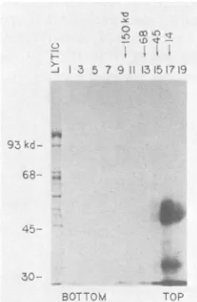
FIG. 9. Sedimentation analysis of ElA proteins. HeLa cells wereinfectedatanMOIof 50 in the presenceof araCand labeled
with32P04at36to38 hp.i. A total of5 x 10 cells werelysed by
method ii, and the extract from 8 x 10⁵ cells was sedimented
through a5to20%glycerol gradientin RIPA bufferasdescribed in
the text. Odd-numbered fractions were immunoprecipitated with
anti-ElA fusion serumand analyzed by gel electrophoresis.
Frac-tion1containedthe CsCl cushion andwasdialyzedbefore
immuno-precipitation. Arrows and molecular weights at the top indicate
sedimentation of markers inaparallel gradient.Gelelectrophoresis markersareindicatedbythe numbersatthe side.Lyticindicates the
latelyticmarker.
r; ik ;*, _ r!A
(D ...\I-SJ) CL.
---E-,F ....}.-1;.1'
-;,~(.IC. /,-\L ,.X
J. VIROL.
on November 10, 2019 by guest
http://jvi.asm.org/
These antibodies have been used to examine proteins
pro-duced in adenovirus-infected cells and have been shown to
recognize the predictedtumorantigensofElA (Fig. 5 to8). Plasmids were also constructed which produce fusion
pro-teins of the trpEgene witheitherthe E1B 55-kdorthe21-kd
protein. These fusion proteins alsoelicited arapidantibody
response, specific forthe appropriate E1B tumorantigens. The immunoprecipitation ofElA proteins from d1313, a
deletion mutant lacking 70 C-terminal amino acids of the ElA proteins, has revealed thatin additionto being shorter the proteins are lessabundant and less phosphorylated than those of wild-type virus. We are currently investigating the decreased phosphorylation in d1313 and other El mutants.
We are alsofurther analyzing the multiple forms of the ElA proteins seen in infected cells, using the specific ElA antisera. The reduced E1B 21-kd protein level in hrl-infected cells (defective inthe ElA289-amino acid protein), but not in pm975-infected cells (defective in the ElA 243-amino acidprotein), is consistent withaspecificrolefor the 289-amino acid protein in induction ofearly viral transcrip-tion (42, 48).
When infectedcellextractswereprepared bythemodified method of Manley et al. (38) described here, the ElA proteins were found to sediment in RIPA buffer as
mono-mers, withoutanyobservable association with othercellular or viral proteins. The procedure extracts >90% of the ElA proteins, as determined by comparing high-speed
superna-tant and pellet fractionsby Western immunoblot analysis or
autoradiography oftwo-dimensional gels of labeledextracts
(Tsukamoto and Berk, unpublished data). This contrasts
with thelow-salt extraction procedureused by Feldman and Nevins which did not extract a large fraction ofthe ElA proteins
(10).
The ElA289-amino acidprotein extracted by their proceduire rapidly pelleted through glycerol gradients preparedin RIPA buffer(10). The discrepancy in sedimenta-tion properties of the ElA proteins in these two studies might be explained by the incomplete dissociation of ionic interactions between thehighlychargedElAproteins(pl4.5 to 5.0; 11, 48)and cellularcomponentsinthe low-saltbuffer. Feldman and Nevins also reported that the ElA 289-amino acid protein was not solubilized from nuclear fractions by extraction with2MNaCl and therefore raised the possibility that the protein is tightly associated with the nuclearmatrix (10). We havealso observed that theElAproteinsarepoorlyextracted by very high-salt buffers (Tsukamoto and Berk, unpublished data). However, the ability to extractthe ElA proteins in soluble form with buffers of intermediate salt concentrations indicates that the proteins are not bound to
nuclear structures through salt-resistant interactions. It was surprising to observe that the ElA proteins which have predictedmolecular weights of27,000to32,000 cosedi-mentedwithlysozyme (14 kd) in RIPAbuffer (Fig. 9). One possibleexplanationwhichcanbe tested is that theproteins have avery extended conformation in this buffer.
The methodofantibody productionreported here israpid and inexpehsive;we havebeen abletoobtain high-titer
anti-ElA antiserum throughout 5 months of immunization. In
addition, immunoprecipitates of mammalian cells, using these antibodies, have alow background since the antigens for immunization are produced in bacterial cells. Antisera producedagainsttheElA bacterialfusion proteinare higher
in titer thanthosedirected againstan ElA synthetic peptide
and are directed against specific gene products. These
antibodies are being used to study proteins of various
adenovirus mutantsand also will be usefulin thepurification and study of the adenovirus tumor antigens.
Theantisera
to
theE1B
55-kdfusion proteinappear tobe of lower titerthan those to the ElA andE1B
21-kd protein.This may be because the fusion protein used for
immuniza-tion contains only 60 amino acids of the adenovirus se-quence. Attempts toinducetrpE-adenovirus fusion
proteins
from plasmids encoding 485amino acidsof the 55-kdprotein
have been unsuccessful. This is
probably
because theresult-ing fusion proteinwould beapproximately 90kd;trpEfusion
proteins of >76 kd have not been obtained (Spindler and Berk, unpublished data; S. Watanabe, J. Konopka, and C. Dieckmann, personal communication). The construction of plasmid derivatives of
pKRS101
with a polylinkerregion
from pUC13 in all three reading frames has extended the usefulness of this expression vector system (A. Tzagoloff
and T. J. Koerner, personal communication). Such vectors allow the rapid construction ofplasmids which can express trpEfusion proteins for biochemical and serological uses.
ACKNOWLEDGMENTS
We thank Rob Gunsalus for advice on growth and induction of
trp-containing
plasmids, Kenn Buckley for many suggestions on cloning in expression plasmids, Sarah French forpurified anthrani-late synthetase, Ann Tsukamoto, Bert Semler, and Owen Witte for helpful discussions on immunization and immunoprecipitations,and Philip Branton and FrankGraham
for providing hamster antitumor sera used as controls in the early patt of this work. We are grateful to Debra Bomar for preparation of the manuscript.This work was supported by Public Health Service grants from the
Natidnal
Cancer Institute, RO1 CA 25235 and P01 CA 32737. A.J.B. is supported by a Faculty Research Award from the Ameri-can Cancer Society. K.R.S. was supported by postdoctoral fellow-ships from the Damon Runyon-Walter Winchell Cancer Fund and the Public Health Service National Institutes of Health (1 F32 CA 06925-01).LITERATURE CITED
1. Baker, C. C., and E. B.Zift.1981. Promotersandheterogeneous 5' termini of the messenger RNAsof adenovirus serotype 2. J. Mol. Biol. 149:189-221.
2. Berk, A., and P. Sharp. 1978. Structureof the adenovirus 2early mRNAs. Cell 14:695-711.
3. Berk, A. J., F. Lee, T. Harrison, J. Williams, and P. A. Sharp. 1979. Pre-early adenovirus 5 gene product regulatessynthesis of early viral messenger RNAs. Cell 17:935-944.
4. Bos, J. L., L. J.Polder, R. Bernards, P.I.Shrier, P. J.vanden
Elsen,
A. J. van der Eb, and H. van Ormondt. 1981. The2.2 kb Elb mRNA of human Adl2 and AdS codes for two tumor antigens starting at different AUG triplets. Cell 27:121-131. 5. Brackmann, K. H., M. Green, W. S. M. Wold, M. Cartas, T.Matsuo, and S. Hashimoto. 1980. Identification and peptide mapping of human adenovirus type 2-induced early polypep-tides isolated by two-dimensional gel electrophoresis and im-munoprecipitation. J. Biol. Chem. 255:6772-6779.
6. Braithwaite, A. W., B. F. Cheetham, P. Li, C. R.Parish, L. K.
Waldron-Stevens, and A. J. D. Bellett. 1983.
Adenovirus-in-duced
alterations of the cell growth cycle: a requirement for expression ofElA but not ofE1B. J. Virol. 45:192-199. 7. Burnette, W. N. 1981. "Western blotting": electrophoretictransfer of proteins from sodium dodecyl sulfate-polyacryl-amide gels tounmodified nitrocellulose and radiographic detec-tion with antibody and radioiodinated protein A. Anal. Bio-chem. 112:195-203.
8. Chow, L. T., T. R. Broker, and J. Lewis. 1979. Complexsplicing
patterns
of RNAs from the early region of adenovirus-2. J.Mol. Biol. 134:265-303.9. Doolittle, W. F., and C. Yanofsky. 1968. Mutants ofEscherichia coli with analteredtryptophanyl-transfer ribonucleicacid syn-thetase. J. Bacteriol. 95:1283-1294.
10. Feldman, L. T., and J. R. Nevins. 1983. Localization of the adenovirus E1Aa protein, a positive actingtranscriptional fac-tor, in infected cells. Mol. Cell. Biol. 3:829-838.
on November 10, 2019 by guest
http://jvi.asm.org/
11. Gaynor, R. B., A. Tsukamoto, C. Montell, and A. J. Berk. 1982. Enhanced expression of adenovirus transforming proteins. J. Virol. 44:276-285.
12. Gilead, Z., Y.-H. Jeng, W.S. M. Wold, K. Sugawara, H. M. Rho, M. L. Harter, and M. Green. 1976. Immunological identifi-cation of two adenovirus 2-induced early proteins possibly involved incell transformation. Nature (London) 264:263-266. 13. Goeddel, D. V., D. G.Kleid, F. Bolivar, H. L. Heyneker, D. G. Yansura, R. Crea, T. Hirose, A. Kraszewski, K. Itakura, and A. D. Riggs. 1979. Expression in Escherichia coli of chemically synthesized genes for human insulin. Proc. Natl. Acad. Sci. U.S.A. 76:106-110.
14. Graham, F. L., T. Harrison, and J. Williams. 1978. Defective transforming capacity of adenovirus type5 host-range mutants. Virology 86:10-21.
15. Graham, F. L., J. S. Smiley, W. C. Russell, and R. Nairn. 1977. Characteristics of a human cell line transformed by DNA from humanadenovirus type 5. J. Gen. Virol. 36:59-72.
16. Graham, F. L., A. J. van der Eb, and H. L. Heijneker. 1974. Size and location of the transforming region in human adenovi-rus DNA. Nature (London) 251:687-691.
17. Green, M., W. S. M. Wold, K. H. Brackmann, and M. A. Cartas. 1979. Identification of families of overlapping polypep-tides coded by early region 1 of human adenovirus type 2. Virology 97:275-286.
18. Halbert, D. N., and H. J. Raskas. 1982. Tryptic and chymotryp-tic methionine peptide analysis of the in vitro translation prod-ucts specified by the transforming region of adenovirus type 2. Virology 116:406-418.
19. Halbert, D. N., D. J. Spector, and H. J. Raskas. 1979. Invitro translation products specified by the transforming region of adenovirus type 2. J. Virol. 31:621-629.
20. Harrison, T., F. Graham, and J. Williams. 1977. Host-range mutants of adenovirus type5 defective for growth in HeLa cells. Virology 77:319-329.
21. Harter, M. L., G. Shanmugam, W. S. M. Wold, and M. Green. 1976. Detection of adenovirus type 2-induced early polypeptides using cyclohexamide pretreatment to enhance viral protein synthesis. J. Virol. 19:232-242.
22. Houweling, A., P. J. van denElsen,and A. J. van der Eb. 1980. Partial transformation of primary rat cells by the leftmost 4.5% fragment of adenovirus 5 DNA. Virology 105:537-550. 23. Johansson, K., H. Persson, A. M. Lewis, U. Pettersson, C.
Tibbetts,and L. Philipson. 1978. Viral DNA sequences and gene products in hamster cells transformed by adenovirus type 2. J. Virol. 27:628-639.
24. Jones, N., and T. Shenk. 1979. An adenovirus type5 early gene function regulates expression of other early viral genes. Proc. Natl. Acad. Sci. U.S.A. 76:3665-3669.
25. Jones, N., and T. Shenk. 1979. Isolation of AdS host-range deletion mutants defective for transformation of rat embryo cells. Cell 17:683-689.
26. Katze, M. G., H. Persson, and L. Philipson. 1982. A novel mRNA and a low molecular weight polypeptide encoded in the transforming region of adenovirus DNA. EMBO J. 1:783-789. 27. Kessler, S. W. 1975. Rapid isolation of antigens from cells with
a staphylococcal protein A-antibody adsorbent: parameters of the interaction of antibody-antigen complexes with protein A. J. Immunol. 115:1617-1624.
28. Kleid, D. G., D. Yansura, B. Small, D. Dowbenko, D. M. Moore, M. J. Grubman, P. D. McKercher, D. 0. Morgan, B. H. Rob-ertson, and H. L. Bachrach. 1981. Cloned viral protein vaccine for foot-and-mouth disease: responses in cattle and swine. Science 241:1125-1129.
29. Laemmli, U. K.1970.Cleavage of structural proteins during the assembly of the head of bacteriophage T4. Nature (London) 270:680-685.
30. Lassam, N. J., S. T. Bayley, and F. L. Graham. 1979. Tumor antigens of humanAdS in transformed cells and in cells infected with transformation-defective host rangemutants. Cell 18:781-791.
31. Lassam, N. J., S. T. Bayley, F. L. Graham, andP. E. Branton. 1979. Immunoprecipitation of protein kinase activity from
ade-novirus 5-infectedcells using antiserumdirectedagainsttumor
antigens. Nature(London) 277:241-243.
32. Levinson, A. D., and A.J.Levine.1977.The groupCadenovirus tumorantigens: identification in infected and transformed cells anda peptide map analysis. Cell 11:871-879.
33. Lewis, J. B., J. F. Atkins, P. R. Baum, R.Solem, R. F. Geste-land,and C. W. Anderson. 1976. Location and identificationof the genesforadenovirus type2earlypolypeptides. Cell 7:141-151.
34. Lewis, J. B., H. Esche,J. E. Smart, B. Stillman, M.L.Harter, and M. B. Mathews. 1979. Organization and expression of the left third of the genome of adenovirus. Cold Spring Harbor Symp. Quant. Biol. 44:493-508.
35. Lupker, J. H., A. Davis, H. Jochemsen, andA. J. vander Eb. 1981. In vitro synthesis of adenovirus type 5 T antigens. I. Translationofearly region 1-specificRNAfrom lytically infect-ed cells. J. Virol. 37:524-529.
36. Malette, P.,S.-P. Yee, and P. E. Branton. 1983. Studies onthe phosphorylation ofthe 58,000 dalton early region1B protein of humanadenovirus type 5. J. Gen. Virol. 64:1069-1078. 37. Maniatis, T., E. F. Fritsch, and J. Sambrook. 1982. Molecular
cloning. Cold Spring Harbor Laboratory, Cold Spring Harbor, N.Y.
38. Manley, J.L., A. Fire, A. Cano, P. A. Sharp, and M. L.Gefter. 1980. DNA-dependent transcription of adenovirus genes in a soluble whole cell extract. Proc. Natl. Acad. Sci. U.S.A. 77:3855-3859.
39. Markwell, M. A. K. 1982. A new solid-state reagent toiodinate proteins. Anal. Biochem. 125:427-432.
40. Maxam, A. M., and W. Gilbert. 1977. A new method for sequencing DNA. Proc. Natl. Acad. Sci. U.S.A. 74:560-564. 41. Messing, J., and J. Vieira. 1982. A new pair of M13 vectors for
selecting either DNA strand ofdouble-digest restriction frag-ments. Gene 19:269-276.
42. Montell, C., E. F. Fisher, M. H. Caruthers, and A.J. Berk. 1982. Resolving thefunctions of overlappingviral genesby site-specific mutagenesis at a mRNA splice site. Nature (London) 295:380-384.
43. Osborne, T. F., and A. J. Berk. 1983. Far upstream initiation sites for adenovirus early region 1A transcription are utilized after the onset of viral DNAreplication. J. Virol. 45:594-599. 44. Osborne, T. F., R. B. Gaynor, and A. J.Berk. 1982. The TATA
homology and the mRNA 5' untranslated sequence are not required for expression of essential adenovirus ElAfunctions. Cell 29:139-148.
45. Perricaudet, M., G. Akusjarvi, A. Virtanen, and U. Pettersson. 1979. Structure of two spliced mRNAs from the transforming region of human subgroup C adenoviruses. Nature (London) 281:694-696.
46. Perricaudet, M., J.-M. LeMoullec, and U. Pettersson. 1980. Predicted structure of two adenovirus tumor antigens. Proc. Natl. Acad. Sci. U.S.A. 77:3778-3782.
47. Persson, H., M. G. Katze, and L.Philipson. 1982.Purificationof a native membrane-associated adenovirus tumor antigen. J. Virol. 42:905-917.
48. Ricciardi, R. P., R. L. Jones, C. L. Cepko, P. A. Sharp, and B. E. Roberts. 1981. Expression of early adenovirus genes requires a viral encoded acidic polypeptide. Proc. Natl. Acad. Sci. U.S.A. 78:6121-6125.
49. Ross, S. R., A. J.Levine, R. S. Galos, J.Williams,and T. Shenk. 1980. Early viral proteins inHeLacellsinfectedwith adenovirus 5 host range mutants. Virology 103:475-492.
50. Saborio, J. L., and B.Oberg. 1976. In vivo and in vitrosynthesis of adenovirus type 2 earlyproteins. J. Virol. 17:865-875. 51. Sarnow, P., Y. S. Ho, J. Williams, and A.J. Levine. 1982.
Adenovirus Elb-58kd tumor antigen and SV40 large tumor
antigen are physically associated with the same 54 kd cellular protein in transformed cells. Cell28:387-394.
52. Sarnow, P., C. A. Sullivan,andA. J.Levine. 1982. A monoclo-nal antibody detecting the adenovirus type 5 Elb-58Kdtumor
antigen: characterization of the Elb-58Kd tumor antigen in adenovirus-infected and -transformed cells. Virology 120:510-517.
on November 10, 2019 by guest
http://jvi.asm.org/
53. Schrier,P. I., P.J.vandenElsen, J. J.L.Hertoghs,and A.J. van der Eb. 1979. Characterization oftumorantigens in cells transformed byfragmentsof adenovirustype5 DNA.Virology
99:372-385.
54. Shenk,T., N.Jones,W.Colby,andD. Fowlkes. 1979.Functional analysis of adenovirus type 5 host range deletion mutants
defective for transformation ofratembryo cells. Cold Spring Harbor Symp. Quant. Biol. 44:367-375.
55. Smart, J. E., J. B. Lewis, M. B. Mathews, M. L. Harter, and C. W. Anderson. 1981. Adenovirustype 2early proteins:
assign-mentof theearly region1Aproteins synthesizedin vivoandin vitrotospecificmRNAs.Virology112:703-713.
56. Stow,N. D. 1982. Theinfectivityof adenovirusgenomeslacking
DNA sequences from their left-hand termini. Nucleic Acids
Res. 10:5105-5119.
57. Tooze, J.1981. DNAtumorviruses. ColdSpring Harbor
Labo-ratory,Cold Spring Harbor, N.Y.
58. vanderEb, A. J., C. Mulder, F. L.Graham,and A.Houweling.
1977. Transformation with specific fragments of adenovirus DNAs. I. Isolation of specific fragments with transforming
activity ofadenovirus 2 and5 DNA. Gene 2:115-132. 59. vanOrmondt,H., J. Maat, A. Dewaard, and A. J.vander Eb.
1978. The nucleotide sequence of the transforming Hpal-E
fragment ofadenovirus type5 DNA. Gene 4:309-328. 60. vanOrmondt, H., J. Maat, and C. P. vanBeveren. 1980.The
nucleotide sequence of the transforming early region El of adenovirustype5 DNA. Gene 11:299-309.
61. Yanofsky, C., T. Platt, I. P. Crawford, B. P. Nichols, G. E.
Christie,H. Horowitz, M. VanCleemput, and A. M. Wu. 1981. The completenucleotide sequenceof the tryptophanoperonof
Escherichiacoli. Nucleic Acids Res. 9:6647-6668.
62. Yee, S.-P., D. T. Rowe, M. L. Tremblay, M. McDermott, and
P.E. Branton. 1983. Identification of human adenovirus early region 1 products using antisera against synthetic peptides corresponding to the predicted carboxy termini. J. Virol. 46:1003-1013.


![FIG. 7.infectedpolyacrylamidewith Immunoprecipitation of phosphorylated El proteins. HeLa cells were infected at an MOI of 20 in the presence of araC and labeled [35S]methionine (lanes 1 to 3) or 32P04 (lanes 5 to 9, 11 to 18) at 36 h p.i](https://thumb-us.123doks.com/thumbv2/123dok_us/1438855.96289/6.612.136.225.75.171/infectedpolyacrylamidewith-immunoprecipitation-phosphorylated-proteins-infected-presence-labeled-methionine.webp)