Comparative Analysis on Image Compression Techniques for Medical Images
Full text
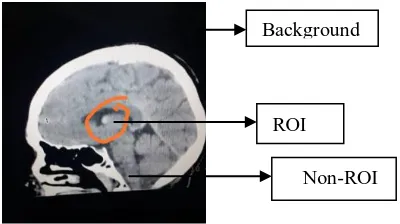
Figure
![Figure 2.[1] wavelet transform image compression process](https://thumb-us.123doks.com/thumbv2/123dok_us/8204889.261880/2.595.304.558.611.839/figure-wavelet-transform-image-compression-process.webp)
![Figure 6:.Results of decoding by fractal technique.[5]](https://thumb-us.123doks.com/thumbv2/123dok_us/8204889.261880/4.595.305.530.145.274/figure-results-of-decoding-by-fractal-technique.webp)
Related documents
STUDY PROTOCOL Open Access Induction of labour versus expectant monitoring for gestational hypertension or mild pre eclampsia between 34 and 37 weeks? gestation (HYPITAT II) a
Evans, Nicole M., "The Process of Exercise Participation in the Community for Functional Recovery Post Formal Rehabilitation among Survivors of Stroke: a grounded theory
Dual roles of voltage gated sodium channels in development and cancer FAHEEMMUDDEEN PATEL and WILLIAM J BRACKENBURY* Department of Biology, University of York, Heslington, York, UK
Pneumoperitoneum for laparoscopic surgery following autologous deep inferior epigastric (DIEP) perforator flap breast reconstruction may prove difficult as a result of loss of
For assessing the complex biomechanical behavior on incisal and contralateral molar loadings, first molar dis- placement bilaterally, the maximum von Mises stress in the
These steps include standardizing the nursing education programs (university and vocational) and examinations to reflect the competencies required and conducting studies on the
