ribosomal frameshifting. These levels of TK can suffice to permit reactivation from latently infected mouse ganglia, but in a majority of ganglia, especially with the G9 virus, reactivation of virus that has reverted to the TK-positive phenotype predominates. To help address the relative contributions of translational mechanisms and reversion in reactivation, we generated viruses with a base either inserted or deleted just downstream of the G string. Both of these viruses had a TK-low phenotype similar to that of the G8 and G9 viruses but with less reversion. Both of these viruses reactivated from latently infected trigeminal ganglia, albeit inefficiently, and most viruses that reactivated had a uniformly TK-low phenotype. We also generated viruses that have one insertion in a run of six C’s or one deletion in a run of five C’s. These viruses lack measurable TK activity. However, they reactivated from latently infected ganglia, albeit inefficiently, with the reactivating viruses having reverted to the wild-type TK phenotype. Therefore, for G-string mutants, levels of active TK as low as 0.25% generated by translational mechanisms can suffice for reactivation, but reversion can also contribute. For viruses that lack TK activity due to mutations on other homopolymeric sequences, reactivation can occur via reversion.
Although acyclovir (ACV) is an effective antiviral agent, herpes simplex virus (HSV) that is resistant to treatment can be a serious clinical problem in immunocompromised patients.
It has been estimated that ACV-resistant (ACVr) HSV
ap-pears in as many as 5% of this population upon treatment (4, 10). Clinically significant drug resistance implies evasion of drug action but retention of pathogenesis. The vast majority of mutations associated with clinical drug resistance occur in the viral thymidine kinase (TK) gene (tk), which encodes the en-zyme that selectively activates ACV. The phenotypes of clinical isolates with mutant TKs typically fall into one of three
cate-gories: altered substrate specificity, low TK activity (TKL), or
lack of measurable TK activity (TK⫺) (11, 12, 26). That viruses
lacking TK activity are pathogenic has been puzzling. Although TK is not required for growth in cell culture, it is essential for pathogenesis in mouse models of HSV infection, in particular, for reactivation of laboratory strains of HSV-1 from latently infected mouse trigeminal ganglia (3, 6, 9, 21). Many of the mutations associated with the mutant TK phenotypes are
in-sertions or deletions of bases in homopolymeric sequences in the gene (11, 12, 26).
The most common mutation in ACVrviruses is a single G
insertion into a run of seven G’s known as the “G string” (11, 12, 19, 26). Double G insertions into the G string are also frequently observed (11–13, 17). Viruses that have either of
these genotypes have a TKLphenotype (14, 15, 19). The active
TK from viruses with a single G insertion occurs via an unusual ribosomal frameshift on the G string that is solely dependent on the G-rich nature of the G string and is possibly the result of a direct interaction between the G string and the rRNA (18; A. Griffiths and D. M. Coen, unpublished results). Preliminary
in vitro evidence suggests that ⫺1 ribosomal frameshifting
occurs via a similar mechanism on a G string with a double G insertion (A. Griffiths and D. M. Coen, unpublished results). We have shown previously that viruses with the single G in-sertion reactivated from latency in mouse trigeminal ganglia,
and occasionally, the virus that reactivated was uniformly TKL
(14). These studies showed that the low levels of TK generated from viruses with the single G insertion were sufficient to support reactivation but that most of the viruses that reacti-vated contained some virus with the wild-type TK phenotype
(TK⫹), suggesting that reversion may also play a role in the
reactivation of viruses with the G8 genotype (14). Indeed, a
virus that is TK⫹should have a strong growth advantage in the
ganglion. Reversion was especially evident with viruses that carried double G insertions into the G string, with about 3% of
plaques apparently TK⫹(13, 15), and this is consistent with the
genetic instability of a homopolymeric sequence increasing with its length (22). All virus populations that reactivated from
* Corresponding author. Mailing address: Department of Biological Chemistry and Molecular Pharmacology, Harvard Medical School, 250 Longwood Ave., Boston, MA 02115. Phone: (617) 432-1691. Fax: (617) 432-3833. E-mail: [email protected].
† Present address: Department of Virology and Immunology, South-west Foundation for Biomedical Research, 7620 NW Loop 410, San Antonio, TX 78227.
‡ Present address: Harvard Medical School at Beth Israel Deaconess Medical Center, Boston, MA 02215.
§ Present address: Department of Paediatrics, Royal Berkshire Hos-pital, London Road, Reading RG1 5AN, United Kingdom.
6568
on November 8, 2019 by guest
ganglia latently infected with a G9 virus contained TK⫹virus.
It was therefore not possible to ascertain whether the TKL
phenotype of the G9 virus, presumably generated by a net⫺1
ribosomal frameshift, was sufficient to support reactivation. The mixed TK phenotypes of these viruses draw an interesting
parallel to those of clinical isolates; many, if not all, ACVr
clinical isolates are comprised of mixtures of viruses with mul-tiple TK phenotypes (11, 24).
While viruses carrying one of the three mutations listed above are among the most frequently observed in isolates from
patients, mutations on other homopolymeric sequences intk
are also associated with drug-resistant HSV disease (11, 12). However, it is not known whether these viruses generate active TK or reactivate from latency.
In this paper, we address the contributions of ribosomal frameshifting and reversion toward the reactivation of mutants with altered sequences around the G string. Additionally, we
have generated recombinant viruses that carry ACVr
muta-tions on other homopolymeric sequences and analyzed their TK activities and reactivation from latency.
MATERIALS AND METHODS
Plasmid construction.Plasmid pAG5 contains the entire BamHI P fragment from HSV-1 strain KOS in pBluescript SK(⫹) (Promega) (15). Plasmids engi-neered to carry the ACVrmutations were generated by site-directed mutagenesis using the QuikChange kit (Stratagene) according to the manufacturer’s instruc-tions. For each plasmid, two complementary oligonucleotides (Integrated DNA Technologies) were synthesized to facilitate mutagenesis; however, only the forward primers are listed here: pTKG7aC, GTTCTGGCTCCTCATATCGGG GGGGAGGCCTGGGAGCTCACATGC; pTKG7dG, GCCGTTCTGGCTCC TCATATCGGGGGGGAGCTGGGAGCTCAC; pTKC6⫹1C, GGGCAGCAT GACCCCCCCAGGCCGTGCTGGCG; and pTK2C5⫺1C, GGCCCCCGAGT TGCTGGCCCCAACGGCGACCTGTATAACG. The presence of desired mutations and the absence of unwanted mutations were confirmed by sequencing of thetkgene.
Cells and viruses.African green monkey (Vero) and TK⫺human osteosar-coma (143B) cells were obtained from the American Type Culture Collection and maintained in Dulbecco’s modified Eagle’s medium, supplemented with 10% bovine fetal serum, at 37°C and 5% CO2. Viruses that are not novel to this
study are as follows: wild-type HSV-1 strain KOS, clinical isolate 615.9, and KOS-derived mutants KG111, LS-95/-85, LS-111/-101/-56/-46, 615.9, LS-29/-18, andtkLTRZ1 (1, 5–8, 20, 24).
Construction of recombinant viruses.The method used to generate recombi-nant viruses has been described previously (15). Briefly, plasmid midi-prep DNA (Wizard Prep; Promega), virion “mini-prep” (7)tkLTRZ1 DNA, and transfec-tion reagent (Effectene; QIAGEN) were added to 50% confluent Vero cells. The use oftkLTRZ1, a recombinant KOS strain that has an insertion intkof the Moloney murine leukemia virus long terminal repeat (LTR) upstream oflacZ, permits blue/white screening for recombinant viruses. In addition,tkLTRZ1 does not reactivate from explanted mouse trigeminal ganglia (references 3, 14, 15, and 21 and this study). The entire BamHI “P” fragment of this virus has been sequenced and shown to be identical to that of KOS, except for the LTR-lacZ
sequences (14). Following the appearance of sufficient cytopathic effect, the cells were harvested. Recombinants were cloned by limiting dilution using a blue/ white screen in 96-well trays, requiring two rounds of screening until a single “white” plaque was observed in a well. Virus from this well was amplified, DNA was prepared, and thetkgene was sequenced to confirm the presence of the mutation. Two independently isolated recombinant viruses were generated from separate transfections with each mutant plasmid: TKG7aC.1 and TKG7aC.2, TKG7dG.1 and TKG7dG.2, TK6C⫹1C.1 and TK6C⫹1C.2, and TK2C5⫺1C.1 and TK2C5⫺1C.2.
Plaque autoradiography.Quantitative plaque autoradiography was performed as described previously (15), using [3H]thymidine (Moravek) to radioactively label plaques. The assay was calibrated with viruses that generate known amounts of active TK polypeptide.
Assays of acute and latent infection in mice.Male 8-week-old randomly bred CD-1 mice (Charles River Laboratories) were infected on scarified corneas with 2⫻106
PFU of strain KOS or 7⫻107
PFU of mutant, as described previously (5, 23). Acute viral replication was monitored by assaying virus in the tear film at 1 day postinfection (p.i.) and in ganglion homogenates at 3 days p.i. Reactivation from latently infected ganglia was measured by enzymatically dissociating ganglia harvested at 30 days p.i. and culturing on Vero cells (23). These cells were screened for 10 days for the appearance of cytopathic effect, replated, and screened for a further 7 days. Viruses that reactivated were amplified on Vero cells and subjected to plaque autoradiography. Additionally, viral DNA was prepared from these viruses and thetkgenes were sequenced as previously described (14, 15).
RESULTS
[image:2.585.81.503.68.249.2]Construction of viruses that have mutations immediately downstream of the G string.We have shown previously that
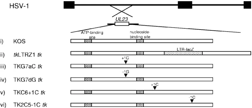
FIG. 1. Structure of thetkgenes of viruses used in this study. The top two lines represent the HSV genome and the location of thetkgene (UL23). Below are schematic diagrams of the tkgenes of the viruses used in this study. Above the arrowheads are the mutations, and the arrowheads indicate the approximate position of the mutation intk. (i) KOS (cross-hatched boxes, functional sites of TK); (ii)tkLTRZ1 (tkwith LTR-lacZinserted into the PstI site [dotted box]); (iii) TKG7aC (tkwith a single C inserted immediately downstream of the G string of KOStk); (iv) TKG7dG (tkwith a single G deleted immediately downstream of the G string of KOStk); (v) TKC6⫹1C (tkwith a single C inserted into the C chord); (vi) TK2C5⫺1C (tkwith a single C deleted in a run of five C’s).
VOL. 80, 2006 FRAMESHIFT MUTATIONS AND HSV REACTIVATION 6569
on November 8, 2019 by guest
http://jvi.asm.org/
viruses with single G (G8) or double G (G9) insertions into the G string generated low levels of TK (15, 19) and that the low levels of TK expressed by the G8 virus can be sufficient to support reactivation from latency (14). However, viruses that had reverted to the wild-type TK phenotype were frequently observed following reactivation from latency with the G8 virus, and the frequency of reversion was so high with the G9 viruses studied by us and others that it made it impossible to
deter-mine whether the TKLphenotype contributed to reactivation
(13–15). As it seemed likely that that the length of the G string affected its genetic stability, we hypothesized that reversion
could be decreased while retaining a TKLphenotype by
main-taining the G string at its wild-type length, seven G’s. In vitro
data suggested that the net⫹1 ribosomal frameshift functions
on a wild-type G string (18). Two viruses were generated that each required a ribosomal frameshift to generate active TK while maintaining the wild-type length of the G string (indepen-dent isolates were generated for each mutation) (Fig. 1). Virus TKG7aC has a single C inserted immediately downstream of
the G string, thereby requiring a net⫹1 ribosomal frameshift
for the generation of active TK, as does the G8 virus. Virus TKG7dG has a single G deleted immediately downstream of
the G string, thereby requiring a net⫺1 ribosomal frameshift
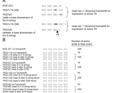
for the generation of active TK. Although in vitro experiments demonstrate that the ribosomal frameshift occurs on the G string (18; A. Griffiths and D. M. Coen, unpublished results), as yet we do not know on which nucleotide the ribosomal frameshifts occur. Therefore, there are several possible amino acid sequences that could be synthesized following a frame-shift, and these sequences are listed in Fig. 2. However, we considered that a small number of amino acid changes would be tolerated with minimal effect on enzyme activity as, based on the crystal structure of TK, the amino acids encoded by the G string are physically distant from the nucleoside and triphos-phate binding sites (2, 27, 28). Also, it has been shown that viruses that have three nucleotides added to the G string (G10) and, therefore, an extra amino acid have wild-type TK activity (13, 15).
[image:3.585.105.480.75.384.2]Viruses with mutations downstream of the G string generate low levels of active TK.The levels of TK synthesized in cells
FIG. 2. Sequences of viruses with mutations immediately downstream of the G string. (A) The first line shows the sequence of the G string in the wild-type virus. The nucleotides representing translated codons are indicated by spaces between the triplets. The second line shows the sequence in a virus that has a single G insertion in the G string, which requires a net⫹1 ribosomal frameshift for expression of active TK. The third line shows the sequence in a virus that has a single C added downstream of the G string, such that a net⫹1 ribosomal frameshift is required for expression of active TK. The fourth line shows the sequence in a virus that has a double G insertion in the G string, which requires a net⫺1 ribosomal frameshift for expression of active TK. The fifth line shows the sequence in a virus that has a single G deleted downstream of the G string, such that a net⫺1 ribosomal frameshift is required for expression of active TK. Inverted nucleotides are shown in boldface type. (B) Possible sequences of amino acids translated following ribosomal frameshifts on the G string. The left column lists the virus and a possible site of ribosomal frameshift. The middle column lists the amino acid sequence that would be translated. The right column shows the number of amino acids to the stop codon in the translated frame.
on November 8, 2019 by guest
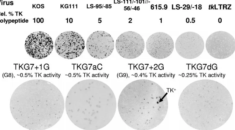
infected with viruses TKG7aC and TKG7dG were measured using quantitative plaque autoradiography, calibrated using viruses that generate known levels of active TK (Fig. 3). Virus
TKG7aC exhibited⬃0.5% of KOS TK activity (the previously
reported G8 virus has⬃0.5% of activity [14]). No TK⫹plaques
were observed from⬎500 plaques examined. Virus TKG7dG
exhibited ⬃0.25% of KOS TK activity (most of the plaques
from TK⫺cells infected with the previously reported G9 virus
have⬃0.4% of activity [15]). No TK⫹plaques were observed
from⬎500 plaques examined. Thus, as anticipated, there was
little reversion of these mutants containing a seven-nucleotide G string.
Replication of viruses TKG7aC and TKG7dG in mouse gan-glia.Separately, two independent isolates of each mutant virus were inoculated onto the scarified corneas of mice. Virus har-vested in eye swabs was titrated at 1 day p.i., and few or no differences in replication were observed among the mutant
viruses, KOS, andtkLTRZ1 (Table 1). At 3 days p.i., ganglia
were harvested and titrated. Although robust replication of KOS was observed, virus was not detected in ganglia from
animals infected with either TKG7aC or TKG7dG. Very little virus was detected in ganglia from animals infected with tkLTRZ1, which could be the result of low-level replication in the ganglia or contamination from the eye during the dissec-tion of the ganglia (Table 1). To establish whether viruses TKG7aC and TKG7dG were able to reactivate from latency
despite a frameshift mutation within tk, ganglia were
har-vested at 30 days postinoculation, enzymatically dissociated, and plated onto Vero cells that were monitored for the ap-pearance of plaques. Two of 18 ganglia latently infected with TKG7aC.1 and 0 of 18 infected with TKG7aC.2 yielded virus (2/36 total), compared to all of those infected with KOS and
none of those infected withtkLTRZ1 (Table 1). Both of the
viruses that reactivated from ganglia infected with TKG7aC were amplified on Vero cells and analyzed by plaque autora-diography (Fig. 4). Both of these viruses appeared to be
uni-formly TKL. Sequencing confirmed that the G7aC mutation
was maintained in the reactivated viruses.
[image:4.585.99.488.71.287.2]Two of 14 ganglia latently infected with TKG7dG.1 and 0 of 14 infected with TKG7dG.2 yielded virus (2/28 total) (Table 1).
FIG. 3. Quantitative plaque autoradiography of viruses. The top line shows the virus used. The line below shows the amount of active TK expressed by each mutant as a percentage of that expressed by wild-type strain KOS. Below, the images of the plates are presented. The next line shows the names of mutant viruses above the TK activities associated with these mutants (data for TKG7⫹1G have been published previously [14], and data for TKG7⫹2G have been published previously [15]). Below, the images of the plates are presented. Arrow, example of a plaque with the TK⫹phenotype on the TKG7⫹2G plate. Rel., relative.
TABLE 1. Replication and reactivation of viruses with TKLphenotypes
Virus Inoculum (PFU)
Titer of virus in eye swabs at 1 day p.i. (mean PFU⫾SE)b
Titer of virus in trigeminal ganglia at 3 days p.i. (mean PFU⫾SE)b
No. of ganglia with reactivating virus/ total no. of ganglia
TK phenotype(s) of reactivating virus
KOS 2⫻106 4.9⫻105⫾1.6⫻105(4) 1.1⫻104(1) 30/30 TK⫹
tkLTRZ1 7⫻107 1.8⫻106⫾7.2⫻105(4) 2.7⫾2.7 (3) 0/34 NDa
TKG7aC.1 7⫻107 2.0⫻105⫾1.4⫻105(2) 0 (2) 0/18 ND
TKG7aC.2 7⫻107 1.3⫻106⫾3.3⫻105(2) 0 (2) 2/18 TKL
TKG7dG.1 7⫻107 1.1⫻106⫾5.2⫻105(2) 0 (1) 2/14 TKLand mixed TK⫹/TKL
TKG7dG.2 7⫻107 ND ND 0/14 ND
aND, not done.
bThe number of samples titrated for each group is shown in parentheses.
VOL. 80, 2006 FRAMESHIFT MUTATIONS AND HSV REACTIVATION 6571
on November 8, 2019 by guest
http://jvi.asm.org/
[image:4.585.44.544.617.708.2]Both of the viruses that reactivated from ganglia infected with TKG7dG were amplified on Vero cells and analyzed by plaque
autoradiography (Fig. 4). One appeared to be uniformly TKL,
and sequence analysis confirmed that this virus contained the
G7dG genotype. The other appeared to be mixed TK⫹and
TKL, and this was consistent with sequencing data.
Generation of recombinant viruses carrying mutations at sites other than the G string intkassociated with clinical drug resistance. The use of ACVr HSV clinical isolates with a
known mutation in tk to investigate a phenotype is difficult
without a pretherapy strain for comparison, e.g., different iso-lates with the same mutation have been reported to have dif-ferent TK phenotypes (11). We therefore engineered into the laboratory HSV-1 strain KOS two other frameshift mutations that have been observed in isolates taken from patients receiv-ing ACV therapy (Fig. 1). As we have done previously, we chose strain KOS, as it is known to be dependent upon TK for reactivation from latently infected mouse trigeminal ganglia (3, 6, 14, 15, 21). Two independent isolates were generated for each mutation. The generation of the recombinant viruses used a blue/white screening procedure, rather than ACV
se-lection, to reduce the chance of selecting for an ACVr
muta-tion at a second site. Importantly, the use oftkLTRZ1 as a
starting point in the construction of the viruses eliminated a
potential source of contaminating TK⫹virus.
TK phenotypes of recombinant viruses. To assess whether these viruses were able to synthesize active TK despite the introduction of mutations into homopolymeric sequences in the gene, as has been observed with other mutations, they were analyzed by plaque autoradiography. We have modified this technique to make it extremely sensitive—approximately 0.25% of wild-type TK activity can be detected. No TK activity
could be detected from plaques infected with virus TKC6⫹1C
or TK2C5⫺1C (Fig. 5). No TK⫹plaques were observed from
⬎300 plaques that were examined from each virus.
Reactivation of TKC6ⴙ1C and TK2C5ⴚ1C from latency.
To establish whether viruses TKC6⫹1C and TK2C5⫺1C were
able to reactivate from latency despite a frameshift mutation
withintk, ganglia were harvested from latently infected mice,
enzymatically dissociated, and plated onto Vero cells that were monitored for the appearance of plaques. One of 13 ganglia
latently infected with TKC6⫹1C.1 and 0 of 12 infected with
TKC6⫹1C.2 yielded virus (1/25 total), compared to all of those
infected with KOS and none of those infected withtkLTRZ1
(Table 2). One of 16 ganglia latently infected with
TK2C5⫺1C.1 and 0 of 16 infected with TK2C5⫺1C.2 yielded
virus (1/26 total) (Table 2). The viruses that reactivated were amplified on Vero cells, analyzed by plaque autoradiography,
and shown to be TK⫹(Fig. 5). Sequencing of thetkgenes from
these viruses showed that they had each reverted to the
wild-typetkgenotype.
FIG. 4. Plaque autoradiography of viruses isolated following reactiva-tion of TKLviruses. In the image of the mixed population, one example of
[image:5.585.81.242.67.294.2]a TK⫹plaque and one of a TK⫺ plaque are shown (black and white arrowheads, respectively).
FIG. 5. Plaque autoradiography of viruses TKC6⫹1C and TK2C5⫺1C and the viruses isolated following reactivation. Each row shows an image of a plate infected with the original virus on the left and a plate with the corresponding virus that reactivated on the right.
TABLE 2. Reactivation of viruses lacking measurable TK activity from latently infected mouse trigeminal ganglia
Virus Inoculum
(PFU)
No. of ganglia with reactivating virus/ total no. of ganglia
TK phenotype of reactivating
virus
KOS 2⫻106 30/30 TK⫹
tkLTRZ1 7⫻107 0/34 NDa
TKC6⫹1C.1 7⫻107 1/13 TK⫹
TKC6⫹1C.2 7⫻107 0/12 ND
TK2C5⫺1C.1 7⫻107 1/16 TK⫹
TK2C5⫺1C.2 7⫻107 0/16 ND
aND, not done.
on November 8, 2019 by guest
[image:5.585.330.512.68.289.2]DISCUSSION
We are interested in mechanisms that permit HSV to evade antiviral chemotherapy yet retain pathogenicity. We have re-ported previously that ribosomal frameshifting and an internal ribosome entry site can support the expression of low levels of TK (15, 16, 19), an important phenotype that appears to permit pathogenesis in immunocompromised individuals while not ac-tivating effective amounts of drug. It has been noted that
cer-tain of these mutations that confer a TKLphenotype to the
virus, particularly the G9 mutation, render the virus disposed
to reversion to the TK⫹phenotype (13, 15, 25). Indeed,
al-though we have previously shown that virus reactivating from
some G8 mutant-infected ganglia were completely TKL, it has
been difficult to study these two mechanisms separately.
Herein, we have addressed the contribution of the TKL
phe-notype to pathogenesis, in the absence of reversion, by asking whether a wild-type G string supports the expression of TK originally reported for mutant sequences. This study shows
that the TKL phenotype is sufficient to support reactivation
from latently infected mouse trigeminal ganglia, even when the
level of TK is as low as⬃0.25% of that of the wild-type virus.
Reactivation was also observed under conditions that mini-mized the frequency of reversion. Conversely, we also wanted to address the contribution of reversion to pathogenesis in the absence of TK. This study shows that for two viruses that lack
measurable TK activity, TKC6⫹1C and TK2C5⫺1C, reversion
to the TK⫹phenotype is sufficient to permit reactivation from
latently infected mouse trigeminal ganglia. We discuss below the importance of these observations with regard to the patho-genesis of drug-resistant HSV and the biology of the wild-type virus.
The wild-type-length G string supports ribosomal frame-shifting.In vitro data using dual-reporter constructs translated in rabbit reticulocyte lysate showed that the G7 sequence of
wild-typetkwas able to support net⫹1 ribosomal
frameshift-ing (18). We have now shown that a wild-type-length G strframeshift-ing behaves similarly in the context of viral infection. Additionally, the expression of active TK following an insertion immediately downstream of the G string suggests that the wild-type G string
may support net⫺1 ribosomal frameshifting. These
observa-tions could mean that translation of the wild-type tk gene
results in the generation of several polypeptides via ribosomal
frameshifting into both the⫺1 and⫹1 reading frames. We are
currently investigating this possibility. Also, we have previously suggested that because of the relatively simple sequence
re-quirements of the G8 G-string net⫹1 ribosomal frameshift,
there may be other hitherto-unrecognized polypeptides gener-ated in mammalian genomes (18). We now propose an in-crease in the number of potential polypeptides that may be generated following frameshifting on G strings, given that seven G’s can suffice for ribosomal frameshifting and the
frameshifts appear to be into either the ⫺1 or ⫹ 1 reading
frames.
Active TK generated despite single base insertions or dele-tions downstream of the G string can support reactivation from latently infected trigeminal ganglia.We have previously shown that low levels of TK generated via ribosomal frame-shifting, from a virus with a single G insertion (G8), was suf-ficient to support reactivation from latently infected mouse
trigeminal ganglia (11 of 35 ganglia reactivated) (14). We
ad-dressed the possibility that reactivation of a TKLvirus could be
the result of a second-site mutation by introducing a mutation into the reactivated virus such that it did not synthesize active TK; this virus did not reactivate from latently infected mouse ganglia (14). A limitation of studying viruses with insertions into the G string was that virus populations that reactivated
from some ganglia contained TK⫹virus, presumably a result of
reversion. In this study, we maintained the wild-type-length G string, added a base immediately downstream, and observed reactivation in only 2 of 36 ganglia infected with TKG7aC viruses. The virus that reactivated from both of these ganglia
was uniformly TKL. Although these two experiments were not
performed at the same time, the data suggest that the differ-ence in frequency of reactivation between the two genotypes may be due largely to the increased instability of the G8 G string versus that of the G7 G string.
Viruses with a double G insertion in the G string (G9) were
remarkably unstable, with ⬃3% of plaques appearing TK⫹
(15). The remaining⬃97% of plaques were TKL. In that study,
virus populations that reactivated from all ganglia contained
TK⫹virus, leaving us uncertain as to whether active TK
gen-erated via a net ⫺1 ribosomal frameshift was sufficient to
support reactivation from latently infected trigeminal ganglia. Grey and colleagues (13) also reported that a virus with a double G insertion into the G string is prone to reversion to the
TK⫹phenotype. Those authors also detected low levels of TK
activity in lysates of cells infected with a clinical isolate carrying the double G mutation but concluded that the TK activity was
likely a result of viruses that had reverted to the TK⫹
pheno-type. Given that enzyme assays, unlike plaque autoradiogra-phy, are unable to discriminate between viruses with different TK phenotypes within a population, it remains at least possible that the clinical isolate studied by Grey and colleagues has a
TKLphenotype, in addition to being prone to reversion.
Pre-viously, we observed that of 35 ganglia latently infected with G9 virus, 20 reactivated, and the two populations that reacti-vated that were analyzed by plaque autoradiography contained
TK⫹virus (15). In this study, virus was observed to reactivate
from only 2 of 28 ganglia latently infected with virus TKG7dG (Table 1). As noted above, although the experiments were not performed at the same time, the data strongly suggest that the difference in frequency of reactivation between the two geno-types may be due largely to the increased instability of the elongated G string.
We interpret the low frequency of reactivation of these vi-ruses as being a reflection of the very low levels of TK ex-pressed. Although it is possible that viruses appearing to
reac-tivate as uniformly TKLpopulations could contain low levels of
TK⫹ virus, we consider this highly unlikely, given that we
detected⬍0.2% of TK⫹virus in these populations and given
the strong selective advantage that TK⫹viruses would have in
the ganglion.
A virus with an insertion into the C chord lacks TK activity.
We have recently shown that a virus that has a deletion in a run
of six C’s intk, known as the “C chord,” has a TKLphenotype
and the TK activity is dependent on an unusual internal
ribo-some entry site (IRES) intk(16). Interestingly, the C chord
itself was not necessary for the synthesis of the polypeptide generated via the IRES. It was therefore surprising that virus
VOL. 80, 2006 FRAMESHIFT MUTATIONS AND HSV REACTIVATION 6573
on November 8, 2019 by guest
http://jvi.asm.org/
shown such viruses to be TKL, rather than TK⫺(references 15
and 19 and this study). In this report, we have shown that recombinant viruses that lack measurable TK activity due to mutations in other homopolymeric sequences are indeed able to reactivate from latently infected mouse trigeminal ganglia.
The reactivating virus in each case was TK⫹. Although we
cannot rule out the possibility that these viruses generate levels of TK below our detection levels, the data suggest that rever-sion due to the inability of the DNA polymerase to faithfully replicate homopolymeric sequences plays an important role in
the pathogenesis of many ACVrviruses.
It seems clear that both the TKLphenotype and reversion to
the TK⫹phenotype can suffice to support reactivation from
latently infected mouse ganglia. However, further experiments are needed to quantify the relative contributions of reversion and low TK activity toward reactivation and to determine at what stage (establishment or reactivation) these phenotypes make their contributions.
ACKNOWLEDGMENTS
We dedicate this paper to the memory of Steve Sacks, who was a pioneer in studies of reversion in the pathogenesis of acyclovir-resis-tant muacyclovir-resis-tants and who was a warm and wonderful colleague.
We thank Jean Pesola for assistance with the animal experiments. This work was supported by grants PO1 NS35138, RO1 AI26126, and T32 AI07245 from the National Institutes of Health.
REFERENCES
1.Bo¨ni, J., and D. M. Coen.1989. Examination of the roles of transcription factor Sp1-binding sites and an octamer motif in transinduction of the herpes simplex virus thymidine kinase gene. J. Virol.63:4088–4092. 2.Brown, D. G., R. Visse, G. Sandhu, A. Davies, P. J. Rizkallah, C. Melitz,
W. C. Summers, and M. R. Sanderson. 1995. Crystal structures of the thymidine kinase from herpes simplex virus type-1 in complex with deoxy-thymidine and ganciclovir. Nat. Struct. Biol.2:876–881.
3.Chen, S. H., W. J. Cook, K. L. Grove, and D. M. Coen.1998. Human thymidine kinase can functionally replace herpes simplex virus type 1 thy-midine kinase for viral replication in mouse sensory ganglia and reactivation from latency upon explant. J. Virol.72:6710–6715.
4.Christophers, J., J. Clayton, J. Craske, R. Ward, P. Collins, M. Trowbridge, and G. Darby.1998. Survey of resistance of herpes simplex virus to acyclovir in northwest England. Antimicrob. Agents Chemother.42:868–872. 5.Coen, D. M., A. F. Irmiere, J. G. Jacobson, and K. M. Kerns.1989. Low
levels of herpes simplex virus thymidine-thymidylate kinase are not limiting
12.Gilbert, C., J. Bestman-Smith, and G. Boivin.2002. Resistance of herpesvi-ruses to antiviral drugs: clinical impacts and molecular mechanisms. Drug Resist. Updates5:88–114.
13.Grey, F., M. Sowa, P. Collins, R. J. Fenton, W. Harris, W. Snowden, S. Efstathiou, and G. Darby.2003. Characterization of a neurovirulent aciclo-vir-resistant variant of herpes simplex virus. J. Gen. Virol.84:1403–1410. 14.Griffiths, A., S. H. Chen, B. C. Horsburgh, and D. M. Coen.2003.
Transla-tional compensation of a frameshift mutation affecting herpes simplex virus thymidine kinase is sufficient to permit reactivation from latency. J. Virol.
77:4703–4709.
15.Griffiths, A., and D. M. Coen.2003. High-frequency phenotypic reversion and pathogenicity of an acyclovir-resistant herpes simplex virus mutant. J. Virol.77:2282–2286.
16.Griffiths, A., and D. M. Coen.2005. An unusual internal ribosome entry site in the herpes simplex virus thymidine kinase gene. Proc. Natl. Acad. Sci. USA102:9667–9672.
17.Harris, W., P. Collins, R. J. Fenton, W. Snowden, M. Sowa, and G. Darby.
2003. Phenotypic and genotypic characterization of clinical isolates of herpes simplex virus resistant to aciclovir. J. Gen. Virol.84:1393–1401.
18.Horsburgh, B. C., H. Kollmus, H. Hauser, and D. M. Coen.1996. Transla-tional recoding induced by G-rich mRNA sequences that form unusual structures. Cell86:949–959.
19.Hwang, C. B., B. C. Horsburgh, E. Pelosi, S. Roberts, P. Digard, and D. M. Coen.1994. A net⫹1 frameshift permits synthesis of thymidine kinase from a drug-resistant herpes simplex virus mutant. Proc. Natl. Acad. Sci. USA
91:5461–5465.
20.Irmiere, A. F., M. M. Manos, J. G. Jacobson, J. S. Gibbs, and D. M. Coen.
1989. Effect of an amber mutation in the herpes simplex virus thymidine kinase gene on polypeptide synthesis and stability. Virology168:210–220. 21.Jacobson, J. G., S. H. Chen, W. J. Cook, M. F. Kramer, and D. M. Coen.
1998. Importance of the herpes simplex virus UL24 gene for productive ganglionic infection in mice. Virology242:161–169.
22.Kunkel, T. A., and K. Bebenek.2000. DNA replication fidelity. Annu. Rev. Biochem.69:497–529.
23.Leib, D. A., D. M. Coen, C. L. Bogard, K. A. Hicks, D. R. Yager, D. M. Knipe, K. L. Tyler, and P. A. Schaffer.1989. Immediate-early regulatory gene mutants define different stages in the establishment and reactivation of herpes simplex virus latency. J. Virol.63:759–768.
24.Sacks, S. L., R. J. Wanklin, D. E. Reece, K. A. Hicks, K. L. Tyler, and D. M. Coen.1989. Progressive esophagitis from acyclovir-resistant herpes simplex. Clinical roles for DNA polymerase mutants and viral heterogeneity? Ann. Intern. Med.111:893–899.
25.Sasadeusz, J. J., and S. L. Sacks.1996. Spontaneous reactivation of thymi-dine kinase-deficient, acyclovir-resistant type-2 herpes simplex virus: masked heterogeneity or reversion? J. Infect. Dis.174:476–482.
26.Sasadeusz, J. J., F. Tufaro, S. Safrin, K. Schubert, M. M. Hubinette, P. K. Cheung, and S. L. Sacks.1997. Homopolymer mutational hot spots mediate herpes simplex virus resistance to acyclovir. J. Virol.71:3872–3878. 27.Wild, K., T. Bohner, A. Aubry, G. Folkers, and G. E. Schulz.1995. The
three-dimensional structure of thymidine kinase from herpes simplex virus type 1. FEBS Lett.368:289–292.
28.Wild, K., T. Bohner, G. Folkers, and G. E. Schulz.1997. The structures of thymidine kinase from herpes simplex virus type 1 in complex with substrates and a substrate analogue. Protein Sci.6:2097–2106.