HEPATOPROTECTIVE ACTIVITY OF BASELLA RUBRA LINN AGAINST
ETHANOL INDUCED HEPATOTOXICITY IN MALE WISTAR
ALBINO RATS
Dissertation submitted to
THE TAMIL NADU DR.M.G.R.MEDICAL UNIVERSITY, CHENNAI
In partial fulfillment for the award of the degree of
MASTER OF PHARMACY
in
PHARMACOLOGY
by
MEENA G
Register No: 261525005
Under the Guidance of
Dr. P.Amudha, M. Pharm., Ph.D
Assistant Professor
DEPARTMENT OF PHARMACOLOGY
C.L.BAID METHACOLLEGE OF PHARMACY
(AN ISO 9001-2008 CERTIFIED INSTITUTION)
CHENNAI – 600097
Dr.P.
AMUDHA, M.Pharm., Ph.D.
Assistant Professor
Department of Pharmacology,
CERTIFICATE
This is to certify that the dissertation entitled “HEPATOPROTECTIVE
ACTIVITY OF
BASELLA RUBRA LINN
AGAINST ETHANOL INDUCED
HEPATOTOXICITY IN MALE WISTAR ALBINO RATS”
submitted by
Register No: 261525005 in partial fulfillment for degree of Master of Pharmacy
in Pharmacology in partial fulfillment of the course for the award of the degree of
Master of Pharmacy in Pharmacology.
It was carried out at Department of
Pharmacology in C.L. Baid Metha College of Pharmacy, Chennai-97 under my
guidance during the academic year 2015-2017
Place: Chennai Dr.P.AMUDHA., M.Pharm.Ph.D
Date:
Prof.Dr.GRACE RATHNAM, M.Pharm., Ph.D.,
Principal
CERTIFICATE
This is to certify that the dissertation entitled “HEPATOPROTECTIVE
ACTIVITY OF
BASELLA RUBRA LINN
AGAINST ETHANOL INDUCED
HEPATOTOXICITY IN MALE WISTAR ALBINO RATS”
submitted by
Register No: 261525005 in partial fulfillment for degree of Master of Pharmacy
in Pharmacology in partial fulfillment of the course for the award of the degree of
Master of Pharmacy in Pharmacology.
It was carried out at Department of
Pharmacology in C.L. Baid Metha College of Pharmacy, Chennai-97 under the
supervision of
Asst Professor Dr.P.Amudha
during the academic year 2015-2017
Date: Prof.Dr.GRACE RATHNAM.,M.Pharm.Ph.D
Dr.P.
MURALIDHARAN, M.Pharm., Ph.
D. Professor & HeadDepartment of Pharmacology,
CERTIFICATE
This is to certify that the dissertation entitled “HEPATOPROTECTIVE
ACTIVITY OF
BASELLA RUBRA LINN
AGAINST ETHANOL INDUCED
HEPATOTOXICITY IN MALE WISTAR ALBINO RATS”
submitted by
Register No: 261525005 in partial fulfillment for degree of Master of Pharmacy
in Pharmacology in partial fulfillment of the course for the award of the degree of
Master of Pharmacy in Pharmacology.
It was carried out at Department of
Pharmacology in C.L. Baid Metha College of Pharmacy, Chennai-97 under my
guidance during the academic year 2015-2017
Place: Chennai Dr.P.MURALIDHARAN.,M.Pharm., Ph.D.
DECLARATION
Register No. 261525005,
hereby declare that this dissertation entitled,
“HEPATOPROTECTIVE ACTIVITY OF
BASELLA RUBRA LINN
AGAINST ETHANOL INDUCED HEPATOTOXICITY IN MALE
WISTAR ALBINO RATS”
has been originally carried out by me under the
guidance and supervision of Prof. Dr.P.Amudha, M.Pharm,. PhD, Asst
professor for the department of pharmacology, C.L. Baid Metha College of
Pharmacy, Chennai-97 for the academic year 2016-2017. This work has not
been submitted in any other degree at any other university.
Place: Chennai-97
ACKNOWLEDGEMENT
It is my proud privilege to release the feeling of my gratitude to several people who
helped me directly or indirectly to conduct this research work. I express my heart full indebtness
and owe a deep sense to my teachers and friends. I would like to extend my sincere thanks to all
of them.
I am highly indebted to my mentor, philosopher and guide Dr. P.Amudha, M.pharm
PhD. Asst Professor, PHARMACOLOGY, C.L. Baid Metha College of Pharmacy, Chennai -97
for his guidance and constant supervision as well as for providing necessary information
regarding the project & also his support in completing the project.
I wish to express my sincere thanks to Dr. P. Muralidharan M.pharm.PhD, Professor
and Head of pharmacology department of C.L. Baid Metha College of Pharmacy, Department of
Pharmacology, Chennai - 97, for her guidance regarding my dissertation work.
I consider it as a great honor to express my deep sense of gratitude and indebtedness to
our Principal, Dr. Grace Rathnam., M Pharm., Ph.D, of C.L Baid Metha College of Pharmacy,
Chennai-97 for providing the necessary facilities to carry out this work.
I submissively express my deep sense of gratitude and sincere thanks to Mr. Clement
Atlee M.Pharm., Assistant Professor and Animal house in-charge, Department of
Pharmacology, C.L.Baid Metha College of Pharmacy, Chennai-97, for his encouragement and
timely provision of animals to carry out and complete this work.
I am extremely thankful to Mr.Srinivasa Ragavan, M.Com, store in-charge and lab
attendees Mr. Rubanathan and Mr. Anand, C.L.Baid Metha College of Pharmacy, Chennai-97,
for their timely help and supply of all necessary chemicals required for my project work and I
also extend my thanks to our security in charge Mr.Ganesh Bahadur.
I would like to express gratitude towards my parents and siblings for their encouragement
I cannot ignore the support and help which I continuously received from my dear Mom
and Dad Malliga & Gurunathan and thanks and appreciation also goes to my classmates
especially Sumi, Keerthi, Ashish, Ramya and for their valuable suggestion, support and help to
complete this work
It is my privilege to thank my beloved friends Sudhakar, Blessy, Saravanan, Suganya
for their moral support during my work.
On behalf of humanity I am so grateful for those animals that gave their supreme life for
the sake of my study
My humble thanks to Almighty, who gave me strength and confidence all along. Thanks
again to all who helped me.
INDEX
CONTENTS
PAGE NUMBER
1.INTRODUCTION
1
2. REVIEW OF LITERATURE
5
2.1 Liver 5
2.2 Structure 5
2.3. Lobes 6
2.4.Facts about liver 7
2.5. Causes of liver disease 9
2.6.Liver functions 10
2.7. Symptom 13
2.8. Diagnosis 14
2.9. Liver disease 16
2.9.1. Alcoholic liver disease:
2.9.2. Non-Alcoholic fatty liver disease
2.9.3. Inborn Errors of Metabolism
2.9.4.Pediatric liver disease
2.9.5. Liver necrosis
17
2.10. Risk Factor of hepatotoxicity 23
2.11. Hepatotoxicity
2.11.1. Mechanisms of liver toxicity
2.11.2.Classification of hepatotoxins
26
2.12. Prevention:
38
3.1.INVITRO STUDY 42
4. Herbal Wealth of India
4.1. A Review of Plants Used in the Treatment of Liver Disease
4.2 Hepatic injury
43
5.Plan of work
49
6. Plant introduction
50
7. MATERIALS AND METHODS
54
7.1.Preliminary phytochemical studies
55
7.2. Column chromatography
60
7.3. Physiochemical analysis 63
7.4. Instruments and chemicals:
7.4.1. Mechanism of Action of silymarin
7.4.2. Alcohol Hepatotoxicity
65
7.5. Animals
7.5.1.Acute toxicity studies:
7.5.2. Experimental procedure:
7.5.3. Assessment of liver function:
7.5.4.Assessment of Hepatoprotective activity
70
8.RESULTS AND DISCUSSION
LIST OF TABLES
S.No TITLE
1 Classification of hepatotoxins
2 Direct hepatotoxins and its effects
3 Indirect hepatotoxins:
4 Animal Models in Liver Diseases
5 Extraction values of basella rubra
6 Nature of phytoconstituents present in the basella rubra
7 Effect of extracts of EEBRleaves on SGOT
8 Effect of extracts of EEBR leaves on SGPT
9 Effect of extracts of EEBR leaves on Bilirubin
LIST OF ABBREVIATIONS
ALP Alkaline phosphatase
ALT Alanine aminotransferase
ANOVA Analysis of Variance
AST Aspartate aminotransferase
CAT Catalase
Conc Concentration
DMSO Di methyl sulfoxide
DPPH 2, 2-diphenyl-1-picrylhydrazyl
EDTA Ethylen diamine tetra acetic acid
GGT Gamma-Glutamyltransferase
GSH Glutathione
MDA Malondialdehyde
NAD* Nicotinamide adenine dinucleotide (Oxidised)
NADH Nicotinamide adenine dinucleotide (reduced)
EEBR Ethanolic extract Basella rubra
ROS Reactive oxygen species
SGOT Serum Glutamic oxaloacetate Transaminase
SGPT Serum Glutamic Pyruvic Transaminase
SOD Superoxide Dismutase
TLC Thin layer chromatography
TMS Tetramethylsilane
UV Ultra violet
μg Microgram
μm Micromloar
mg Milligram
SEM Standard error of mean
v/v Volume by volume
1. INTRODUCTION
Early in the twenty century herbal medicine was a prime healthcare system as antibiotics
or analgesics were not available. With the development of allopathic systems of medicine, herbal
medicine gradually lost its popularity among people and it was based on the fast therapeutic
actions of synthetic drugs. Almost a century has passed and we have witnessed limitations of
allopathic systems of medicine. Lately herbal medicine has gained momentum and it is evident
from the fact that certain herbal remedies peaked at par with synthetic drugs.
It can be concluded that knowledge of Alternative and Complementary Systems of
Medicines like Ayurveda, botany, pharmacognosy and phytochemistry, biochemistry, ethno
pharmacology and toxicology is integral part of herbal medicine.
Recently we have witnessed explosive growth of herbal drug industry. Data and
meta-analysis have shown that more and more people are consulting herbal practitioners. It‟s cheering
that the World Health Organization has also identified importance of herbal medicine. According
to a study from U.S., 60-70% patients living in rural areas are dependent on herbal medicine for
their day to day diseases. (Singh A; 2007)
Several authors have reported favorable results with herbal drugs (mostly in form of
extracts) either in animal or in human studies. Ginkgo biloba L., Echinacea purpurea L.,
Hypericum perforatum L. and Cimcifuga racemosa (L.) Nutt, were subjected to clinical trials.
Silybum marianum L., the reputed hepatoprotective, has remained a golden standard in
the treated of liver ailments. Several years have passed but status of this herbal drug remains
unquestioned. In India, a study reported that Picrorrhiza kurroa Royle. Is more potent than
Silybum marianum as hepatoprotective agent (however, this study is not complete in all aspects).
(Singh A; 2007)
Herbal drugs are significant source of hepatoprotective drugs. Mono and poly-herbal
preparations have been used in various liver disorders. According to one estimate, more than 700
mono and poly-herbal preparations in the form of decoction, tincture, tablets and capsules from
addressing hepatotoxic potential of herbal drugs. These studies suggest that the drugs that were
claimed to be hepatoprotective are actually hepatotoxic.
In India, several steps have been taken to improve quality of Ayurvedic medicines. Good
manufacturing practice (GMP) guidelines have been introduced so as to ensure quality control.
Medicinal plant boards have been constituted at state and center level to inspire people,
particularly the farmers for adopting cultivation of medicinal plants. Herbal gardens have been
developed to make the common man conversant with the rich heritage of Indian system of
medicine. Various institutes like NIPER, NBRI, CIMAP and CDRI are playing pivotal role in
laying down standards for Ayurvedic system of medicine.
To conclude it may be said that herbal drugs have provided us with potent weapons like
atropine, codeine, taxol, vincristine and vinblastine. In the modern scenario, diseases are
becoming drug-resistant and scientists are studying possible roles of plant based drugs for
screening life saving drugs. The herbal system of medicine is a fully fledged system of medicine
and it cannot be ruled out as quackery. Backing up this system is the fact that ancient findings
and documentation have through the centuries provided us with leads on the development of
life-saving drugs.
Treatment options for common liver diseases such as cirrhosis, fatty liver, and chronic
hepatitis are problematic. The effectiveness of treatments such as interferon, colchicine,
penicillamine, and corticosteroids are inconsistent at best and the incidence of side-effects
profound. All too often the treatment is worse than the disease. Conservative physicians often
counsel watchful waiting for many of their patients, waiting in fact for the time when the disease
has progressed to the point that warrants the use of heroic measures. Physicians and patients are
in need of effective therapeutic agents with a low incidence of side-effects. Plants potentially
constitute such a group.
Several hundred plants have been examined for use in a wide variety of liver disorders.
Just a handful has been fairly well researched. The latter category of plants include: Silybum
marianum (milk thistle), Picrorhiza kurroa (kutkin), Curcuma longa (turmeric), Camellia sinensis
(green tea), Chelidonium majus (greater celandine), Glycyrrhiza glabra (licorice), and Allium
kurroa will be reviewed in Part One. Curcuma longa, Camellia sinensis, Chelidonium majus,
Glycyrrhiza glabra, and Allium sativa
There are number of phytoconstituents from plants which have exhibited antihepatotoxic activity
A number of recent reviews have focused on the adverse effects of herbal products. In the
current review, we will highlight on herbs known to be hepatoprotective, mechanisms of
hepatoprotectivity, and clinical documentation. In fact some herbal products claiming to be
Hepatoprotective may actually be having some components with hepatotoxic potential.
Silybum marianum, Picrrorhiza kurroa, Andrographis paniculata, Phyllanthus niruri, and
Eclipta Alba are proven Hepatoprotective medicinal herbs, which have shown genuine utility in
liver disorders. These plants are used widely in Hepatotprotective preparations and extensive
studies have been done on them. Their discussion is beyond the scope of the article.
India is known as a botanical garden in world and the largest producer of herbal
medicines. India recognizes more than 3000 plant as medicinal use. It is estimated that more than
6000 plants in India are in use in traditional and herbal system of medicines. Herbal medicines
are used in various forms in indigenous system such as Unani, Ayurveda, and
Siddha.(Farnsworth NR et al;1991)
Around 25,000 effective herbal formulations are used in traditional and folk medicine in
India. The demand for plant products is increased throughout the world and the pharmaceutical
companies are currently carrying out research on plant material for the potential medicinal
components. Even though they are not able to prove the therapeutic effects of many plants,
research continues to screen the active ingredients which form the basis of drugs to fight disease
like psychological disorder, neuro-developmental disorder, diabetes, cancer, AIDS and various
more chronic disease.(Prakash KC et al;2007)
Herbal drug is the oldest form of health care known to mankind. Herbs had been used by
all the cultures throughout the history. In modern civilization herbal drug is an integral part of
the development. Primitive man observed and appreciated the great diversity of plants available
to him. The most use of medicinal plant has been developed through observation of wild animal
area based on their knowledge. They collected the information on herbs based on the method and
well-defined it in herbal pharmacopoeia. Indeed, well into the 20th century most of the
pharmacopoeia of scientific medicine was derived from the herbal lore of native place. Much of
the drug commonly use now a day is of herbal origin. Most civilized country USA dispensed
about 25% of prescription which contains at least one active ingredient derived from plant
materials. Some are made from plant extract others are synthesized to mimic the natural plant
compounds.
From last five thousand years human being has relied on natural product as the primary
source of medicines. However, the last two centuries have brought an explosion to understand
how the natural products are produced and how they react with other organisms. The World
Health Organization (WHO) estimates that 80% of the world health populations presently use
herbal medicines for some aspect of primary health care (Kokate CK et al; 2011)
In recent years synthetic drugs are showing more adverse affect, to overcome this
problem researchers are trying to avoid this risk of those drugs. Whenever a drug is prescribed to
a patient they are facing risk of side effect, so long term use of these drugs patient should be
careful. But in herbal medicine the toxic effects are negligible, so the uses of herbal industry are
growing up. Indian, Chinese are using plant as medicine, as whole plant or its extract. Toxicity of
herbal drugs is less when compared with the synthetic medicines. (Farnsworth NR et al; 1991)
2. REVIEW OF LITERATURE
2.1 Liver
The liver is a vital organ only found in vertebrates. The liver has a wide range of
functions, including detoxification of various metabolites, protein synthesis, and the production
of biochemical‟s necessary for digestion. It also plays a role in metabolism, regulation of
glycogen storage, decomposition of red blood cells and hormone production.
Terminology related to the liver often starts in hepat- from the Greek word for liver
2.2 Structure
A human liver normally weighs 1.44–1.66 kg (3.2–3.7 lb) and has a width of about 15
cm. It is the heaviest internal organ and the 2nd largest gland in human body. Then it is located in the right upper quadrant of the abdominal cavity, it rests just below the diaphragm, to the right of
the stomach and overlies the gallbladder.
Reddish - brown wedge-shaped organ with four lobes of unequal size and shape.
Functional units of the liver are lobules and it is made up of hepatic cells (hepatocytes) which are
the basic metabolic cells.
The liver is connected to two large blood vessels: the hepatic artery and the portal vein
The lobules are held together by a fine dense irregular fibro elastic connective tissue layer which
extends into the structure of the liver, by accompanying the vessels (veins and arteries), ducts
and nerves through the hepatic portal, The whole surface of the liver is covered in a serous coat
derived from peritoneum and this has an inner fibrous coat (Gilson‟s capsule) to hitch it is firmly
adhered. The fibrous coat is of areolar tissue and follows the vessels and ducts to support them.
There are, on the surface, four lobes: right, left, caudate and quadrate. The Falciform
ligament divides the liver into two main lobes, right and left, with the right lobe being the larger
and is sub- divided into the right lobe proper, the caudate lobe and the quadrate lobe.
Gross anatomy traditionally divided the liver into two portions – a right and a left lobe, as
viewed from the front (diaphragmatic) surface; but the underside (the visceral surface) shows it
to be divided into four lobes and includes the caudate and quadrate lobes. (Erwin k et al; 2009)
The falciform ligament, visible on the front of the liver, divides the liver into a left and a
much larger right lobe. From the visceral surface, the two additional lobes are located between
the right and left lobes, one in front of the other. A line can be imagined running from the left of
the vena cava and all the way forward to divide the liver and gallbladder into two halves. This
line is called "Cantlie's line".
Other anatomical landmarks exist, such as the ligamentum venosum and the round
ligament of the liver (ligamentum teres), which further divide the left side of the liver in two
sections. An important anatomical landmark, the porta hepatis, also known as the transverse
fissure of the liver, divides this left portion into four segments, which can be numbered starting at
the caudate lobe as I in an anticlockwise manner. From this visceral view, seven segments can be
Fig. No 1: Liver lobes
2.4. Facts about liver
Theliver performs over 500 different functions including fights off infection, neutralizing
toxins, manufacturing proteins and hormones, controlling blood sugar and helping to clot the
blood.
The liver is the largest internal and most metabolically complex organ in humans.
The liver is the only organ that can regenerate itself thus making it possible for one
person to donate part of their liver to another person. When a portion of the liver is transplanted,
the donor's liver will regenerate back to its original size while the transplanted portion will grow
to the appropriate size for the recipient.
The liver has an enormous task of maintaining the body„s metabolic homeostasis. This
includes, the processing of dietary amino acids, carbohydrates, lipids, and vitamins; synthesis of
pollutant xenobiotics. Hepatic disorders have far reaching consequences, given the critical
dependence of other organs on the metabolic functions of the liver. Liver injury and its
manifestations tend to follow characteristic patterns. In some instances, the diseased process is
primary to the liver. In others, the hepatic involvement is secondary, often to some of the most
Common diseases in humans, such as cardiac decompensation, alcoholism and extra hepatic
infections with progression of diffused disease or strategic disruption of circulation or bile
flow.
1) Inflammation: Injury to hepatocytes associated with an influx of acute or chronic
inflammatory cells into the liver is termed hepatitis. Attack of viable antigen- expressing liver
cells by sensitized T-cells is a common cause of liver damage. Inflammation may be limited to
portal tract or may spill over into the parenchyma.
[image:20.612.74.537.296.658.2]E.g., viral hepatitis due to hepatitis A virus (HAV), HBV, HCV, HDV and HEV.
2) Degeneration: The hepatocytes get damaged due to toxic or immunological insult and show
an edematous appearance. Degeneration can also be in the form of stetosis, where in there is
accumulation of fat droplets within the hepatocytes. e.g., hepatic degeneration can be due to
genetic diseases or exogenous substance such as alcohol.
3) Cell death:
Cell death which is toxic or immunologically mediated occurs via apoptosis wherein the
hepatocytes become shrunken, pyknotic, and intensely eosinophilic. Alternatively, hepatocytes
may also undergo lytic necrosis (osmotically swell and rupture). The other types are centrilobular
necrosis, bridging necrosis, submassive necrosis and massive necrosis.
4) Fibrosis:
Fibrotic tissue is formed in response to inflammation or direct toxic insult to the liver.
Deposition of collagen has lasting consequences on hepatic pattern of blood flow and perfusion
of heapatocytes. Initially fibrosis may develop within or around portal tracts or the central
vein or may be deposited directly with in the sinusoids. Progressively, these fibrous strands link
regions of the liver (portal-to-portal, portal-to-central, central-to-central),a process called
bridging fibrosis. Fibrosis is generally considered as an irreversible consequence of hepatic
damage.
5) Cirrhosis:
Cirrhosis with continuing fibrosis and parenchyma injury, the liver is subdivided into
nodules of degenerating hepatocytes surrounded by scar tissue, termed cirrhosis and is an end
[image:21.612.204.410.549.676.2]stage form of liver.
The clinical consequences of liver diseases are hepatic dysfunction in the form of
jaundice, hypoalbuminemia, hyperammonemia, hyperglycemia, fector hepatitis, palmar
erythema, spider angiomas, hypogonadism, gynecomastia, weight loss, muscle wasting, and
portal hypertension from cirrhosis. If these are not treated promptly, they will lead to life
threatening complications like hepatic failure in the form of hepatic encephalopathy,
hepatorenal-syndrome; or portal hypertension from cirrhosis, Malignancy with chronic disease
and hepatocellular carcinoma
2.5. Causes of liver disease:
Liver disease can be caused by a variety of factors. Causes include:
• Congenital birth defects, or abnormalities of the liver present at birth
• Metabolic disorders, or defects in basic body processes
• Viral or bacterial infections
• Alcohol or poisoning by toxins
• Certain medications that is toxic to the liver
• Nutritional deficiencies
• Trauma, or injury
2.6. Liver functions:
The liver is a metabolically active organ responsible for many vital life functions. The
primary functions of the liver are include bile production, metabolic functions, blood
detoxification or purification and storage of vitamins and minerals
The liver is thought to be responsible for up to 500 separate functions,usually in combination
with other systems and organs
The liver forms part of the gastrointestinal system, which is responsible for breaking
below the ribcage. It is a large organ with many different functions, including: (Vander AJ;
2004)
• Production and secretion of bile and bile salts to help digestion and absorption.
• Production of insulin-like growth factor (IGF-I).
• Production of clotting factors.
• Release of glucose into the blood to provide energy for cells.
• Production of urea, a waste product.
• Cholesterol production.
Behind the liver there‟s a small organ called the gallbladder, which function‟s to store
bile produced by the liver and empty it into the small intestine to aid digestion and
absorption.(Starr C;2008)
Bile or gall is a dark green to yellowish brown fluid, produced by the liver of most
vertebrates that helps the digestion of lipids in the small intestine. In humans, bile is produced by
the liver (liver bile), and stored and concentrated in the gallbladder (gallbladder bile). Volume
secreted per day is about 600-1000 ml and ph is around 8 and also bile helps in the
emulsification of fats.
Bile salts are derived from bile acids. These are synthesized in the liver from cholesterol
by hepatocytes. The two important bile acids are cholic acid and chenodeoxy cholic acid which
are produced in the liver from cholesterol.
Blood purification
Blood from the stomach and intestines is filtered by the liver. To prevent contaminants
from circulating in the bloodstream, the liver removes a plethora of toxic waste from our
circulation, such as bacteria, antigens, imperfect or no-longer functioning blood cells e.g.
Metabolic function
1) Carbohydrate metabolism
Maintenance of normal blood glucose level:
When blood glucose is low the liver breaks stored glycogen down into glucose and release
into the blood stream.
When blood glucose is high the liver converts glucose to glycogen and triglycerides (for
storage).
2) Protein Metabolism
The most critical aspects of protein metabolism that occur in the liver are:
• Deamination and transamination of amino acids, followed by conversion of the
non-nitrogenous part of those molecules to glucose or lipids. Several of the enzymes used in
these pathways (for example, alanine and aspartate aminotransferases) are commonly
assayed in serum to assess liver damage.
• Removal of ammonia from the body by synthesis of urea. Ammonia is very toxic and if
not rapidly and efficiently removed from the circulation, will result in central nervous
system disease. A frequent cause of such hepatic encephalopathy in dogs and cats are
malformations of the blood supply to the liver called portosystemic shunts.
• Synthesis of non-essential amino acids.
• Hepatocytes are responsible for synthesis of most of the plasma proteins. Albumin, the
major plasma protein, is synthesized almost exclusively by the liver. Also, the liver
synthesizes many of the clotting factors necessary for blood coagulation.
3) Lipid metabolism
The liver is the center of lipid metabolism. It manufactures nearly 80% of the cholesterol
synthesized in the body from acetyl-CoA via a pathway that connects metabolism of
carbohydrates with that of lipids. Moreover, the liver can synthesize, store, and export
triglycerides. The liver is also the site of keto acid production via the pathway of fatty acid
In the process of controlling the body's level of cholesterol and triglycerides, the liver
assembles, secretes, and takes up various lipoprotein particles. Some of these particles (very
low-density lipoproteins [VLDL]) serve to distribute lipid to adipose tissue for storage as fat or to
other tissues for immediate use. In the course of these functions, the structure of VLDL particles
is modified by loss of lipid and protein components. The resulting low-density lipoprotein (LDL)
particles are then returned to the liver by virtue of their affinity for a specific receptor, the LDL
receptor, found on the surface of various cells of the body, including hepatocytes. Other
lipoprotein particles (high-density lipoproteins [HDL]) are synthesized and secreted from the
liver. They scavenge excess cholesterol and triglycerides from other tissues and from the
bloodstream, returning them to the liver where they are excreted. Thus, secretion of HDL and
removal of LDL are both mechanisms by which cholesterol in excess of that needed by various
tissues is removed from the circulation
4) Hematological functions (Haematopoeisis and coagulation)
1. Production of fibrinogen, prothrombin, heparin, and other clotting factors VII, VIII,
IC and C.
2. Destruction of erythrocytes. (at the end of their respective life span)
5) Circulatory function
1. Transfer of blood from portal to systemic circulation
2. Blood storage (regulation of blood volume)
6) Detoxification and protective functions
1. Kupffer cells remove foreign bodies from blood (phagocytosis).
2. Detoxification by conjugation, methylation, oxidation and reduction.
3. Removal of ammonia.
Removal of ammonia
When exposed to harmful substances, toxins may enter in our body like pesticides;
For example, when our body digests protein, ammonia is released and your liver converts
it into the less toxic substance called urea that is eliminated through urine. If any wastage it can
either carried by bile into your small intestines or carried by the blood to your kidneys.
2.7. A symptom of liver diseases includes:
Symptoms may begin slowly and slowly get worse. They may also begin suddenly and be severe
from the start. (Nevah MI et al; 2016)
Early symptoms may be mild and include:
• Breath with a musty or sweet odor
• Change in sleep patterns
• Changes in thinking
• Confusion that is mild
• Forgetfulness
• Mental fogginess
• Personality or mood changes
• Poor concentration
• Poor judgment
• Worsening of handwriting or loss of other small hand movements
More severe symptoms may include:
Abnormal movements or shaking of hands or arms
Agitation, excitement, or seizures (occur rarely)
Disorientation
Drowsiness or confusion
Strange behavior or severe personality changes
Slowed or sluggish movement
People with hepatic encephalopathy can become unconscious, unresponsive, and possibly
enter a coma.
A rare but severe form of the liver infection called acute fulminant hepatitis causes liver
failure. Symptoms of liver failure include:
An enlarged and tender liver
Enlarged spleen
Susceptibility to bleeding
Encephalopathy, which is a disorder that affects how the brain functions Changes in mental status or level of consciousness
Ascites, which is an accumulation of fluid inside the abdomen Edema or swelling under the skin
Aplastic anemia, a condition in which the bone marrow cannot make blood cells
2.8. Diagnosis
Many further tests may also be used to support the diagnosis. These include blood tests, such as:
Liver function tests, which are blood tests that check a wide variety of liver enzymes and
byproducts
A complete blood count (CBC), which looks at the type and number of blood cells in the body
Abdominal X-rays
Ultrasounds, to show size of abdominal organs and the presence of masses
An upper GI study, which can detect abnormalities in the esophagus caused by liver disease
Liver scans with radio tagged substances to show changes in the liver structure
ERCP, or endoscopic retrograde cholangiopancreatography. A thin tube called an
endoscope is used to view various structures in and around the liver.
Abdominal CT scan or abdominal MRI, which provide more information about the liver
Fig No:4 Dignosis
Diagnosis of Drug-Related Hepatotoxicity.
There is no single test, including liver biopsy that can be used to diagnose drug-related
Hepatotoxicity. Other causes of liver injury must first be considered with the use of a
combination of serologic tests, imaging studies, and clues from the patient‟s history. CT denotes
computed tomography, MRI magnetic resonance imaging, MRCP magnetic resonance
cholangiopancreatography, ERCP endoscopic retro grade cholangiopancreatography, AST
aspartate aminotransferase, ALT alanine aminotransferase, TIBC totaliron-binding capacity, and
2.9. Liver disease
One way to classify liver disease is by their duration. A chronic disorder lasts for more
than 6 months; a sub acute disorder lasts for 3 to 6 months, while an acute disorder occurs over a
period less than 3 months. A very severe disorder that leads to liver failure within 6 weeks is
termed fluminant. There are more than 100 different types of liver disease, which together affect
at least 2 million people in the UK.
Diseases caused by viruses, such as hepatitis A, hepatitis B, and hepatitis C
Diseases caused by drugs, poisons, or too much alcohol. Examples include fatty liver disease,
hepatic encephalopathy, and cirrhosis.
Liver cancer
Inherited diseases, such as hemochromatosis and Wilson disease.
Liver injury is defined as an alanine aminotransferase (ALT) level of more than three times the
upper limit of the normal range, an alkaline phosphatase (ALP) level of more than twice the
upper limit of normal, or a total bilirubin (TBL) level of more than twice the upper limit of
normal if associated with any elevation of the alanine aminotransferase or alkaline phosphatase
level. Liver injury is further characterized as Hepatocellular when there is a predominant initial
elevation of the alanine aminotransferase level or as cholestatic when there is a predominant
initial elevation of the alkaline phosphatase level; a mixed pattern comprises elevations of both
the alanine aminotransferase and alkaline phosphatase levels. Recognizing the pattern of liver
injury helps to categorize it, since drugs tend to create injury predominantly in one or another
pattern. The injury patterns are not mutually exclusive, and a mixed pattern of injury may occur
in many instances of drug-related hepatotoxicity. HAART denotes highly active antiretroviral
2.9.1. Alcoholic liver disease:
It is the major cause of liver disease in Western countries. Although alcoholic
steatosis (fatty liver), alcoholic hepatitis, alcoholic cirrhosis will develop in any individual who
consumes a large quantity of alcoholic over a long period of time.
Fat can accumulate in the liver in excessive amounts, thus resulting in a fatty liver; the
accumulation of fat in alcoholic steatosis may also be accompanied by a progressive
inflammation of the liver (hepatitis), called steatohepatitis. This more severe condition may be
termed either alcoholic steatohepatitis
2.9.2. Non-Alcoholic fatty liver disease:
Non-alcoholic fatty liver disease (NAFLD) is one of the types of fatty liver which occurs
when fat is deposited (steatosis) in the liver due to causes other than excessive alcohol
use. NAFLD has a number of causes, including being overweight, diabetes, high blood fats,
and high blood pressure.
a) Non-alcoholic steatohepatitis (NASH) is the most extreme form of NAFLD.
Long term effects of the disease:
Long- term effects depend on the type of liver disease present. For example, chronic hepatitis
can lead to:
Cirrhosis of the liver
Liver failure
Illnesses in other parts of the body, such as kidney damage or low blood counts
Other long-term effects of liver disease may include:
Gastrointestinal bleeding. This includes bleeding esophageal varices, which are
abnormally enlarged veins in the esophagus and/or the stomach.
Encephalopathy, which is deteriorating brain function that may progress to a
coma
Peptic ulcers, which erode the stomach lining
Liver cancer
Cirrhosis is most commonly caused by alcohol, hepatitis B, hepatitis C, and
non-alcoholic fatty liver disease. Cirrhosis is a condition in which the liver does not function
properly due to long-term damage. This damage is characterized by the replacement of normal
liver tissueby scar tissue. Typically, the disease develops slowly over months or years. Early on,
there are often no symptoms. As the disease worsens, a person may become tired, weak, itchy,
have swelling in the lower legs, develop yellow skin, bruise easily, have fluid buildup in the
abdomen, or develop spider-like blood vessels on the skin. Cirrhosis is the end result of chronic
liver damage.
Alcoholic cirrhosis, like all forms of cirrhosis, is often life threatening. The disease is
characterized by regenerative nodules of hepatic tissue completely surrounded by fibrous scar
tissue. The scar tissue grows faster than liver cells can regenerate, and the growing network of
scar tissue inhibits blood flows. Once cirrhosis develops, the risk of liver cancer elevates
substantially, even if the patient abstains from drinking for several years.
c) Hepatic encephalopathy
Hepatic encephalitic can occur in those with acute or chronic liver disease. Episodes can
be triggered by infections, Gastro intestinal bleeding, constipation, electrolyte problems, or
certain medications. This problem may occur suddenly or develop slowly over time.
Loss of brain function occurs when the liver is unable to remove toxins from the blood.it
is neuropsychiatric syndrome associated with acute liver failure, chronic parenchymal liver
disease or portal systemic shunting. The spectrum of symptoms may vary from subtle mental
changes with recurrent disturbances in consciousness, impairment of intellectual function,
neuromuscular dysfunction, elevated arterial blood ammonia concentration (Chu et al., 2001).
Hepatitis
Hepatitis is a liver disease characterize by swelling and inadequate functioning of liver.
Hepatitis may be acute or chronic. In severe conditions, it may lead to liver failure and death.
Hepatitis is caused by viruses, bacteria poisons, autoimmune disease drug abuse, alcohol, some
therapeutic drugs and inheritance from mother during parturition. Viral hepatitis is of five types
namely, hepatitis A, B, C, D and E.
Hepatitis A and E are caused mostly by intake of water and food contaminated with
hepatitis virus. Generally these two types of hepatitis are not life threatening.
Hepatitis B, C and D are caused by sharing needles with infected person, accidental prick
by infected needle, having unprotected sex with infected person, inheritance from mother during
[image:32.612.72.542.241.397.2]parturition and blood transfusion from infected donors.
Figure 5 : Hepatitis
These three forms of hepatitis are serious diseases when compared to hepatitis A and E.
Among these, hepatitis B is more common and considered more serious because it may lead to
cirrhosis and cancer of liver
Alcoholic hepatitis is inflammation of the liver, and can exist as either acute or chronic
conditions. Symptoms can vary greatly, from asymptomatic to severe fever, nausea, and
abdominal pain. Acute hepatitis can often cause death, and the chronic form often leads to
cirrhosis. On the bright side, alcoholic hepatitis is also potentially reversible, if recovery occurs
and the patient abstains from drinking.
Jaundice (Sembulingam K; 2004)
This is the yellow pigmentation of the skin, mucous membrane and deeper tissues due to
increased bilirubin level in blood. The normal serum bilirubin level is 0.5 to 1.5 mg%. When this
serum bilirubin level exceeds 2 mg %, jaundice occurs.
Jaundice is classified into three type„s namely haemolytic jaundice, hepatocellular
jaundice, and obstructive jaundice.
a) Hemolytic Jaundice
Hemolytic jaundice is also called prehepatic jaundice. During this, the excretory function
of liver is normal. But, there is excessive destruction of red blood cells and thus the bilirubin
level in blood is increased the liver cells cannot excrete much bilirubin rapidly. So, it
accumulates in the blood resulting in jaundice. In this type of jaundice the free bilirubin level
increases in blood. Increased in formation of urobilinogen in resulting in the excretion of more
amount of urobilinogenin urine. Any condition that causes hemolytic anemia can lead to
hemolytic jaundice.
It represents the inability of the liver to excrete bilirubin due to various defects in the liver like
a) Criggler Najjar Disease
b) Dubin-Johnson Syndrome
c) Gilbert's disease.
d) Neonatal Physiological Jaundice.
b) Hepatocellular Jaundice
The jaundice due to the damage of liver cells is called Hepatocellular or hepatic jaundice.
It is also called hepatic cholestatic jaundice. Here, bilirubin is conjugated. But the conjugated
bilirubin cannot be excreted. So, it returns to the blood. The damage of liver cells occurs because
of toxic substances (toxic jaundice) or by infection (infective jaundice). Commonly liver is
affected by virus resulting in hepatitis.
c) Obstructive Jaundice
This is otherwise called extra hepatic cholestatic jaundice or post hepatic jaundice. It is
due to the obstruction of bile flow at any level of biliary system. The bile cannot be poured into
small intestine and bile salts and bile pigments enter the circulation. In this, blood contains more
conjugated bilirubin.
This is caused due to obstruction of biliary tract, ductal occlusions by stones or
compression by neoplastic diseases. There is an increase in the levels of conjugated bilirubin in
Hemochromatosis is a condition in which too much iron is contained in the body. It is the most
common genetic disease. Chronic hemochromatosis can lead to cirrhosis, cancer, impotence and
heart problems. Iron damages the body through its promotion of oxidation, increasing the level
of free radicals in the body. Harmful levels of iron can be accumulated in body simply by eating
too of the wrong foods and supplements. The human body uses approximately I to 2 milligrams
of iron daily. However, the average diet contains between 10 and 20 milligrams of iron. In
hemochromatosis, the body cannot absorb iron as effectively, and also cannot detect when iron
levels are too high. This excess iron is then absorbed into the body's organs, particularly the
liver. Hemochromatosis is treated by lowering the level of iron in the body. The most common
method is via phlebotomies. A phlebotomy is purposefully removing blood from the body. Diet
is also very important to patients with hemochromatosis. Iron needs to be kept to a minimum, as
well as alcohol and medications that may do further damage to the liver.
Gallstones
Cholesterol secreted by liver into the bile, may precipitate in the gall bladder to produce
gallstones. Occasionally a gallstone may pass out of the gall bladder and enter the cystic duct,
blocking the release of bile. Such a condition interferes with normal digestion, and often
cholecystectomy is carried out (Guyton and Hall, 2002).
1.4.11. Gilbert's Syndrome
It is a fairly common, mild liver disorder. People with this disorder have a moderate,
fluctuating increase in serum bilirubin, and further increase in bilirubin may produce jaundice
(Crawford, 2004).
2.9.3. Inborn Errors of Metabolism
Antitrypsin Deficiency
It is an inherited condition. In this condition, the alpha antitrypsin, which is produced by
the body, is abnormal with no protective activity and is not released in sufficient amount from
the liver.
Wilson Disease
Wilson disease is a relatively rare hereditary condition in which excessive amounts of
copper accumulate in the body including liver which leads to cirrhosis.
Hereditary hemochromatosis refers to an HLA- linked autosomal recessive disease
characterized by excessive accumulation of body iron, most of which is deposited in liver
(Crawford, 2004).
Liver diseases most likely to be seen in children include:
Galactosemia: an inherited disease in which the body can not tolerate certain sugars in milk.
These sugars can build up, causing serious damage to the liver and other organs of the body.
Alagille's syndrome: a condition in which the bile ducts narrow and deteriorate, especially
during the first year of life
Alpha 1- antitrypsin deficiency: a genetic liver disease in children that can lead to hepatitis and
cirrhosis of the liver
Neonatal hepatitis: which is hepatitis that occurs in a newborn during the first few months of
life
Tyrosinemia: a disorder that causes serious problems with liver metabolism
Hemorrhagic telangiectasia: a condition in which thin blood vessels allow frequent and easy
bleeding of the skin and digestive tract
Reye's syndrome: a condition that causes a buildup of fat in the liver. This condition has been
linked in some cases to use of aspirin, especially in conjunction with chickenpox, influenza, or
other illnesses with fever.
Wilson's disease: an inherited condition that causes a buildup of the mineral copper in the liver
Tthalassemia: a group of hereditary anemias, or low red blood cell counts
Biliary atresia: a condition in which the bile ducts extending from the liver to the intestine are
too small in diameter or are missing
Chronic active hepatitis: an inflammation of the liver that causes severe scarring and
interference with liver function
Like other parts of your body, cancer can affect the liver. You could also inherit a liver disease
such as hemochromatosis.
2.9.4.Pediatric liver diseases
Reye Syndrome
Reye syndrome is a rare disease characterized by fatty infiltration and encephalopathy. It
primarily affects children younger than 4 years. Laboratory findings include elevated blood
Neonatal Hepatitis
It is an inflammation of the liver that occurs in early infancy, usually one to two months
after birth. Viruses, which can cause neonatal hepatitis in infants, include cytomegalovirus,
rubella (measles), and hepatitis A, B and C (Crawford, 2004).
Hepatotoxicity
A large variety of chemical compounds have identified as hepatotoxins. Liver injury
induced by chemicals has been recognized as a toxicological problem for more than 100 years.
"Liver injury is not a single entity; the lesion observed depends only on the chemical agents
involved, but also on the period of exposure. The vulnerability of the liver to chemically induced
damage is a function of
a) Its anatomical proximity to the blood supply from digestive tract
b) Its ability to concentrate and biotransform foreign chemicals.
c) Its role in the excretion of xenobiotics or their metabolites in bile (Plaa and
Charbonneau, 1994).
2.9.5. Liver necrosis:
[image:36.612.74.535.310.707.2]Liver necrosis is divided into 3 types.
Fig No 5: Liver cancer
When there is extensive and diffuse necrosis of the liver involving all the cells in group
of lobules it is termed as diffuse or submassive to massive necrosis[11]. Most commonly caused
by the viral hepatitis and drug toxicity.
Zonal necrosis:
It is a necrosis of hepatocytes in 3 different zones of the hepatic lobules. Accordingly it is
of 3 types:
i. Centrilobular necrosis is the commonest type involving hepatocytes in zone-3.
Centrilobular necrosis is the characteristic feature of ischemic injury such as in shock and
CHF.
ii. Midzonal necrosis is uncommon and involves zone-2 of hepatic lobule. This pattern of
necrosis is seen in yellow fever and viral hepatitis.
iii. Periportal necrosis is seen in zone-1 involving the parenchyma closest to the arterial and
portal blood supply. It is almost vulnerable to the effects of circulating hepatotoxins.
Focal necrosis:
This form of necrosis involves small groups of hepatocytes irregularly distributed in the
hepatic lobule. Focal necrosis is generally caused by microbiological infections.
2.10. Risk Factor of hepatotoxicity
Several conditions are Responsible for development of Hepatotoxicity. Liver injury
involve certain risk factors that can be genetic, non genetic or environmental [41]. Adverse effect
of drug or metabolites (like, antiretroviral drug nad alcholol) and certain health condition such as
age, sex, disease (HIV or diabetes) are coupled with each other [42]. Some Resent research on
hepatic injury and various disease condition Show increased hepatotoxicity among HIV, Diabetic
Fig No6:Risk factors
Race: Some drugs appear to have different toxicities based on race. For example, blacks and
Hispanics may be more susceptible to isoniazid (INH) toxicity. The rate of metabolism is under
the control of P-450 enzymes and can vary from individual to individual.
Age: Apart from accidental exposure, hepatic drug reactions are rare in children. Elderly persons
are at volume. In addition, poor diet, infections, and multiple hospitalizations are important
reasons for drug-induced hepatotoxicity.
Sex: Although the reasons are unknown, hepatic drug reactions are more common in females.
Alcohol Ingestion: Alcoholic persons are susceptible to drug toxicity because alcohol induces
liver injury and cirrhotic changes that alter drug metabolism. Alcohol causes depletion of
glutathione (hepatoprotective) stores that make the person more susceptible to toxicity by drugs.
Liver disease: In general, patients with chronic liver disease are not uniformly at increased risk
of hepatic injury. Although the total cytochrome P-450 is reduced, some may be affected more
than others. The modification of doses in persons with liver disease should be based on the
knowledge of the specific enzyme involved in the metabolism. Patients with HIV infection who
are co-infected with hepatitis B or C virus are at increased risk for hepatotoxic effects when
treated with antiretroviral therapy. Similarly, patients with cirrhosis are at increased risk of
Other comorbidities: Persons with AIDS, persons who are malnourished, and persons who are
fasting may be susceptible to drug reactions because of low glutathione stores.
Drug formulation: Long-acting drugs may cause more injury than shorter-acting drugs.
Host factors that may enhance susceptibility to drugs, possibly inducing liver disease
a) Female - Halothane, nitrofurantoin, sulindac
b) Male - Amoxicillin-clavulanic acid (Augmentin)
c) Old age - Acetaminophen, halothane, INH, amoxicillin-clavulanic acid
d) Young age - Salicylates, valproic acid
e) Fasting or malnutrition - Acetaminophen
f) Large body mass index/obesity - Halothane
g) Diabetes mellitus - Methotrexate, niacin
h) Renal failure - Tetracycline, allopurinol
i) AIDS - Dapsone, trimethoprim-sulfamethoxazole
j) Hepatitis C - Ibuprofen, ritonavir, flutamide
k) Preexisting liver disease - Niacin, tetracycline, methotrexate
Genetic factor
Idiosyncratic drug-induced liver injury (DILI) significantly associate with certain genetic
traits and Genetic studies conducted till now abundantly have been hypothesis-driven only,
Which involved a candidate compound–candidate gene approach . According to Jae Woo-kwon
et al genetic variations in thioredoxin reductase 1 gene (TXNRD1) leads toward development of
DILI.(Aggarwal BB et al;2007) Variation in activity of drug metabolizing enzyme, (such
asCYPS3A, CYP2C9, CYP2C19) which require for drug metabolism lead to pathogenesis of
idiosyncratic DILI [229]. Polymorphism of bioactivities pathway through CYP450 enzymes,
detoxification reactions and excretion/transport reaction together with immunological factors
(HLA class II antigen, cytokines ) are major genetic factor that can effect Hepatotoxicity(Wilke
RA et al;2007)
Environmental factors like Age, gender, Drug dose, alcohol abuse and some disease
condition like HIV, and TB also associate with Hepatotoxicity. For specific medication age is
important risk factor for Hepatotoxicity such as use of aspirin in younger age increase risk to
liver injury [50, 51]. Large scale study on age factor in US patient has been done, shows between
25-34 age Hepatotoxicity is 4.4 par 1000 patients where as it increase vigorously to 20.83 per
1000 patient at the age of 50 or older (Lucena MI et al;2006)
Women and men have different susceptibility for drug induced hepatotoxicity. For
example according to Hyman and Zimmermanin in 1978 women are more susceptible for drugs
like isoniazid, nitrofurantoin, chlorpromazine etc where as men are mostly affected azathioperine
induced hepatotoxicity [53]. Other factor like obesity (increased expression of CYP2E1
Associated condition), also responsible for liver injury [228] acute and chronic alcohol
consumption associated hepatotoxicity has been reported in various experiment. Some disease
condition like HIV tuberculosis, Hepatitis B and C associate disease always increase of liver
injury.(Ekstedt M et al;2007)
2.11. Hepatotoxicity
Many xenobiotics like chemicals, Drugs, house hold things, herbs and environmental chemicals
have been known to induce hepatotoxicity. Most important for xenobiotic-induced hepatic
damage, the centrilobular (zone three) hepatocytes are the primary sites of cytochrome P450
enzyme activity, which frequently makes them most susceptible to xenobiotic-induced liver
injury. Carbontetrachloride (CCl4), N-nitrosodiethylamine (NDEA), Acetylaminofluorene
(2-AAF), Galactosamine, d-Galactosamine /Lipopolysachharide (GalN/LPS), Thioacetamide,
antitubercular drugs, paracetamol, arsenic etc. are used to induce experimental hepatotoxicity in
laboratory animals. Some list of chemicals is follow that are responsible for
hepatotoxicity(Zimmerman HJ;1978) Industrial chemical: CCl4, Trtra chloroethane Di
phenyleoxide Chloroform, Ethylene dichloride, Arsenic, Antimony, Copper, Hydrezines House
hold thing: Antifreeze Dry cleaning fluids Glue, Stamping Ink Paint Products, Polishes, Paint
remover, Wax Pesticides: Orgenochloride, insecticide Herbicide, fungicide Thallium, warfarin
Copper salt DDT Pollutant chemical in food and water: Polychloridated Biphenyls
Polybrominated biphenyls Chloroalkane Plant Extract: Pyrrolizidine alkaloids, Pennyroyal, Kava
AAP, APAP, Acetophenazine Maleate AmrinoneLactate, Azacitidine, Asparaginase, Blenoxane
Anti Tuberculosis drug: Isoniazid, Rifampicin, Rifabutin Pyrazinamide Ethionamide,
Prothionamide Para-aminosalicylic acids
Hepatotoxicants:
A) Producing zonal Hepatocellular alterations:
Carbon tetrachloride
Chloroform
Phosphorus
Tannic acid
Ethionine
Ethanol
B) Producing bilary dysfunction
Phenothiazine derivatives
Antimicrobial agents
Anabolic steroids
Oral hypoglycemic
C) Producing Hepatocellular necrosis
Iproniazid
MAO inhibitcn
Halothane
2.11.1. Mechanisms of liver toxicity (Chun LJ et al;2009)
The pathophysiologic mechanisms of hepatotoxicity are still being explored and include
both hepatocellular and extracellular mechanisms. The following are some of the mechanisms
Disruption of the hepatocyte: Covalent binding of the drug to intracellular proteins can cause a
decrease in ATP levels, leading to actin disruption. Disassembly of actin fibrils at the surface of
the hepatocyte causes blebs and rupture of the membrane.
Disruption of the transport proteins: Drugs that affect transport proteins at the canalicular
membrane can interrupt bile flow. Loss of villous processes and
interruption of transport pumps such as multidrug resistance–associated protein 3 prevent the
excretion of bilirubin, causing cholestasis.
Cytolytic T-cell activation: Covalent binding of a drug to the P-450 enzyme acts as an
immunogen, activating T cells and cytokines and stimulating a multifaceted immune response.
Apoptosis of hepatocytes: Activation of the apoptotic pathways by the tumor necrosis
factor-alpha receptor of Fas may trigger the cascade of intercellular caspases, which results in
programmed cell death.
Mitochondrial disruption: Certain drugs inhibit mitochondrial function by a dual effect on both
beta-oxidation energy production by inhibiting the synthesis of nicotinamide adenine
dinucleotide and flavin adenine dinucleotide, resulting in decreased ATP production.
Bile duct injury: Toxic metabolites excreted in bile may cause injury to the bile duct epithelium.
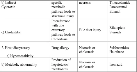
[image:42.612.68.547.581.711.2]2.11.2. Classification of hepatotoxins:
Table 1. Classification of hepatotoxins
Category of agent Mechanism Histological
Lesion
Examples
1. Intrinsic toxicity
a) Direct
Membrane injury destruction of structural basis of cell
metabolism
Necrosis and / or steatosis
CCl4, CHCl3, Phosphorus
b) Indirect Cytotoxic
specific metabolic
pathway leads to structural injury
necrosis Thioacetamide
Paracetamol Ethanol c) Cholestatic Intereference with bile excretory
pathway leads to Cholestasis
Bile duct injury Rifampicin
Steroids
2. Host idiosyncrasy
a) Hypersensitivity
Drug allergy Necrosis or
cholestasis
Sulfonamides Halothane
b) Metabolic abnormality
Production of hepatotoxic metabolites
Necrosis or
cholestasis Isoniazid
[image:43.612.67.538.71.321.2]Direct hepatotoxins and their effects:
Table 2. Direct hepatotoxins and its effects
Name Morphological alterations
Carbon tetrachloride Decreases glycogen and protein levels and
increases the content of lipid
the protein and lipid content
Thioacetamide Decreases the glycogen and protein levels
without effecting any significant change in
lipid level
Paracetamol Decreases the liver glycogen and protein
contents severely and elevates lipid level
Galactosamine Decreases the content of glycogen and protein
with marked elevation in lipid profile
Fulvine Produces edema and congestion, damaging
effect of the parenchyma
Phalloidin Damages plasma membrane of the hepatocytes,
as well as their active filaments
Ethyl alcohol Causes hepatocyte degeneration, collagen
deposition and necrosis
Alfa toxins Thymidine incorporation into DNA of
regenerating liver, synthesis of RNA
Lanthanum chloride Elevates the level of lipid , protein, α- and β
globulinsin liver and decreases albumin in
Indirect hepatotoxins:
Drug Class of agent
Methyl testosterone Anabolic steroid
Methimazole Antithyroid
Erythromycin estolate Chemotherapeutic
Norethynoderal with mestranol Oral contraceptive
Chlorpropamide Oral contraceptive
Chlorpromazine Tranquilizer
Tetracycline Chemotherapeutic
Valporic acid Anticonvulsant
Halothane Anesthitic
Phenytoin Anticonvulsant
Methyl dopa Antihypertensive
Isoniazid Chemotherapeutic
Chlorthiazide Diuretic
Oxyphenisatin Laxative
Paracetamol Analgesic
Phenylbutazone Antiinflammatory
Sulfonamides Chemotherapeutic
Allopurinol Xanthine oxidase inhibitor
Rifampicin Antitubercular
Mimosa pudica
Polygala chinensis (polygalaceae) Teucriumchamaedrys (Labiatae)
Anti inflammatory Sedative
Atiobesity
Piper methysticum (Piperaceae) Antiaxiety, Antipsychotic
Callilepsislaureola (Asteraceae) Anthelmentic, Antitussive, Antiimpotence
Symphytumofficinale (Boraginaceae) Antigastrointestinal ailments
Tussilagofarfara (Asteraceae) Treatment of lung and gastric diseases
Lycopodiumserratum (Lycopodiaceae) Analgesic
Tripterygiumwilfordii (Celasteraceae) Male contraceptive
Alcohol induced toxicity:
Chronic excessive use of alcohol
Continues drinking
Fatty liver Liver Fibrosis
Continues
CURED Continues drinking episode drinking
Abstinence
Alcoholic hepatitis Liver cirrhosis
DEATH Hepatocellular carcinoma
[image:46.612.74.541.95.516.2]
Fig : Schematic representation of Alcoholic liver diseases and progression process
Alcohol is metabolized by liver. This process produces a number of potentially dangerous
byproduct.
Alcohol is converted to acetaldehyde by the enzyme Alcohol dehydrogenase (ADH).The formed
acetaldehyde is highly toxic. Normally the enzyme Aldehyde dehydrogenase (ALDH) rapidly
oxidizes acetaldehyde to acetate. Both these enzymes ADH and ALDH are also involved in
metabolism of vitamin A.
Apart from ADH, the enzyme CYP2E1 ( microsomal ethanol oxidizing system; MEOS) is also
CYP2E1 produces a toxic byproduct N-acetyl-p-benzo-quinone imine (NAPQI) which is
[image:47.612.72.540.108.434.2]responsible for damaging the hepatic protein
Fig No 8: Liver disease
Paracetamol induced liver toxicity:
Paracetamol is a non-steroidal anti-inflammatory drug which is available as OTC(over the
counter) drug. The caution of this acetaminophen (Paracetamol) is its active metabolite is injury
to liver (i.e) leads to liver damage
Normal dose of the drug- 4000mg per day (Maximum) (Franciscus A;2012)
Paracetamol
Glucornosyltransfarase Cytochrome P
Sulfotransfarase
N – acetyl p-benzoquioneimine
( NAPQI)
Sulfate and glucuronide Glutachione
Conjunction
Conjunction Covalent binding liberation of to
hepatic proteins free radicals
Detoxification Adduct formation Lipid
peroxidation
Eliminated from the body
TOXICITY
The active metabolite of acetaminophen is N-acetyl p-benzoquinoneimine(NAPQI)This NAPQI
sulfate conjucation and then excreted in the urine. 5-10% is metabolized by cytochrome P450,
mainly by CYP2E1 which produces NAPQI.
CCl4 induced toxicity:
CC14
Smooth endoplasmic reticulum
CYP2E1, CYP2B1, CYP2B2, CYP3A
CC13
Oxygen (O2)
CC13 O2
Lipid per oxidation
Endoplasmic reticulum Soluble lipid peroxides
Damage
Damage to
Decreased apportion membrane
Fa