JOURNAL OFVIROLOGY, Oct. 2005, p. 12592–12596 Vol. 79, No. 19 0022-538X/05/$08.00⫹0 doi:10.1128/JVI.79.19.12592–12596.2005
Copyright © 2005, American Society for Microbiology. All Rights Reserved.
Conversion of Mouse Mammary Tumor Virus to a
Lymphomagenic Virus
Sanchita Bhadra, Mary M. Lozano, and Jaquelin P. Dudley*
Section of Molecular Genetics and Microbiology and Institute for Cellular and Molecular Biology, The University of Texas at Austin, Austin, Texas 78712
Received 29 April 2005/Accepted 5 July 2005
Type B leukemogenic virus is a variant of mouse mammary tumor virus (MMTV) that causes thymic lymphomas rather than mammary tumors in mice. We demonstrate that conversion of a mammotropic MMTV to a T-cell-tropic virus requires two alterations in the long terminal repeat: (i) acquisition of a T-cell-specific enhancer and (ii) loss of transcriptional repression through deletion of negative regulatory elements (NREs) or by suppression of NRE activity after appropriate positioning of the enhancer.
Mouse mammary tumor virus (MMTV) is an oncogenic retrovirus that induces mammary tumors in mice (8), whereas a T-cell-tropic variant, type B leukemogenic virus (TBLV), causes thymic lymphomas (1). Compared to the MMTV long terminal repeat (LTR), the TBLV LTR has a 443-bp deletion that eliminates a negative regulatory element (NRE) (2, 3, 9–11) and a 124-bp substitution that generates a triplicated element, which serves as a T-cell enhancer (12). The deletion and substitution truncate the superantigen (sag) gene (2) to eliminate the C-terminal interaction with the T-cell receptor (TCR) (22, 24). All T-cell-tropic MMTV variants isolated from thymic lymphomas harbor partial or complete NRE deletions, and some reportedly also contain T-cell enhancers (7, 13, 21). The MMTV NRE has been shown to repress viral expression in a number of cell types, including lymphoid cells (3, 11, 19, 25), and mutation of a SATB1-binding site within the NRE increases MMTV LTR-reporter gene expression in the lym-phoid tissues of transgenic mice relative to that in the mam-mary gland (3, 11).
To determine the contribution of the MMTV LTR to viral disease specificity, various mutant proviruses have been con-structed by our laboratory and others. Viruses produced using an MMTV molecular clone, HYB-MTV (20) (Fig. 1A), which contained the entire LTR from thymotropic MMTVs MA and DL8 (23) or a 440-bp segment (encompassing the deleted and substituted region) of the TBLV LTR (14) induced thymic lymphomas instead of mammary tumors. These studies
sug-gested thatcis-acting LTR elements determine viral
tumorige-nicity since TBLV Sag is dispensable for lymphomagenesis in BALB/cJ mice (14). To distinguish the relative contributions of the NRE and the T-cell enhancer to MMTV-induced disease, we analyzed additional mutants.
The TBLV T-cell enhancer masks NRE-mediated transcrip-tional repression. Two different MMTV LTRs were engi-neered by insertion of the TBLV T-cell enhancer (E) at the StuI site upstream of the NRE (MTVStuE LTR), similar to its
position in the TBLV LTR (2), or at the AflII site downstream of the NRE (MTVAflE LTR) (Fig. 1B). In both cases, the
NRE remained intact. Enhancer insertion truncated thesag
gene in the MTVStuE LTR but not the MTVAflE LTR. The
third mutant LTR, MTV⌬NRE, has been described previously
(12) and lacks the entire NRE and the flanking T-cell-en-hancer sequences (Fig. 1B).
The mutant LTRs were linked to the firefly luciferase gene and used in transient-transfection assays of human Jurkat T cells. Wild-type TBLV and MTVAflE LTRs demonstrated similar transcriptional strengths that were ca. 700-fold higher than that of the MMTV LTR (Fig. 1C). As previously
dem-onstrated, deletion of the NRE alone (MTV⌬NRE) resulted
in a 3-fold increase in reporter gene activity (12) (data not shown), whereas insertion of the TBLV T-cell enhancer up-stream of the NRE (MTVStuE) resulted in a 250-fold eleva-tion of luciferase activity (Fig. 1C). These results indicate that the placement of the TBLV enhancer between the MMTV NRE and the LTR promoter abolishes NRE activity in Jurkat T cells.
Since the TBLV enhancer contains a triplication of the hor-mone-responsive element (12), we also analyzed the transcrip-tional activity of the mutant LTRs in mouse mammary cells expressing functional glucocorticoid receptors. All LTRs,
ex-cept for that of MTV⌬NRE, had statistically similar
transcrip-tional efficiencies after transient transfections of HC11 cells grown in the absence of a glucocorticoid, dexamethasone (DEX), suggesting that T-cell enhancer addition (12) con-ferred no transcriptional advantage in mammary cells (Fig. 1D). As anticipated, the wild-type MMTV LTR gave a fivefold increase in reporter activity in the presence of DEX.
Glucocor-ticoid induction of the MTV⌬NRE LTR was not statistically
significant (P⬎0.25), presumably because NRE deletion
ele-vates basal MMTV transcription approximately fourfold (Fig. 1D). Enhanced glucocorticoid induction of the TBLV, MTVAflE, and MTVStuE LTRs may be attributed to altered spacing and triplication of a hormone-responsive element within the TBLV enhancer (2). However, the overall expres-sion from the MTVStuE LTR appeared to be diminished com-pared to that from the MTVAflE LTR, both of which con-tained the NRE, suggesting that the NRE may serve as a position-dependent insulator (4). Placement of the TBLV
en-* Corresponding author. Mailing address: Section of Molecular Ge-netics and Microbiology, The University of Texas at Austin, One Uni-versity Station, A5000, 24th Street and Speedway, ESB 226, Austin, TX 78712-0162. Phone: (512) 471-8415. Fax: (512) 471-7088. E-mail: [email protected].
12592
on November 8, 2019 by guest
http://jvi.asm.org/
hancer downstream of the NRE (MTVAflE) may provide the same effect as NRE deletion (TBLV).
Lymphomagenic MMTVs require NRE loss and T-cell en-hancer acquisition. AvrII-to-SstI segments of the mutant MMTV LTRs were replaced in the corresponding region of
the HYB-MTV 3⬘LTR (Fig. 1A). Jurkat T cells stably
express-ing wild-type (Jurkat/HYB-MTV) or mutant viruses (Jurkat/ HYB-MTVStuE, MTVAflE, and
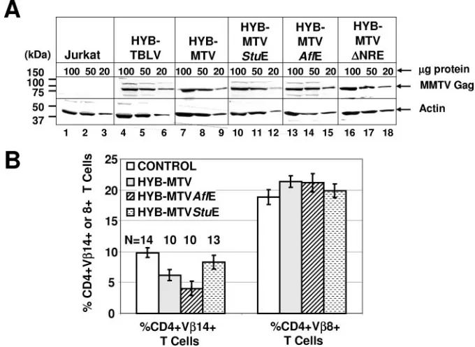
Jurkat/HYB-MTV⌬NRE) were isolated. Comparable MMTV Gag
expression levels of transfected cells were verified by Western
blotting (Fig. 2A) prior to intraperitoneal inoculation of 2⫻
107 cells into weanling BALB/cJ mice. Sag-reactive CD4⫹
V14⫹T cells were deleted at 3 months postinoculation with
Jurkat/HYB-MTV and Jurkat/HYB-MTVAflE (Fig. 2B),
while nonreactive CD4⫹ V8⫹ T cells remained unaffected.
The significantly faster T-cell deletion in HYB-MTVAflE-in-fected mice might be due to the higher viral loads resulting from enhanced viral transcription and replication in T cells (Fig. 1C). Deletion of Sag-reactive T cells was not detectable in mice injected with HYB-MTVStuE transfectants, consistent
with our previous data that truncation of Sag by more than a few amino acids is incompatible with T-cell deletion (22).
HYB-MTV induced mammary tumors with an incidence of
100% and an average latency of 7.3 ⫾ 1.7 months.
HYB-MTV⌬NRE also induced only mammary tumors in 20% of the
inoculated female mice within 10⫾2.8 months, indicating that
NRE deletion alone is insufficient for lymphomagenesis. Our previous data indicated that HYB-TBLV induced exclusively T-cell lymphomas, with an incidence of 50% and an average
latency of 5.7 ⫾ 1.5 months (14). However, 50% of
HYB-MTVStuE-infected mice developed mammary tumors within
8.7⫾2.1 months, whereas 30% developed thymic lymphomas
within 8 ⫾ 2.6 months. One animal infected with
HYB-MTVStuE (mouse 4) developed both tumor types. HYB-MTVAflE infection induced a 17% incidence of mam-mary tumors and a 22% incidence of thymic lymphomas, with
latencies of 6 months and 6.8 ⫾ 1.1 months, respectively.
[image:2.585.113.469.65.342.2]Kaplan-Meier plots followed by Wilcoxon or log rank tests indicated that the mammary tumor incidence for each of the mutants was significantly different than that for wild-type
FIG. 1. Construction and characterization of mutant MMTV LTRs. (A) Diagram of the hybrid proviral clone of MMTV, HYB-MTV. The 5⬘ portion of the provirus, including the LTR, is derived from the endogenous MMTV strainMtv1 (white), while the 3⬘portion, including the LTR, is derived from the exogenous C3H MMTV (gray). The coiled extension at the 3⬘end of the provirus denotes the flanking cellular DNA at the site of proviral integration. The recognition sites of restriction enzymes used in this study are also shown. (B) Diagram of the MMTV, TBLV, and mutant MMTV LTRs. The arrow on the MMTV LTR shows the transcription start site at the U3-R border.sagcoding potential is shown as a black
(full-length Sag) or gray (truncated Sag) box under each LTR. (C) Transcriptional efficiencies of wild-type MMTV, mutant MMTV, and TBLV LTRs in transient-transfection assays of Jurkat T cells. Luciferase (LUC) activity is reported in light units normalized for DNA uptake as measured by cotransfection with theRenillaluciferase expression plasmid, pRL-TK. LUC activities from the TBLV and mutant MMTV LTRs are depicted relative to that from the MMTV LTR (assigned a value of 1). Standard deviations from the means of triplicate assays are shown. (D) Transcrip-tional efficiencies of wild-type MMTV, mutant MMTV, and TBLV LTRs in HC11 mouse mammary cells. HC11 cells were grown in the absence (white bars) or presence (gray bars) of 10⫺6M DEX for 24 h prior to assays. LUC activity is reported in light units normalized for DNA uptake
as measured by cotransfection with the pRL-TK reporter plasmid. LUC activities from the TBLV and mutant MMTV LTRs are depicted relative to that from the MMTV LTR in the absence of DEX (assigned a value of 1). Standard deviations from the means of triplicate assays are shown. Significance was determined by Student’sttest.
on November 8, 2019 by guest
http://jvi.asm.org/
HYB-MTV (Fig. 3A), but the difference among the various mutants was not statistically significant. Lymphomas have never been observed after infection with HYB-MTV (Fig. 3B), even after the inoculation of much larger numbers of animals (22, 26). The lower incidence of mammary and T-cell tumors induced by MTVStuE viruses may be due to a suboptimal
arrangement ofcis-acting sequences in the LTRs, leading to
lower transcriptional activity (Fig. 1C) and favoring recombi-nation with endogenous MMTVs. The lower incidence and latency of tumors induced by MTVAflE did not lead to lower transcriptional activity or recombination with endogenous MMTVs; however, Sag-mediated deletion by this virus was significantly faster statistically than that by HYB-MTV (Fig. 2B). Thus, altered tumorigenesis by MTVAflE might result from the deletion of T cells required for dissemination of the virus or tumor targets, as suggested by Papiernik et al. (16).
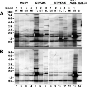
To determine the LTR structure of newly integrated provi-ruses, PstI-digested tumor DNA was analyzed by Southern blotting using probes specific for either the MMTV LTR (Fig. 4A) or the NRE (Fig. 4B). Most integrated proviruses in HYB-MTVAflE-induced lymphomas contained the NRE (Fig. 4A and B, lanes 5 and 7). Furthermore, these LTRs retained their original size, indicative of an intact T-cell enhancer with a 62-bp triplication. Shorter LTRs might harbor fewer enhancer
copies or a partial deletion of the NRE. Growth of TBLV-induced lymphomas appears to select proviral integrations with suboptimal numbers of LTR enhancer repeats to modu-late oncogene expression (6). LTRs of predominant acquired proviruses in HYB-MTVStuE-induced lymphomas lacked the entire NRE and were detected only by the MMTV LTR probe (Fig. 4A and B, lanes 10 and 11). The reduced size of these LTRs is consistent with complete NRE deletion and retention of the T-cell enhancer. NRE-lacking recombinants were not observed in HYB-MTVStuE-induced mammary cancer (Fig. 4A and B, lanes 8, 9, and 12), even in an animal that developed both tumor types (compare lanes 11 and 12). The presence of the T-cell enhancer in lymphomagenic HYB-MTVAflE and HYB-MTVStuE viruses was verified by reverse transcription-PCR, cloning, and sequencing (data not shown).
[image:3.585.121.461.70.318.2]These results suggest that two events are required for MMTV-induced lymphomagenesis: loss of NRE-mediated transcriptional repression and acquisition of a T-cell enhancer. Selection of proviruses lacking the NRE in HYB-MTVStuE-induced lymphomas may be driven by the incomplete suppres-sion of NRE activity by the upstream T-cell enhancer, as ob-served in transient-transfection assays in T cells. HYB-MTVAflE is not subject to this selection pressure, since the T-cell enhancer located downstream of the NRE appears to
FIG. 2. Protein expression and Sag-mediated deletion induced by wild-type MMTV and mutant viruses. (A) Western blot analysis comparing viral Gag expression levels in Jurkat T cells expressing wild-type (Jurkat/HYB-MTV) or mutant (Jurkat/HYB-MTVStuE, Jurkat/HYB-MTVAflE, and Jurkat/HYB-MTV⌬NRE) MMTV proviruses. Stably transfected Jurkat cells were derived by electroporation with the appropriate proviral constructs carrying the hygromycin resistance cassette, followed by growth selection in 250g/ml hygromycin. Three different amounts (100, 50, and 20g) of whole-cell lysates were analyzed on 8% sodium dodecyl sulfate-containing polyacrylamide gels. The Gag precursor (arrow) was detected with an MMTV capsid-specific monoclonal antibody (17) using a protocol described previously (15). The same amounts of cellular lysates were incubated with antibodies specific for actin as a control for protein loading. (B) Flow cytometric analysis of Sag-mediated peripheral deletion of T cells. Injected mice were analyzed at 3 months after injection with Jurkat T cells stably expressing wild-type or mutant MMTV proviruses. CD4⫹V14⫹and CD4⫹V8⫹peripheral T cells were detected using phycoerythrin-conjugated anti-mouse CD4 monoclonal antibody (RM4-5) and fluorescein-conjugated anti-mouse TCR V14 (14-2) or V8.1 and 8.2 (MR5-2) monoclonal antibodies from Pharmingen (San Diego, Calif.). The cells were analyzed using a FACSCalibur (Becton Dickinson, Franklin Lakes, N.J.) and CELLQuest software. The percentages of TCR V⫹ cells in the gated CD4⫹T-cell populations were calculated. The number of animals (N) analyzed from each group is indicated above each bar. The mean percentage (⫾standard deviation) of CD4⫹TCR V⫹T cells in each group is shown.
12594 NOTES J. VIROL.
on November 8, 2019 by guest
http://jvi.asm.org/
FIG. 3. Kaplan-Meier plots of incidence of mammary tumors and thymic lymphomas induced by wild-type and mutant MMTV proviruses. Female mice were caged to maintain continuous breeding and lactation. Mice that died from nontumor causes (e.g., birthing problems) before the average latency of tumor induction were excluded from the study. (A) Development of mammary tumors. Numbers in parentheses indicate the numbers of animals with tumors/total numbers of injected females. The circles indicate one or more animals that died without any detectable tumors. (B) Development of thymic lymphomas. Numbers in parentheses indicate numbers of animals with tumors/total numbers of injected males and females. One mouse injected with HYB-MTVStuE developed both a mammary tumor and a thymic lymphoma after 11 months of latency.
FIG. 4. LTR structure of acquired proviruses in wild-type and mutant MMTV-induced mammary tumors (MT) and thymic lymphomas (TL). (A) Southern blotting of tumor DNA using the full-length MMTV LTR probe. (B) Southern blotting of tumor DNA with the MMTV NRE probe (StuI-to-AflII fragment of the MMTV LTR). Genomic DNA was digested with PstI, separated on 0.8% agarose gels, transferred to nitrocellulose, and hybridized as previously described (9). Southern blotting of genomic DNA from an uninfected BALB/cJ mouse liver (LI) was used to distinguish the bands originating from the three endogenousMtvs in the BALB/cJ strain. White asterisks (lanes 5, 7, 10, 11, and 13) indicate the LTR fragments of predominant acquired proviruses in virus-induced tumors.
12595
on November 8, 2019 by guest
[image:4.585.142.439.373.671.2]mask transcriptional repression. The ability of HYB-MTVAflE to induce lymphomas also indicates that Sag truncation is dis-pensable for this process, in agreement with our previous data (14). Interestingly, the LTRs of acquired proviruses in the
HYB-MTV⌬NRE-induced mammary tumor had reacquired
the NRE, suggesting that the presence of this region might favor mammary tumorigenesis (Fig. 4A and B, lane 13).
Fur-thermore, the low tumor incidence of HYB-MTV⌬NRE might
be dependent on the appearance of such recombinants. In conclusion, development of lymphomagenic MMTVs re-quires alterations of transcriptional regulatory elements in the LTR. Loss of NRE activity and T-cell enhancer acquisition allow increased viral expression in T cells, resulting in a higher probability of insertional mutagenesis. The viral enhancer also may be critical for transcriptional upregulation of selected
cellular oncogenes, such as c-mycandrorc, in T cells (5, 6, 18).
We acknowledge helpful suggestions from the Dudley lab. We also acknowledge support from NIH grants P01 CA77760 and R01 CA34780.
REFERENCES
1.Ball, J. K., and G. A. Dekaban.1987. Characterization of early molecular biological events associated with thymic lymphoma induction following in-fection with a thymotropic type-B retrovirus. Virology161:357–365. 2.Ball, J. K., H. Diggelmann, G. A. Dekaban, G. F. Grossi, R. Semmler, P. A.
Waight, and R. F. Fletcher.1988. Alterations in the U3region of the long
terminal repeat of an infectious thymotropic type B retrovirus. J. Virol. 62:2985–2993.
3.Bramblett, D., C.-L. L. Hsu, M. Lozano, K. Earnest, C. Fabritius, and J. Dudley.1995. A redundant nuclear protein binding site contributes to neg-ative regulation of the mouse mammary tumor virus long terminal repeat. J. Virol.69:7868–7876.
4.Brasset, E., and C. Vaury.2005. Insulators are fundamental components of the eukaryotic genomes. Heredity94:571–576.
5.Broussard, D. R., M. M. Lozano, and J. P. Dudley.2004. Ror␥(Rorc) is a common integration site in type B leukemogenic virus-induced T-cell lym-phomas. J. Virol.78:4943–4946.
6.Broussard, D. R., J. A. Mertz, M. Lozano, and J. P. Dudley.2002. Selection for c-mycintegration sites in polyclonal T-cell lymphomas. J. Virol.76:2087– 2099.
7.Dudley, J., and R. Risser.1984. Amplification and novel locations of endog-enous mouse mammary tumor virus genomes in mouse T-cell lymphomas. J. Virol.49:92–101.
8.Dudley, J. P.1999. Mouse mammary tumor virus, p. 965–972.InR. G. Webster and A. Granoff (ed.), Encyclopedia of virology. Academic Press Ltd., London, United Kingdom.
9.Hsu, C.-L. L., C. Fabritius, and J. Dudley.1988. Mouse mammary tumor virus proviruses in T-cell lymphomas lack a negative regulatory element in the long terminal repeat. J. Virol.62:4644–4652.
10.Lee, J. W., P. G. Moffitt, K. L. Morley, and D. O. Peterson.1991. Multipartite
structure of a negative regulatory element associated with a steroid hor-mone-inducible promoter. J. Biol. Chem.266:24101–24108.
11.Liu, J., D. Bramblett, Q. Zhu, M. Lozano, R. Kobayashi, S. R. Ross, and J. P. Dudley.1997. The matrix attachment region-binding protein SATB1 partic-ipates in negative regulation of tissue-specific gene expression. Mol. Cell. Biol.17:5275–5287.
12.Mertz, J. A., F. Mustafa, S. Meyers, and J. P. Dudley.2001. Type B leuke-mogenic virus has a T-cell-specific enhancer that binds AML-1. J. Virol. 75:2174–2184.
13.Michalides, R., E. Wagenaar, and P. Weijers.1985. Rearrangements in the long terminal repeat of extra mouse mammary tumor proviruses in T-cell leukemias of mouse strain GR result in a novel enhancer-like structure. Mol. Cell. Biol.5:823–830.
14.Mustafa, F., S. Bhadra, D. Johnston, M. Lozano, and J. P. Dudley.2003. The type B leukemogenic virus truncated superantigen is dispensable for T-cell lymphomagenesis. J. Virol.77:3866–3870.
15.Mustafa, F., M. Lozano, and J. P. Dudley.2000. C3H mouse mammary tumor virus superantigen function requires a splice donor site in the enve-lope gene. J. Virol.74:9431–9440.
16.Papiernik, M., A. C. Wache, C. Pontoux, and B. Nabarra.1997. Massive mammary gland infection and pregnancy-dependent mammary tumor devel-opment in mice infected neonatally with mouse mammary tumor virus (SW) but not in mice infected as adults, despite a dramatic local response. Eur. J. Immunol.27:2145–2151.
17.Purdy, A., L. Case, M. Duvall, M. Overstrom-Coleman, N. Monnier, A. Chervonsky, and T. Golovkina.2003. Unique resistance of I/LnJ mice to a retrovirus is due to sustained interferon gamma-dependent production of virus-neutralizing antibodies. J. Exp. Med.197:233–243.
18.Rajan, L., D. Broussard, M. Lozano, C. G. Lee, C. A. Kozak, and J. P. Dudley.2000. The c-myc locus is a common integration site in type B retrovirus-induced T-cell lymphomas. J. Virol.74:2466–2471.
19.Ross, S. R., C.-L. L. Hsu, Y. Choi, E. Mok, and J. P. Dudley.1990. Negative regulation in correct tissue-specific expression of mouse mammary tumor virus in transgenic mice. Mol. Cell. Biol.10:5822–5829.
20.Shackleford, G. M., and H. E. Varmus.1988. Construction of a clonable, infectious, and tumorigenic mouse mammary tumor virus provirus and a derivative genetic vector. Proc. Natl. Acad. Sci. USA85:9655–9659. 21.Theunissen, H. J. M., M. Paardekooper, L. J. Maduro, R. J. A. M.
Micha-lides, and R. Nusse.1989. Phorbol ester-inducible T-cell-specific expression of variant mouse mammary tumor virus long terminal repeats. J. Virol. 63:3466–3471.
22.Wrona, T. J., M. Lozano, A. A. Binhazim, and J. P. Dudley.1998. Mutational and functional analysis of the C-terminal region of the C3H mouse mam-mary tumor virus superantigen. J. Virol.72:4746–4755.
23.Yanagawa, S.-I., K. Kakimi, H. Tanaka, A. Murakami, Y. Nakagawa, Y. Kubo, Y. Yamada, H. Hiai, K. Kuribayashi, T. Masuda, and A. Ishimoto. 1993. Mouse mammary tumor virus with rearranged long terminal repeats causes murine lymphomas. J. Virol.67:112–118.
24.Yazdanbakhsh, K., C. G. Park, G. M. Winslow, and Y. Choi.1993. Direct evidence for the role of COOH terminus of mouse mammary tumor virus superantigen in determining T cell receptor Vspecificity. J. Exp. Med. 178:737–741.
25.Zhu, Q., K. Gregg, M. Lozano, J. Liu, and J. P. Dudley.2000. CDP is a repressor of mouse mammary tumor virus expression in the mammary gland. J. Virol.74:6348–6357.
26.Zhu, Q., U. Maitra, D. Johnston, M. Lozano, and J. P. Dudley.2004. The homeodomain protein CDP regulates mammary-specific gene transcription and tumorigenesis. Mol. Cell. Biol.24:4810–4823.
12596 NOTES J. VIROL.