THE TAMILNADU DR.M.G.R. MEDICAL UNIVERSITY
DISSERTATION ON
A PROSPECTIVE COMPARATIVE STUDY ON
LAPAROSCOPIC CHOLECYSTECTOMY VS OPEN
CHOLECYSTECTOMY
submitted for
In partial fulfillment of the requirements for the degree of
M.S. GENERAL SURGERY – BRANCH I
DEPARTMENT OF GENERAL SURGERY
THANJAVUR MEDICAL COLLEGE AND HOSPITAL
A PROSPECTIVE COMPARATIVE STUDY ON
LAPAROSCOPIC CHOLECYSTECTOMY VS OPEN
CHOLECYSTECTOMY
BY
DR.SRINATH. R
Dissertation submitted to the
THE TAMILNADU DR. M. G. R. MEDICAL UNIVERSITY
In partial fulfillment of the requirements for the degree of
M.S. GENERAL SURGERY – BRANCH I
Under the guidance of
Prof. Dr.V.KOPPERUNDEVI M.S.,
DEPARTMENT OF GENERAL SURGERY
THANJAVUR MEDICAL COLLEGE AND HOSPITAL
DECLARATION BY THE CANDIDATE
I solemnly declare that this Dissertation “A PROSPECTIVE
COMPARATIVE STUDY ON LAPAROSCOPIC CHOLECYSTECTOMY VS OPEN CHOLECYSTECTOMY”was done by me in the Department of General Surgery, Thanjavur Medical College, and Hospital, Thanjavur under the Guidance and
Supervision of my Professor Dr.V.KOPPERUNDEVI M.S. Department of General
Surgery, Thanjavur Medical College, Thanjavur between 2016 and 2017.
This Dissertation is submitted to the Tamilnadu Dr. M.G.R Medical University,
Chennai in partial fulfillment of University requirements for the award of M.S Degree
(GENERAL SURGERY).
DR.SRINATH. R
Postgraduate Student,
Thanjavur Medical College.
CERTIFICATE BY THE GUIDE
This is to certify that this dissertation titled “A PROSPECTIVE
COMPARATIVE STUDY ON LAPAROSCOPIC CHOLECYSTECTOMY VS OPEN CHOLECYSTECTOMY” is a bonafide research work done by
Dr.SRINATH. R in partial fulfillment of the requirement for the degree of
M.S.GENERAL SURGERY – BRANCH I.
Dr.V.KOPPERUNDEVI M.S., Associate Professor
Dept. of General Surgery
Thanjavur Medical College and Hospital
(Guide)
Date:
CERTIFICATE
This is to certify that the dissertation titled “A PROSPECTIVE
COMPARATIVE STUDY ON LAPAROSCOPIC CHOLECYSTECTOMY VS OPEN CHOLECYSTECTOMY” is a bonafide research work done by
DR.SRINATH. R under the guidance of Dr.V.KOPPERUNDEVI M.S., ( Associate Professor, Department of General Surgery) Thanjavur Medical College Hospital,
Thanjavur.
Dr.M.ELANGOVAN M.S., The Dean, Professor and HOD,
ACKNOWLEDGEMENT
I am grateful to Dr. JEYAKUMAR M.S.,M.CH., Dean for giving me permission and opportunity to conduct study and data collection at Thanjavur Medical
College and Hospital.
I am deeply grateful to my professor and Head of the Department of General
Surgery, Prof. Dr. ELANGOVAN.M. M.S., for his encouragement and suggestions in preparing this work.
I owe my sincere and grateful acknowledgement to my beloved chief, teacher
and guide Prof. Dr.V.KOPPERUNDEVI M.S., Associate Professor of General Surgery who inspired me to take this topic of “A PROSPECTIVE
COMPARATIVE STUDY ON LAPAROSCOPIC CHOLECYSTECTOMY VS OPEN CHOLECYSTECTOMY”. I extend my grateful acknowledgement to my
teachers, Dr.V.PANDIYAN M.S., Asst. Prof. of General Surgery,Dr.THIVAGAR M.S., Asst. Prof. of General Surgery, Dr.SARAVANAN M.S., Asst. Prof of general surgery in guiding me to complete my dissertation.
I express my gratitude and sincere acknowledgement for the help and support
extended to me by the radiologist, my seniors, junior PGs, house surgeons and all
laboratory members. I wish to express my whole hearted thanks to all the patients who
participated in the study. Completion of this work would not have been possible
without their co-operation.
CONTENTS
1. INTRODUCTION
2. AIM OF THE STUDY
3. MATERIALS AND METHODS
4. REVIEW OF LITERATURE
5. RESULTS
6. CONCLUSION
7. PROFORMA
8. BIBLIOGRAPHY
1
A PROSPECTIVE COMPARATIVE STUDY ON LAPAROSCOPIC CHOLECYSTECTOMY VS OPEN CHOLECYSTECTOMY
INTRODUCTION
Gall stone disease is common in India, that too affecting mostly middle
age group men and women.The classical open cholecystectomy and the
minimally invasive laparoscopic cholecystectomy are two alternative
operations for removal of the gallbladder. There are no significant differences
in mortality and complications between the laparoscopic and the open
techniques. The laparoscopic operation has advantages over the open operation
regarding duration of hospital stay and convalescence. With the advent of
laparoscopic cholecystectomy the scenario of surgical management of
cholelithiasis has changed drastically. It has opened new horizons in the
management of gallstones. Theoretical benefits of laparoscopic approach
include reduced hospitalization and cost, decreased pain, avoidance of large
incision with improved cosmetic purpose and reduced post-operative recovery
time with an early return to work.
Although it showed early promising results, recent trials show an
increase in the incidence of operative complications, especially common bile
duct injury. Expensive instruments, specialized training and long learning
curve also limit the use of laparoscopy. This has led to a lot of soul searching
2
vsopen cholecystectomy. Recent upsurge in practice of laparoscopic surgery
and other form of minimal access surgery has ushered a new era of surgical
3
AIM OF THE STUDY
O To compare time taken for surgery.
O To compare and discuss the various operative complications in both the
surgery.
O To compare the postoperative complications.
O To compare postoperative hospital stay.
4
MATERIALS AND METHODS
My study is a prospective randomized study included 88 patients with
gallstones, who were admitted to Thanjavur medical college and hospital from
September 2016 to September 2017. The selection of procedure of open
cholecystectomy or laparoscopic was decided depending up on the inclusion
and exclusion criteria, exception when patient consent was not given for a
particular procedure. Information was collected from the patients after a written
valid informed consent from them. The study was approved by IEC. Patients
between 20 years to 60 years with acute cholecystitis, or chronic cholecystitis and gall stones without pain abdomen, were included in the study. Patient’s
written valid informed consent for the particular procedure was taken. Patients
less than 20 years and more than 60 years or those with Gall bladder cancer and
Choledocholithiasis were excluded. This study involved preoperative
assessment, intraoperative practice and post-operative management and follow
up till 3 months. All the patients were studied with reference to duration of
surgery, post-operative analgesia, post-operative stay and morbidity, intra
operative and post-operative complications.
Patients were admitted a day prior to surgery in case of elective
cholecystectomy from OPD after complete investigations performed required
for general anesthesia. Some patients were admitted from emergency
5
patients were investigated for the same. Investigations performed in these
patients include complete Haemogram, Blood sugar level, Urine examination,
Liver function test, Blood urea and serum creatinine, Chest x-ray, ECG
andUltrasonography of abdomen, CT abdomen. The patients were studied with
respect to their clinical presentation and were grouped as patients with
asymptomatic Gall stones, acute calculus cholecystitis and chronic calculus
cholecystitis.
After complete investigations and after satisfying the inclusion and
exclusion criteria for my study, patients were subjected to either open or
laparoscopic cholecystectomy. First dose of antibiotics administered to the
patient just prior to incision, immediately after intubation. General anesthesia was administered to all the patients. Foleys Catheterization and Ryle’s tube
insertion was done in all patients. Post-operative management included nil per
oral tillbowel sounds were heard. Intravenous fluids in the form of crystalloids,
Broad-spectrum antibiotics (Injcefotaxim) were administered. Injection
Amikacin and Injection Metronidazole were added in cases of bile leak.
Analgesics in the form of Injection Tramadol were given. Top-up analgesia in
the form of intramuscular Injection Diclofenac Sodium was given, whenever it
was required. Discharge after start of oral diet and without any signs of
postoperative wound infection after first dressing change. If sign of wound
infection were present then pus from wound was taken and sent for
6
after reports and wound care taken accordingly. Follow up in OPD for stitch
removal after 7 days, if operative wound is healthy. All laparoscopic
cholecystectomy converted to open cholecystectomy were considered as
7
REVIEW OF LITERATURE
GALLBLADDER ANATOMY
The gallbladder is a pear-shaped, hollow structure located under the
liver and on the right side of the abdomen. It has an inferior peritoneal surface
and a superior hepatic surface that is closely applied to the gallbladder bed in
the liver. The cystic plate is a condensation of fibro-areolar tissue that separates
the gallbladder from the liver parenchyma. Small bile ducts may drain from
liver parenchyma to the gallbladder through the cystic plate (ducts of Luschka),
this may result in bile leak and collection, even without an apparent bile duct
injury, during cholecystectomy. The part of the gallbladder projecting beyond
the undersurface of the liver is called the fundus, which continues into the main
body of the gallbladder, which lies in a fossa on the undersurface of the liver.
The body of the gallbladder narrows into an infundibulum, which leads through
the neck to the cystic duct. The cystic duct has spiral folds of mucosa called
"valves" of Heister. An inferior sacculation (outpouching) of the gallbladder
infundibulum or neck is sometimes present, this is called the Hartmann pouch.
The Calot's triangle is bounded by the cystic duct on the right, common hepatic
duct (CHD) on the left, and undersurface of the liver above; the cystic artery
and cystic lymph node lie in the Calot triangle. A peritoneal fold
(cholecysto-duodenal fold) connects the gallbladder neck to the first part of the duodenum.
The right and left hepatic ducts join outside the liver in its hilum (portahepatis)
8
form the common bile duct (CBD), which travels downward in the HDL along
with the hepatic artery to its right and the portal vein behind it. The upper limit
of the diameter of the normal CBD is 6-7 mm.
The CBD has supraduodenal, retroduodenal (behind the first part of the
duodenum), infra or retropancreatic (in a groove or sulcus behind or a tunnel
through the upper half of the head of the pancreas) and intraduodenal
(intramural) parts. The gallbladder is part of the biliary tract. The terminal part
of the CBD is joined by the terminal part of the pancreatic duct in the head of
pancreas to form a common channel (called the hepatopancreatic ampulla),
which runs through the medial duodenal wall and opens on the dome of the
greater duodenal papilla, on the medial wall of the middle segment of the
second part of the duodenum. A smooth muscle sphincter called p of Oddi is
present around the common channel of the CBD and the main pancreatic duct
and prevents reflux of duodenal juice into the 2 ducts. Two other individual
smooth muscle sphincters are present around the terminal parts of the CBD
(sphincter of Boyden) and the main pancreatic duct before they join; these
prevent reflux of pancreatic juice into the CBD and bile into the main
9
10
11 FUNCTION
The gallbladder serves as a reservoir for bile while it’s not being used
for digestion. The gallbladder's absorbent lining concentrates the stored bile.
When food enters the small intestine, a hormone called cholecystokinin is
released, signaling the gallbladder to contract and secrete bile into the small
intestine through the common bile duct.
The bile helps the digestive process by breaking up fats. It also drains
waste products from the liver into the duodenum, a part of the small intestine.
PRESENTATION OF GALLBALDDER DISEASE
BILIARY COLIC (intermittent duct blockage)/ABDOMINAL PAIN - The most common symptom of a gallbladder problem is pain. This pain usually occurs suddenly and rapidly increasing in the right upper abdomen or epigastric area. Some will have pain radiating to the right
shoulder (or back pain in the tip of the scapula) and also develop nausea
and vomiting. The pain usually subsides in about 1 to 5 hours although a
mild ache may persist for about a day. It can be mild and intermittent. It can be quite severe and frequent.
NAUSEA AND VOMITING - These are common symptoms of all
types of gallbladder problems. Chronic gallbladder disease may cause digestive problems, such as regurgitation or acid reflux, nausea and vomiting.
12
If you have an infection, you need treatment before it worsens and becomes very severe. The infection can become life-threatening if it spreads to other parts of the body.
CHRONIC DIARRHEA - Having more than four bowel movements
per day for at least three months may be a sign of chronic gallbladder disease.
JAUNDICE – it may be a sign of a common bile duct block or stone.
UNUSUAL STOOLS OR URINE - light colored and high colored
urine are possible signs of a common bile duct block.
Disease of gall bladder
CHOLELITHIASIS(Gallstones)- For unknown reasons, substances in
bile gets crystallize in the gallbladder, forming gallstones. It is common
and it isharmless,but gallstones can sometimes cause pain, nausea, or
inflammation.
Gall Stones
Types:
o Cholesterol gallstones
Cholesterol gallstones are made primarily of cholesterol.
They are the most common type of gallstone, comprising
80% of gallstones in individuals from Europe and the
Americas. Cholesterol is one of the substances that liver
13
Pigment gallstones
Pigment gallstones are the second most common type of gallstone.
o There are two types of pigment gallstones 1) black pigment
gallstones, and 2) brown pigment gallstones.
Black pigment gallstones: too much bilirubin in bile
Brown pigment gallstones: If there is reduced contraction
of the gallbladder or obstruction to the flow of bile
through the ducts, bacteria may ascend from the
duodenum into the bile ducts and gallbladder. The bacteria
alter the bilirubin in the ducts and gallbladder, and the
altered bilirubin then combines with calcium to form
pigment.
Pathogenesis for gall stone disease:
- metabolic
- infections and infestations
- bile stasis
- increased bilirubin production
- saints triad: gall stones,
diverticulosis of colon,hiatus hernia.
Factors affecting cholesterol to bile salts ratio:
14
- ocp
- ileal disease
- ileal resection
- alteredenterohepatic circulation
Gall stone colic:
Sudden , severe , colicky abdominal pain in right hypochondrium which
radiates to back and shoulder.
-tendernesss in right hypochondrium
-aggravated by lying position in night
-there is reflux pylorospasm causing vomiting.
MIRIZZI syndrome:
-gall stone gets impacted in the gall bladder wall and compress causing
pressure necrosis
- type 1: compression of CBD without lumen narrowing,
- type 2: compression causing lumen narrowing,
- type 3 : compression causing CBD wall necrosis
15
16
17
18
GALL STONE OBSTRUCTING NECK OF GB
A
c
a
l
c
u
l
o
19
20
CHOLECYSTITIS - Infection of the gallbladder, often due to a gallstone in
the gallbladder. Cholecystitis causes severe pain and fever, and can require
surgery when infection continues or recurs.
Acute cholecystitis:
- occurs following chronic cholecystitis
- usual cause is impacted gall stone in hartmanns pouch obstructing
cystic duct.
Types
Acute calculouscholecystitis
Acute acalculouscholecystitis
Complications:
perforation
Peritonitis
Pericholecystitis abscess
Empyema GB
Cholangitis
Septicemia
ACALCULOUS CHOLECYSTITIS
Acalculouscholecystitis is an inflammatory disease of the gallbladder
without evidence of gallstones or cystic duct obstruction. It is a severe illness
that is a complication of various other medical or surgical conditions. Duncan
21
complicating an incarcerated hernia was reported. The condition causes
approximately 5-10% of all cases of acute cholecystitis and is usually
associated with more serious morbidity and higher mortality rates than
calculouscholecystitis.
It is most commonly observed in the setting of very ill patients (eg, on
mechanical ventilation, with sepsis or severe burn injuries, after severe trauma
.In addition, acalculouscholecystitis is associated with a higher incidence of
gangrene and perforation compared to calculous disease.
The usual finding on imaging studies is a distended acalculous
gallbladder with thickened walls (>3-4 mm) with or without pericholecystic
fluid. Acalculouscholecystitis can be observed in patients with human
immunodeficiency virus (HIV) infection, although it is a late manifestation of
this disease. Acalculouscholecystitis can also be found in patients on total
parenteral nutrition (TPN), typically those on TPN for more than 3 months
PATHOPHYSIOLOGY:
The main cause of this illness is thought to be bile stasis and increased
lithogenicity of bile. Critically ill patients are more predisposed because of
increased bile viscosity due to fever and dehydration and because of prolonged
absence of oral feeding resulting in a decrease or absence of
cholecystokinin-induced gallbladder contraction. Gallbladder wall ischemia that occurs because
of a low-flow state due to fever, dehydration, or heart failure may also play a
22 ETIOLOGY
The main cause of acalculouscholecystitis is gallbladder stasis with
resulting stagnant bile. This is observed most commonly in patients with sepsis,
patients in intensive care units, patients on long-term total parenteral nutrition
(TPN), those with cardiovascular disease, patients with diabetes (occasionally),
or other patients with gallbladder dysmotility. The condition has been reported
during pregnancy, as a complication of hepatitis A.It has been rarely reported
in children, also as a complication of hepatitis A,with a favorable course with
conservative treatment. This disease has also been reported as associated with
aortic dissection.
PROGNOSIS
The prognosis of patients with acalculouscholecystitis is guarded. The
mortality and morbidity rates associated with acalculouscholecystitis can be
high. The illness is frequently observed in patients with sepsis or other serious
conditions. The reported mortality range is 10-50% for acalculouscholecystitis
as compared to 1% for calculouscholecystitis.
COMPLICATIONS
Perforation or gangrene of the gallbladder and extrabiliary abscess
23 CHRONIC CHOLECYSTITIS
CAUSES
This condition usually begins with the formation of gallstones in the gallbladder. Gallstones may depend on several factors, including:
genetic predisposition
weight
gallbladderinactivity
dietary habits
Gallstones form when substances in the bile form crystal-like particles. They can range from the size of a grain of sand to the size of a golf ball. The presence of gallstones causes pressure, irritation, and may cause infection. The walls of the gallbladder begin to thicken over time. Eventually, the gallbladder starts to shrink. These changes make it harder for the gallbladder to function properly.
In addition to gallstones, cholecystitis can be due to:
Infection of the CBD drainage system
CBD blockage
Excess cholesterol in the gallbladder, which can happen during pregnancy after rapid weight loss
Decreased blood supply to the gallbladder because of diabetes
Tumors in the liver or pancreas
24
Repeated or prolonged attacks of cholecystitis, it becomes a chronic condition.
SYMPTOMS
Symptoms of cholecystitis can appear suddenly or develop slowly over a period of years. Most of the time these symptoms appear after a meal that is high in fat.
Symptoms include:
Severe abdominal pains that may feel sharp or dull
Abdominal cramping and bloating
Pain radiates to back or below right shoulder blade
Fever
Chills
Nausea
Vomiting
Loose, light-colored stools
Jaundice
Itching
25
Complications can include:
Pancreatitis, an inflammation of the pancreas
Perforation of the gallbladder as a result of infection
Enlarged gallbladder due to inflammation
Infection may cause the bile to build up
Cancer of the gallbladder (this is a rare, long-term complication)
Death of gallbladder tissue (this can lead to a tear and ultimately a burst of the organ)
TREATMENT
The symptoms of cholecystitis can be treated at home with pain medication and rest, if have been properly diagnosed. Take antibiotics and avoid fatty foods.
Treatment options for cholecystitis
The specific cause for attack will determine the course of treatment. The options include:
broad-spectrum antibiotics for fighting infection
oral dissolution therapy using medications to help dissolve gallstones (this is typically a last resort, reserved for individuals who cannot undergo surgery)
pain relievers for controlling pain during treatment
26
surgery is an outpatient procedure, which means a shorter recovery time.
Emphysematous cholecystitis:
Emphysematous cholecystitis is a rare form of acute cholecystitis where
gallbladder wall necrosis causes gas formation in the lumen or wall. It is a
surgical emergency, due to the high mortality from gallbladder gangrene and
perforation.
EPIDEMIOLOGY
Men are affected twice as commonly as women (reverse is true in most
cases of acute cholecystitis).The majority of patients are between 50 and 70
years of age and have underlying diabetes mellitus.
CLINICAL PRESENTATION
Clinical manifestation is often insidious and may then progress rapidly. Up to
one-third of patients may be afebrile and localised tenderness is often not a
dominant clinical feature.
PATHOLOGY
Vascular compromise of the cystic artery is thought to play a significant role in
causing emphysematous cholecystitis. It is associated with
acalculouscholecystitis (present in ~50% of cases 9-11) and carries a higher
incidence of gallbladder perforation. Commonly isolated organisms include
Clostridium welchii/ perfringens, Escherichia coli and Bacteroidesfragilis.
RADIOGRAPHIC FEATURES
27
demonstration of air in the gallbladder wall +/- biliary ducts, in the absence of
an abnormal communication with thegastrointestinal tract .
ULTRASOUND
Ultrasonography may demonstrate highly echogenic reflectors with low-level
posterior shadowing and reverberation artifact ("dirty" shadowing and
"ring-down" artifact) .
A less common but more specific finding is small, non-shadowing echogenic
foci rising up from the dependent portions of the gallbladder lumen, similar to
effervescing bubbles in a glass of champagne (champagne sign).
CT
CT is considered the most sensitive and specific imaging modality for
identifying gas within the gallbladder lumen or wall . The presence of a
pneumoperitoneum indicates perforation.
NUCLEAR MEDICINE
Hepatobiliary nuclear imaging may demonstrate non-visualisation of the
gallbladder, along with a region of increased hepatic activity adjacent to the
gallbladder fossa. This feature is sometimes termed the rim sign.
Treatment and prognosis
Treatment is emergent surgical intervention. Overall mortality rate is ~20%
(range 15-25%), compared with <5% in uncomplicated cases of acute
28
29
30
ACUTE CHOLECYSTITIS IN PREGNANCY
In most cases, cholecystitis pregnancy may be due to the trapping of
gallstones at the opening of the gallbladder. When pregnant, there is a decrease
in gallbladder motility. There is also an increase in the cholesterol saturation of
bile. An infection in the bile can also cause an inflammation of the gall bladder.
The following conditions can also increase the risk of cholecystitis:
• Obesity or sudden weight gain (that can be the case in pregnancy)
• Sudden loss in weight (that can also be the case in certain pregnancies)
• Diabetes
• Crohn’s disease
• Hyperlipidemia
• Family history of gallstones, especially on the maternal side
SYMPTOMS
Symptoms of cholecystitis during pregnancy are overlooked, as they may be
similar to regular pregnancy aches. However, the pain can soon turn worse and
cause much discomfort.
Some of the common symptoms of cholecystitis are the following:
Pain in the upper right part of your abdomen. The pain can also
radiates towards back or right shoulder blade.
There may be persistent feeling of nausea or vomiting. As these two
31
and ignore it initially.
Fever
TREATMENT
Laparoscopic cholecystectomy is a treatment option in the second
trimester for patients suffering from acute cholecystitis.
It can be quite difficult to treat cholecystitis during pregnancy. Adopt a
disciplined lifestyle and healthy eating habits to prevent the condition from
getting worse. Do not ignore the symptoms as part of your pregnancy.
GALLBLADDER DISEASE WITHOUT STONES
Gallstones don’t cause every type of gallbladder problem. Gallbladder disease without stones, also called acalculous gallbladder disease, can occur. In this case, patients experience symptoms commonly associated with gallstones without actually having stones.
COMMON BILE DUCT INFECTION(cholangitis)
An infection may develop if the common bile duct is obstructed either due to stones or tumor. Treatment for this condition is successful if the infection is found early. If it’s not, the infection may spread and become fatal.
EMPYEMA (Abscess of the gallbladder)
32
and treated, it can become life-threatening as the infection spreads to other parts of the body.
GALLSTONE PANCREATITIS
An impacted gallstone blocks the ducts that drain the pancreas. Inflammation
of the pancreas occurs.
GALLBLADDER POLYPS
These growths are typically benign, or noncancerous. Small gallbladder polyps may not need to be removed. In most cases, they don’t pose any risk to you or your gallbladder. But larger polyps may need to be surgically removed before they develop into cancer or cause other problems.
PORCELAIN GALLBLADDER
A healthy gallbladder has very muscular walls. Over time, calcium deposits can stiffen the gallbladder walls, making them rigid. This condition is called porcelain gallbladder. People with this condition have a high risk of developing gallbladder cancer.
GALLBLADDER CANCER
Although rare, cancer can affect the gallbladder. It is difficult to diagnose
and usually found at late stages when symptoms appear. Symptoms may
resemble those of gallstones
GALLBLADDER TESTS
ULTRASOUND ABDOMEN - a noninvasive test in which a probe on
33
Ultrasound is an excellent test for gallstones and to check the
gallbladder wall.
The most sensitive US finding in acute cholecystitis is
The presence of cholelithiasis in combination with the
sonographic Murphy sign.
I. Gallbladder wall thickening (>3 mm)
II. pericholecystic fluid are secondary findings.
Other less specific findings include gallbladder distension and sludge.
Every effort should be made to demonstrate the obstructing stone in the
34
35
36
COMPUTED TOMOGRAPHY OF ABDOMEN
Although less sensitive than ultrasound, CT findings include
• Cholelithiasis: isodense to bile will be missed on CT
• Gallbladder distension
• Gallbladder wall thickening
• Mural or mucosal hyperenhancement
• Pericholecystic fluid and inflammatory fat stranding
• Enhancement of the adjacent liver parenchyma due to reactive
hyperaemia
• Tensile gallbladder fundus sign
Fundus bulging the anterior abdominal wall
~75% sensitivity and ~95% specificity for acute cholecystitis in the
absence of any other CT features
Diagnostic criteria on CT as proposed by Mirvis et al include :
Major Criteria
Gallstones
Thickened Gallbladder Wall
Pericholecystic Fluid Collections
SubserosalOedema
Minor Criteria
Gallbladder Distention
37
diagnosis of acute cholecystitis can be supported if one major and two minor
38
CT PICTURE OF ACUTE CHOLECYSTITIS WITH
39
40
HIDA SCAN (cholescintigraphy) - In this nuclear medicine test,
radioactive dye is injected intravenously and is secreted into the bile.
Cholecystitis is likely if the scan shows bile doesn’t make it from the
liver into the gallbladder.
ENDOSCOPICRETROGRADECHOLANGIOPANCREATOGRA
PHY(ERCP) - Using a flexible tube inserted through the mouth,
through the stomach, and into the small intestine, a doctor can see
through the tube and inject dye into the bile system ducts. Tiny surgical
tools can be used to treat some gallstone conditions during ERCP.
MAGNETIC RESONANCE
CHOLANGIOPANCREATOGRAPHY(MRCP) - An MRI scanner
provides high-resolution images of the bile ducts, pancreas, and
gallbladder. MRCP images help guide further tests and treatments.
EUS (Endoscopic ultrasound)- A tiny ultrasound probe on the end of a
flexible tube is inserted through the mouth to the intestines. Endoscopic
ultrasound can help detect choledocholithiasis and gallstone pancreatitis.
XRAY OF ABDOMEN - Although they may be used to look for other
problems in the abdomen, X-rays generally cannot diagnose gallbladder
disease. However, X-rays may be able to detect gallstones.
41
Treatment of cholecystitis depends on the severity of the condition and
the presence or absence of complications. Uncomplicated cases can often be
treated on an outpatient basis. Complicated cases may necessitate a surgical
approach. In patients who are unstable, percutaneous
transhepaticcholecystostomy drainage may be appropriate. Antibiotics may be
given to manage infection. Definitive therapy involves cholecystectomy or
placement of a drainage device.
In acute cholecystitis, the initial treatment includes bowel rest,
intravenous hydration, correction of electrolyte abnormalities, analgesia, and
intravenous antibiotics. For mild cases of acute cholecystitis, antibiotic therapy
with a single broad-spectrum antibiotic is adequate. Regimens include a
third-generation cephalosporin plus metronidazole
Bacteria that are commonly associated with cholecystitis include
Escherichia coli and Bacteroidesfragilis, as well as Klebsiella,
Enterococcus, and Pseudomonas species.
Emesis can be treated with antiemetics and nasogastric suction.
Because of the rapid progression of acute acalculouscholecystitis
to gangrene and perforation, early recognition and intervention
are required.
Supportive medical care should include restoration of
hemodynamic stability and antibiotic coverage for gram-negative
enteric flora and anaerobes if biliary tract infection is suspected.
42 CHOLECYSTITIS
Outpatient treatment may be appropriate for cases of uncomplicated
cholecystitis. If a patient can be treated as an outpatient, discharge with
antibiotics, appropriate analgesics, and definitive follow-up care. Criteria for
outpatient treatment include the following:
Afebrile with stable vital signs
No evidence of obstruction by laboratory values
No evidence of common bile duct obstruction on ultrasonography
No underlying medical problems, advanced age, pregnancy, or
immunocompromised condition
Adequate analgesia
Reliable patient with transportation and easy access to a medical facility
Prompt follow-up care
SURGICAL MANAGEMENT
TREATMENT
Most common treatment is surgical management.
Two common ways of surgical treatment
Open cholecystectomy
Laparoscopic cholecystectomy
43
conventionally treated surgically. The gold standard of management is
cholecystectomy, utilizing either the open or laparoscopic approach. However,
early surgical intervention may result in increased morbidity and mortality in
cases involving the elderly, patients with multiple comorbidities, or those with
advanced cholecystitis. In such patients, urgent or early gallbladder drainage
(GBD) can be used either as a temporary measure prior to surgery or as the
definitive treatment.
Percutaneous trans-hepatic gallbladder drainage (PTGBD) is an
effective measure for the temporary decompression of the gallbladder.
However, this procedure is limited in patients with severe coagulopathy,
thrombocytopenia, or anatomically inaccessible gallbladders. Additional
associated risks include catheter dislodgment, cellulitis, pneumothorax,
bleeding, fistulas, and infection. Frequently, the external catheter can cause
significant pain and cosmetic disfigurement that can adversely affect the
patient's quality of life. PTGBD may also need to be performed repeatedly, as
the stent may need to be upsized. Moreover, there is a high recurrence rate of
cholecystitis when the catheter is removed.
Two endoscopic methods, the endoscopic transpapillary gallbladder
drainage (ETGD) and endoscopic ultrasound-guided transmural GBD
(EUS-GBD) have been described in patients who are poor surgical candidates. In
retrospective studies, ETGD has a pooled technical success rate of 80.9%. This
technique may not be feasible if the cystic duct cannot be opacified during a
44
into the gallbladder due to tortuosity or obstruction. As an alternative,
EUS-GBD has been proposed as a safe and effective method for draining the
gallbladder. EUS-GBD involves using EUS to perform a transmural puncture
of the gallbladder usually by the transgastric or transduodenal route with the
placement of either a drain or stent in the fistula tract in order to facilitate
drainage.
Laparoscopic cholecystectomy is the standard of care for the surgical
treatment of cholecystitis.
The guidelines include detailed recommendations for making the
decision to operate, performing the procedure, and managing postoperative
care, with the patient's safety always the primary consideration.
Recommendations are as follows
Preoperative antibiotics should be considered only to reduce the
possibility of wound infection in high-risk patients, and then limited to
one preoperative dose.
Intraoperative cholangiography may improve injury recognition and
decrease the risk of bile duct injury.
If bile duct injury occurs, the patient should be referred to an
experienced hepatobiliary specialist before any repair is undertaken,
unless the primary surgeon has experience with biliary reconstruction.
Early operation within 72 hours of admission has both medical and
45
surgeons with adequate experience in laparoscopic cholecystectomy.
Immediate cholecystectomy or cholecystotomy is usually reserved for
complicated cases in which the patient has gangrene or perforation.
STEPS OF CHOLECYSTECTOMY OPEN CHOLECYSTECTOMY Indication
If laparoscopic cholecystectomy is contraindicated
Intra operative conversion during laparoscopic cholecystectomy
Gallbladder carcinoma
Cholecystitis
Cholelithiasis
Contraindication:
Contraindicated if laparoscopic cholecystectomy is indicated and can be
performed.
Advantages:
Better exposure during surgery
Less risk of wound infection
Less days off work
Disadvantages:
46
Longer hospital stay
≥ 5 days.
Longer recovery time.
Post-operative pain.
PRE-OPERATIVE MANAGEMENT AND ANESTHESIA
NPO over night .
Place the patient under general anesthesia, supine position, with arms extended.
Place a Foley catheter and sequential compression devices before the operation
begins, If indicated, administer preoperative antibiotics within 60 minutes of
skin incision.
Incision.
Types of Incision:
• Kocher's Incision.
• Modified Kocher's Incision.
• Transverse Incision.
47
48
allows excellent exposure of the gallbladder bed and cystic duct. Alternatively,
an upper midline incision can be used when other concomitant operations are
planned and a wider exposure is needed. Typically, the midline incision
remains above the umbilicus, still allowing adequate exposure of the
gallbladder with appropriate retraction. A right paramedian incision is another
option but is not often used at present.
Start the subcostal incision approximately 1 cm to the left of the
lineaalba, about two fingerbreadths below the costal margin (~4 cm). Extend
the incision laterally for 10-15 cm, depending on the patient's body habitus.
Incise the anterior rectus sheath along the length of the incision, and
divide the rectus and lateral muscles (external oblique, internal oblique, and
transversusabdominis) with the electrocautery. Then, incise the posterior rectus
sheath and peritoneum and enter the abdomen.
Inspection
To the extent possible, perform a thorough manual and visual inspection
to evaluate for concomitant pathology or anatomic abnormalities. Place a
retracting device as needed for adequate exposure.
Palpate and inspect the liver, and admit air into the subphrenic space to
49
downward displacement is needed, laparotomy pads can be placed above and
lateral to the liver to facilitate exposure. 2 wet mops are placed.One Wet Mop
is placed to displace the duodenum, the transverse colon and coils of small
intestine.Second Wet Mop is placed slightly to the left of the Common Bile
Duct (CBD) to displace the stomach to the left.
Retract the duodenum inferiorly to expose the portahepatis.Palpate the
gallbladder for stones or masses. The portahepatis can be assessed by inserting
the left index finger into the foramen of Winslow and using the thumb to
palpate anteriorly on the portahepatis/common bile duct (CBD) for stones or
tumors.
Grasp the dome of the gallbladder with a Kelly clamp, and elevate it
superiorly. Adhesions to the undersurface of the gallbladder from the
transverse colon or duodenum are typically encountered; these can be lysed
with sharp dissection or judicious use of the electro cautery.
Dissection of the gallbladder can be performed in two ways.
Duct first method:
Cystic Duct and Artery are dissected first and divided, after which
Gallbladder is removed.
Indication:
50 Contraindication:
Contraindicated in the presence of adhesions or exudates in CBD, CHD, CD.
Advantages:
Less chance to injure:
CBD.
Right Hepatic Artery.
Fundus First method:
Dissection is started from Fundus of the Gallbladder and gradually proceeded
towards the Cystic Duct, which is divided last of all.
Indication:
Indicated in the presence of adhesions or exudates in CBD,
CHD, CD.
Contraindication:
Contraindicated in no presence of adhesions or exudates in
CBD, CHD, CysticDuct due to disadvantages.
Advantages:
Gallbladder can be removed in the presence of adhesions or
exudates in CBD, CHD, CD.
Disadvantages:
51 CBD
Right Hepatic Artery.
In the retrograde approach, incise the visceral peritoneum overlying of
the gallbladder fundus approximately 1 cm from its attachment to the liver.
Grasp the fundus of the gallbladder with a Kelly clamp. Using a right-angle
clamp or suction device, develop the plane on both the lateral and the medial
side of the gallbladder, and carry the incision along the gallbladder parallel to
the liver. This dissection ensures complete mobilization of the gallbladder from
the liver bed before dissection within the triangle of Calot.
The dissection plane is typically avascular, with only small cholecystic
veins that must be divided. Occasionally, more prominent veins may be
encountered that must be ligated, especially in the presence of portal
hypertension or gallbladder distention. If significant bleeding occurs, the
dissection has likely been too deep entering the liver parenchyma.
Division of cystic duct and artery
When the cystic duct and artery are correctly identified and completely
dissected, they are ligated. Various techniques for ligation have been described,
including using suture, staplers, and clips.
52
however, they are not recommended for biliary-enteric anastomosis or
choledochotomy suture lines, because they can be lithogenic and incite a
chronic inflammatory reaction. Absorbable sutures, such as polyglactin or
polydioxanone, are traditionally used for ligation of the cystic duct. Metallic
(titanium) clips or locking (Weck) clips can also be used.
If the cystic duct is large and inflamed, mechanical staplers may be
used, as well. The cystic artery can be ligated with ties (absorbable or
nonabsorbable), suture ligature, or clips. Mechanical staplers are rarely
necessary for ligation of the cystic artery.
DIVISION OF CYSTIC DUCT AND CYSTIC ARTERY AND
53 Complications
54
mortality,open cholecystectomy does carry a risk of potential complications.
Traditionally, the complication rate for this procedure has been reported to be
in the range of 6-21%, Though this has likely decreased in the current era
patients with Child-Pugh class A or B cirrhosis who are undergoing
cholecystectomy for symptomatic cholelithiasis, laparoscopic cholecystectomy
has decreased complication rates and increased recovery as compared with
open cholecyestectomy.
Infection
Some people develop a wound or internal infection after a gallbladder
removal.Signs of a possible infection include increasing pain, swelling or
redness, and pus leaking from a wound. Ifthese symptoms develop, may need
a short course of antibiotics according to culture and sensitivity.
Bleeding
Bleeding can occur after operation, although this is rare. If it does occur,
it may require a further operation to stop it.
Bile leakage
When the gallbladder is removed, special clips are used to seal the tube
that connects the gallbladder to the main bile duct.Bile leaks can be caused by
clips or ligatures slipping off the cystic duct stump, injuries to the bile ducts,
55
persistent abdominal pain, nausea, vomiting, and elevated liver function test
results. Once diagnosed, an endoscopic retrograde cholangiopancreatography
(ERCP) can help further characterize the leak as well as provide therapeutic
options.
Sometimes this can be managed conservatively when this leak is in very
small amount, which may come from the minor biliary radicals from the liver
bed. Occasionally, surgery is required to drain the bile and repair the leak.
Bile leakage occurs in around 1% of cases.
Injury to the bile duct
In around 1 in 500 cases, the bile duct is damaged during a gallbladder
removal.If this happens during surgery, it may be possible to repair it straight
away. In some cases, further surgery is needed after your original operation.
Injury to the intestine, bowel and blood vessels
The surgical instruments used to remove the gallbladder can also injure
surrounding structures, such as the intestine, bowel and blood vessels.
This type of injury is rare, occurring in around 1 in 1,000 cases, and can
usually be repaired at the time of the operation. Sometimes injuries are noticed
afterwards and a further operation is needed.
56
Some people are at a higher risk of developing blood clotsafter surgery.
This is known as deep vein thrombosis (DVT) and usually occurs in a leg vein.
This can be serious because the clot can travel around the body and could block
the flow of blood into the lungs (pulmonary embolism). Compression stockings
wear after the operation to prevent this happening.
Risks from general anesthesia
There are several serious complications associated with having a general
anesthesia, but these are very rare.Complications include allergic reaction and
death. Being fit and healthy before your operation reduces the risk of any
complications occurring.
Post-cholecystectomy syndrome
Some people experience symptoms similar to those caused by gallstones after
surgery, including:
Abdominalpain
Indigestion
Diarrhea
Yellowing of the eyes and skin
Fever
This is known as post-cholecystectomy syndrome (PCS) and it's thought
to be caused by bile leaking into areas such as the stomach or by gallstones
57
In most cases symptoms are mild and short-lived, but they can persist for many
months. If you do have persistent symptoms, may benefit from a procedure to
remove any remaining gallstones, or medication to relieve your symptoms.
LAPAROSCOPIC CHOLECYSTECTOMY Supine position
Head end up and right sided
In general anesthesia
Contraindications to laparoscopic cholecystectomy include the following:
• High risk for general anesthesia
• Morbid obesity
• Signs of gallbladder perforation, such as abscess, peritonitis, or fistula
• Giant gallstones or suspected malignancy
• End-stage liver disease with portal hypertension and severe
coagulopathy
PORTS
10mm port in umbilicus, to pass 10mm telescope
10 mm port in epigastrium as working channel
Two 5 mm port at mid clavicular and anterior axillary line at subcostal
region
58 PROCEDURE
59
10mm umblical port is inserted, telescope is passed
All other ports are passed under vision
With lateral 5mm port Gallbladder grasper forceps is passed and fundus
of the gall bladder is held and pushed up
With 5mm port grasper, hold the hartman’s pouch.
Calot’s triangle is dissected
Cystic duct is identified
Adhesions are released posterior dissection is completed.
Cystic duct is clipped cystic artery is also clipped.
GB is dissected off the liver bed using cautery.
GB is removed through 10mm working port using sterile bag
Wound closed in layers
60
61
63 Bileduct injury
Bleeding
Bile leak
Infection
Cholangitis
Septicemia
Subphrenic abscess
Injury to the other organs
64
Out of 88 patients in the study 56 patients did not present with pain
abdomen. They are diagnosed in USG for the complaints of vague abdominal
symptoms like epigastric fullness, early satiety, vomiting after fatty meal. 12
patients presented with features suggestive of acute cholecystitis that includes
emphysematous cholecystitis. 20 patients were diagnosed earlier who had few
episodes of acute cholecystitis and were treated elsewhere.
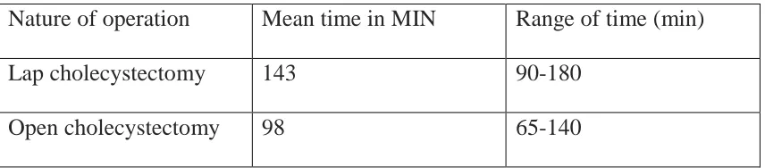
Median operating time for lap cholecystectomy was 90180 min (mean
-143 min) and open cholecystectomy was 65-140 min (mean- 98min). during
study operating time for lap cholecystectomy showed tendency to be shorter.
Use of analgesics on lap cholecystectomy is considerably less than open
cholecystectomy. Conversion rate in our study is 11.4 %(10 cases), inspite of
[image:72.595.86.514.549.643.2]being teaching and training institution.
Table :1 – operating time
Nature of operation Mean time in MIN Range of time (min)
Lap cholecystectomy 143 90-180
65
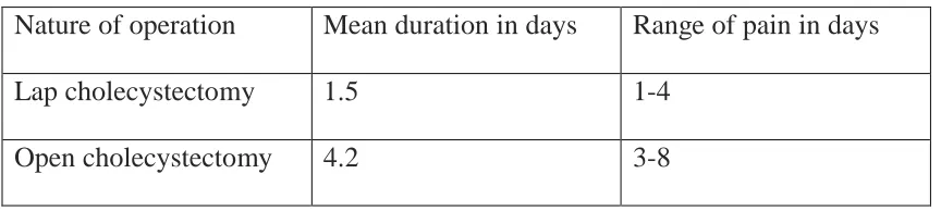
It has been observed that duration of post op pain and analgesia required were
[image:73.595.84.516.189.286.2]significantly less in Lap cholecystectomy than open cholecystectomy group
Table: 2 – Pain duration
Nature of operation Mean duration in days Range of pain in days
Lap cholecystectomy 1.5 1-4
Open cholecystectomy 4.2 3-8
Mean postoperative hospital stay was 3.7 days in lap cholecystectomy and 6.2
days after open cholecystectomy
Table : 3- Hospital stay duration
Nature of operation Mean duration in days Range of stay in days
Lap cholecystectomy 3.38 2-5
Open cholecystectomy 6.7 4-10
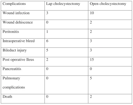
Complications were significantly more in open cholecystectomy group
than lap cholecystectomy group. Wound infection and dehiscence,
intraoperative bleeding, abdominal infection, bile duct injury, pancreatitis,
postoperative ileus, death and others were the complications taken into
[image:73.595.82.516.433.525.2]66
infection where as only 3 developed wound infection in case of Lap
cholecystectomy group that too occurred in difficult cases only.
Intraoperative bleeding and bile duct injury are more common in Lap
cholecystectomy group than open cholecystectomy especially in case of
beginners.
Post op Ileus as found in about 15 cases in case of open
cholecystectomy group, which warranted nasogastric tube insertion. In case of
open cholecystectomy Lung infection has occurred in about 5 cases as is
bedridden due to pain because of which patient could not be ambulated, hence
67 Table :4 – complications
Complications Lap cholecystectomy Open cholecystectomy
Wound infection 3 10
Wound dehiscence 0 2
Peritonitis 1 2
Intraoperative bleed 6 3
Bileduct injury 5 3
Post operative Ileus 2 15
Pancreatitis 0 0
Pulmonary
complications
0 5
Death 0 2
In our study series, about 10 cases of lap cholecystectomy were
converted to open surgery because of bileduct injury, which warranted open
repair like suturing of the bileduct which was difficult in laparoscopy and
hepaticojejunal anastomosis that was done due to unrepairable injury. Other
reasons for conversion are intra operative bleeding that cannot be controlled
laparoscopically, retained stones that cannot be retrievedlaparoscopically,
68
Time taken for starting oral feeds plays a major role in improving
patient general condition and discharge. The time taken for starting oral feeds
was less in laparoscopic surgery than open surgery. This improved the patient’s
general condition, wound healing, ambulation and also promoted early
[image:76.595.78.519.392.757.2]discharge of the patient from the hospital.
Table: 5- time taken for starting oral feeds
Type of surgery Mean time in days Range of time in days
Lap cholecystectomy 2.2 1-4
Open cholecystectomy 4 2-6
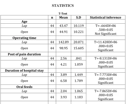
STATISTICS T-Test
n Mean S.D Statistical inference Age
Lap 44 43.47 10.119 T=-.664Df=86
.508>0.05 Not Significant
Open 44 44.91 10.221
Operating time
Lap 44 142.89 20.071 T=11.428Df=86 .000<0.05 Significant
Open 44 98.95 15.605
Post of pain duration
Lap 44 2.56 .841 T=-8.131Df=86
.000<0.05 Significant
Open 44 4.21 1.059
Duration of hospital stay
Lap 44 3.89 1.449 T=-7.773Df=86
.000<0.05 Significant
Open 44 6.58 1.789
Oral feeds
Lap 44 2.04 1.065 T=-7.865Df=86
.000<0.05 Significant
69
CONCLUSION
Minimally invasive surgery is better than open cholecystectomy in terms
of postoperative pain, analgesic requirement, early return to work. However
open cholecystectomy is preferred method for surgeons in beginning of their
70
PROFORMA
NAME
AGE/SEX
History of abdominal pain
History of fever
History of vomiting
History of jaundice
PHYSICAL EXAMINATION
Pulse
BP
Respiration
Temperature
LOCAL EXAMINATION
Tenderness, rigidity, distension.
INVESTIGATIONS
Hb
Blood group and typing
Blood urea, S.creatinine, and blood sugar
Liver function test
Plain X ray abdomen, chest
Ultra sonogram
71 TREATMENT
OPERATIVE PROCEDURE:
OPERATING TIME
NO. OF DAYS OF PAIN
TIME OF STARTING ORAL FEEDS
INTRAOPERATIVE BLEEDING – YES / NO
BILE DUCT INJURY – YES / NO
NO. OF DAYS WITH POST OP ILEUS
NO. OF DAYS OF HOSPITAL DAY
FOLLOW UP
IMMEDIATE POST OP
72
BIBILIOGRAPHY
• Lujan JA, Parrilla P, Robles R, Marin P, Torralba JA, et al. (1998)
Laparoscopic cholecystectomy vs open cholecystectomy in the
treatment of acute cholecystitis: a prospective study. [Internet]. Archives
of surgery 173-175.
• Kane RL, Lurie N, Borbas C, Morris N, Flood S, et al. (1995) The outcomes
of elective laparoscopic and open cholecystectomies. J Am CollSurg
180: 136-145.
• Dubois FBerthelot GLevard H Laparoscopic cholecystectomy: historic
perspective and personal experience. SurgLaparoscEndosc.
57
• Spaw ATReddick EJOlsen DO Laparoscopic laser cholecystectomy:
analysis of 500 procedures. SurgLaparoscEndosc. 1991;12- 7
• Cameron JCGadacz TR Laparoscopic cholecystectomy. Ann Surg.
1991;214251
• Fried GMSigman HHMeakins J et al. Management of acute cholecystitis
by laparoscopic cholecystectomy. SurgEndosc. 1992;688
• Lujan JAParrilla PRobles R et al. Laparoscopic cholecystectomy in the
73
• Clavien PRSanabria JRStrasberg SM Proposed classification of
complications of surgery with examples of utility in
cholecystectomy. Surgery. 1992;111518- 526Ellis H (2009) John
StoughBobbs: father of gall bladder surgery. Br J Hosp Med (Lond) 70:
650.
• Traverso LW (1976) Carl Langenbuch and the first cholecystectomy.
[Internet]. American journal of surgery p 81-82.
• Paulino-Netto A (1993) A review of 391 selected open cholecystectomies for
comparison with laparoscopic cholecystectomy. Am J Surg 166: 71-73.
• Cheslyn-Curtis S, Russell RC (1991) New trends in gallstone
management. Br J Surg 78: 143-149.
• Villanova N, Bazzoli F, Taroni F, Frabboni R, Mazzella G, et al. (1989)
Gallstone recurrence after successful oral bile acid treatment. A
12-year follow-up study and evaluation of long-term post dissolution
treatment. Gastroenterology 97: 726–731.
• Della Bianca P, Bonvin B (1990) [Lithotripsy of biliary calculi by shock
waves. Current possibilities and perspectives]. HelvChirActa 56:
913-916.
• Khuroo MS, Mahajan R, Zargar SA, Javid G, Sapru S (1989) Prevalence of
biliary tract disease in India: a sonographic study in adult population in
74
• Cuschieri ADubois FMouiel J et al. The European experience with
laparoscopic cholecystectomy. Am J Surg. 1991;161385- 388
• Fromm H (1989) Gallstone dissolution therapy with ursodiol. Patient
selection. Dig Dis Sci 34: 36S-38S.
• Miller REKimmelstiel FM Laparoscopic cholecystectomy for acute
cholecystitis. SurgEndosc. 1993;7296- 299
• Phillips EHCarroll BJFallas MJ Laparoscopically guided cholecystectomy:
a detailed report of the first 453 cases performed by one surgical
team. Am Surg. 1993;59235- 242
• Lujan JAParrilla PRobles R et al. Laparoscopic cholecystectomy in the
treatment of acute cholecystitis. J Am Coll Surg. 1995;18175- 77
• Clavien PRSanabria JRStrasberg SM Proposed classification of
complications of surgery with examples of utility in
cholecystectomy. Surgery. 1992;111518- 526Ellis H (2009) John
StoughBobbs: father of gall bladder surgery. Br J Hosp Med (Lond) 70:
650.
• Goco IR, Chambers LG (1988) Dollars and cents: minicholecystectomy and
early discharge. South Med J 81: 161-163.
• Ware R, Filston HC, Schultz WH, Kinney TR (1988) Elective
75
outcome using a preoperative transfusion regimen. Annals of Surgery
208: 17-22.
• Alponat A, Kum CK, Koh BC, Rajnakova A, Goh PM (2002) Predictive
factors for conversion of laparoscopic cholecystectomy. American
journal of surgery 21: 254-258.
• O’Dwyer PJ, McGregor JR, McDermott EW, Murphy JJ, O’Higgins NJ
(1992) Patient recovery following cholecystectomy through a 6 cm or
15 cm transverse subcostal incision: a prospective randomized clinical
trial. Postgraduate Medical Journal. BMJ Group 68: 817-819.
• Mühe E (1992) Long-term follow-up after laparoscopic cholecystectomy.
Endoscopy 24: 754-758.
• Jani K, Rajan PS, Sendhilkumar K, Palanivelu C (2006) Twenty years after
Erich Muhe: Persisting controversies with the gold standard of
laparoscopic cholecystectomy. Journal of Minimal Access Surgery 2:
49-58
• Friedman GD (1993) Natural history of asymptomatic and symptomatic
gallstones. Am J Surg 165: 399-404.
• McSherry CK, Ferstenberg H, Calhoun WF, Lahman E, Virshup M (1985)
The natural history of diagnosed gallstone disease in symptomatic and
76
• Meshikhes AW (2002) Asymptomatic gallstones in the laparoscopic era.
Journal Of The Royal College Of Surgeons Of Edinburgh 47: 747-478.
• Lowenfels AB, Domellöf L, Lindström CG, Bergman F, Monk MA, et al.
(1982) Cholelithiasis, cholecystectomy, and cancer: a case-control study
in Sweden. Gastroenterology 83: 672-676.
• Lam CM, Murray FE, CuschieriA (1996) Increased cholecystectomy rate
after the introduction of laparoscopic cholecystectomy in Scotland. Gut
38: 282-284.
• Escarce JJ, Chen W, Schwartz JS (1996) Falling cholecystectomy thresholds
since the introduction of laparoscopic cholecystectomy. Jama The
Journal Of The American Medical Association 91: 2653-2654.
• Mulvihill SJ (2003) Surgical management of gallstone disease and
postoperative complications. SeminGastrointest Dis 14: 237-244.
• Aucott JN, Cooper GS, Bloom AD, Aron DC (1993) Management of
gallstones in diabetic patients. Arch Intern Med 153: 1053-1058.
• Thompson JS, Philben VJ, Hodgson PE (1984) Operative management of
incidental cholelithiasis. Am J Surg 148: 821-824.
• Goldfarb A, Grisaru D, Gimmon Z, Okon E, Lebensart P, et al. (1990) High
incidence of cholelithiasis in older patients with homozygous beta-
77
• Domínguez LC, Rivera A, Bermúdez C, Herrera W (2011) [Analysis of
factors for conversion of laparoscopic to open cholecystectomy: a
prospective study of 703 patients with acute cholecystitis]. Cir Esp 89:
300-306.
• Vecchio R, MacFadyen BV, Latteri S (1998) Laparoscopic cholecystectomy:
an analysis on 114,005 cases of United States series. IntSurg 83:
215-219.
• Butt A (2006) Conversion of laparoscopic to open cholecystectomy-six years
experience at Shalamar Hospital, Lahore. Ann King Edward Medcoll
12: 536-538.
• Barone JE, Lincer RM (1991) Correction: A prospective analysis of 1518
laparoscopic cholecystectomies. N Engl J Med 325: 1517-1518.
• Miller REKimmelstiel FM Laparoscopic cholecystectomy for acute
cholecystitis. SurgEndosc. 1993;7296- 299
• Phillips EHCarroll BJFallas MJ Laparoscopically guided cholecystectomy:
a detailed report of the first 453 cases performed by one surgical