“A STUDY OF POST OPERATIVE COMPLICATIONS
IN
THYROID SURGERIES”
Dissertation submitted to
THE TAMILNADU Dr. M. G. R. MEDICAL UNIVERSITY
in partial fulfillment of the regulations for the award of the degree of
M. S. GENERAL SURGERY (BRANCH I)
1 CHENGALPATTU MEDICAL COLLEGE,
CHENGALPATTU
CERTIFICATE
This is to certify that this dissertation titled “A STUDY OF POST PERATIVE COMPLICATIONS IN THYROID SURGERIES” has been prepared by DR. PALANISAMY.B, under my supervision in the Department of General Surgery, Chengalpattu Medical College, Chengalpattu, during the
academic period 2015 – 2018, and is being submitted to The Tamilnadu Dr.
M.G.R. Medical University, Chennai, in partial fulfillment of the University
regulation for the award of the Degree “Master Of Surgery” (M. S., General
Surgery) and his dissertation is a bonafide work.
Prof.Dr.A.PRABAKAR.M.S ,. Prof.Dr.RAMULA.M.S, DGO,.
PROFESSOR, of general surgery, PROFESSOR & HEAD,
Department Department of general surgery, Chengalpattu Medical College , Chengalpattu Medical College, Chengalpattu-01 Chengalpattu-01
Prof.Dr.USHA SADHASIVAM.M.D,.
DEAN,
DECLARATION
I, Dr. PALANISAMY.B , solemnly declare that the dissertation “A STUDY OF POST OPERATIVE COMPLICATIONS IN THYROID SURGERIES”is a bonafide work done by me in the Department of General Surgery, Chengalpattu Medical College, Chengalpattu, Under the able guidance
of PROF. Dr.PRABAKAR., M.S, Professor , Department of General Surgery , Chengalpattu Medical College , Chengalpattu .
Place:Chengalpattu
ACKNOWLEDGEMENT
I wish to express my sincere thanks to Dr, Dean, Chengalpattu Medical
College & Hospital, Chengalpattu, for having kindly permitted me to utilize the
hospital facilities.
I wish to express my heartfelt gratitude to:
The Prof. Dr. RAMULA M.S,,DGO., Professor & Head of the Department of General surgery, Chengalpattu Medical College & Hospital, Chengalpattu for
her immense help,encouragement,guidance and constant supervision .
I express my heartfelt gratitude to my old Chief Dr. T.RAGHUPATHY. M.S.,and acting chief Dr.PRABAKAR. M.S., and Dr.SANTHAKUMAR. M.S., for their ever valuable guidance ,motivation and immense support and for their constructive suggestions.
I Am Greatly Thankful To Our Asst. Professors Dr. P.SANKARLINGAM. M.S., Dr.LAKSHMYPATHY. M.S., Dr.KARTHIK ARUMUGAM. M.S., Dr.VARAGUNAPANDIAN. M.S., and
Dr.MALATHY. M.S., for their Valuable Suggestions and guidance and great care and attention to prepare this dissertation .
I owe great debt of gratitude to all the Assistant Professors and senior
PGs- Dr.SANDHYA.M.S., Dr.ASWINI.M.S., and DR.Vaithiswaran.M.S..,
for their able help and support. They have been a source of great encouragement
junior post graduates and House surgeon for their willing co-operation and
assistance in assimilating the clinical material.
I Wish to thank the theatre personnel without whom the study would not
have materialized. I thank all the patients who took part in my study and their
CONTENTS
S.NO TITLES PAGE NO
1 INTRODUCTION 1
2 AIMS AND OBJECTIVES 3
3 MATERIALS AND METHODS 4
4 SURGICAL ANATOMY
5 OPERATIVE SURGERY ON THE THYROID
GLAND
22
6 STANDARD SURGICAL APPROACH TO
THE THYROID
25
7 POST OPERATIVE COMPLICATIONS AND
THEIR MANAGEMENT
38
8 OBSERVATION AND RESULTS
9 DISCUSSION
CONCLUSION
BIBLIOGRAPHY
ANNEXURES
PROFORMA
CONSENT FORM
INTRODUCTION
Thyroidectomy is a common surgery with an extremely low
mortality1, with specific morbidities which are related to the experience of the
surgeon2. Very low morbidity rates with specialised centers. Thyroid surgery
is associated with few complications and no fatality. Post-operative
complications may be as insignificant as edema of the flap or as dangerous
and life threatening as hemorrhage or respiratory obstruction. Complications
are less with sound surgical technique and good preoperative preparation.
With proper preoperative preparation , the patient will be euthyroid at the
time of surgery. In hyperthyroid patient , laryngeal edema may result,
producing respiratory difficulties . Improper technique may result in
massive post operative haemorrhage, recurrent laryngeal nerve paralysis ,
both, causing respiratory difficulty. Improper technique and inexperience .
may involve removal of too little or too much thyroid tissue
or possibly all parathyroids, resulting in myxedema, recurrent
hyperthyroidism, or parathyroid deficiency.Complication of thyroid surgery
The present study analyse the clinical audit of thyroid surgery for
adult patients undertaken at the Chengalpattu Medical college hospital
Chengalpattu.. The complications of Thyroidectomy are highlighted and
AIMS AND OBJECTIVES
1. The aim of the study is to compare complication rates of Bilateral
sub total thyroidectomy (SBT) , near total thyroidectomy (NTT)
Hemithyroidectomy (Total lobectomy and isthmusectomy), and Total
thyroidectomy (TT) in cohort of patients undergoing surgery for
various thyroid disorders.
2. To compare complication rates after thyroidectomy for benign
diseases and malignant diseases.
MATERIALS AND METHODS
One hundred patients who underwent thyroid surgery, for various
thyroid disorders, at the Chengalpattu Medical College Hospital,
Chengalpattu, Tamilnadu, India between September 2016 to August 2017 were
studied. Operations were performed by various professors, Assistant
professors and also by surgical post graduates supervised by senior
surgeons using various surgical techniques. Indications for surgery in this study
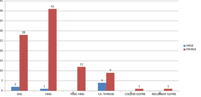
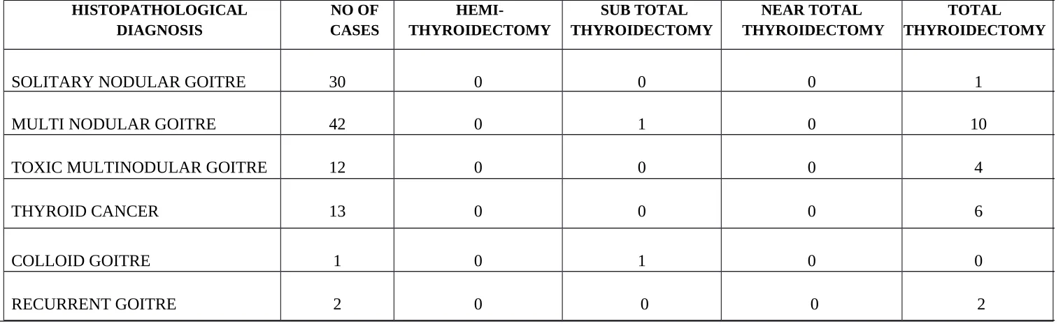
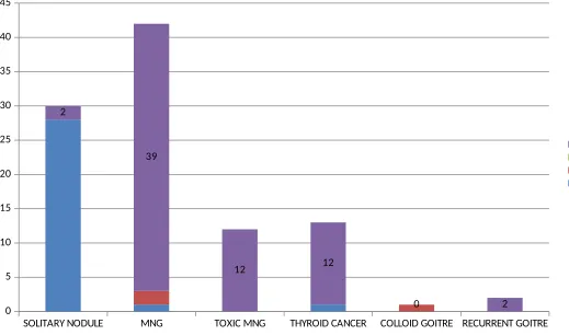
group include forty two patients with non toxic multinodular goitre (MNG),
twelve patients with toxic multinodular goitre, thirty patients with nontoxic
solitary nodular goitre, thirteen patients with carcinoma, two patients with
recurrent goitre and one patient with colloid goitre.
Of these one hundred patients ,ninety three patients were female and
seven patients were male. Patients were broadly divided in to two categories
based on the diagnosis and treatment modality.
For all selected patients a thorough history , complete physical
examination , basic biochemical and hematological investigations were done.
Special investigations like thyroid hormone profile and serum calcium
estimation done. Vocal cords were examined pre operatively by indirect
laryngoscope in all the patients and post operative vocal cord examination
Patients were classified as having hypocalcaemia (hyperparathyroidism)
if both clinical and biochemical (a fall in corrected serum calcium
concentration below 8 mg/dl and or the need for calcium
supplementation )23 supportive evidence were present .
FNAC done to all patients. Based on the Final diagnosis, the treatment
was advised by the Assistants and The Chief and the further management
planned.
The details of patients were documented as shown in the
proforma (ANNEXURE I).
THYROID EMBRYOLOGY
The understanding of embryology of thyroid is must for the surgeon
dealing with thyroid abnormalities must be familiar with their embryologic
antecedents. Only through this knowledge surgeons can understand the
variations in glandular location, shape or number and rationale for surgical
maneuvers be appreciated. The thyroid gland develops as an endodermal
bud of cells at the foramen caecum at the junction of the anterior two-third
of the tongue with its posterior one-third. In the floor of the pharynx,
between the levels of the first and second pharyngeal pouches, gradually a
median diverticulum is formed in the later half of the fourth week. It grows
divides into a series of double cellular 7 plates from which the isthmus and the
lateral lobes of thyroid gland are developed.
As the thyroid primordium descends, it acquires mesodermal
contributions such as the parafollicular C -cells which ultimately secrete
calcitonin. These parafollicular C cells are derived from a bud which is
known as Ultimo-bronchial body which arises from a diverticulum of the
fourth pharyngeal pouch of each side and amalgamates with the
corresponding lateral lobe of thyroid. Except the distal part of the
thyroglossal duct which usually differentiates to form the pyramidal lobe of
thyroid, the rest of the duct disappears or sometimes replaced as a
muscle-levator, glandulae thyroidea. A portion of the thyroglossal duct
may persist and give rise to the formation of cyst in the
midline of the neck called as thyroglossal cyst. Sometimes
thyroglossal fistulas or ectopic thyroid tissue may develop. Total agenesis
of one thyroid lobe may occur. This is rare but can be clinically important,
leading to confusion in diagnosis, especially in toxic glands, when it could
be diagnosed as a secreting nodule.
GROSS ANATOMY
The thyroid gland occupies the centre of the visceral compartment of the
neck, lying astride the trachea just above the thoracic inlet with average
as a result of its relation to the anterolateral portions of the cervical
vertebra and trachea from the level of the thyroid cartilage to the fifth or sixth
tracheal ring. They reside in a bed between the trachea and larynx medially
and the two carotid sheaths and the sternocleidomastoid muscles laterally.
The right lobe is often larger than the left and the lobes are joined
together across the midline by a thin isthmus plastered quite firmly to the
anterior surface of the trachea at the level of the 2nd and 3rd tracheal rings. A
small pyramidal lobe arises from the isthmus somewhere along its upper
border near the midline, represents a vestige of the embryonic thyroglossal
duct and can be demonstrated in about 80 percent of patients .
The thyroid gland is enveloped by a thickened fibrous capsule, the deep
cervical fascia which divides into an anterior and posterior sheath, creating a
false capsule for the thyroid. Anteriorly, the thyroid lobes are in relation to
the strap muscles. Situated on the posterior surface of the lateral lobes of the
gland are the parathyroid glands. The recurrent laryngeal nerves, which lie in
a cleft between the trachea and esophagus just medial to the lateral lobes.
THE SKIN
The skin of the neck may reveal several transverse folds that may be of
value in deciding the site of incision. Of the folds just above the sternum
THE MUSCULOFASCIAL COVERINGS
The platysma is a thin, sheet like muscle immediately beneath the
skin in the subcutaneous plane , spreading like fan from below the clavicle to
laterally to the mandibles above. When the skin flaps are turned up to
expose the underlying muscles, the skin, subcutaneous fat, and platysma
should be dissected as a single layer. By this plane , unnecessary bleeding
will be avoided, since the fascial plane between the platysma and the fascia
covering the prethyroid muscles is quite avascular. The muscle that must
be dealt with are the two lateral sternocleidomastoid muscles, forming
the boundaries, and the strap muscles namely the sternohyoid and
sternothyroid muscles, forming the superficial and anterior boundaries.
Both the sternohyoid, lying more anterior, and the sternothyroid, just
beneath and covering the thyroid capsule, arise from the sternum. The
sternohyoid attaches above to the hyoid, and the sternothyroid attaches to
the thyroid cartilage. The fascia of the anterio- medial border of the
sternocleidomastoid fuses with fascia of the lateral border of the
sternohyoid. This is divided longitudinally during exposure of the thyroid to
allow retraction of the sternocleidomastoid laterally and the sternohyoid
ANSA CERVICALIS .
This nerve descends along the lateral border of the sternohyoid and
enters the muscles low in the neck . The sternohyoid and sternothyroid are to be
divided transversely to facilitate access to the gland, they must be transected
high to preserve the motor function of this nerve.
The other important implication of the musculofascial covering of the
gland is that at the end of the thyroid operations the divided fascial
envelope is resutured in the midline and this again closes the visceral
space. If there is post operative haemorrhage into this closed space,
respiratory embarrassment from the tracheal compression results and
requires immediate release of the sutures to restore the airway.
THE VASCULAR SUPPLY
The blood supply of the thyroid gland is very rich in the hyperthyroid
state and there may be an enormous increase in the volume of blood circulating
through the gland.The principle blood supply to the thyroid gland are
external to the capsule and can be ligated before the gland is entered. The
superior and inferior thyroid arteries constitute the main arterial supply.
Occasionally a branch from the aorta or innominate artery, the lowest
inferior thyroid veins (vena thyroidea ima) enter from below. The veins, in
general, drain the corresponding arteries.
THE ARTERIES
THE SUPERIOR THYROID ARTERY
Superior thyroid artery is the first branch from the external
carotid artery at about the level of the bifurcation of the common carotid
artery. It runs downward and medially on the surface of the inferior
constrictor muscles, entering the upper pole of the thyroid on its
anterosuperior surface. It is just inferior and lateral
to the superior laryngeal nerve. The superior laryngeal nerve usually turns
medial from the superior thyroid artery about one cm above the pole. Then,
care must be taken in ligating the superior thyroid artery close to its
entrance to the gland to avoid injury to the superior laryngeal nerve.
In many pathologically enlarged thyroid glands there is a lingula of
thyroid tissue above and lateral to the point of entry of the superior thyroid
artery and the superior laryngeal nerve. If this lingula is mistaken for the
upper pole, the artery may be ligated high with possible damage to the
superior laryngeal nerve. Occasionally a branch of the superior thyroid
artery to the pyramidal lobe and isthmus may arise high and bleeding may
because the artery was ligated below the origin of this branch to the
pyramidal lobe.
THE INFERIOR THYROID ARTERY
It arises from the thyrocervical trunks shortly after their origin from
the subclavian arteries and ascend upward behind the jugular veins and
carotid arteries to a level above the inferior pole of the thyroid. Then they
make a loop downward and medially and enter the gland at its mid
position, not at the inferior pole.
The surgeon’s first glimpse of the inferior thyroid artery
is usually when the thyroid gland is retracted medially and the jugular vein
laterally. Before entering the thyroid, the inferior thyroid artery
may be divided into one or more branches and be intimately associated
with recurrent laryngeal nerve. One branch of the inferior thyroid artery may
supply the inferior parathyroid. Great care must be used in ligating the
inferior thyroid artery in this region to avoid injury to the recurrent
laryngeal nerve. It should not be ligated until the recurrent nerve and its
branches are visualized; then it should be ligated as close to the gland as
possible individually.
Arteia thyroidea ima
It arises from the aorta or innominate artery and passes directly upward
ligated with the inferior thyroid veins. A few unnamed arteries from the
trachea also supply the thyroid gland. After partial thyroidectomy, they
maintain the blood supply of the remaining glandular tissue.
THE VEINS
THE SUPERFICIAL VEINS lies beneath the platysma. They are
easily visualized.
The EXTERNAL JUGULAR VEINS are lateral and cross over the belly
of the sternocleidomastoid.
The ANTERIOR JUGULAR VEINS immediately overlie the
sternohyoid muscles. A plexus of communicating veins may be present
between the external and anterior jugular veins. In large goiters the superficial
veins may be of considerably large. Care should be exercised in turning up the
skin flaps with platysma so that the veins are not cut. If the surgeon carefully
divides the fascia between the sternocleidomastoid and the sternohyoid, the
communicating veins may be ligated and divided. The external jugular vein
may then be retracted laterally along with the sternocleidomastoid. The
anterior jugular is divided and ligated safely . Venous plexus forms under
the capsule and contribute to confluences forming the superior thyroid vein
at the upper pole, the middle or lateral thyroid vein in the middle of the lobe,
arising from the lower pole are the inferior thyroid veins. THE DEEP
mainly at the superior and inferior poles and the lateral aspect of the gland.
They are less constant than the arteries. The deep veins may be a serious
threat during thyroid surgery because they are numerous and may easily be torn
with ensuing hemorrhage. This is particularly true in patient with a large,
substernal goiter. In such patients the clavicle may act as a tourniquet
and produce great dilatation and increased pressure in the veins. If
uncontrolled hemorrhage occurs, the lower pole and the substernal
extension of the goiter must be quickly mobilized out of the mediastinum.
Release of the tourniquet effect of the clavicle will allow the veins to collapse
and the hemorrhage will be brought under control.
THE SUPERIOR THYROID VEINS leaves the gland at the superior pole just anterior and lateral to the superior thyroid artery and empties into the
cricothyroid tributary of the internal jugular vein. It can be ligated along with
the superior thyroid artery during thyroidectomy.
THE MIDDLE or LATERAL THYROID VEINS vary greatly in number,
pass directly from the lateral border of the lobes and enter into the internal
jugular vein. During thyroidectomy they must be divided to allow access to
the lateral compartment. In pathologically enlarged, the jugular vein and the
lateral thyroid veins may be pushed close to the capsule of the thyroid. In
such instances they may be mistaken for veins in the thyroid
be difficult to find. The lateral veins may override the capsule and must be
identified and divided before an attempt is made to enucleate the gland.
THE INFERIOR THYROID VEIN leaves the lower pole in one or more trunks, frequently forming a plexus of veins. The inferior thyroid vein is
not adjacent to the inferior thyroid artery, which is in the region of the middle
third of the gland. The inferior thyroid artery is more closely associated
with the lateral thyroid vein. The inferior thyroid vein empties into the
internal jugular vein and occasionally directly into the innominate vein. Vein
of thyroidea ima drains the isthmus, passing downward on the trachea to
enter the left innominate vein. The inferior thyroid veins may be closely
interwoven with the recurrent laryngeal nerve as it ascends. This is a
vulnerable area for injury to the recurrent laryngeal nerve.
THE LYMPHATIC SYSTEM
Knowledge of the lymphatic vessels draining the thyroid is essential in
planning radical surgical procedures. In general, the lymphatics
accompany the veins. Collecting lymph channels draining the intra
glandular capillaries are found beneath the thyroid capsule. These
channels drain into the lymph vessels associated with the capsule and may
THE SUPERIOR LYMPH VESSELS drain the isthmus and the medial superior portion of the thyroid lobes, ascending in front of the larynx
and terminating in the sub digastric lymph nodes of the internal jugular
chain.
THE MEDIAN INFERIOR LYMPH VESSELS descend with the
inferior vein to the pretracheal nodes. The lateral collecting group, above,
follows the superior thyroid vessels to the anterior and superior nodes of
the internal jugular chain and, below, follows the lateral thyroid and inferior
thyroid veins to the lateral and inferior nodes of the internal jugular vein.
These internal jugular pretracheal and anterior jugular nodes can usually
be excised surgically by a radical neck dissection. Tumor invading the
pretracheal nodes, may spread downward into the anterosuperior
mediastinal nodes out of reach of the usual radical neck dissection44.
Another group of lymphatics draining the posterosuperior aspect of the
thyroid leaves the posterior capsule and passes to the retropharyngeal
nodes Rouviere found this to be present in one fifth of his dissections.
Obviously, if the retropharyngeal nodes are involved in tumor, they are not
eradicated by a radical neck dissection.45
The knowledge of the complexity of this lymphatic drainage and the
anatomical distribution of the regional nodes becomes important in
of the nodes, standard radical neck dissection has been changed into
regional node dissection.
THE NERVES
The gland receives its innervation from sympathetic and
parasympathetic divisions of the autonomic nervous system. The
sympathetic fibers arise from the cervical ganglion and enter with blood
vessels, while the parasympathetic fibers are derived from the vagus and
reach the gland via branches of the laryngeal nerves.
THE RECURRENT LARYNGEAL NERVES arise from the vagus at
different levels on the two sides. The right recurrent laryngeal nerve arises
where the vagus crosses the first portion of the subclavian artery. The
nerve hooks around the lower and posterior aspect of the subclavian artery and
ascends lateral to the trachea, entering the larynx posterior to the thyroid
at the cricothyroid articulation. The left recurrent laryngeal nerve
leaves the vagus as the vagus crosses over the arch of aorta; it hooks
around the aorta and ascends again, as the right recurrent nerve does,
lateral to the trachea to its terminal branches within the laryngeal muscles.
The nerves usually lie in the tracheo-esophageal groove and then bear a
variable relationship to the branches of the inferior thyroid artery before
entering the larynx. In the majority of cases, the nerve is found easily in the
be anomalous and it may be much more lateral as emphasized by Fowler
and Hanson 46. In very rare instances, because of failure of development
of the 4th arch vessel and a resultant anomalous right subclavian artery, the
nerve on that side will be non-recurrent and then passes directly medially
at a much higher level from the vagus to the larynx. In this position it could
be in danger at the time of ligation of the middle thyroid vein, though the
difference between these two structures should be easily apparent. the level of
the hyoid cornu, divides into two branches. The internal branch
is sensory; it penetrates the thyrohyoid membrane and may anastomose
with the sensory branch of the recurrent laryngeal nerve to complete the
LOOP OF GALEN. The external branch of the superior laryngeal nerve lies on the lateral surface of the inferior pharyngeal constrictor muscle and 2
descends to innervate the cricothyroid muscle. Both branches lie
immediately adjacent to the superior thyroid artery and may be injured if
the superior thyroid artery is ligated “in bulk” too high above its entrance
into the thyroid gland. If the recurrent nerve is injured, paralysis of the vocal
cord occurs on the ipsilateral side. The laryngeal muscles (five on each
side) controlling motion of vocal cords are the abductors (the internal
arytenoids and the thyroarytenoid) and the adductors (the lateral and the
posterior cricoarytenoids) and are innervated by the recurrent laryngeal
tensors of the vocal cord), is innervated by the internal branch of superior
laryngeal nerve.
Semon-Rosenbach law postulated that the abductor fibers
of the recurrent nerve are more susceptible to pressure than the adductor
fibers47,48. The Wagner-Grossmann theory stated that paralysis of the vocal
card in the paramedian position was the result of injury of the recurrent
laryngeal nerve and paralysis of the vocal card in the intermediate position .
THE SUPERIOR LARYNGEAL NERVE arises from the vagus close
to the base of the skull, descends medially to the carotid vessels, and at was
the result of injury of the recurrent laryngeal nerve and the superior
OPERATIVE SURGERY ON THE THYROID GLAND
The mortality rate of thyroidectomy as reported in several large
series approaches are very rare.. This can be accomplished by a well
performed standardized technique. The morbidity rate should be less than 5
percent. A well-executed thyroidectomy will keep this morbidity to a
minimum. The step-by-step technique of thyroidectomy is recommended for
satisfactory results. Surgery is a part in the successful treatment of thyroid
disease.if the patient is brought into the operation theater in either a hypo
thyroid or hyper thyroid state, disaster may follow. If the patient is
thyrocardiac the services of the cardiologist are mandatory. If the patient is
euthyroid but proper anaesthesia is not available, complications may occur. If
an expert in diagnosing thyroid pathology from a frozen section is lacking, the
wrong procedure may be elected. For the best results, treatment of the
thyroid patient demands the services not only from the surgeon but also
of the internist, the cardiologist, the anaesthesiologist and the
pathologist-with all experts in thyroid disease.
PRE OPERATIVE PREPARATION
Preparation is must to ensure safe induction of anaesthesia and a trouble
free intra and post operative course.Haemoglobin estimation, chest
radiography,and an electrocardiogram are mandatory. Blood transfusion is
patients. If thyroid enlargement is massive or retrosternal and if the patient
shows clinical signs of respiratory embarrassment or superior vena caval
obstruction, a CT scan of the neck and thoracic inlet will indicate the
possible need to enter the chest and potential problems that may be
encountered on intubation also.
The vocal cords should always be examined by indirect
laryngoscopy; this is especially important when the voice is compromised,
malignancy is suspected, or when previous thyroid surgery has been
undertaken. A small proportion of patients have unsuspected palsy of the
recurrent laryngeal nerve; it is of clinical and medico legal importance to both
the patient and the surgeon to determine before surgery whether or not the
vocal cards are moving normally.
Thyrotoxic patients need to be rendered euthyroid or the peripheral
effects of high circulating concentrations of thyroxin blocked. The majority
of thyrotoxic patients referred for surgery have already received one or
more courses of anti thyroid drugs.
Where residual toxicity is modest and surgery can be undertaken
quickly, it is standard practice in many centers to stop anti thyroid drugs 10
days before operation and switch to oral propranolol in a dose of 40 to 120
mg every 6 to 8 hrs. The dose is adjusted to keep the patients sleeping
is approximately 6hrs, it is important to administer medication
right up to induction of anaesthesia and to continue thereafter, especially if
the patient develops tachycardia. B-blockers are contraindicated in patients
with bronchial asthma, sinus bradycardia, or congestive heart failure.
Patients with severe thyrotoxicosis who require relatively early surgery
should receive a 6 to 8 week course of carbimazole (Neo-mercazole 10 to 15
mg 8 hourly). In the event of an adverse drug reaction, propylthiouracil (100
mg three times daily) may be substituted for carbimazole. Extended use of
thiourea drug may cause prothrombin deficiency (hence the advisability of
stopping the drug 10 days before surgery), leucopenia or profound bone
marrow depression. Full blood count and the international normalized ratio
should be checked if reduced resistance to infection or impaired haemostasis
SURGICAL APPROACH TO THE THYROID
ANAESTHESIA AND POSITION ON THE OPERATINGTABLE
General anaesthesia is administered through an endotracheal tube .The
patient is supine position with the table tilted up 15 degree at the head end to
reduce venous engorgement. A sand bag is placed under the shoulders and
the neck is extended to make the thyroid gland more prominent and apply
tension to skin, platysma and strap muscles, which makes dissection easier.
The patient head is supported on a head ring to avoid rotation of the head.
The neck from the chin to the suprasternal notch is exposed.
THE INCISION
The skin incision can be marked by pressing a length of thread onto
the skin along langhans line.. A gently curved skin crease low collar kocher
incision is made, 2 finger breadth above the sternum, extending to the
lateral borders of the two sternomastoids. With large goiters the incision is
made a little higher so as to provide better access to the superior thyroid
pole.
ELEVATION OF THE FLAP
The skin, subcutaneous fat, and platysma are elevated as one layer. The
platysma is divided at a slightly higher level than the skin. The fascial plane
bleeding. The platysmal fibers are most easily identified at the lateral margins
of the incision and its fibers divided exposing the fascia of the prethyroid
muscles. The superficial veins are not elevated with the flap, and thus they act
as a deep landmark to keep the proper fascial plane.By applying upward
traction, the upper flap is raised well above the thyroid notch by a
combination of sharp and blunt dissection in a relatively avascular plane. The
lower flap may be freed from the underlying fascia down to the level of the
suprasternal notch.
DIVISION OF THE PRETHYROID MUSCLES
Although this maneuver is not universally accepted, the prethyroid
muscles should be divided in all cases to obtain adequate exposure and to
perform a thyroidectomy safely. If the muscles are severed high,
preserving their innervation from the ascending branch of the ansa
hypoglossal nerve, which enters the muscle low, and if they are later
carefully sutured, no disability or disfigurement results. Disfiguring atrophy of
the prethyroid muscles, with the sunken neck and prominent trachea, is the
result of dividing the prethyroid muscles and their innervation too low in the
neck.
The prethyroid muscles are mobilized for transaction by freeing their
lateral and medial fascial attachments. The fascia of the lateral border of
sternocleidomastoid muscle. If this fascia between the two muscles is
divided along the medial edge of the sternocleidomastoid muscle, the
anterior jugular and external jugular veins can easily be visualized, ligated,
and divided. The medial borders of the sternohyoid and sternothyroid
muscles are best identified in the midline low in the neck. The midline of
the trachea above the suprasternal notch should be determined by
palpation and this area explored. Often a small amount of free fatty tissue
is a clue to the midline. The finding of the midline before the midline fascia
is divided is important in order to visualize, ligate, and divide the
communicating veins crossing the midline from the anterior jugular of one
side to the anterior jugular of the other.
sternohyoid and leave the sternothyroid attached to the thyroid
capsule. Both prethyroid muscles are then separated from the gland by
blunt dissection. While the medial border of the sternocleidomastoid is
retracted downward and outward by the assistant to push the anterior jugular
out of the way, the surgeon applies kocher clamps across the upper aspects of
the prethyroid muscle from the median side toward the lateral side. The
prethyroid muscles are then divided between clamps.
Then retract the upper portion of the divided prethyroid muscles to
expose the upper pole and the superior thyroid vessels. This is aided by
muscles are freed laterally and retracted downward. Both lobes are
exposed in the same manner, and the anterior exposure of the thyroid
completed.
If the anterior exposure has been performed properly, the remainder of
the operation should go smoothly. Difficulty encountered with
mobilization, hemorrhage, and identification of the recurrent nerves and the
parathyroids is usually due to the result of poor anterior exposure. With the
borders of the prethyroid muscle mobilized by blunt dissection, their
posterior sheaths are separated from the underlying thyroid capsule. This
procedure is accomplished better and bleeding is avoided if it is started over
the lower section of the thyroid rather than in the region of the upper poles.
MOBILIZATION OF THE GLAND
The mobilization of the thyroid gland involves the division of the
lateral thyroid veins so that the internal jugular vein and carotid artery can
be retracted laterally and the thyroid displaced up and out of its bed. This
gives access to the inferior thyroid aspect of the gland so that the inferior
thyroid artery can be isolated and the recurrent laryngeal nerve identified.
The numbers of lateral veins are variable. Gentle upward and medial
traction on the gland places the lateral veins on the stretch so that by
meticulous dissection they may be clamped and severed. As the lateral
flood the lateral compartment, making later identification of the inferior
thyroid artery and the recurrent laryngeal nerve more difficult. In large
goiters the lateral veins should be divided close to the capsule. If the gland
is rotated anteriorly and medially, the cleavage plane will be apparent, the
internal jugular vein visualized and retracted laterally, and the remaining
lateral veins severed.
ISOLATION OF THE INFERIOR THYROID ARTERY
The most vulnerable location for injury to the recurrent laryngeal
nerve is the point at which the inferior thyroid artery approaches the lateral
aspect of the thyroid. Before entering the thyroid gland, the inferior thyroid
artery may divide into one or more branches and be intermittently
associated with the recurrent laryngeal nerve and its branches. For easy
identification of the inferior thyroid artery and the recurrent laryngeal nerve,
the entire posterior lateral edge of the thyroid is exposed and this
compartment should be free of blood and blood stained tissue. By
meticulous scissors dissection in this avascular field, the inferior thyroid
artery will be visualized coming from beneath the carotid artery, in most
instances at the level of the mid portion of the thyroid gland. As the inferior
thyroid artery is followed to the capsule of the gland, attention is directed to
the recurrent laryngeal nerve; in most cases, this passes inferior to the
damage to the recurrent laryngeal nerve the inferior thyroid artery is not
ligated until the nerve is isolated and out of the way. The artery is then
ligated in continuity,free of surrounding tissue as far laterally as possible.
Tying the inferior thyroid artery in continuity reduces the risk of a
blown-off tie, a serious complication. Although tying the inferior thyroid
artery laterally may occlude the branch of the artery supplying the inferior
parathyroid, most of the surgeon have not experienced any parathyroid
deficiency. Hemorrhage from a torn inferior thyroid artery is inexcusable.
Obviously it cannot be controlled with pressure on the carotid artery. If this
catastrophe occurs, the carotid sheath must be retracted laterally and
ISOLATION OF THE RECURRENT LARYNGEAL NERVE
Most often the recurrent nerve is identified at the time the inferior
thyroid artery is isolated. If not, it is sought low in the neck in its usual
position in the groove between the esophagus and the trachea. As it
approaches the lower border of the thyroid it may, as it ascends, turn as
much as one cm, lateral to this groove and be intimately associated with a
plexus of delicate inferior thyroid veins. Bleeding is avoided by meticulous
scissors dissection; the scissors are always opened in the direction of the
course of the nerve. The recurrent laryngeal nerve may have one or more
extralaryngeal branches. Only one, however, contains the motor fibers
supplying the laryngeal muscles. From a practical point of view, all ranches
must be considered as possible motor branches and spared injury. Once
identified, the nerve is followed to its junction with the inferior thyroid artery.
The inferior thyroid artery is then ligated in continuity.
MOBILIZATION OF THE INFERIOR POLE
With the inferior thyroid artery ligated and the recurrent nerve
visualized, the lower pole may be mobilized safely. The midline of the
trachea is demonstrated below the isthmus. The entire plexus of vessels
below the isthmus and inferior poles may be cross-clamped. The clamps are
applied from the midline out. Use two hemostats below and one above
possibility of the vessels slipping from one clamp and escaping into the
mediastinum, where they may be difficult to retrieve with the vessels
divided, the lower pole is gently detached from the trachea and pulled
upward.
DIVISION OF THE SUPERIOR THYROID VESSELS AND
MOBILIZATION OF THE UPPER POLE.
The superior thyroid vessels must be ligated under direct vision. With
high elevation of the prethyroid muscle and downward and inward traction of
the superior pole, they can be separated from the inferior constrictor
muscle. They may be hidden beneath a few fibers of the divided
sternothyroid. It is best to ligate the vessels with a ligature passing from
inside out. The vessels are doubly ligated above and divided between the
ligatures and a clamp placed below. In hypertrophic glands a lingual or tongue
of the tissue may ascend high, lateral to the entry of the vessels into the gland.
In such instances two mistakes may be made.
1. A blind attempt may be made to pass a ligature around the vessels
around the lingual.
2. The lingula may be transected with the vessels leaving remaining
thyroid tissue intact, a possible and frequent focus for recurrence in
Graves’ disease and a difficult problem to remedy at a later date.
vessels at a lower level; then, by excising the thyroid capsule, this
extension of thyroid tissue made by inward and downward traction
may be enucleated. Before complete enucleation, small posterior
veins entering the gland should be clamped.
IDENTIFICATION OF THE PARATHYROIDS
Every attempt is made to preserve at least one parathyroid on each
side. As familiarity is gained with their appearance -split pea-sized, molded
edges, tan to mahogany brown in color, appearing as distinct organs close
to the thyroid capsule - they become more readily identified. Operative
trauma or bruising of the parathyroids or adjacent lymphnodes may result
in some capsular hemorrhage, staining the tissues so that the thyroid,
parathyroid fat and lymphatic tissues are not clearly differentiated. Once the
superior pole is rotated downward and inward, the superior parathyroid may
usually be seen at about the junction of the upper and middle thirds of the
thyroid glands along its lateral posterior aspect. The inferior parathyroids are
usually found just below the junction of the inferior thyroid artery and the
recurrent laryngeal nerves. Frequently a branch of the inferior thyroid artery
leads to the inferior parathyroid. Clamps should be inserted into the thyroid
capsule anterior to the parathyroids and the capsule and the parathyroids
should be gently dislodged from the thyroid gland so that they are not
DIVISION OF THE ISTHMUS
The isthmus is freed from its attachment to the trachea in the midline
from below upward by blunt dissection and divided between a series of
hemostats. Care must be excised in separating the isthmus from the
underlying pretracheal fascia to avoid puncture of the trachea by the sharp
points of this straight clamp.
At the upper aspect of the isthmus, the suspensory ligament is
divided to complete the mobilization of the gland. A series of clamps is
placed along the medial capsule and the capsule is incised above the
clamps, producing a cuff that outlines the medial border of the resection. At this
moment in thyroidectomy, it is necessary to determine the
amount of gland to resect or, conversely, the amount of gland to be left.
This is determined by the pathologic condition for which thyroidectomy is
being performed. With the superior and inferior poles mobilized and the
gland retracted medially, hemostats are applied to the lateral capsule, outlining
the amount of thyroid to be removed. The capsule is incised above the line
of hemostats. The thyroid tissue will appear to bulge out of the capsule.
preserve the parathyroids and their blood supply. A cup of thyroid capsule
and thyroid tissue is developed that will clearly establish the lateral border
of the remnant and will later be sutured to the medial cuff to reconstruct the
remnant.The thyroid tissue between the lateral and medial lines of hemostats
now outlines the boundaries of the thyroid to be excised. By the clamp and
cut technique within the thyroid capsule, the gland is excised to free it from
the remaining remnant. All vessels are tied and hemostasis is assured. The
remnant is reconstructed by suturing the lateral thyroid capsule to the
medial capsule. If the medial capsule is not well defined, the suture may be
placed in the pretracheal fascia to buttress the cut edge of the gland
against the trachea. To avoid injury to the recurrent laryngeal nerve,
sutures should not be placed deep in the tissue of the remnant.
CLOSURE OF THE WOUND
The head is slightly flexed to remove tension on the prethyroid
muscles. The prethyroid muscles are sutured with mattress sutures. The anterior
jugular vein, if unusually large, should be ligated separately. After
proper hemostasis and positioning suction drainage to the deep cervical
fascia {for any sero sanguineous collection}, the wound is closed in layers.
The excellence of the scar depends upon careful approximation of the skin
following extubation and record the findings in his notes, for future
reference.
For first 12 hours, the patient is kept in propped up [FOWLER’S]
position and liquid diet started after 6 hours or on the first post operative
day. The drain is removed after 48 hours and the skin sutures [or clips]
after 96 hours.
POST OPERATIVE COMPLICATIONS AND THEIR MANAGEMENT
ANATOMICAL ENTITIES
Vascular structures
Nerves
Thyroid parenchyma
Parathyroid
Aerodigestive tract
---Arteries Veins
--- Superior laryngeal Recurrent
laryngeal
---Trachea Oesophagus
DURATION
Early complication
Late complication
CLINICAL ENTITIES Non metabolic complication
A) Neural
B) Non neural
Metabolic complication
A) Hypocalcaemia without hypoparathyroidism i) Temporary
ii)Permanent
iii) Spurious
i)Temporary
ii) Permanent
HAEMORRHAGE
Failure to secure the superior thyroid vessels efficiently, preferably
with a transfixation suture, is associated with the risk of serious blood loss.
Inadequate control of the inferior and middle thyroid vein may also have
serious consequences. Haemorrhage may be of two types (1).Immediate or
(2).Delayed.
(1).Immediate haemorrhage is the more serious and must be recognized easily. It frequently occurs during the post anesthetic period when the
endotracheal tube is removed. Its origin may be arterial or venous, such as
from a tear in a large vein. The patient may cough or vomit, producing
increased venous pressure, which allows insecure ligatures to dislodge or
insignificant vessels to bleed profusely. The surgeon or the assistant must be
remains with the patient until the endotracheal tube is removed and the patient
is breathing quietly.
Profuse haemorrhage may also occur during the several hours after
surgery. It becomes apparent from swelling of the neck and from strider.
Major hemorrhage deep to the strap muscles must be recognized
quickly; this causes pressure on the airway within a confined space and
may lead to rapid respiratory difficulty and stridor. Medical and nursing
staff needs to be aware of the significance of pallor, respiratory difficulty,
stridor, and swelling of the wound. The absence of blood loss from drains
TREATMENT
Any haematoma should be evacuated immediately, and intubation or
tracheostomy may be necessary to avert a potentially life-threatening
situation. Clip removers and a pair of artery forceps should be readily
available at the bedside of all patients after thyroidectomy. These will allow
the incision and the strap muscles to be opened. If strider persists, skilled
anaesthetic help is needed to perform intubation, which may be difficult in
the presence of oedema. In the absence of such help a mini tracheostomy
or large Medicut needle and cannula (no. 12 blue) inserted percutaneously
through the cricothyroid membrane or between the tracheal rings should
stabilize the patient until haemorrhage can be arrested. The patient should
be returned to the operating room if respiratory embarrassment is present.
Delayed bleeding may occur two or three days after operation and is
usually the result of oozing from small veins. The neck will appear swollen,
and the patient may complain of tightness in the neck. Usually, respiratory
difficulty is not evident. Serum and whole blood may be evacuated through an
opening in the incision at its lateral aspects. The potential disasters that may
arise from such post-thyroidectomy haemorrhage can almost be eliminated
by meticulous attention to haemostasis at operation, careful monitoring in
the recovery room, appreciation of the subtle signs of respiratory distress
and hypoxia (restlessness and mild degrees of stridor),. Two further technical
are both drains and dressings. The presence of drains tends to decrease the
concern of the recovery room and surgical teams about haemorrhage and
hematoma; their absence heightens concern about the patient’s status and thus
leads to earlier willingness to reexplore the operative area as well as to
stimulate a more thorough attention to haemostasis by the surgeon. Frequently a
clot forms and prevents passage of blood below the area of the drains.
Many patients returned to the operating room for haemorrhage have had
drains in place. concerns arise about the completeness of operative
haemostasis, use medium Ready vac suction catheters brought out through
separate stab wounds at the ends of the collar incision and placed beneath
the muscles and into the deep compartment of the neck. The second technical
modification is to avoid all dressings so that the incision and the contour of the
neck may be inspected and evaluated quickly, closely, and repeatedly.
Wound infections originate not from the incision once sutures or clips have
been placed but from the nasopharynx or the skin of the operating team or
from the resident or circulating bacteria of the patient while the wound is
open. So the absence of dressings has major advantages and no theoretic or
RESPIRATORY OBSTRUCTION
Respiratory obstruction may result from collapse or kinking of the
trachea, the wall of which has become softened due to chondromalacia,
after removal of a long-standing goiter .Most cases are due to laryngeal
edema and also edema of vocal cards and uvula in patients with
hypothyroidism, particularly secondary to chronic thyroiditis, or in
patients who have been over prepared with anti thyroid drugs. The most
important cause of laryngeal
edema is a tension hematoma. Trauma to the larynx by anaesthetic
intubation and surgical manipulation are important contributory
factors,
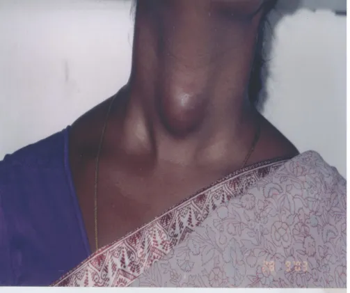
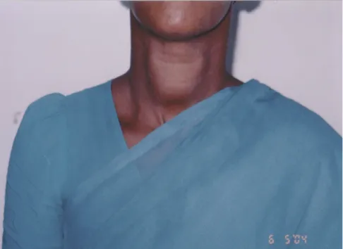
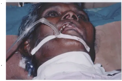
FIG 7(A): A CASE OF ANAPLASTIC CARCINOMA OF THYROID WHO UNDERWENTPREOPERATIVE RADIOTHERAPY FOR
[image:51.595.97.531.390.662.2].
.
‘
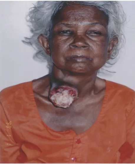
FIG 7(B): THE SAME PATIENTS ON ENDOTRACHEAL TUBE WITH OXYGEN SUPPLEMENTATION POST
[image:52.595.69.495.72.356.2]particularly if the goiter is very vascular, and may cause laryngeal
edema without a tension hematoma. Unilateral or bilateral recurrent nerve
paralysis will not cause immediate post operative respiratory obstruction
unless laryngeal edema is also present, but they will aggravate the
obstruction. If releasing the tension hematoma does not immediately
relieve airway obstruction, the trachea should be intubated at once. An
endotracheal tube can be left in place for several days; steroids are given
to reduce edema and a tracheostomy is rarely necessary. Intubation in the
presence of laryngeal edema may be very difficult and should be carried
out by an experienced anaesthetist. Repeated unsuccessful attempts may
aggravate the problem and, in a crisis, it is safer for the inexperienced
surgeon to perform a needle tracheostomy as a temporary measure; a
Medicut 12G needle (diameter 2.3 mm) is highly satisfactory. Whenever
the trachea is markedly shift and narrow, an elective tracheostomy should
be performed. On occasion, a patient will be anxious and nervous and will
complain of difficulty in breathing with no evidence of strider. Steam
inhalation and sedation may be all that is required. A patient with thyrocardiac
disease will not tolerate any degree of respiratory embarrassment. In such
NEURAL COMPLICATIONS A). RECURRENT LARYNGEAL NERVE DAMAGE
Recurrent laryngeal nerve is susceptible for injury most commonly
Recurrent nerve paralysis may be unilateral or bilateral, transient or
permanent. Transient paralysis 23 occurs in about 2-3% of patients
recovers in 3 weeks to 3 months. .Permanent paralysis23 is extremely
rare (less than 0.1 %). It is largely avoidable if the surgeon routinely seeks
to identify the nerve on each side during all operations on the gland.Mechanism
of injury of the nerves are, ligation of the nerve,
transection of the nerve, clamping (crushing) of the nerve, stretching of the
nerve, and using diathermy. Three most common areas of recurrent
laryngeal nerve injury (TRIPLE STRUCTURE CONCEPT) are inferior
thyroid vein (since recurrent nerve may be anteriorly displaced to it),
ligament of Berry (since tunneling of the recurrent nerve within the
ligament), and inferior thyroid artery (since recurrent nerve can be lie
between branches). By proceeding with meticulous care, deliberate
exposure of the nerves to locate them anatomically; and avoidance of
routine total thyroidectomies, recurrent nerves can be protected from injury. In
the presence of cancer, if preoperative inspection of the vocal
cards demonstrates that paralysis is already present, presumably from
nerve involvement, the nerve may be sacrificed for the sake of more
preserve both recurrent laryngeal nerves, even if this means slightly less
thorough resection of cancerous tissue.Loss of vocal power and huskiness is
often evident for 2 or 3 days after surgery; this is most likely to be due to
edema and is relieved by local anaesthetic lozenges and/ or humidified air.
Persistence of symptoms may indicate neuropraxia, caused by stretching or
crushing of the nerve; this is reversible and recovers over several weeks or
months. Permanent damage will result if the nerve is divided or ligated and is
more likely to occur when the anatomy is distorted, for example with recurrent
or malignant goiters. Unilateral injury may be asymptomatic and pass
undetected due to compensatory hyper adduction of the unaffected cord
unless routine post operative laryngoscopy is performed. Symptomatic
unilateral paralysis improves if the affected cord is stabilized in adducton by
the submucous injection of Teflon under direct laryngoscopy. The effects of
bilateral nerve injury are likely to be temporary but pose an immediate
problem when the patient is extubated at the end of surgery since the
unopposed adductor action the cricothyroid muscles closes the glottis to such
an extent that the least exertion results in obstruction. So look for
inspiratory strider, dyspnoea and minimal dysphon4a and the patient should
be returned 4 promptly to the operating room and emergency endotracheal
intubation should be done and ventilated whilst hydrocortisone is given, 100
mg three times daily, to combate the edema and inflammatory response. The
fails tracheostomy is required. Once a tracheostomy tube has been placed for
unilateral paralysis, it may be plugged when the immediate accompanying
post operative edema has subsided; in most patients the tracheostomy
tube can be removed within 1 or 2 weeks if respiratory distress is not
persistent.
If the tracheostomy tube needs to be retained for a longer time, as in case
of bilateral nerve injury, particularly in the presence of cancer, use of a
TUCKER valve makes wearing of the tracheostomy tube infinitely more
acceptable. Patients with this flap valve may inhale through the
tracheostomy tube but exhale through the larynx so that adequate
phonation may occur, so he can speak adequately.Other surgical options
available are arytenoidopexy or cordectomy and arytenoidectomy. The
permanent nature of laryngeal damage should not be accepted unless it lasts
for more than 9 months. Exploration and resuture of the nerve using
microsurgical anastomosis with 7 or 8:0’ nylon /prolene will prevent cord
atrophy. Epineural anastomosis is superiorfeasible,as in the anastomosis of
the Ansa hypoglossi to the recurrent laryngeal nerves. Alternatively Ansa
neuromuscular pedicle may also be transfered to re innervate the
thyroarytenoids.
B). SUPERIOR LARYNGEAL NERVE PARESIS
A change in the voice, such as loss of high pitch, huskiness, voice
analysis using a visipitch oscilloscope will help to confirm this damage,
which may occur in up to 25% of patients. The majority will recover, since
the nerve has only been stretched. If no improvement is evident after 3
months, it is unlikely to occur. Isolated injury to external branch will lead to no
interference with cord function. To prevent superior laryngeal nerve injury,
upper pole pedicle should be identified and downward traction on the gland
applied, when ligating it. Leave alone if isolated injury occurred.
C).CERVICAL SYMPATHETIC DAMAGE
This rare complication results from deep, forceful retraction on the
carotid sheath, producing Horner’s syndrome. This is notable by the absence
of the vascular dilatation component. The resulting myosis and ptosis are
frequently permanent.
HYPOPARATHYROIDISM
Parathyroid insufficiency is due to removal of the parathyroid glands
or infarction through damage to the parathyroid end artery; often, both
factors occur together. Vascular injury is probably far more important than
inadvertent removal. The incidence of this condition should be less than
0.5%, when a total thyroid lobectomy is performed on one side, exceeding
caution is utilized on the opposite lobe, if operated on at all, to identify and
glands into muscle with some success should not alter this philosophy, as
no data indicate that total removal of the thyroid gland is either desirable or
necessary except for medullary carcinoma. Thus, the mere ability to
compensate for an iatrogenic disaster should not remove our obligation to
prevent such problems by means of more limited but still altogether
successful surgery. (Pathogenesis of temporary hypocalcaemia- is given as a
flow chart). Most cases present dramatically 2-5 days after operation but, very
rarely, the onset is delayed for 2-3 weeks or a patient with marked
hypocalcaemia is asymptomatic. The serum calcium concentration should
be monitored post operatively as patients without overt hypoparathyroidism
may develop vague lethargy,depression, insidious cataract, mental
deterioration, and psychosis. Hypocalcaemia due to parathyroid deficiency
will usually be evident within one week of operation and should be
suspected if the patient appears unduly agitated or depressed, or
hyperventilates. Circum oral tingling is generally the first and most sensitive
indicator of low serum calcium; parasthesia in the fingers and toes
preceding frank tetany is seen when hypocalcaemia is profound. Tapping
over the fascial nerve will cause contraction of the facial muscles
(Chvosteks sign); Carpo pedal spasm, provoked by occlusion of the
circulation to the arm by inflating the sphygmomanometer around the
brachial, above the systolic pressure for 3 minutes, (Trousseau’s sign),
parathyroidism is in distinguishing transient from permanent deficiency23. In
the immediate post operative period, every attempt is made to avoid
intravenous calcium administration, and only rarely is the calcium fall so
rapid or early symptoms so severe that parenteral calcium salts are
required. If intra venous calcium is given, the hypocalcaemia should be
documented by a preliminary blood sample drawn for low calcium
demonstration. Once exogenous calcium is started, the decision to stop
may be difficult; therefore, documentation is required, as episodes of
hyperventilation may symptomatically mimic hypocalcemia . After collecting
a blood sample for calcium estimation, 10 ml of 10 % calcium gluconate (given
slowly to avoid cardiac arrest in systolic) should be given intra
venously, which immediately relieves the symptoms. Subsequently, if the
calcium is above 8.0 mg /100 ml by the sixth post operative day but shows no
signs of improvement and is accompanied by mild symptoms. We would begin
oral effervescent calcium alone in the form if glubionate calcium or calcium
lactate power mixed in a warm liquid,4 to 6 grams daily, depending on
response. If the calcium is over 8.0 mg/100ml, or calcium with vitamin
D3 combinations can be tried.gradually increasing or stable, and not
accompanied by significant symptoms, no medication is given, since the
best stimulus to resumption of parathyroid haemostasis is transient
hypocalcaemia. The patients should be evaluated later in follow up studies,
the calcium is less than 7.5mg/100ml on the sixth post operative day,
particularly if accompanied by a phosphorus of over 5mg/100ml, and such
significant symptoms as carpopedal spasm, we would begin oral vitamin D
in the form of Ergocalciferal (calciferal), 50,000 to 100,000 units per day, as
well as the oral calcium 2 to 3 gram per day are given until a normocalcaemic
state is achieved. Signs and symptoms of hypocalcaemia will recur in these
patients at times of metabolic stress, such as pregnancy or the menopause.
Since most hypoparathyroidism will be transient, patients given vitamin D
should stop receiving that medication and the oral calcium after about 3
months, and they should be observed for a prolonged period. Vitamin D
activity may persist for several months; therefore, repeated calcium and
phosphorus determinations should be performed over a period of atleast a
further 6 months before final determination is made regarding permanent
hypoparathyroidism.
THYROID CRISIS OR STORM
Fortunately thyroid storm is a rarity nowadays, thanks to effective
and the improved methods of control of thyrotoxicosis. It is an acute
exacerbation of hyperthyroidism, due to sudden release of thyroid
hormones into circulation, with secondary effects of increased
catacholamines, explaining the whole spectrum of clinical features, such as high
fever, extreme tachycardia (atrial fibrillation ), extreme restlessness,