B-cell antigen receptor signaling is initiated upon binding of the antigen to membrane-bound immunob-lobulin (Ig), and the anti-Ig antibody (Ab) mimics this signaling. In B cells latently infected with Epstein-Barr virus (EBV), the same signals induce virus activation. We examine here whether rheumatoid factors (RFs), autoantibodies directed against the Fc portion of IgG, induce EBV and B-cell activation. As a source of RFs, RF-producing lymphoblastoid cell line (LCL) clones were isolated from peripheral blood mononuclear cells (PBMC) and synovial cells from patients with rheumatoid arthritis (RA) by EBV transformation. Burkitt’s lymphoma-derived Akata cells, which are highly responsive to EBV activation by anti-Ig Abs, were used for the assay of EBV activation. Akata cells expressed IgG3 as membrane-bound Ig. RFs from a synovium-derived LCL were directed to IgG3 and induced EBV activation in 16 to 18% of Akata cells, whereas RFs from another synovium-derived LCL were directed to IgG1 and did not induce EBV activation. Pretreatment of RFs with the purified Fc fragment of human IgG completely abolished EBV activation. Furthermore, B-cell activation was assessed by incorporation of [3H]thymidine. RFs from synovium-derived LCLs efficiently induced B-cell
activation, and the addition of CD40 ligand had a synergistic effect. On the other hand, RFs from PBMC-derived LCLs were polyreactive, had a lower affinity to IgG, and did not induce EBV and B-cell activation. The present findings imply a possible role for RFs as EBV and B-cell activators.
Epstein-Barr virus (EBV) is a human herpesvirus, which infects the majority of the human population and is the caus-ative agent of infectious mononucleosis. After primary infec-tion, EBV persists in B cells in a latent state for the life of the host (15). Various reagents have been found to induce virus activation in latently EBV-infected B cells in vitro. They in-clude halogenated pyrimidine (13), phorbol ester (42), anti-immunoglobulin (Ig) antibody (Ab) (9, 32, 37), and butyrate (20). Although we do not know the physiological stimuli that control activation of the virus productive cycle and the switch from the latency in vivo, anti-Ig treatment, which activates B-cell antigen receptor (BCR) signaling (39), would serve as a more physiologically relevant activator.
Rheumatoid arthritis (RA) is a chronic inflammatory dis-ease of unknown cause (17), and EBV has long been suggested as a causative agent. RA patients have a higher frequency and/or higher levels of antibodies to EBV in serum than do normal individuals (1, 7, 11, 30). It has been reported that the number of circulating B cells infected with EBV is increased in RA patients (36). More recently, we have reported that there is an extremely high EBV load and that EBV replication oc-curs in the synovial tissue of RA patients (34). Besides EBV activation, B cells are also activated in RA patients (18). Rheu-matoid factors (RFs) are autoantibodies directed against the
Fc portion of IgG (14, 21) and are found in the sera of most patients with RA. It is well established that there is a direct correlation between the severity of the disease and the titer of these antibodies in patients with RA (19, 43). Although the precise pathogenic role of RF in RA has not been defined, a great deal of evidence suggests its participation in sustaining inflammatory synovitis (5, 38, 43).
Anti-Ig Abs efficiently induce lytic virus replication in some Burkitt’s lymphoma (BL) cell lines, including Akata (32, 33), which expresses a limited number of EBV latent gene prod-ucts, including EBV-determined nuclear antigen 1 (EBNA1), two EBV-encoded small RNAs known as EBER1 and EBER2, the rightward transcripts from the BamHI A region (BARTs), and a very small amount of latent membrane protein 2A (LMP2A) (termed type I latency) (15, 31). On the other hand, EBV-immortalized lymphoblastoid cell lines (LCLs) are unre-sponsive to EBV induction by anti-Ig Abs. LMP2A, which is expressed in a high amount in LCLs, is known to interfere with EBV activation after BCR cross-linking (23, 24). Therefore, low LMP2A expression is important for efficient EBV activa-tion in anti-Ig-treated cells (16). The high-level expression of LMP2A in LCLs is caused by transactivation of the EBNA2 protein (41). On the other hand, BL cells are negative for EBNA2 expression and express little or no LMP2A. The anal-ysis of peripheral blood lymphocyte by PCR showed that only EBNA1 and LMP2A were expressed in EBV latency in vivo (8, 25, 27, 35). Although the level of LMP2A expression in pe-ripheral lymphocytes has not been measured quantitatively, the absence of EBNA2 expression suggests a low level LMP2A
* Corresponding author. Mailing address: Department of Tumor Virology, Institute for Genetic Medicine, Hokkaido University, N15 W7, Kita-ku, Sapporo 060-0815, Japan. Phone: 81-11-706-5071. Fax: 81-11-706-7540. E-mail: kentaka@igm.hokudai.ac.jp.
9918
on November 8, 2019 by guest
expression in these cells. Therefore, BL cells with type I latency are likely to represent in vivo latency. Hence, by using Akata cells, we investigated whether RF could induced virus activa-tion. The results indicated that RFs induce EBV and B-cell activation.
MATERIALS AND METHODS
Cell culture.BL-derived EBV-positive Akata cells were maintained in RPMI 1640 medium containing 10% fetal bovine serum and antibiotics at 37°C in 5% CO2.
RF-producing cell lines.RF-producing cell lines were generated by EBV transformation of peripheral blood mononuclear cells (PBMC) or single cells from the synovial tissue of RA patients. The cells were incubated in the culture supernatant of the B95-8 cell line and resuspended in GIT medium (Nihonsei-yaku Co., Ltd., Tokyo, Japan). The cells were transferred to 96-well plates (Costar Corp., Cambridge, Mass.) at 200 cells/well for PBMC and 2,000 cells/well for synovial cells. X-ray-irradiated (5,000 rads) allergenic PBMC were added as feeder cells at 5⫻104cells/well. Cells were cultured for 4 weeks. RFs were
purified from the culture supernatant by using HiTrap IgM purification columns (Amersham Pharmacia Biotech AB, Uppsala, Sweden).
Abs and reagents.The Abs used for EBV activation and B-cell activation were a rabbit polyclonal Ab to human IgG (␥-chain-specific) (Dako, Copenhagen, Denmark), F(ab⬘)2fragment of mouse monoclonal Ab (MAb) to the Fc
frag-ment of human IgG (Jackson ImmunoResearch, West Grove, Pa.), and the F(ab⬘)2fragment of mouse MAb to the Fab fragment of human IgG (Jackson
ImmunoResearch). Ordinarily, they were used at a concentration of 7.5g/ml. Other Abs included mouse MAb to phosphotyrosine (Cell Signaling, Beverly, Mass.), rabbit polyclonal Abs to phospho-Syk and phospho-ERK (Cell Signal-ing), a mouse MAb to EBV BZLF1 (Dako), and an MAb to EBV gp350 (C1, kindly provided by T. Sairenji). The purified Fc fragment of human IgG and purified Fab fragment of human IgG were purchased from Jackson Immuno-Research, and CD40 liganc (CD40L) was from PEPRO Tech (Rocky Hill, N.J.). Immunofluorescence assay.Expression of EBV lytic antigens was examined on acetone-fixed cells by the indirect immunofluorescence method with MAb C1 specific to the EBV glycoprotein gp350. The second Ab was a fluorescein iso-thiocyanate (FITC)-conjugated F(ab⬘)2fragment of rabbit Ab to mouse IgG
(Dako).
Determination of calcium mobilization.Akata cells (106/ml) were loaded with
4M fluo-3AM (Molecular Probes, Eugene, Oreg.) at room temperature for 30 min. Cells were washed twice with phosphate-buffered saline and resuspended at the same concentration in 500l of fresh medium for each sample. Baseline calcium release was measured for 30 s and 500l of purified IgM RF or medium containing 15g of anti-Fc Abs/ml was then added to the cell suspension. The intracellular calcium levels were measured by using flow cytometry (Becton Dickinson, Franklin Lakes, N.J.).
Immunoblot analysis.Twenty micrograms of cell lysate was separated by sodium dodecyl sulfate–10% polyacrylamide gel electrophoresis and transferred to a nitrocellulose membrane (Schleicher & Schuell, Dassel, Germany). The membrane was incubated overnight at 4°C with the first Ab and then reacted with horseradish peroxidase-conjugated sheep Ab to mouse IgG (diluted at 1:5,000) or horseradish peroxidase-conjugated donkey Ab to rabbit IgG (diluted at 1:2,000) (Amersham Bioscience Corp., Piscataway, N.J.).
Determination of IgG subclass in Akata cells.Akata cells were incubated with FITC-conjugated goat anti-human IgG1, IgG2, IgG3, or IgG4 at a concentration of 1:50 in 37°C for 1 h. The cells were washed with PBS containing 1% bovine serum albumin, followed by flow cytometric analysis.
Affinity determination by inhibition ELISA.To determine the affinities of RFs and the anti-Fc Ab, we used a standard competitive inhibition enzyme-linked immunosorbent assay (ELISA), in which soluble human IgG Fc from 10⫺10M to
10⫺5M were preincubated with a fixed amount of RFs at 4°C overnight. This
mixture of IgG Fc-RF was then centrifuged, and the supernatant was transferred to a plate coated with human IgG Fc (MBL, Tokyo, Japan). The plates were incubated at 37°C for 2 h, followed by a wash with washing buffer. Then, 100l of alkaline phosphatase-conjugated goat anti-human IgM or goat anti-mouse IgG Abs was added to each well at a dilution of 1:6,000 and incubated for 30 min at 37°C. Plates were washed, and the substrate was added. The results were plotted as the percent bound versus the concentration of the competitor. The percent bound was calculated by using the optical density and taking the reading without a competitor as 100% bound.
B-cell purification and assay of B-cell activation.PBMC were separated from adult peripheral blood by using Histopaque (Sigma, St. Louis, Mo.). B cells were purified from PBMC with anti-human CD19 magnet beads (Dynal ASA). A total of 200l of a cell suspension (2⫻105/well) was cultured in RPMI 1640 medium
supplemented with 10% fetal bovine serum and antibiotics in 96-well tissue culture plates. Cells were stimulated with 30g of anti-IgG, anti-Fab, or anti-Fc Abs/ml or 200g of RFs/ml for 2 h, and then 3g of CD40L/ml was added to the culture. After 48 h of incubation, entry into the cell cycle was assessed by measurement of incorporation of [3H]thymidine (ICN Biomedicals Canada, Inc.,
Mississauga, Ontario, Canada) after a 16- h pulse with 0.5Ci of [3H]thymidine.
RESULTS AND DISCUSSION
Activation of latently infected EBV by anti-Fc Ab.By using Akata cells, we investigated whether RF could induce virus activation. First, we studied whether an MAb to the Fc portion of human IgG (anti-Fc Ab) could induce EBV activation. Akata cells were treated with an anti-Fc Ab for 24 h, and the expression of viral glycoprotein gp350, which is expressed at a late stage of EBV lytic infection (15), was examined by immu-nofluorescence assay. As shown in Fig. 1A, the anti-Fc Ab
FIG. 1. Anti-Fc Ab induces EBV activation in the latently EBV-infected B-cell line Akata as efficiently as anti-Fab Ab does. (A) Immuno-fluorescence assay showing induction of an EBV lytic protein, gp350, after treatment with 7.5g of anti-IgG, anti-Fab, and anti-Fc Abs/ml for 24 h. (B) Dose response of gp350 expression showing that anti-Fc Ab induces gp350 expression as efficiently as anti-Fab Ab.
on November 8, 2019 by guest
induced lytic infection as efficiently as the anti-IgG polyclonal Ab and an MAb against the Fab portion of human IgG (anti-Fab Ab). Anti-Fc and anti-(anti-Fab Abs gave similar dose-response curves in their ability to induce EBV activation (Fig. 1B), suggesting that signals from the Fab and Fc portions of IgG were equally potent as EBV activators.
Activation of latently infected EBV by RFs.Based on these findings, we studied whether RF could induce EBV activation. To obtain RFs, B cells from PBMC and single cells prepared from the synovial tissue of patients with RA were infected with EBV (B95-8 strain) and cultured for 4 weeks in the wells of 96-well plates at 200 and 2,000 cells/well, respectively (3, 12). Two of each of the EBV-transformed LCLs producing RFs were chosen as a source of RFs. RFs were purified from the culture supernatant by using an IgM affinity column and were added to the Akata cell culture at 100 g/ml. After 24 h of cultivation, the expression of gp350 was examined by using an immunofluorescence assay. As shown in Fig. 2A, RFs pro-duced from a synovium-derived LCL (SY1) inpro-duced gp350 in ⬃8% of Akata cells, whereas RFs produced from another synovium-derived LCL (SY2) and RFs from two PBMC-de-rived LCLs (PBMC1 and PBMC2) had no substantial gp350 induction (⬍0.2% [data not shown]). The dose-response ex-periment indicated that SY1 RFs gave a maximum gp350 in-duction of ca. 16 to 18% at concentrations of 180 to 220g/ml
(Fig. 2B). EBV activation by SY1 RFs was further confirmed by detection of an EBV lytic protein, BZLF1 (15), by immu-noblot analysis (Fig. 2C). Pretreatment of SY1 RFs with the purified Fc fragment of human IgG completely abolished BZLF1 induction, whereas pretreatment with the Fab frag-ment of human IgG did not (Fig. 2C), suggesting that EBV activation was induced by specific binding of RFs to the Fc portion of cell membrane Ig.
Activation of BCR signaling by RFs.It is known that cross-linking of BCR is followed by phosphorylation of tyrosine kinases and elevation of the intracellular calcium concentra-tion (10, 29), and both are required for EBV activaconcentra-tion (9). We therefore studied whether RFs could stimulate these pathways. Immunoblot analysis revealed that SY1 RFs induced phos-phorylation of tyrosine kinases, including Syk and ERK (Fig. 3A). Calcium mobilization of SY1 RF-treated Akata cells was also examined by flow cytometry. The results showed that SY1 RFs induced enhanced calcium influx (Fig. 3B).
Lower affinity of RFs to Fc fragment of IgG than anti-Fc Ab.
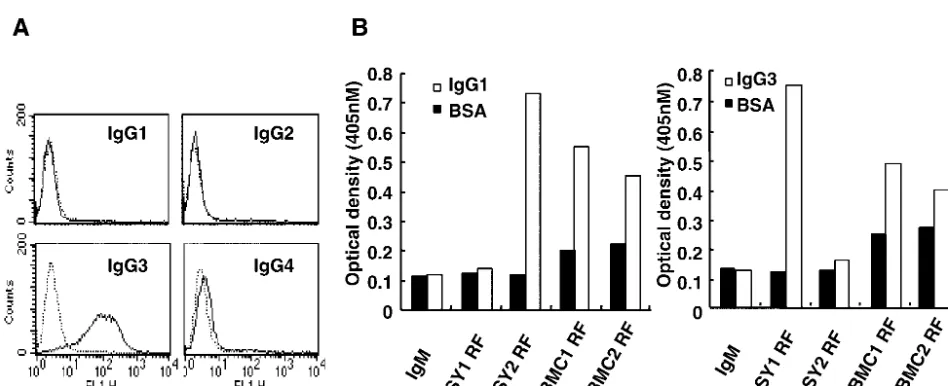
[image:3.603.117.487.93.367.2]The flow cytometric analysis indicated that Akata cells ex-pressed IgG3 among four subclasses of IgG: IgG1, IgG2, IgG3, and IgG4 (Fig. 4A). RFs used in the present studies were examined to determine their reactivities with IgG subclasses. The results indicated that SY1-RFs, which induced EBV acti-vation, reacted with IgG3 and not with IgG1, whereas SY2
FIG. 2. RFs produced from a synovium-derived LCLs (SY1) induce EBV activation in latently EBV-infected B-cell line Akata. Anti-Fc Ab is used as a positive control. (A) Immunofluorescence assay showing induction of an EBV lytic protein, gp350, after treatment with synovium-derived RFs, SY1 RFs, at 100g/ml for 24 h; (B) dose response of gp350 expression showing that SY1 RFs induce maximum expression of gp350 at 180 to 220g/ml; (C) immunoblot analysis showing induction of an EBV lytic protein, BZLF1, after treatment with SY1 RFs for 24 h. Pretreatment of SY1 RFs with the Fc fragment of IgG completely abolished the BZLF1-inducing ability of RFs.
on November 8, 2019 by guest
RFs, which did not induce EBV activation, reacted with IgG1 but not with IgG3 (Fig. 4B). On the other hand, PBMC-de-rived RFs reacted with both IgG1 and IgG3, and weakly re-acted with bovine serum albumin as well. These results are consistent with previous reports that most RFs produced from PBMC are polyreactive (3, 12).
Although anti-Fc Abs induced lytic infection in ca. 70% of Akata cells, SY1 RFs induced EBV activation in 16 to 18% of the cells, and PBMC-derived RFs could not induce EBV acti-vation in spite of their reactivity with IgG3. Furthermore, dose-response analysis indicated that the maximum EBV induction by SY1 RFs was obtained at a concentration of 180 g/ml, whereas the maximum induction by the anti-Fc Ab was ob-tained at a concentration of 7.5g/ml. To examine the possi-bility that RFs had a lower affinity to Fc fragments of IgG than
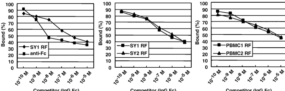
the anti-Fc Ab, competitive inhibition ELISA was performed. RFs or anti-Fc Abs were first incubated with purified Fc frag-ment of human IgG at various concentrations. The concentra-tion of free RFs or anti-Fc Ab was then determined by an indirect ELISA. As a result, the affinities of SY1 RF and SY2 RF were 7.1⫻ 10⫺7M and 1.9⫻10⫺6M, respectively, and
were ca. 100 times lower than that of the anti-Fc Ab (8.9⫻ 10⫺9 M), and the affinities of PBMC1 RF and PBMC2 RF
were 4.5⫻10⫺6M and 7.0⫻10⫺6M, respectively, and were
ca. 1,000-times lower than that of the anti-Fc Ab (Fig. 5).
[image:4.603.115.478.72.244.2]B-cell activation by RFs.Finally, we examined whether RFs could induce B-cell activation. B cells were purified from adult peripheral blood by using anti-human CD19 magnetic beads and were treated with RFs (200g/ml) with or without addi-tion of CD40L, which is necessary for efficient activaaddi-tion of
FIG. 3. RFs produced from synovium-derived LCLs (SY1) induce phosphorylation of tyrosine kinases and elevation of the intracellular calcium concentration in latently EBV-infected B-cell line Akata. Anti-Fc Ab is used as a positive control. (A) Immunoblot analysis showing phosphor-ylation of tyrosine kinases after treatment with SY1 RFs (100g/ml); (B) flow cytometry showing elevation of intracellular calcium levels after treatment with SY1 RFs (100g/ml).
FIG. 4. Synovium-derived RFs are monoreactive and LCL-derived RFs are polyreactive for IgG subclasses. (A) Flow cytometric analysis showing that Akata cells express the IgG3 subclass on the cell membrane; (B) RFs produced by synovium-derived LCLs (SY1 and SY2) react with IgG3 or IgG1, and PBMC-derived RFs (PBMC1 and PBMC2) react with both IgG1 and IgG3.
on November 8, 2019 by guest
[image:4.603.56.530.500.693.2]peripheral B cells (2, 18). After 48 h of treatment, the cells were incubated with [3H]thymidine for 16 h, and its
incorpo-ration was measured. As shown in Fig. 6, synovium-derived RFs alone efficiently induced B-cell activation, and the addi-tion of CD40L had a synergistic effect. On the other hand, PBMC-derived RFs did not induce B-cell activation (data not
shown). Figure 6 also shows that anti-Fc and anti-Fab Abs were equally potent as B-cell activators.
We report here that RFs can stimulate EBV lytic replication and polyclonal B-cell activation. This finding is quite conceiv-able because anti-Fc Abs induce EBV and B-cell activation as efficiently as anti-Fab Abs do. Although the degree of EBV and B-cell activation by SY RFs was lower than that by the anti-Fc Ab, this can be attributed to the difference in the specificity and the affinity to the Fc fragment of IgG; i.e., SY1 RFs reacted with IgG3, SY2 RFs reacted with IgG1, and the affinity of SY RFs was ca. 100 times lower than that of anti-Fc Ab. The 1,000-fold-lower affinity of PBMC-derived RFs com-pared to that of anti-Fc Abs would explain why they could not induce EBV and B-cell activation. Cross-linking the RFs by anti-human IgM Abs did not increase their ability to activate EBV (data not shown). These results also indicate that the differences in affinity of the various RFs described here could account for the failure of some RF to activate B cells.
It has been reported that RFs derived from RA patients include monoreactive and polyreactive RFs (4). Monoreactive RFs bind with relatively high affinity and have specificity for the IgG Fc fragment, whereas polyreactive RFs bind to a number of different self (e.g., ssDNA, Ins, Tg, etc) and non-self (e.g., TT and bacterial lipopolysaccharide) antigens (26). Al-though the molecular basis of these cross-reactivities has not been conclusively answered (6), many studies indicate that the VHand VLgene segments of monoreactive high-affinity Abs
harbor more somatic mutations than the VH and VL gene
segments of polyreacitve low-affinity Abs, which is close to unmutated “germ line” genes (22, 28, 40).
Although we tested 10 RF-positive sera from patients with RA, only two sera induced EBV activation (5 and 1%, respec-tively) (data not shown). Since RFs in sera are bound by serum IgG, they must have lower IgG-binding capabilities. Moreover, several reports demonstrated that RFs produced by rheuma-toid synovial cells had greater reactivity to the IgG3 subclass, in contrast to serum RF, which had greater reactivity to the hu-man IgG1 subclass (29). Further observations indicated that synovium-derived RFs had greater affinity than PBMC-derived RFs (3). Because the synovium is the central site of
patholog-FIG. 5. RFs have a lower affinity to Fc fragment of IgG than anti-Fc Ab. Competitive inhibition ELISA showing that synovium-derived RFs (SY1 and SY2) and PBMC-derived RFs (PBMC1 and PBMC2) have a lower affinity to the Fc fragment of IgG than anti-Fc Ab.
FIG. 6. RFs induce peripheral B-cell activation. Purified B cells (5 ⫻ 105) from adult peripheral blood were incubated with RFs (200
g/ml) and anti-IgG, anti-Fab, and anti-Fc Abs (30 g/ml) with or without the addition of CD40L (3g/ml) for 48 h and then pulsed with 0.5Ci of [3H]thymidine for 16 h. (Upper panel) Synovium-derived
RFs, SY1 RF and SY2 RF, induce B-cell activation; (lower panel) anti-Fc Ab induces B-cell activation as efficiently as anti-Fab Ab.
on November 8, 2019 by guest
[image:5.603.57.530.70.222.2]of patients with rheumatoid arthritis. J. Clin. Investig.67:1134–1140. 2. Armitage, R. J., W. C. Fanslow, L. Strockbine, T. A. Sato, K. N. Clifford,
B. M. Macduff, D. M. Anderson, S. D. Gimpel, T. Davis-Smith, C. R. Mal-iszewski, E. A. Clark, C. A. Smith, K. H. Grabstein, D. Cosmon, and M. K. Spriggs.1992. Molecular and biological characterization of a murine ligand for CD40. Nature357:80–82.
3. Burastero, S. E., P. Casail, R. L. Wilder, and A. L. Notkins.1988. Monore-active high affinity and polyreMonore-active low affinity rheumatoid factors are pro-duced by CD5⫹B cells from patients with rheumatoid arthritis. J. Exp. Med. 168:1979–1992.
4. Burastero, S. E., M. Cutolo, V. Dessi, and F. Celada.1990. Monoreactive and polyreactive rheumatoid factors produced by in vitro Epstein-Barr virus-transformed peripheral blood and synovial B lymphocytes from rheumatoid arthritis patients. Scand. J. Immunol.32:347–357.
5. Carson, D. A., J. L. Pasquali, C. D. Tsoukas, S. Fong, S. F. Slovin, S. Lawrance, L. Strominger, and J. H. Vaughan.1981. Physiology and pathol-ogy of rheumatoid factors. Springer Semin. Immunopathol.4:161–179. 6. Carayannopoulos, M. O., K. N. Potter, Y. Li, J. B. Natvig, and J. D. Capra.
2000. Evidence that human immunoglobulin M rheumatoid factors can be derived from the nature antibody pool and undergo an antigen driven im-mune response in which somatically mutated rheumatoid factor have lower affinities for immunoglobulin G Fc than their germline counterparts. Scand. J. Immunol.51:327–336.
7. Catalano, M. A., D. A. Carson, S. F. Slovin, D. D. Richman, and J. H. Vaughan.1979. Antibodies to Epstein-Barr virus-determined antigens in normal subjects and in patients with seropositive rheumatoid arthritis. Proc. Natl. Acad. Sci. USA76:5825–5828.
8. Chen, F., J. Zou, L. di Renzo, G. Winberg, L. Hu, E. Klein, G. Klein, and I. Ernberg.1995. A subpopulation of normal B cells latently infected with Epstein-Barr virus resembles Burkitt lymphoma cells in expressing EBNA-1 but not EBNA-2 or LMP1. J. Virol.69:3752–3758.
9. Daibata, M., R. E. Humphreys, K. Takada, and T. Sairenji.1990. Activation of latent EBV via anti-IgG-triggered, second messenger pathways in the Burkitt’s lymphoma cell line Akata. J. Immunol.144:4788–4793. 10. DeFranco, A. L.1997. The complexity of signaling pathways activated by the
BCR. Curr. Opin. Immunol.9:296–308.
11. Ferrell, P. B., C. T. Aitcheson, G. R. Pearson, and E. M. Tan.1981. Sero-epidemiological study of relationships between Epstein-Barr virus and rheu-matoid arthritis. J. Clin. Investig.67:681–687.
12. Hakoda, M., T. Ishimoto, S. Hayashimoto, K. Inoue, A. Taniguchi, N. Ka-matani, and S. Kashiwazaki.1993. Selective infiltration of B cells committed to the production of monoreactive rheumatoid factor in synovial tissue of patients with rheumatoid arthritis. Clin. Immunol. Immunopathol.69:16–22. 13. Hampar, B., J. G. Derge, L. M. Martos, and J. L. Walker.1972. Synthesis of Epstein-Barr virus after activation of the viral genome in a “virus-negative” human lymphoblastoid cell (Raji) made resistant to 5-bromodeoxyuridine. Proc. Natl. Acad. Sci. USA69:78–82.
14. Jefferis, R., and R. A. Mageed.1989. The specificity and reactivity of rheu-matoid factors with human IgG. Monogr. Allergy26:45–60.
15. Kieff, E., and A. B. Rickinson.2002. Epstein-Barr virus, p. 2579–2582.In
B. N. Fields, D. M. Knipe, and P. M. Howley (ed.), Fields virology, 4th ed. Lippincott-Raven, Philadelphia, Pa.
16. Konishi, K., S. Maruo, H. Kato, and K. Takada.2001. Role of Epstein-Barr virus-encoded latent membrane protein 2A on virus-induced immortaliza-tion and virus activaimmortaliza-tion. J. Gen. Virol.82:1451–1456.
17. Krause, A., T. Kamradt, and G. R. Burmester.1996. Potential infectious agents in the induction of arthritides. Curr. Opin. Rheumatol.8:203–209.
Bolen, and E. Kieff.1995. Integral membrane protein 2 of Epstein-Barr virus regulates reactivation from latency through dominant-negative effects on protein-tyrosine kinases. Immunity2:155–166.
25. Miyashita, E. M., B. Yang, G. J. Babcock, and D. A. Thorley-Lawson.1997. Identification of the site of Epstein-Barr virus persistence in vivo as a resting B cells. J. Virol.71:4882–4891.
26. Nakamura, M., S. E. Burastero, A. L. Notkins, and P. Casali.1988. Human monoclonal rheumatoid factor-like antibodies from CD5 (Leu-1)⫹B-cell are polyreactive. J. Immunol.140:4180–4186.
27. Qu, L., and D. Rowe.1992. Epstein-Barr virus latent gene expression in uncultured peripheral blood lymphocytes. J. Virol.66:3715–3724. 28. Radoux, V., P. P. Chen, J. A. Sorge, and D. A. Carson.1986. A conserved
human germline Vgene directly encodes rheumatoid factor light chains. J. Exp. Med.164:2119–2124.
29. Robbins, D. L., W. F. Benisek, E. Benjamini, and R. Wister.1987. Differ-ential reactivity of rheumatoid synovial cells and serum rheumatoid factors to human immunoglobulin G subclasses 1 and 3 and their CH3 domains in rheumatoid arthritis. Arthritis Rheum.30:489–497.
30. Sculley, T. B., P. J. Walker, D. J. Moss, and J. H. Pope.1984. Identification of multiple Epstein-Barr virus-induced nuclear antigens with sera from pa-tients with rheumatoid arthritis J. Virol.52:88–93.
31. Shimizu, N., H. Yoshiyama, and K. Takada.1996. Clonal propagation of Epstein-Barr virus (EBV) recombinants in EBV-negative Akata cells. J. Vi-rol.70:7260–7263.
32. Takada, K.1984. Cross-linking of cell surface immunoglobulins induces Epstein-Barr virus in Burkitt lymphoma lines. Int. J. Cancer33:27–32. 33. Takada, K., and Y. Ono.1989. Synchronous and sequential activation of
latently infected Epstein-Barr virus genomes. J. Virol.63:445–449. 34. Takeda, T., Y. Mizugaki, L. Matsubara, S. Imai, T. Koike, and K. Takada.
2000. Lytic Epstein-Barr virus infection in the synovial tissue of patients with rheumatoid arthritis. Arthritis Rheum.43:1218–1225.
35. Tierney, R. J., N. Steven, L. S. Young, and A. B. Rickinson.1994. Epstein-Barr virus latency in blood mononuclear cells: analysis of viral gene tran-scription during primary infection and in the carrier state. J. Virol.68:7374– 7385.
36. Tosato, G., A. D. Steinberg, R. Yarchoan, C. A. Heilman, S. E. Pike, V. De Seau, and R. M. Blaese.1984. Abnormally elevated frequency of Epstein-Barr virus-infected B cells in the blood of patients with rheumatoid arthritis. J. Clin. Investig.73:1789–1795.
37. Tovey, M. G., G. Lenoir, and J. Begon-Lours.1978. Activation of latent Epstein-Barr virus by antibody to human IgM. Nature276:270–272. 38. Vaughan, J. H.1993. Pathogenetic concepts and origins of rheumatoid factor
in rheumatoid arthritis. Arthritis Rheum.36:1–6.
39. Weiss, A., and D. R. Littman.1994. Signal transduction by lymphocyte antigen receptors. Cell76:263–274.
40. Williams, D. G., S. P. Moyes, and R. A. Mageed.1999. Rheumatoid factor isotype switch and somatic mutation variants within rheumatoid arthritis synovium. Immunology98:123–136.
41. Zimber-Strobl, U., E. Kremmer, F. Grasser, G. Marchall, G. Laux, and G. W. Bornkamm.1993. The Epstein-Barr virus nuclear antigen 2 interacts with an EBNA2 responsivecis element of the terminal protein 1 gene promoter. EMBO J.12:167–175.
42. zur Hausen, H., F. J. O’Neill, U. K. Freese, and E. Hecker.1978. Persisting oncogenic herpesvirus induced by the tumor promoter TPA. Nature272: 373–375.
43. Zvaifler, N. J.1973. The immunopathology of joint inflammation in rheu-matoid arthritis. Adv. Immunol.16:265–336.