0022-538X/06/$08.00⫹0 doi:10.1128/JVI.80.4.1724–1733.2006
Copyright © 2006, American Society for Microbiology. All Rights Reserved.
Characterization of the Role of Very Late Expression Factor 1 in
Baculovirus Capsid Structure and DNA Processing
Adam L. Vanarsdall, Kazuhiro Okano, and George F. Rohrmann*
Department of Microbiology, Oregon State University, Corvallis, Oregon 97331-3804
Received 26 September 2005/Accepted 15 November 2005
Very late expression factor 1 (VLF-1) ofAutographa californicamultiple nucleopolyhedrovirus is a putative tyrosine recombinase and is required for both very late gene expression and budded virus production. In this report, we show that avlf-1knockout bacmid was able to synthesize viral DNA at levels similar to that detected for agp64knockout bacmid that served as a noninfectious control virus. Additionally, analysis of replicated bacmid DNA by field-inversion gel electrophoresis indicated that VLF-1 is not required for synthesizing high-molecular-weight intermediates that could be resolved into unit-length genomes when cut at a unique restriction site. However, immunoelectron microscopic analysis revealed that in cells transfected with avlf-1
knockout bacmid, aberrant tubular structures containing the capsid protein vp39 were observed, suggesting that this virus construct was defective in producing mature capsids. In contrast, rescuing thevlf-1knockout bacmid construct with a copy of VLF-1 that carries a mutation of a highly conserved tyrosine (Y355F) was sufficient to restore the production of nucleocapsids with a normal appearance, but not infectious virus production. Furthermore, the results of a DNase I protection assay indicated that the DNA packaging efficiency of the VLF-1(Y355F) virus construct was similar to that of thegp64knockout control. Finally, a recombinant virus containing a functional hemagglutinin epitope-tagged version of VLF-1 was constructed to investigate the association of VLF-1 with the nucleocapsid. Analysis by immunoelectron microscopy of Sf-9 cells infected with this virus showed that VLF-1 localized to an end region of the nucleocapsid. Collectively, these results indicate that VLF-1 is required for normal capsid assembly and serves an essential function during the final stages of the DNA packaging process.
The familyBaculoviridaeconsists of a diverse group of
rod-shaped viruses that contain circular covalently closed (ccc) double-stranded DNA genomes that range in size from 80 to 180 kbp. Baculovirus infections are restricted to invertebrates,
with the most well-studied example, Autographa californica
multiple nucleopolyhedrovirus (AcMNPV), being pathogenic for insects of the order Lepidoptera. Upon entry of AcMNPV into a susceptible host, replication occurs in cell nuclei to generate two virus phenotypes. Budded virions (BV) are pro-duced from nucleocapsids that become enveloped during egress through the plasma membrane, which is modified by the viral fusion protein GP64. In contrast, occlusion-derived viri-ons are produced from nucleocapsids that remain in the nu-cleus, where they are enveloped prior to becoming occluded within a crystalline matrix comprised of polyhedrin. BV are associated with systemic infection, whereas occlusion-derived virions mediate lateral transmission between insects when re-leased into the environment upon the death of the host.
It has been determined through the use of a transient rep-lication assay that six baculovirus gene products are required for viral DNA replication (12, 28). These include an activator of transcription (ie-1), a helicase (p143), a DNA polymerase
(dnapol), and three late expression factors (lef genes), i.e., a
primase (lef-1), a primase accessory factor (lef-2), and a
single-stranded DNA binding protein (lef-3). More recently, thelef-11
gene product was shown to be required for the replication of
bacmid DNA in tissue culture (18). Although these data pro-vide insight into the putative baculovirus replisome required to synthesize viral DNA, very little is known about the mode by which baculoviruses replicate their DNA or about the gene products involved in processing DNA intermediates to gener-ate infectious genomes. Previous reports have suggested that replication may proceed via a rolling-circle mechanism that could generate large head-to-tail concatemers (14, 24). Addi-tionally, the fact that many baculovirus genomes contain ho-mologous regions and encode enzymes related in both
se-quence and function to theRed homologous recombination
system entices one to envisage that baculoviruses employ a recombination-based replication strategy like that described for other large double-stranded DNA viruses that generate highly branched intermediate structures (6, 13, 16, 19, 20). Shared between both models is the requirement of processing DNA intermediates in order to generate monomeric genomes that can be packaged. A viral protein that might be involved in this process is very late expression factor 1 (VLF-1). Found in all baculovirus genomes sequenced to date, VLF-1 was origi-nally identified from a temperature-sensitive mutant defective in occlusion body formation and subsequently shown to serve as a transcriptional activator by stimulating the expression of
two very late genes,p10andpolh(21, 31). Sequence analysis
indicated that VLF-1 is a member of the tyrosine recombinase
family of proteins, represented by the integrase (int) of
phage, that function to catalyze DNA rearrangements through recombination (1, 21). A common feature among members of this family is an absolutely conserved nucleophilic tyrosine responsible for forming a covalent phosphodiester bond with substrate DNA and initiating strand exchange between two
* Corresponding author. Mailing address: Department of Microbi-ology, Nash Hall Room 220, Oregon State University, Corvallis, OR 97331-3804. Phone: (541) 737-1793. Fax: (541) 737-0496. E-mail: [email protected].
1724
on November 8, 2019 by guest
http://jvi.asm.org/
homologous target sites. Attempts to rescue a virus containing a mutation of this conserved tyrosine in VLF-1 were unsuc-cessful, suggesting that VLF-1 likely retains the catalytic activ-ity that defines this family of proteins (32). Investigations into this putative activity through in vitro studies revealed that VLF-1 could bind to DNA substrates that mimicked recombi-nation junctions in a non-sequence-specific manner, but they failed to exhibit enzymatic activity (22).
Previous characterization of a bacmid lacking VLF-1 indi-cated that it was not infectious when transfected into Sf-9 cells due to an apparent defect in budded virus production, al-though DNA synthesis and late gene expression were observed (17, 29). This phenotype suggests that VLF-1 may be involved in DNA transactions during the late stages of the replication cycle, namely, processing of DNA intermediates, or may facil-itate packaging into viral capsids. Therefore, to assist in eluci-dating the essential function of VLF-1 and to advance our understanding of the mechanisms involved in baculovirus rep-lication, we continued our investigation of VLF-1 using the AcMNPV bacmid system. The results described herein dem-onstrate that VLF-1 is not required for normal levels of DNA synthesis or for the production of replication intermediates that display a similar pattern to that of a wild-type (wt) control virus when analyzed by field-inversion gel electrophoresis (FIGE). However, VLF-1 is required for the production of normal capsid particles that contain nuclease-resistant viral DNA, and this requirement appears to be exclusive of its activity as a putative recombinase. In addition, our results indicate that the association of VLF-1 with the nucleocapsid is localized to an end region.
MATERIALS AND METHODS
Cells and antibiotics.Spodoptera frugiperda(Sf-9) cells were cultured in Sf-900 II serum-free medium (Invitrogen) with added penicillin G (50 units/ml), strep-tomycin (50 units/ml; Whittaker Bioproducts), and amphotericin B (Fungizone [375 ng/ml]; Invitrogen) as previously described (10).
Bacmid construction, purification, and transfection.Avlf-1knockout bacmid was generated using theRed homologous recombination system inEscherichia coliand has been described previously (29). For this study, the same methods were used to construct agp64knockout bacmid. Briefly, primers gp64catF (5⬘ -TACAATTTTTTATTATTACATTAATAATGATACAACATATGAATATC CTCCTTAG-3⬘) and gp64catR (5⬘-TACTAGTAAATCAGTCACACCAAGGC TTCAATAAGGTGTAGGCTGGAGCTGCTTC-3⬘) were used to generate a linear chloramphenicol acetyltransferase (CAT) marker cassette from plasmid pKD3 (7) and to include 50 nucleotides of sequence homology to the 5⬘and 3⬘
regions of thegp64open reading frame (ORF). This fragment was subsequently electroporated intoE. coliDH10B cells containing bacmid bMON14272 (In-vitrogen) and plasmid pKD46 encoding theRed recombination genesgam,
beta, andexo(7). Potential bacmid knockout clones were selected on LB plates containing chloramphenicol, and isolated colonies were screened by PCR with primers Ac107700 (5⬘-GCCCTATTCCTATGGCCATA-3⬘) and Ac110240 (5⬘ -TGAGCGCCCATAAGCAAGTC-3⬘).
Several transfer plasmids were constructed for transposing gene sequences into the polyhedrin (polh) locus of bacmid constructs following the Bac-to-Bac protocol (Invitrogen), and these are outlined in Fig. 1A. The transfer plasmid pfbie-GFP was used to transpose a green fluorescent protein (GFP) marker gene intovlf-1andgp64knockout bacmids to generate constructsvlf-1-KO andgp64 -KO, respectively. This entailed generating a 1.3-kb fragment from pegfp (25) with primers ieGFPF (5⬘-GGTACCGATGTCTTTGTGATGCGCG-3⬘) and ieGFPR (5⬘-GGTACCTTACTTGTACAGCTCGTCC-3⬘). This fragment in-cluded a GFP reporter gene derived from plasmid pEGFPN-1 (Clontech) under the control of the AcMNPV immediate early (ie-1) promoter (AcMNPV coor-dinates 126600 to 127197). The resulting fragment was cloned into pCR2.1-TOPO (Invitrogen), excised by KpnI digestion, and ligated into the KpnI site of pFastBac Dual (Invitrogen), which had previously been digested with SmaI and
BamHI to remove thepolhandp10promoters. The transfer plasmid pfbie-GUS(Eco81I) was used to transpose a-glucuronidase (GUS) reporter gene, in addition to an Eco81I restriction site, into thevlf-1andgp64knockout bacmids to generate the constructsvlf-1-KO(Eco81I)andgp64-KO(Eco81), respectively. The
transfer plasmid pfbie-GUS (29) was modified to include an Eco81I restriction site that is not otherwise found in the AcMNPV bacmid. (Previously, AvrII has been used for such analyses [22], but the bacmid construct contains two such sites.) This was accomplished by annealing the oligonucleotide 5⬘-TCGACCCT AAGGG-3⬘, followed by ligation into the XhoI restriction site of pfbie-GUS. To generate an epitope-tagged VLF-1 repair construct (VLF-1–HA), avlf-1 knock-out bacmid was transposed with the transfer plasmid pfbvlf-1 as described pre-viously (22). A second VLF-1 repair construct, VLF-1(Y335F), was made in which tyrosine 355 of VLF-1 was changed to phenylalanine. For this construct, a region of thevlf-1ORF was excised from plasmid pXA7(Y355F) (32) by diges-tion with HpaI and BstEII, ligated into plasmid pfbvlf-1, described above, to generate the transfer plasmid pfbvlf-1(Y355F), and transposed into avlf-1 knock-out bacmid. To generate the gp64 repair construct (gp64-repair), primers Ac108179 (5⬘-TTAATATTGTCTATTACGGT-3⬘) and Ac110240 (5⬘-TGAGC GCCCATAAGCAAGTC-3⬘) were used to amplify thegp64ORF and native promoter region. This PCR fragment was cloned into plasmid pCR2.1-TOPO (Invitrogen), excised by XbaI and HindIII digestion, inserted into pfbie-GFP as described above to generate the transfer plasmid pfbgp64-repair, and subse-quently transposed into agp64knockout bacmid. The table in Fig. 1A summa-rizes the transfer plasmids used to transpose DNA fragments into parental bacmids to generate the virus constructs used for this study.
Bacmid DNAs were purified from 0.5-liter cultures by using a Large-Construct purification kit (QIAGEN) according to the manufacturer’s instructions, and 2
g of purified DNA was used for transfecting Sf-9 cells (0.9⫻106cells/well)
seeded in a six-well plate via a cationic liposome method (4). Briefly, bacmid DNA was mixed with 200l of Sf-900 II medium containing 10l of liposomes and incubated at 27°C for 30 to 45 min. After incubation, the DNA solution was increased to 1 ml with Sf-900 II medium, overlaid onto freshly plated Sf-9 cells, transferred to 27°C, and allowed to incubate for 4 h. After 4 h of incubation, the transfection medium was removed, and the cells were replenished with 2 ml of fresh Sf-900 II medium and returned to 27°C.
Q-PCR DNA replication assay.To assess viral DNA replication, a quantitative real-time PCR (Q-PCR) assay was performed as previously described (30). To prepare total DNA for analysis, transfected Sf-9 cells were harvested in 1 ml phosphate-buffered saline (PBS), lysed in 500l cell lysis buffer (10 mM Tris, pH 8.0, 100 mM EDTA, 20g/ml RNase A, 0.5% sodium dodecyl sulfate), and incubated for 30 min at 37°C before the addition of 80g/ml of proteinase K and continued incubation overnight at 65°C. Total DNA was phenol extracted, eth-anol precipitated, and suspended in 300l of water. Prior to PCR, 10l of total DNA from each time point was digested with 10 units of DpnI restriction enzyme (Fermentas) for 24 h in a 50-l total reaction volume. Quantitative PCR was performed with 1l of digested DNA added to Platinum SYBR green qPCR SuperMix UDG (Invitrogen) according to the manufacturer’s instructions and was analyzed on an ABI Prism 7000 sequence detection system under the fol-lowing conditions: 50°C for 2 min, 95°C for 2 min, and 45 cycles of 95°C for 30 s and 60°C for 30 s, with a 500 nM concentration of each primer.
Field inversion gel electrophoresis.Sf-9 cells were transfected as described above, and at the indicated time points, cells were harvested, washed once with PBS, and mixed with 1% low-melting-point agarose to a final cell density of 3⫻ 106cells/200l. The mixture was poured into a plug mold, resulting in a 20- by
9- by 1.5-mm agarose plug that was cut into 8 to 10 pieces. The agarose plugs were treated with 10 mM Tris-HCl (pH 8.0), 100 mM EDTA, 1%N-lauroyl sarcosine, and 200g/ml proteinase K at 50°C overnight. After being washed several times with 10 mM Tris-HCl (pH 8.0), samples were stored at 4°C until further use. Digestion of DNA was performed with 20 U of DpnI and 30 U of Eco81I per sample in a 100-l reaction volume and incubated at 37°C overnight. Finally, the plugs were inserted into the loading wells of a 1% pulsed-field-certified agarose gel (Bio-Rad) in 0.5⫻TBE buffer (45 mM Tris-borate, pH 8.0, 1 mM EDTA). DNAs were separated by FIGE using an MJ Research PPI-200 programmable pulse inverter with program 4 (initial reverse time, 0.05 min; reverse increment, 0.01 min; initial forward time, 0.15 min; forward increment, 0.03 min; number of steps, 81; reverse increment, 0.001 min; forward increment, 0.003) run at 8 V/cm for 17 h at 4°C. MidRange PFG marker I (New England Biolabs) was used as a DNA size marker. The DNAs were transferred to a nylon membrane and hybridized with viral genomic DNA as described previously (29).
Immunoelectron microscopy.Sf-9 cells were either transfected as stated above or infected at a multiplicity of infection of⬃5, harvested at 72 h posttransfection (hpt) or postinfection (hpi), and prepared for immunoelectron microscopy as described previously (26). A mouse monoclonal antibody to the hemagglutinin
on November 8, 2019 by guest
http://jvi.asm.org/
(HA) epitope tag (Babco) was used at a dilution of 1:50, and the monoclonal antibody to AcMNPV, vp39 (a generous gift from Loy Volkman), was used as an undiluted tissue culture supernatant. The goat anti-mouse immunoglobulin G 10-nm-gold-conjugated secondary antibody was used at a dilution of 1:50. Images were obtained with a Phillips EM 300 electron microscope.
DNase I protection assay.Sf-9 cells were transfected in triplicate and har-vested in PBS as described above, and each cell pellet was initially suspended in 50l of supplemented RSB buffer (20 mM Tris-HCl [pH 7.4], 10 mM KCl, 1.5 mM MgCl, 1 mM CaCl, 0.5% NP-40, 100g/ml phenylmethylsulfonyl fluoride [PMSF], 20g/ml RNase A), split into two equal 25-l portions that were either left untreated (total DNA) or treated with DNase I (MP Biomedicals) at 100
g/ml (encapsidated DNA), and incubated at 37°C for 1 h. For BV controls, virions were collected in triplicate from 6 ml of budded virus stock by centrifu-gation at 12,000⫻gat 4°C and suspended in 300l RSB buffer. Each sample was subsequently split into three 100-l aliquots that were either untreated, treated with DNase I (100g/ml), or treated with proteinase K (80 mg/ml) for 20 min followed by treatment with DNase I (100g/ml). (PMSF was included in the RSB buffer, except for the final aliquot, in which PMSF was added after incu-bation with proteinase K.) For an intracellular capsid control, a 50-ml culture of Sf-9 cells (⬃1.5 ⫻106
cells/ml) was infected with wt-GUS virus stock at a multiplicity of infection of⬃2, harvested at 72 hpi, suspended in 5 ml of sup-plemented RSB buffer, and disrupted by sonication (three 10-s bursts). The cell lysate was then clarified by centrifugation at 8,000⫻g for 20 min, and the supernatant was removed and loaded onto a 25% sucrose column. The nucleo-capsids were collected by ultracentrifugation through the 25% sucrose column at 24,000 rpm in a Beckman SW28 rotor at 4°C, suspended in 350l of supple-mented RSB buffer, and divided into six 50-l aliquots. Three samples were left untreated, and three were treated with DNase I as described above for trans-fected cells. For all samples, after the 1-h incubation at 37°C, the suspension volume was increased twofold with RSB buffer and with EDTA to a final con-centration of 40 mM. Samples were then treated with an equal volume of 2⫻cell lysis buffer (20 mM Tris-HCl [pH 7.4], 80 mM EDTA, and 1% sodium dodecyl sulfate) plus 80g/ml proteinase K and incubated for 4 h at 65°C. DNAs were extracted with phenol-chloroform, precipitated with an equal volume of 2-pro-panol, and suspended in 201 water. Viral DNAs were quantified by using the conditions described for the Q-PCR DNA replication assay, with 1l of the DNA sample added to the reaction mixture.
RESULTS
Construction and characterization of gp64 knockout
bac-mid.To construct agp64knockout bacmid, a chloramphenicol
resistance marker gene was designed to recombine with the
gp64ORF via theRed recombination system inE. coli.The
target site corresponded to AcMNPV coordinates 108179 to
109729, with the intention of deleting the entiregp64coding
sequence. Deletion of the gp64 ORF from the bacmid was
confirmed by PCR analysis with primers designed to amplify genomic DNA spanning the recombined locus outlined in Fig. 1B. The results of this analysis indicated that when PCR was
performed with agp64 knockout bacmid as the template, a
1.9-kb PCR product was generated, whereas a 2.5-kb product
FIG. 1. Organization of transfer plasmids used to generate virus constructs and confirmation of agp64knockout bacmid. (A) Organi-zation of transfer plasmids used for transposing gene sequences into thepolhlocus of bacmid constructs. The diagrams outline the transfer plasmids containing the selected marker genes, the vlf-1 and gp64 repair genes, and the Eco81I restriction site; pie-1 refers to the AcMNPV immediate-early promoter, ppolhrefers to the AcMNPV polyhedrin promoter, and pgp64refers to the native promoter located upstream of thegp64ORF. The table summarizes the bacmid con-structs used for this report that were derived by transposing parental bacmids with the indicated transfer plasmids. The virus construct
wt-GUS has been described previously (29). (B) Confirmation ofgp64 gene deletion by recombination. The top diagram outlines thegp64 locus in the unmodified bacmid (diagram 1), and the bottom diagram outlines the identical location in thegp64knockout bacmid (diagram 2) after homologous recombination with the chloramphenicol resis-tance marker gene (CAT). The arrows indicate the relative annealing positions for primers Ac107700 and Ac110240, used to amplify this region, and the dashed lines indicate the sizes of the expected PCR products from each template. The panel shows an ethidium bromide-stained agarose gel of the PCR products generated using the afore-mentioned primer set to confirm the deletion of thegp64ORF. Lane 1 is the PCR product generated from the unmodified bacmid, and lane 2 is the PCR product generated from thegp64knockout bacmid. M indicates a 1-kb DNA size marker (Invitrogen).
on November 8, 2019 by guest
http://jvi.asm.org/
was produced when an unmodified (wt) bacmid was used as the
template, confirming that thegp64ORF was replaced with the
CAT marker gene (Fig. 1B).
To confirm that deletion of the gp64ORF eliminated the
ability of the bacmid to propagate in cell culture, thegp64-KO
bacmid containing the GFP marker gene was transfected into Sf-9 cells and monitored for GFP expression. The expression of GFP could only be observed in a small fraction of isolated cells within the monolayer by 96 hpt, and no GFP expression was observed in cells incubated with the transfection superna-tant by 72 hpi (data not shown). To ensure that the inability of this construct to initiate cell-to-cell infection was not due to unintentional mutations acquired during the cloning proce-dures or to a disruption of regulatory elements, Sf-9 cells were
transfected with agp64repair bacmid and monitored for GFP
expression as described above. Cells transfected with thegp64
repair bacmid showed widespread fluorescence in the initial transfection at 96 hpt and in cells incubated with the transfec-tion supernatant at 72 hpi. Additransfec-tionally, a growth curve
anal-ysis showed that BV production of thegp64repair bacmid was
similar to that of a wt control virus (data not shown).
There-fore, these results confirmed that deletion of thegp64ORF in
the context of a bacmid abolished the ability of the virus to propagate in cell culture.
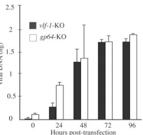
Analysis of viral DNA replication.To determine if VLF-1 is required for generating normal levels of nascent viral DNA within transfected cells, a DNA replication assay was
per-formed, with thegp64knockout bacmid described above
serv-ing as the control virus. Because the viral fusion protein GP64 is required for nucleocapsids to egress from infected cells (23),
this mutant construct is similar to avlf-1knockout bacmid in
that it lacks the ability to initiate cell-to-cell infection, but all other replication processes should be unaffected. DNA repli-cation was assessed using a highly sensitive and quantitative assay involving real-time PCR and DpnI digestion to discrim-inate between input and replicated DNAs. The results of this analysis are shown in Fig. 2 and indicate that although some
variability at early times was observed, the bacmid lackingvlf-1
was able to synthesize similar amounts of nascent DNA as the
bacmid lackinggp64by 72 and 96 hpt. These data confirm that
the phenotype of the vlf-1knockout does not result from a
defect in the level of DNA synthesis.
Analysis of replicated viral DNA by FIGE.Since the data
described above indicated that avlf-1knockout could
synthe-size normal levels of viral DNA, it was then of interest to
characterize the replicated DNA relative to thegp64knockout
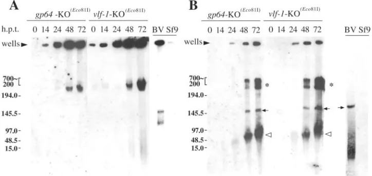
control. For these experiments, FIGE was performed, which is suitable for separating DNA molecules with high molecular weights. Initial experiments were intended to characterize the replicated bacmid DNA in its native form without restriction digestion. An analysis of in situ-processed Sf-9 cells transfected
with thegp64-KO(Eco81)control bacmid from 0 to 72 hpt under
these conditions indicated that as nascent viral DNA accumu-lated, the majority of this DNA remained in the wells of the agarose gel (Fig. 3A). Similarly, this was the case when viral DNA
was analyzed from cells transfected with the vlf-1-KO(Eco81)
bacmid under the same conditions (Fig. 3A). Although a band
was present at 48 and 72 hpt for both the gp64 and vlf-1
knockout samples of the⬎250-kb size marker, this DNA is not
predicted to represent linearized monomeric genomes due to its high molecular weight (Fig. 3A). Similarly, most of the DNA from BV was also trapped in the well (Fig. 3A). Addi-tionally, two bands were present that migrated between 100 and 150 kb for the uncut BV control. However, it should be noted that because bacmid DNA and baculovirus genomes are circular DNA molecules, they are likely impaled by agarose fibers during electrophoresis, which prevents them from enter-ing the gel or causes them to migrate aberrantly, as reported for other circular DNA molecules (5, 15). Therefore, it is not possible to draw conclusions regarding the sizes of patterns for uncut DNA. However, our data do demonstrate similar elec-trophoretic patterns of replicated DNAs from cells transfected
with either thevlf-1knockout or the control.
The next set of FIGE experiments intended to characterize replicated bacmid DNA after treatment with the single-cutting restriction enzyme Eco81I (see Materials and Methods). An
analysis of DNA from cells transfected with agp64knockout
bacmid after Eco81I digestion indicated that a large propor-tion of the viral DNA that was previously trapped in the wells was able to migrate through the gel to produce a distinct band representing unit-length genomic DNA that first appeared at 48 hpt and gradually increased in concentration by 72 hpt (Fig.
3B,gp64-KO(Eco81)). A similar band was observed with DNA
purified from budded virus- but not mock-transfected Sf-9 cells under the same conditions, confirming the classification of this band as unit-length genomic DNA (Fig. 3B, BV lane). Simi-larly, a band representing unit-length genomic DNA was
ob-served when cells transfected with a vlf-1 knockout bacmid
were analyzed, and this band also first appeared by 48 hpt and
increased by 72 hpt (Fig. 3B,vlf-1-KO(Eco81)). Two additional
bands were observed at 48 and 72 hpt for cells transfected with
either thevlf-1orgp64knockout bacmid. These included DNA
that migrated as less than unit length (Fig. 3B,gp64-KO(Eco81)
and vlf-1-KO(Eco81)) and a relatively high-molecular-weight
species that migrated⬎250 kb (Fig. 3B, gp64-KO(Eco81)and
vlf-1-KO(Eco81)), the latter of which has been confirmed to
[image:4.585.94.236.66.200.2]consist of viral DNA that has been completely digested with Eco81I (unpublished data). Therefore, similar to the FIGE analysis of uncut DNA, these FIGE gels demonstrate that after
FIG. 2. Real-time PCR analysis of viral DNA replication in trans-fected Sf-9 cells. At the designated times, total DNAs were isolated from Sf-9 cells transfected with the indicated bacmid constructs, di-gested with the restriction enzyme DpnI to eliminate input bacmid DNA, and analyzed by real-time PCR using SYBR green I. Values are displayed as averages for transfections performed in triplicate, with error bars indicating standard deviations.
on November 8, 2019 by guest
http://jvi.asm.org/
digestion with the single-cutting enzyme Eco81I, thevlf-1and
gp64knockouts produce similar patterns of DNA.
Electron microscopic (EM) analysis of transfected cells.
VLF-1 has previously been shown to be associated with nu-cleocapsids by Western blot analysis, suggesting a possible role in capsid assembly (32). To investigate this possibility, immu-noelectron microscopy was performed with thin sections gen-erated from bacmid-transfected cells. Control experiments
performed with thegp64-KO bacmid revealed cells in which
the nucleus became enlarged and reorganized into an electron-dense virogenic stroma typical of baculovirus infection. As expected, these cells also had rod-shaped nucleocapsids that reacted with a monoclonal antibody to the vp39 major capsid protein, confirming that nucleocapsid synthesis was not inhib-ited by their inability to egress from the cell (Fig. 4A). Nucleo-capsids were also observed in bundle formations that are typ-ically seen in baculovirus-infected cells. Observations of cells
transfected with thevlf-1-KO bacmid also revealed cells with
enlarged nuclei and virogenic stroma; however, in contrast to
cells transfected with thegp64-KO control bacmid, these cells
contained clusters of elongated tube-like structures that local-ized to the inner nuclear membrane and reacted specifically with the vp39 antibody, suggesting that they are related to capsids (Fig. 4B). Additionally, these structures appeared more electron translucent than capsids found in cells
trans-fected with thegp64knockout bacmid, although in some cases
a few electron-dense structures could be discerned within these clusters (Fig. 4C, arrows). A cross section of these structures indicated that they lack an electron-dense core that would be indicative of nucleoprotein (Fig. 4D, arrowheads).
To determine if the aberrant capsid structures observed in
cells transfected with thevlf-1knockout bacmid were the result
of a structural or enzymatic deficiency associated with VLF-1, a VLF-1(Y355F) construct containing a point mutation of the
highly conserved tyrosine 355 was analyzed by electron micros-copy. Although this mutation in VLF-1 has not previously been characterized in the context of a bacmid, our analysis agreed with a previous report that suggested it could prevent infec-tious virus production (32; data not shown). Electron micro-scopic analysis of cells transfected with the VLF-1(Y355F) bacmid revealed the presence of electron-dense rod-shaped nucleocapsids that were morphologically indistinguishable
from nucleocapsids observed in cells transfected with thegp64
control bacmid (Fig. 4E). However, the nucleocapsids in these cells appeared to be more heavily concentrated within the virogenic stroma than those in control transfected cells. We
also observed that, whereas ingp64-KO-transfected cells both
[image:5.585.111.473.71.243.2]single and bundled nucleocapsids could be seen dispersed be-tween the perimeter of the virogenic stroma and the nuclear membrane, no nucleocapsids could be seen in this region in cells transfected with the VLF-1(Y355F) bacmid, suggesting that they remained tethered to the virogenic stroma (compare Fig. 4A and F). Together, these data show that VLF-1 is required for proper capsid assembly and for nucleocapsids to be released from the virogenic stroma.
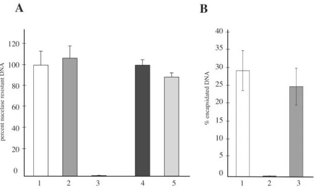
DNase I protection assay. Because the results described above indicated that the VLF-1(Y355F) bacmid appeared as
proficient in nucleocapsid production as agp64knockout
bac-mid, a DNase I protection assay was performed to assess the ability of this repaired virus to encapsidate viral DNA. This involved treating transfected cells with DNase I and quantify-ing the encapsidated nuclease-resistant viral DNA by real-time PCR. To confirm that encapsidated baculovirus DNA is pro-tected from nuclease digestion, control experiments were per-formed with budded virions isolated from infected cell super-natants as well as nucleocapsids purified from infected cell lysates. The results of this assay showed that the level of viral DNA detected from budded virions treated with DNase I was
FIG. 3. Analysis of viral DNA by FIGE. (A) Southern blot of total DNA from in situ-processed Sf-9 cells transfected with either the gp64-KO(Eco81)orvlf-1-KO(Eco81)bacmid. The agarose-embedded plugs were processed and separated in a 1% Tris-borate-EDTA–agarose gel,
using a pulsed-field gel electrophoresis apparatus as described in Materials and Methods. For all samples, the DNA was digested with the restriction enzyme DpnI to eliminate input bacmid DNA. Times posttransfection are indicated above the blot (hpt), and the Mid-Range PFG DNA size marker (New England Biolabs) is indicated in kilobases on the left. (B) An identical Southern blot to that shown in panel A, except that cell samples were treated with the restriction enzymes DpnI and Eco81I during processing. BV indicates viral DNA extracted from budded viruses, and Sf-9 indicates mock-transfected cells. The solid arrows indicate unit-length genomic DNAs, the asterisks indicate viral DNAs that migrated as larger than unit length, and the open arrowheads indicate subgenomic-sized DNA fragments generated after Eco81I digestion. The blots were probed with AcMNPV genomic DNA using the alkaline phosphatase direct labeling system (Amersham).
on November 8, 2019 by guest
http://jvi.asm.org/
FIG. 4. Electron microscopic analysis of transfected Sf-9 cells stained with vp39 antibody to characterize VLF-1 mutants. (A) Image showing a portion of a cell transfected with thegp64-KO bacmid as a control. The arrows indicate nucleocapsid bundles dispersed along the outer regions of the nucleus. The inset shows nucleocapsid bundles observed in the same cell at a higher magnification. (B) Image of the nucleus of a cell transfected with avlf-1-KO bacmid showing tubular capsid-like structures staining for vp39. (C) Image of the nucleus of a cell transfected with a vlf-1-KO bacmid showing electron-translucent tubular structures with a few examples that are electron dense (arrows). (D) Image showing a cross section of a nucleocapsid bundle located in the nucleus of a vlf-1-KO-transfected cell. The arrowheads indicate tubular structures that appear to lack an electron-dense core. (E) Image of the nucleus of a cell transfected with the VLF-1(Y355F) bacmid showing abundant electron-dense nucleocapsids associated with the virogenic stroma. (F) Image showing a portion of the nucleus from a cell transfected with the VLF-1(Y355F) virus indicating that, in contrast to the case for panel A, no nucleocapsids were observed outside the virogenic stroma. For all samples, sections were generated from cells harvested at 72 hpt and stained with a primary antibody to AcMNPV p39 as an undiluted tissue culture supernatant. The secondary gold-conjugated antibody was used at a 1:50 dilution. For images B, C, and D, the bar represents 0.25m; for images A and F, the bar represents 0.5m; and for image E, the bar represents 1m. VS, virogenic stroma; nm, nuclear membrane.
on November 8, 2019 by guest
http://jvi.asm.org/
essentially identical to the level of viral DNA detected from virions that were left untreated (Fig. 5A, bars 1 and 2). In contrast, when the virions were initially digested with protein-ase prior to DNprotein-ase I digestion, the level of nucleprotein-ase-resistant viral DNA was reduced to background levels (Fig. 5A, bar 3). Similarly, nucleocapsids that were purified from cell lysates also showed nearly complete resistance to nuclease when an-alyzed under similar conditions (Fig. 5A, bars 4 and 5). There-fore, these results validate the experimental conditions and confirm that DNAs from both budded virions and intracellular nucleocapsids are resistant to nuclease digestion. This strategy was then extended to bacmid-transfected cells to assess the amount of encapsidated viral DNA relative to the amount of total replicated viral DNA. An analysis of cells transfected with
thegp64knockout bacmid serving as the control showed that
28% of the total replicated viral DNA was encapsidated (Fig. 5B, bar 1). In contrast, and in agreement with the aberrant electron-translucent capsid structures observed by electron mi-croscopy, the level of nuclease-resistant viral DNA in cells
transfected with thevlf-1knockout was⬍1% (Fig. 5B, bar 2).
Interestingly, when the ability of the VLF-1(Y355F) bacmid to encapsidate viral DNA was assessed under these conditions, the amount of encapsidated viral DNA was similar to that
detected for thegp64knockout bacmid, in that⬃24% of the
total replicated viral DNA was resistant to nuclease digestion (Fig. 5B, bar 3). Therefore, although it is essential for budded virus production, both the EM and DNase I protection exper-iments clearly indicate that tyrosine 355 of VLF-1 is not re-quired for viral DNA to be packaged into nucleocapsids.
EM analysis of VLF-1 localization on nucleocapsids. Be-cause VLF-1 has previously been shown to be associated with nucleocapsids of both budded viruses and occlusion-derived
viruses, determining where VLF-1 localizes on the viral parti-cle might provide insight into its essential function. Therefore, immunoelectron microscopy was performed with the virus
VLF-1–HA, in which the wild-typevlf-1open reading frame
was replaced with a version ofvlf-1containing an HA epitope
tag. Notably, this virus was able to generate titers similar to those of an unmodified control virus, indicating that the epitope tag does not interfere with the essential functions of VLF-1 (data not shown). Electron microscopic examination of thin sections generated from cells infected with VLF-1–HA at 72 hpi revealed nucleocapsids with gold labeling localized to one end (Fig. 6A), and this staining pattern could be seen among several nucleocapsids within a given field, indicating that gold labeling of the nucleocapsid was not due to random interactions (Fig. 6B). Additionally, a longitudinal section of nucleocapsids arranged in a bundled formation characteristic of baculovirus-infected cells showed gold labeling at only one end, suggesting that VLF-1 may preferentially localize to ei-ther the apical or basal end region (Fig. 6C). In contrast, although some gold-stained nucleocapsids were observed in sections from cells infected with the wt-GUS control virus that lacks the epitope-tagged version of VLF-1, this staining was less likely to be end specific and was primarily observed in regions with a high background (Fig. 6D). Therefore, these results suggest that the association of VLF-1 on the nucleo-capsid is localized to an end region.
DISCUSSION
[image:7.585.137.450.68.255.2]Previous data indicated that a bacmid lacking VLF-1 could only replicate DNA to 10% the level in a nonmutant control bacmid when transfected into Sf-9 cells (29). This decrease,
FIG. 5. DNase I protection assay to detect encapsidated viral DNA. (A) Control experiments performed with budded virions or nucleocapsids isolated from infected cells. For budded virions, viral DNA was extracted from virions after they were either left untreated as a control (bar 1), treated with DNase I (bar 2), or treated with proteinase K followed by DNase I (bar 3). For purified cellular nucleocapsids, viral DNA was extracted from virions after they were either left untreated (bar 4) or treated with DNase I (bar 5). Viral DNAs were quantified by real-time PCR. The results are displayed as percentages of nuclease-resistant DNA, which were determined by comparing the amounts of DNA from treated samples with the amounts of DNA from the untreated controls, set at 100%. Values represent the averages and standard deviations from triplicate samples. (B) Percentages of nuclease-protected viral DNA from mutant bacmids at 72 hpt. Cells transfected with either thegp64knockout control (bar 1), thevlf-1knockout (bar 2), or the VLF-1(Y355F) repair bacmid (bar 3) were either left untreated (total DNA) or treated with DNase I (encapsidated DNA). The viral DNAs were subsequently isolated and quantified by real-time PCR. The values are represented as percentages of encapsidated DNA and were obtained by dividing the mean amounts of DNase I-resistant DNA by the mean of total viral DNA from triplicate samples.
on November 8, 2019 by guest
http://jvi.asm.org/
however, was at least partly due to the fact that VLF-1 is required for budded virus production and can therefore only replicate in initially transfected cells. To compensate for this limitation and to better understand the effect VLF-1 has on nascent viral DNA synthesis, comparisons in this study were
made to a gp64 knockout bacmid generated to serve as a
“wild-type” control virus that should replicate its DNA
nor-mally but, similar to a bacmid lackingvlf-1, be limited to
ini-tially transfected cells. An analysis of DNA replication by means of a quantitative real-time PCR assay determined that
by the end of a 96-h time course, a bacmid lackingvlf-1could
replicate its DNA to similar levels to those detected for agp64
knockout control, indicating that VLF-1 is dispensable for nor-mal levels of viral DNA synthesis. However, the fact that VLF-1 was not required to synthesize normal levels of viral DNA did not exclude the possibility that VLF-1 is required to generate higher-order intermediates that can then be pro-cessed into unit-length genomes. To investigate this possibility, FIGE was performed to characterize the DNA replicated by a
vlf-1knockout bacmid. Our initial analysis of intact genomes
indicated that DNA isolated from cells transfected with either
thegp64control orvlf-1knockout bacmid failed to migrate into
the gel but instead accumulated in the agarose wells as the time course progressed. A similar observation has been reported for herpes simplex virus type 1 replication intermediates, and it is thought that this results from viral DNA being nonlinear or highly branched (20, 27). Additionally, when FIGE was used to analyze viral DNA from transfected cells after treatment with the single-cutting restriction enzyme Eco81I, similar DNA pat-terns were observed, further confirming that VLF-1 is not required to produce replication intermediates that resemble
those produced by thegp64control virus.
To investigate whether the failure of avlf-1knockout bacmid
to produce budded viruses was due to a defect in nucleocapsid assembly, electron microscopy was performed with bacmid-transfected cells. The results of these experiments
demon-strated that in cells transfected with thevlf-1knockout bacmid,
elongated rod-like structures could be found grouped along the inner nuclear membrane. Since these structures were shown to react specifically with a monoclonal antibody to the major capsid protein vp39, our assessment is that these struc-tures represent incomplete capsid particles. It was initially
FIG. 6. Immunogold localization of VLF-1 in thin sections of infected Sf-9 cells at 72 hpi. (A to C) Images of cells infected with budded viruses from the recombinant bacmid (VLF-1–HA) containing avlf-1ORF fused to an HA epitope tag. (D) Image of a cell infected with budded viruses from a recombinant bacmid containing the nativevlf-1ORF without the epitope tag, serving as a control. For all samples, sections were stained with a primary monoclonal antibody to the HA epitope at a 1:50 dilution and a secondary gold-conjugated antibody at a 1:50 dilution. Bar, 0.25 m.
on November 8, 2019 by guest
http://jvi.asm.org/
thought that these aberrant capsids represent precursor struc-tures awaiting nucleic acids and that the lack of DNA process-ing by VLF-1 inhibited their ability to acquire DNA. This scenario would fit well with a previously proposed model (8) which suggests that viral DNA is packaged into a preassembled capsid sheath. However, these aberrant capsid structures ap-peared to be substantially longer than the average length of individual capsid particles, indicating that further processing may be required to generate legitimate precursors. Addition-ally, it is thought that DNA replication and packaging occur in the nucleus within the centrally located virogenic stroma, yet these aberrant capsid structures remained apart from the vi-rogenic stroma, localizing instead to the inner nuclear mem-brane. Therefore, a likely scenario is that these aberrant capsid structures remain incomplete due to the lack of VLF-1, sug-gesting that VLF-1 is an essential capsid component.
Electron microscopy was then performed with cells trans-fected with the recombinant virus VLF-1(Y355F). This mu-tant, which would be predicted to be inactive in its putative recombinase function, showed abundant nucleocapsid particles within the virogenic stroma, with no discernible difference in phenotype from nucleocapsids observed in cells transfected
with thegp64knockout control. The VLF-1(Y355F) virus also
possessed the ability to package viral DNA with an efficiency
similar to that of thegp64knockout control, as indicated by a
DNase I protection assay. These results are interesting, not only for the fact that they confirm that VLF-1 is required for capsid production, but also because they indicate that the pu-tative enzymatic activity of VLF-1 is not required to process DNA intermediates as a prerequisite for encapsidation. How-ever, the fact that the DNA-containing capsids observed in cells transfected with the VLF-1(Y355F) repair bacmid failed to initiate subsequent infection does confirm that the active-site tyrosine is essential for generating infectious budded virus. Possible explanations for why the repair virus was unable to generate infectious particles may be that the capsids are filled with defective genomes incapable of replicating in new cells or that the capsid-associated DNA requires additional processing by VLF-1 to complete the replication cycle. Because we were unable to detect budded virus particles in the supernatant from cells transfected with the VLF-1(Y355F) virus, the data sug-gest that VLF-1 is involved in a very-late-stage processing mechanism required for nucleocapsid egress. Although the type of processing mechanism this may entail remains unclear, the association of VLF-1 with the end region of the nucleo-capsid suggests that VLF-1 may act in conjunction with the packaging process. It has been proposed that VLF-1 may serve as a site-specific endonuclease that cleaves concatameric DNA to generate unit-length DNA molecules for packaging (22). This would be analogous to the terminase enzyme of herpes-viruses and bacteriophages (2, 3, 11). However, the observation that viral DNA could be encapsidated by the VLF-1(Y355F) bacmid indicates that genomic DNA can be packaged without such activity being performed by VLF-1. Therefore, it is likely that VLF-1 is involved in a very-late-stage processing mecha-nism such as terminating the packaging process. A failure to terminate this process might block nucleocapsid egress and may explain why the nucleocapsids synthesized by the VLF-1(Y355F) bacmid appeared to be heavily concentrated or trapped within the virogenic stroma, as opposed to the more
dispersed pattern observed with the control virus. Similarly, VLF-1 could function as a DNA resolvase on branched DNA structures generated during the replication process that would require processing during packaging in order to incorporate complete genomes, and the failure to resolve such structures would also likely lead to an arrest in packaging. Interestingly, a similar mechanism has been described for endonuclease VII of T4 bacteriophage and has also been shown to interact with a component of the packaging machinery (9).
ACKNOWLEDGMENTS
We thank Loy Volkman for the monoclonal antibody to AcMNPV p39, A. Lorena Passarelli for the pXA7Y355F plasmid, and Victor Mikhailov for comments and suggestions during this research.
This research was supported by National Institutes of Health grant GM060404 (to G.F.R).
REFERENCES
1.Azaro, M., and A. Landy.2002.integrase and theint family, p.118–148.
InN. Craig, R. Craigie, M. Gellert, and A. Lambowitz (ed.), Mobile DNA II. ASM Press, Washington, D.C.
2.Baines, J. D., A. P. Poon, J. Rovnak, and B. Roizman.1994. The herpes simplex virus 1 UL15 gene encodes two proteins and is required for cleavage of genomic viral DNA. J. Virol.68:8118–8124.
3.Becker, A., and M. Gold.1978. Enzymatic breakage of the cohesive end site of phage lambda DNA: terminase (ter) reaction. Proc. Natl. Acad. Sci. USA
75:4199–4203.
4.Campbell, M. J.1995. Lipofection reagents prepared by a simple ethanol injection technique. BioTechniques18:1027–1032.
5.Cole, K. D., and C. M. Tellez.2002. Separation of large circular DNA by electrophoresis in agarose gels. Biotechnol. Prog.18:82–87.
6.Dannenberg, R., and G. Mosig.1983. Early intermediates in bacteriophage T4 DNA replication and recombination. J. Virol.45:813–831.
7.Datsenko, K. A., and B. L. Wanner.2000. One-step inactivation of chromo-somal genes in Escherichia coli K-12 using PCR products. Proc. Natl. Acad. Sci. USA97:6640–6645.
8.Fraser, M. J.1986. Ultrastructural observations of virion maturation in
Autographa californicanuclear polyhedrosis virus infectedSpodoptera frugi-perdacell cultures. J. Ultrastruct. Mol. Struct. Res.95:189–195.
9.Golz, S., and B. Kemper.1999. Association of Holliday-structure resolving endonuclease VII with gp20 from the packaging machine of phage T4. J. Mol. Biol.285:1131–1144.
10.Harwood, S. H., L. Li, P. S. Ho, A. K. Preston, and G. F. Rohrmann.1998. AcMNPV late expression factor-5 interacts with itself and contains a zinc ribbon domain that is required for maximal late transcription activity and is homologous to elongation factor TFIIS. Virology250:118–134.
11.Kanamaru, S., K. Kondabagil, M. G. Rossmann, and V. B. Rao.2004. The functional domains of bacteriophage T4 terminase. J. Biol. Chem.279:
40795–40801.
12.Kool, M., C. Ahrens, R. W. Goldbach, G. F. Rohrmann, and J. M. Vlak.1994. Identification of genes involved in DNA replication of theAutographa cali-fornicabaculovirus. Proc. Natl. Acad. Sci. USA91:11212–11216. 13.Kool, M., J. T. M. Voeten, R. W. Goldbach, J. Tramper, and J. M. Vlak.1993.
Identification of seven putative origins ofAutographa californicaMNPV DNA replication. J. Gen. Virol.74:2661–2668.
14.Leisy, D. J., and G. F. Rohrmann.1993. Characterization of the replication of plasmids containinghrsequences in baculovirus-infectedSpodoptera fru-giperdacells. Virology196:722–730.
15.Levene, S. D., and B. H. Zimm.1987. Separations of open-circular DNA using pulsed-field electrophoresis. Proc. Natl. Acad. Sci. USA84:4054–4057. 16.Li, L., and G. F. Rohrmann.2000. Characterization of a baculovirus alkaline
nuclease. J. Virol.74:6401–6407.
17.Li, Y., J. Wang, R. Deng, Q. Zhang, K. Yang, and X. Wang.2005. vlf-1 deletion brought AcMNPV to defect in nucleocapsid formation. Virus Genes31:275–284.
18.Lin, G., and G. W. Blissard.2002. Analysis of anAutographa californica
nucleopolyhedrovirus lef-11 knockout: LEF-11 is essential for viral DNA replication. J. Virol.76:2770–2779.
19.Little, J.1967. An exonuclease induced by bacteriophage lambda. II. Nature of the enzymatic reaction. J. Biol. Chem.242:679–686.
20.Martinez, R., R. T. Sarisky, P. C. Weber, and S. K. Weller.1996. Herpes simplex virus type 1 alkaline nuclease is required for efficient processing of viral DNA replication intermediates. J. Virol.70:2075–2085.
21.McLachlin, J. R., and L. K. Miller.1994. Identification and characterization of vlf-1, a baculovirus gene involved in very late gene expression. J. Virol.
68:7746–7756.
on November 8, 2019 by guest
http://jvi.asm.org/
22.Mikhailov, V., and G. Rohrmann.2002. Binding of the baculovirus very late expression factor 1 (VLF-1) to different DNA structures. BMC Mol. Biol.
3:14.
23.Oomens, A. G., and G. W. Blissard.1999. Requirement for GP64 to drive efficient budding of Autographa californica multicapsid nucleopolyhedrovi-rus. Virology254:297–314.
24.Oppenheimer, D. I., and L. E. Volkman.1997. Evidence for rolling circle replication of Autographa californica M nucleopolyhedrovirus genomic DNA. Arch. Virol.142:2107–2113.
25.Pearson, M. N., C. Groten, and G. F. Rohrmann.2000. Identification of the
Lymantria disparnucleopolyhedrovirus envelope fusion protein provides evi-dence for a phylogenetic division of theBaculoviridae. J. Virol.74:6126–6131. 26.Russell, R. L., and G. F. Rohrmann.1990. A baculovirus polyhedron
enve-lope protein: immunogold localization in infected cells and mature polyhe-dra. Virology174:177–184.
27.Severini, A., D. G. Scraba, and D. L. J. Tyrrell.1996. Branched structures in the intracellular DNA of herpes simplex virus type I. J. Virol.70:3169–3175. 28.Todd, J. W., A. L. Passarelli, and L. K. Miller.1995. Eighteen baculovirus genes, includinglef-11, p35, 39K, andp47, support late gene expression. J. Virol.69:968–974.
29.Vanarsdall, A. L., K. Okano, and G. F. Rohrmann.2004. Characterization of a baculovirus with a deletion of vlf-1. Virology326:191–201.
30.Vanarsdall, A. L., K. Okano, and G. F. Rohrmann.2005. Characterization of the replication of a baculovirus mutant lacking the DNA polymerase gene. Virology331:175–180.
31.Yang, S., and L. K. Miller.1999. Activation of baculovirus very late promot-ers by interaction with very late factor 1. J. Virol.73:3404–3409. 32.Yang, S., and L. K. Miller.1998. Expression and mutational analysis of the
baculovirus very late factor 1 (vlf-1) gene. Virology245:99–109.