A PHARMACOLOGICAL EVALUATION FOR THE
ETHANOLIC EXTRACT OF ALPINIA CALCARATA
RHIZOME FOR IT
’
S
ANTI - ASTHMATIC, ANTIOXIDANT AND ANTI -
INFLAMMATORY ACTIVITIES
Dissertation work submitted to
The Tamilnadu Dr. M.G.R. Medical University, Chennai
In partial fulfillment for the award of degree of
MASTER OF PHARMACY
IN
PHARMACOLOGY
Submitted by Reg no: 261525752Under the guidance and supervision of
Prof. Dr. R. ANANDAN, M. Pharm., Ph.D.
PROFESSOR
Department of Pharmacology
Padmavathi College of Pharmacy & Research Institute, Peiyanahalli, Dharmapuri.
DEPARTMENT OF PHARMACOLOGY
PADMAVATHI COLLEGE OF PHARMACY & RESEARCH INSTITUTE PERIYANAHALLI - 635 205, DHARMAPURI DIST, TAMILNADU.
CERTIFICATE
This is to certify that this dissertation work entitled “A PHARMACOLOGICAL EVALUATION FOR THE ETHANOLIC EXTRACT OF ALPINIA CALCARATA RHIZOME FOR IT’S ANTI – ASTHMATIC, ANTIOXIDANT AND ANTI –
INFLAMMATORY ACTIVITIES” Constitutes the original work carried out by
Reg.No:261525752, Under the guidance and supervision of Prof.Dr.R.Anandan, M.Pharm.,Ph.D., Professor, Department of Pharmacology,
Padmavathi College of Pharmacy and Research Institute, Periyanahalli,
Dharmapuri, Tamilnadu – 635 205.
PRINCIPAL
Prof. DR. V. Mythili., M.Pharm., Ph.D. Head, Department of Pharmacognosy
Padmavathi College of Pharmacy & Research Institute,
Periyanahalli, Dharmapuri, Tamilnadu – 635 205.
CERTIFICATE
This is to certify that this dissertation work entitled “A PHARMACOLOGICAL EVALUATION FOR THE ETHANOLIC EXTRACT OF ALPINIA CALCARATA RHIZOME FOR IT’S ANTI – ASTHMATIC, ANTIOXIDANT AND ANTI –
INFLAMMATORY ACTIVITIES” Constitutes the original work carried out by Reg.No:261525752, for the partial fulfilment of the requirements for the award of degree of Master of Pharmacy in Pharmacology, was carried out in Department
of Pharmacology, Under the guidance and supervision of
Prof.Dr.R.Anandan, M.Pharm.,Ph.D., Professor, Department of Pharmacology, Padmavathi College of Pharmacy and Research Institute, Periyanahalli,
Dharmapuri, Tamilnadu – 635 205.
HOD
Prof. S. Vannamalar., M.Pharm., MBA., (Ph.D.) CEO of Sapthagiri Padmavathi
Pee gee group of Institutions,
Head of the Department of Pharmacology, Coordinator of Pharm D. Program,
Padmavathi College of Pharmacy & Research Institute,
Dharmapuri, Tamilnadu – 635 205.
CERTIFICATE
This is to certify that this dissertation work entitled A PHARMACOLOGICAL EVALUATION FOR THE ETHANOLIC EXTRACT OF ALPINIA CALCARATA RHIZOME FOR IT’S ANTI – ASTHMATIC, ANTIOXIDANT AND ANTI –
INFLAMMATORY ACTIVITIES” Constitutes the original work carried out by Reg.No:261525752, for the partial fulfilment of the requirements for the award of degree of Master of Pharmacy in Pharmacology, was carried out in Department
of Pharmacology, Under the guidance and supervision of Prof.Dr.R. Anandan, M.Pharm. Ph.D., Professor, Department of Pharmacology, Padmavathi College of Pharmacy and Research Institute, Periyanahalli, Dharmapuri, Tamilnadu – 635
205.Under my guidance and supervision.
GUIDE
Prof. Dr. R. Anandan, M. Pharm., PhD. Professor,
Department of Pharmacology, Padmavathi College of Pharmacy
& Research Institute,
Dharmapuri, Tamilnadu – 635 205.
DECLARATION
I Hereby I declare that this thesis work “A PHARMACOLOGICAL EVALUATION FOR THE ETHANOLIC EXTRACT OF ALPINIA CALCARATA RHIZOME FOR IT’S ANTI – ASTHMATIC, ANTIOXIDANT AND ANTI – INFLAMMATORY ACTIVITIES”, has been originally carried out by myself under the guidance and supervision of Prof. Dr. R. Anandan, M.Pharm. Ph.D., Professor, Department of
Pharmacology, Padmavathi College of Pharmacy and Research Institute, Periyanahalli, Dharmapuri, Tamilnadu – 635 205.This work has not been
submitted for any degree at any university.
Reg, No: 261525752
ACKNOWLEDGEMENT
The task of preparing this dissertation has been fascinating experience and it is really a movement of great pleasure for me to express my hearty gratitude to those who have helped me in success full completion of this dissertation.
First and foremost, I would like to thank Almighty God for showering his immense blessings upon me and granting me the courage, wisdom, health and strength to undertake this thesis work and enabling me to its completion.
I would like to express my sincere thanks to Kalvi Kodai Vallal, Mr. M.G. Sekhar,
B.A., B.L., EX.M.L.A., Chairman, Sapthagiri, Padhmavathi & Pee Gee group of institutions for granting me permission to utilize all the facilities and amenities successfully to achieve this task.
I would like to express my sincere thanks to Prof. S. Vannamalar., M.Pharm., MBA., (Ph.D.)., CEO of Sapthagiri Padmavathi Pee gee group of Institutions, Head, Department of Pharmacology, Coordinator of Pharm D Program, Padmavathi College of Pharmacy & Research Institute.
I would like to express my sincere thanks to Prof.Dr.V. Mythili., M.Pharm., Ph.D., Principal, Head, Department of Pharmacognosy, Padmavathi College of Pharmacy and Research Institute, for permitting to carry out the work in college.
It is a delightful moment for me, to put into words all my deep sense gratitude to
my beloved and esteemed guide, Prof.Dr.R. Anandan., M.Pharm., Ph.D., Professor, Department of Pharmacology, Padmavathi College of Pharmacy & Research Institute, for his unstinted guidance, innovating ideas, constructive criticism and continuous supervision, and also for making the requisite arrangement to enable me to complete my project work.
I would like to thank Prof. M. Saravanan, M.Pharm., Ph.D., Vice Principal, Head, Department of Pharmaceutical Chemistry, his right directions given during the study period helped me to conclude the thesis with right results.
I would like to thank Prof, Raja., M. Pharm and Mrs.Gnanamabigai, M.Pharm. Department of Pharmacognosy to gave me valuable and right direction to my work.
I would like to thank Prof. M. Saravanakumar., M.Pharm., Ph.D., Head, Department of Pharmacy Practice.
I would like to thank Prof. J. Saravanan., M.Pharm., Ph.D., Head, Department of Pharmaceutical Analysis his right direction given during the study period helped
me to conclude the thesis.
I would like to express my sincere thanks to Prof. P. Pannerselvam, M.Pharm., and Mrs.Gomathi, M.Pharm., Department of Pharmacology for their attention and motivation and for being with me whenever I needed them for their valuable guidance.
I would like to thank Mr. R. Suresh, M Pharm., Ph. D., Head, Department of
Pharmaceutics, Mr. S. Rajesh Kumar, M. Pharm., Mrs. Usha, M.Pharm., and
Mr. Sasi Kumar, M. Pharm., gave me valuable suggestions and encouragement
to my project.
I would like to thank Mrs. Usha., Librarian, Padmavathi College of Pharmacy and Research institute. I place on record, sincere acknowledgement to all teaching and non-teaching staff of Padmavathi College of Pharmacy. I would like to thank MV-Tech for their esteemed service. I take this opportunity to express gratitude to
my dearest classmates for all their help and support when I needed them. Words have no power to pay regards to my most beloved parents and siblings for their prayers, love and inspiration bestowed upon me without which I would not have accomplished the completion of my thesis work. I greatly acknowledge my friends and my juniors for generous help during my project work. The chain of gratitude would be definitely incomplete if I would forget to thank the first cause of the chain, using Aristotle s words.
“The prime mover, my deepest and sincere gratitude for inspiring and guiding this humble being.”
EVALUATION CERTIFICATE
This is to certify that dissertation entitled “A PHARMACOLOGICAL EVALUATION FOR THE ETHANOLIC EXTRACT OF ALPINIA CALCARATA RHIZOME FOR IT’S ANTI – ASTHMATIC, ANTIOXIDANT AND ANTI – INFLAMMATORY ACTIVITIES”, constitutes the original work carried out by Mr.Akhil.S.B Reg.No: 261525752, under the guidance and supervision of Prof. Dr. R. Anandan, M.Pharm. Ph.D., Professor, Department of Pharmacology, Padmavathi College of Pharmacy and Research Institute, Periyanahalli,
Dharmapuri, Tamilnadu – 635 205, has been evaluated
on________________________
Evaluators:
INTRODUCTION ABOUT HERBAL DRUGS
The term “medicinal plant” include various types of plants used in herbalism ("herbology" or "herbal medicine"). It is the use of plants for medicinal purposes, and the study of such uses. The word “herb” has been derived from the Latin word, “herba” and an old French word “herbe”. Now days, herb refers to any part of the plant like fruit, seed, stem, bark, flower, leaf, stigma or a root, as well as a non-woody plant. Earlier, the term “herb” was only applied to non-woody plants, including those that come from trees and shrubs. These medicinal plants are also used as food, flavonoid, medicine or perfume and also in certain spiritual activities.
Plants have been used for medicinal purposes long before prehistoric period. Ancient Unani manuscripts Egyptian papyrus and Chinese writings described the use of herbs. Evidence exist that Unani Hakims, Indian Vaids and European and Mediterranean cultures were using herbs for over 4000 years as medicine. Indigenous cultures such as Rome, Egypt, Iran, Africa and America used herbs in their healing rituals, while other developed traditional medical systems such as Unani, Ayurveda and Chinese Medicine in which herbal therapies were used systematically.
Traditional systems of medicine continue to be widely practiced on many accounts. Population rise, inadequate supply of drugs, prohibitive cost of treatments, side effects of several synthetic drugs and development of resistance to currently used drugs for infectious diseases have led to increased emphasis on the use of plant materials as a source of medicines for a wide variety of human ailments.
needs. According to WHO, around 21,000 plant species have the potential for being used as medicinal plants.
As per data available over three-quarters of the world population relies mainly on plants and plant extracts for their health care needs. More than 30% of the entire plant species, at one time or other were used for medicinal purposes. It has been estimated, that in developed countries such as United States, plant drugs constitute as much as 25% of the total drugs, while in fast developing countries such as India and China, the contribution is as much as 80%. Thus, the economic importance of medicinal plants is much more to countries such as India than to rest of the world. These countries provide two third of the plants used in modern system of medicine and the health care system of rural population depend on indigenous systems of medicine.
Treatment with medicinal plants is considered very safe as there is no or minimal side effects. These remedies are in sync with nature, which is the biggest advantage. The golden fact is that, use of herbal treatments is independent of any age groups and the sexes.
The ancient scholars only believed that herbs are only solutions to cure a number of health related problems and diseases. They conducted thorough study about the same, experimented to arrive at accurate conclusions about the efficacy of different herbs that have medicinal value. Most of the drugs, thus formulated, are free of side effects or reactions. This is the reason why herbal treatment is growing in popularity across the globe. These herbs that have medicinal quality provide rational means for the treatment of many internal diseases, which are otherwise considered difficult to cure.
Medicinal plants such as Aloe, Tulsi, Neem, Turmeric and Ginger cure several common ailments. These are considered as home remedies in many parts of the country. It is known fact that lots of consumers are using Basil (Tulsi) for making medicines, black tea, in Pooja and other activities in their day to day life.
Medicinal plants are considered as a rich resources of ingredients which can be used in drug development either pharmacopoeial, non- pharmacopoeial or synthetic drugs. A part from that, these plants play a critical role in the development of human cultures around the whole world. Moreover, some plants are considered as important source of nutrition and as a result of that they are recommended for their therapeutic values. Some of these plants include ginger, green tea, walnuts, aloe, pepper and turmeric etc. Some plants and their derivatives are considered as important source for active ingredients which are used in aspirin and toothpaste etc.
Apart from the medicinal uses, herbs are also used in natural dye, pest control, food, perfume, tea and so on. In many countries different kinds of medicinal plants/ herbs are used to keep ants, flies, mice and flee away from homes and offices. Now a day’s medicinal herbs are important sources for pharmaceutical manufacturing.
Recipes for the treatment of common ailments such as diarrhoea, constipation, hypertension, low sperm count, dysentery and weak penile erection, piles, coated tongue, menstrual disorders, bronchial asthma, leucorrhoea and fevers are given by the traditional medicine practitioners very effectively.
Over the past two decades, there has been a tremendous increase in the use of herbal medicine; however, there is still a significant lack of research data in this field. Therefore, since 1999, WHO has published three volumes of the WHO monographs on selected medicinal plants.
Importance of some herbs with their medicinal values
Herbs such as black pepper, cinnamon, myrrh, aloe, sandalwood, ginseng, red clover, burdock, bayberry, and safflower are used to heal wounds, sores and boils.
cleansers'. Certain herbs improve the immunity of the person, thereby reducing conditions such as fever.
Some herbs are also having antibiotic properties. Turmeric is useful in inhibiting the growth of germs, harmful microbes and bacteria. Turmeric is widely used as a home remedy to heal cut and wounds.
To reduce fever and the production of heat caused by the condition, certain antipyretic herbs such as Chirayta, black pepper, sandal wood and safflower are recommended by traditional Indian medicine practitioners.
Sandalwood and Cinnamon are great astringents apart from being aromatic. Sandalwood is especially used in arresting the discharge of blood, mucus etc.
Some herbs are used to neutralize the acid produced by the stomach. Herbs such as marshmallow root and leaf. They serve as antacids. The healthy gastric acid needed for proper digestion is retained by such herbs.
Indian sages were known to have remedies from plants which act against poisons from animals and snake bites.
Herbs like Cardamom and Coriander are renowned for their appetizing qualities. Other aromatic herbs such as peppermint, cloves and turmeric add a pleasant aroma to the food, thereby increasing the taste of the meal.
Some herbs like aloe, sandalwood, turmeric, sheet raj hindi and khare khasak are commonly used as antiseptic and are very high in their medicinal values.
Ginger and cloves are used in certain cough syrups. They are known for their expectorant property, which promotes the thinning and ejection of mucus from the lungs, trachea and bronchi. Eucalyptus, Cardamom, Wild cherry and cloves are also expectorants.
Herbs such as Chamomile, Calamus, Ajwain, Basil, Cardamom, Chrysanthemum, Coriander, Fennel, Peppermint and Spearmint, Cinnamon, Ginger and Turmeric are helpful in promoting good blood circulation. Therefore, they are used as cardiac stimulants.
Certain medicinal herbs have disinfectant property, which destroys disease causing germs. They also inhibit the growth of pathogenic microbes that cause communicable diseases.
Certain aromatic plants such as Aloe, Golden seal, Barberry and Chirayata are used as mild tonics. The bitter taste of such plants reduces toxins in blood. They are helpful in destroying infection as well.
Certain herbs are used as stimulants to increase the activity of a system or an organ, for example herbs like Cayenne (Lal Mirch, Myrrh, Camphor and Guggul).
A wide variety of herbs including Giloe, Golden seal, Aloe and Barberry are used as tonics. They can also be nutritive and rejuvenate a healthy as well as diseased individual.
Honey, turmeric, marshmallow and liquorice can effectively treat a fresh cut and wound. They are termed as vulnerary herbs.
As our lifestyle is now getting techno-savvy, we are moving away from nature. While we cannot escape from nature because we are part of nature. As herbs are natural products they are free from side effects, they are comparatively safe, eco-friendly and locally available. Traditionally there are lot of herbs used for the ailments related to different seasons. There is a need to promote them to save the human lives.
1.1. ASTHMA
Asthma is a chronic inflammatory lung disease that can cause repeated
episodes of cough, wheezing and breathing difficulty.
During an acute asthma episode, the airway lining in the lungs becomes
inflamed and swollen. In addition, mucus production occurs in the airway and
muscles surrounding the airway spasm. Combined, these cause a reduction in air
flow. 1
1.1.1. Asthma is characterized by:
Airway inflammation: The airway lining becomes red, swollen, and narrow.
Airway obstruction: The muscles encircling the airway tighten causing the
airway to narrow making it difficult to get air in and out of the lungs.
Airway hyper-responsiveness: The muscles encircling the airway respond
more quickly and vigorously to small amounts of allergens and irritants.
1.1.2. Common signs and symptoms of an acute asthma episode include:
• Coughing
• Wheezing
• Breathlessness
• Respiratory rate increased
• Chest tightness
• Chest or abdominal pain
• Fatigue, feeling out of breath
• Agitation
• Increased pulse rate
1.1.3. Causes
Allergens from nature, typically inhaled, which include waste from common
household pests.
Indoor air pollution from volatile organic compounds.
Medications,aspirin,βadrenergic antagonists (beta blockers), and penicillin.
1.1.4. Pathophysiology and pathogenesis of asthma
Airflow limitation in asthma is recurrent and caused by a variety of changes in the airway. These include:
Bronchoconstriction
In asthma, the dominant physiological event leading to clinical symptoms is
airway narrowing and a subsequent interference with airflow. In acute
exacerbations of asthma, bronchial smooth muscle contraction
(bronchoconstriction) occurs quickly to narrow the airways in response to
exposure to a variety of stimuli including allergens or irritants. Allergen-induced
acute bronchoconstriction results from an IgE-dependent release of mediators
from mast cells that includes histamine, tryptase, leukotrienes and prostaglandins
that directly contract airway smooth muscle.
Airway edema
As the disease becomes more persistent and inflammation more progressive,
other factors further limit airflow. These include edema, inflammation, mucus
hypersecretion and the formation of inspissated mucus plugs, as well as structural
changes including hypertrophy and hyperplasia of the airway smooth muscle.
These latter changes may not respond to usual treatment.
Airway hyper responsiveness
This is an exaggerated bronchoconstrictor response to a wide variety of stimuli
is a major, but not necessarily unique, feature of asthma.
Airway remodeling
In some persons who have asthma, airflow limitation may be only partially
reversible. Permanent structural changes can occur in the airway; these are
associated with a progressive loss of lung function that is not prevented by or fully
reversible by current therapy. Airway remodeling involves an activation of many of
1.1.5. Pathophysiologic mechanism in the development of air way inflammation
Inflammation has a central role in the pathophysiology of asthma. As noted in
the definition of asthma, airway inflammation involves an interaction of many cell
types and multiple mediators with the airways that eventually results in the
characteristics pathophysiologic features of the disease, bronchial inflammation and
airflow limitation that result in current episodes of cough, wheeze, and shortness of
breath. The process by which these interactive events occur and leads to asthma are
still under investigation. The pattern of airway inflammation in asthma, however does
not necessarily vary depending upon the disease severity, persistence and duration
of disease. The cellular profile and the response of the structural cells in asthma are
quite consistent.3
1.1.6. Inflammatory Cells
• Lymphocytes
An increased understanding of the development and regulation of airway
inflammation in asthma followed the discovery and description of subpopulations
of lymphocytes, T helper1 cells and T helper2 cells with distinct inflammatory
mediator profiles and asthma as a Th2 disease, recognizing the importance of
number of families of cytokines and chemokines has advanced our understanding
of the development of airway inflammation effects on airway function. After the
discovery of these distinct lymphocyte subpopulations in animal models of allergic
inflammation, evidence emerged that, in human asthma, a shift, or predilection,
toward the Th2-cytokine profile resulted in the eosinophilic inflammation
characteristic of asthma. In addition, generation of Th2 cytokines (e.g.,
interleukin-4 (IL-interleukin-4), IL-5 and IL-3) could also explain the overproduction of IgE, presence of
eosinophils, and development of airway hyperresponsiveness. There also may be
a reduction in a subgroup of lymphocytes, regulatory T cells, which normally
inhibit Th2 cells, as well as an increase in natural killer (NK) cells that release
large amounts of Th1 and Th2 cytokines. T lymphocytes, along with other airway
resident cells, also can determine the development and degree of airway
• Mast cells
Activation of mucosal mast cells releases bronchoconstrictor mediators
(histamine, cysteinyl-leukotrienes, prostaglandin D2). Although allergen activation
occurs through high affinity IgE receptors and is likely the most relevant reaction,
sensitized mast cells also may be activated by osmotic stimuli to account for
exercise induced bronchospasm (EIB). Increased number of mast cells in airway
smooth muscle may be linked to airway hyperresponsiveness. Mast cells also can
release a large number of cytokines to change the airway environment and
promote inflammation even though exposure to allergens is limited.
• Eosinophils
Increased numbers of eosinophils exist in the airways of most, but not all, persons
who have asthma. These cells contain inflammatory enzymes, generate
leukotrienes, and express a wide variety of pro-inflammatory cytokines. Increases
in eosinophils often correlate with greater asthma severity. In addition, numerous
studies show that treating asthma with corticosteroids reduces circulating and
airway eosinophils in parallel with clinical improvement. However, the role and
contribution of eosinophils to asthma is undergoing a reevaluation based on
studies with an anti-IL-5 treatment that has significantly reduced eosinophils but
did not affect asthma control. Therefore, although the eosinophil may not be the
only primary effector cell in asthma, it likely has a distinct role in different phases
of the disease.
• Neutrophils
Neutrophils are increased in the airways and sputum of persons who have severe
asthma, during acute exacerbations, and in the presence of smoking. Their
pathophysiological role remains uncertain; they may be a determinant of a lack of
response to corticosteroid treatment. The regulation of neutrophil recruitment,
activation, and alteration in lung function is still under study, but leukotriene
• Dendritic cells
These cells function as key antigen-presenting cells that interact with allergens
from the airway surface and then migrate to regional lymph nodes to interact with
regulatory cells and ultimately to stimulate Th2 cell production from naive T cells.
Macrophages
Macrophages are the most numerous cells in the airways and also can be
activated by allergens through low-affinity IgE receptors to release inflammatory
mediators and cytokines that amplify the inflammatory response.4
1.1.7. Inflammatory mediators
Chemokines are important in recruitment of inflammatory cells into the airways
and are mainly expressed in airway epithelial cells. Eotaxin is relatively selective
for eosinophils, whereas thymus and activation-regulated chemokines (TARCs)
and macrophage-derived chemokines (MDCs) recruit Th2 cells. There is an
increasing appreciation for the role this family of mediators has in orchestrating
injury, repair, and many aspects of asthma.
Cytokines direct and modify the inflammatory response in asthma and likely
determine its severity. Th2-derived cytokines include IL-5, which is needed for
eosinophil differentiation and survival, and IL-4 which is important for Th2 cell
differentiation and with IL-13 is important for IgE formation. Key cytokines include
IL-1β and tumor necrosis factor-α (TNF-α), which amplify the inflammatory
response, and granulocyte-macrophage colony-stimulating factor (GM-CSF),
which prolongs eosinophil survival in airways. Recent studies of treatments
directed toward single cytokines (e.g., monoclonal antibodies against IL-5 or
soluble IL-4 receptor) have not shown benefits in improving asthma outcomes.
Cysteinyl-leukotrienes are potent bronchoconstrictors derived mainly from mast
cells. They are the only mediator whose inhibition has been specifically
associated with an improvement in lung function and asthma symptoms. Recent
studies have also shown leukotriene B4 can contribute to the inflammatory
Nitric oxide (NO) is produced predominantly from the action of inducible NO
synthase in airway epithelial cells; it is a potent vasodilator. Measurements of
fractional exhaled NO
(FeNO) may be useful for monitoring response to asthma treatment because of
the purported association between FeNO and the presence of inflammation in
asthma).
Immunoglobulin E (IgE) is the antibody responsible for activation of allergic
reactions and is important to the pathogenesis of allergic diseases and the
development and persistence of inflammation. IgE attaches to cell surfaces via a
specific high-affinity receptor. The mast cell has large numbers of IgE receptors;
these, when activated by interaction with antigen, release a wide variety of
mediators to initiate acute bronchospasm and also to release pro-inflammatory
cytokines to perpetuate underlying airway inflammation.5
1.1.8. Treatment
There are number of treatments that can help effectively control or sooth the
asthmatic condition. Treatment is based on two important goals, which are (i) specific
regimens for the treatment of acute attack by opening swollen airways that are
limiting breathing and (ii) prophylactic measures to reduce the inflammation and
airway resistance and to maintain airflow. Treatment and prevention involves a
combination of medicines, life style advices and identifying and then avoiding
potential asthma triggers.
Drugs which are indicated for the treatment of asthma includes the classes of
beta2 agonists, corticosteroids, leukotriene inhibitors and xanthenes. They are
available in the forms of inhalations, tablets, capsules and injections are used based
on medical condition and supervision. Inhalation preparation includes solutions for
nebulization, metered dose inhalers and powdered inhalers. Asthma medicines are
usually given by inhalers, which are the devices that deliver the drug directly into the
Drugs for treating asthma are divided into two categories: (1) Quick-relief
medications (which are used to relief acute asthma) and (2) Long-term asthma
control medications (which are used as prophylactic measures).
1. Quick-relief medications: they are used as needed for rapid, short-term symptom
relief during an asthma attack. Types of quick-relief medications are
a) Short-acting beta2 agonists: these inhaled, quick-relief bronchodilators act
within minutes to rapidly ease symptoms during an asthma attack.
Short-acting beta2 agonists can be taken using a portable, hand-held inhaler or a
nebulizer. Examples are salbutamol and terbutaline.
b) Antimuscarinics: these inhaled antimuscarinics act quickly to immediately relax
the airways, like other bronchodilators, making it easier to breathe. Examples
are ipratropium and tiotropium.
c) Systemic corticosteroids: these systemic corticosteroids (i.e., oral and
intravenous routes) relieve airway inflammation caused by severe asthma.
However, due to serious side effects when used long term, the systemic
routes are used only on a short-term basis to treat severe asthma symptoms.
Examples are prednisone and methyl prednisone.
d) Intravenous xanthines: these xanthines relax smooth muscle and to relieve
bronchial spasm and are indicated for severe asthma attack. Example is
aminophylline.
2. Long-term asthma control medications: they work to reduce the amount of
inflammation in the airways and prevent asthma attacks occurring.
a) Inhaled corticosteroids: they are the most effective preventers, however, you
may need to use these medications for several days to weeks before they
reach their maximum benefit. Examples are fluticasone and budesonide.
b) Long-acting beta2 agonists: these inhaled medications open the airways.
Some research shows that they may increase the risk of a severe asthma
attack, unless they are used in combination with an inhaled corticosteroid.
Examples are salmeterol and formoterol.
c) Leukotriene inhibitors: they act against one of the inflammatory components of
exercise or exposure to allergen or to cold air. Examples of leukotriene
inhibitors include montelukast and zafirlukast.
d) Xanthines : apart from the relaxiation of bronchial muscle and bronchial spasm
they can stimulant effect on respiration and have anti-inflammatory effects.
Example is theophylline.6
1.2. INFLAMMATION
Inflammation is defined as the local response of living mammalian tissue to
injury due to any agent. It is a body defense reaction in order to eliminate or limit the
spread of injurious agent.
1.2.1. Causes of inflammation
Infective agents. E.g.: Bacteria, viruses and their toxins.
Immunological agents. E.g.: Cell mediated and antigen antibody reaction.
Physical agents. E.g.: Heat, cold, radiation, mechanical trauma.
Chemical agents. E.g.: Organic and inorganic poisons
1.2.2. Signs of inflammation
The main signs of inflammation are
Redness (Latin rubor)
Heat (calor)
Swelling (tumor)
Pain (dolor)
1.2.3. Inflammation may be classified as
A) Acute inflammation
A. Acute inflammation
Acute inflammation is immediate and early response to tissue. Main features are
listed below
a) Vasodilation
b) Vascular leakage and edema
c) Leukocyte emigration
a) Vasodilation
Brief arteriolar vasoconstriction followed by vasodilation
Accounts for warmth and redness.
Opens microvascular beds.
Increased intravascular pressure causes an early transudate into interstitium.
b) Vascular leakage
Transudate gives way to exudate (protein‐rich)
Increases interstitial osmotic pressure contributing to edema (water and ions)
Five mechanisms known to cause vascular leakiness
1. Histamines, bradykinins, leukotrienes cause an earl, brief (15-30 min)
immediate transient response, in the form of endothelial cell contraction that widens
intercellular gaps of venules.
2. Cytokines mediators (TNF, IL-1) induce endothelial junction retraction through
cytoskeleton reorganization.
3. Severe injuries may cause immediate direct endothelial cell damage (necrosis,
detachment) making them leaky until they are repaired (immediate sustained
response), or may cause delayed damage as in thermal or UV injury.
4. Marginating and endothelial cell adherent leukocytes may pile-up and damage the
endothelium through activation and release of toxic oxygen radicals and proteolytic
enzymes.
c) Leucocyte emigration
Leukocytes leave the vasculature routinely through the following sequence of events:
Margination and rolling
Adhesion and transmigration
Chemotaxis and activation
Chemical mediators
• Plasma‐derived:
Complement, kinins, coagulation factors
Many in “pro‐form” requiring activation (enzymatic cleavage) 8
• Cell‐derived:
Preformed, sequestered and released (mast cell histamine)
Synthesized as needed (prostaglandin)9
B. Chronic inflammation
Chronic inflammation is defined as prolonged process in which tissue
destruction and inflammation occurs at the same time. The characteristic features of
chronic inflammation are the presence of chronic inflammatory cells such as
lymphocytes, plasma cells and macrophages.10
Causes of chronic inflammation
Chronic inflammation following acute inflammation
Recurrent attacks of chronic inflammation
Characteristic features
Necrosis
Mononuclear cell infiltration
Types of chronic inflammation
a. Non specific
This type of chronic inflammation is occurred with the formation of granulation
tissue E.g: chronic ulcer.
b. Specific
When the injurious agent causes a characteristic histologic tissue response
E.g: syphilis11
Granulomatous inflammation
Granuloma is defined as the tiny lesion about 1 mm diameter, composed
predominantly of collection of modified macrophages called epithelioid cells and
rimmed at periphery by lymphoid cells.
Examples of granulomatous Inflammation-Tuberculosis, leprosy, fungal infections.12
1.3. ANTIOXIDANTS
The ability to utilize oxygen has provided humans with the benefit of
metabolizing fats, proteins, and carbohydrates for energy; however, it does not come
without cost. Oxygen is a highly reactive atom that is capable of becoming part of
potentially damaging molecules commonly called “free radicals.” Free radicals are
capable of attacking the healthy cells of the body, causing them to lose their structure
and function.13
Cell damage caused by free radicals appears to be a major contributor to
aging and to degenerative diseases of aging such as cancer, cardiovascular disease,
cataracts, immune system decline, and brain dysfunction. Overall, free radicals have
been implicated in the pathogenesis of at least 50 diseases. Fortunately, free radical
formation is controlled naturally by various beneficial compounds known as
antioxidants. It is when the availability of antioxidants is limited that this damage can
become cumulative and debilitating.14
Free radicals are electrically charged molecules, i.e., they have an unpaired
substances in order to neutralize themselves. Although the initial attack causes the
free radical to become neutralized, another free radical is formed in the process,
causing a chain reaction to occur. And until subsequent free radicals are deactivated,
thousands of free radical reactions can occur within seconds of the initial reaction.
Antioxidants are capable of stabilizing, or deactivating, free radicals before they
attack cells. Antioxidants are absolutely critical for maintaining optimal cellular and
systemic health and well-being.15
1.3.1. Reactive oxygen species
Reactive oxygen species (ROS) is a term which encompasses all highly reactive,
oxygen-containing molecules, including free radicals. Types of ROS include the
hydroxyl radical, the superoxide anion radical, hydrogen peroxide, singlet oxygen,
nitric oxide radical, hypochlorite radical, and various lipid peroxides. All are capable
of reacting with membrane lipids, nucleic acids, proteins and enzymes, and other
small molecules, resulting in cellular damage.16
ROS are generated by a number of pathways. Most of the oxidants produced
by cells occur as:
• A consequence of normal aerobic metabolism: approximately 90% of the oxygen utilized by the cell is consumed by the mitochondrial electron transport
system.
• Oxidative burst from phagocytes (white blood cells) as part of the mechanism by which bacteria and viruses are killed, and by which foreign proteins
(antigens) are denatured.
• Xenobiotic metabolism, i.e., detoxification of toxic substances.
Consequently, things like vigorous exercise, which accelerates cellular
metabolism; chronic inflammation, infections, and other illnesses; exposure to
allergens and the presence of “leaky gut” syndrome; and exposure to drugs or toxins
such as cigarette smoke, pollution, pesticides, and insecticides may all contribute to
1.3.2. Antioxidant protection
To protect the cells and organ systems of the body against reactive oxygen
species, humans have evolved a highly sophisticated and complex antioxidant
protection system. It involves a variety of components, both endogenous and
exogenous in origin, that function interactively and synergistically to neutralize free
radicals. These components include.18, 19
• Nutrient derived antioxidants like ascorbic acid (vitamin C), tocopherols and
tocotrienols (vitamin E), carotenoids and other low molecular weight compounds such
as glutathione and lipoic acid.
• Antioxidant enzymes, e.g., superoxide dismutase, glutathione peroxidase, and glutathione reductase, which catalyze free radical quenching reactions.
• Metal binding proteins, such as ferritin, lactoferrin, albumin, and ceruloplasmin that sequester free iron and copper ions that are capable of catalyzing
oxidative reactions.20
• Numerous other antioxidant phytonutrients present in a wide variety of plant foods.
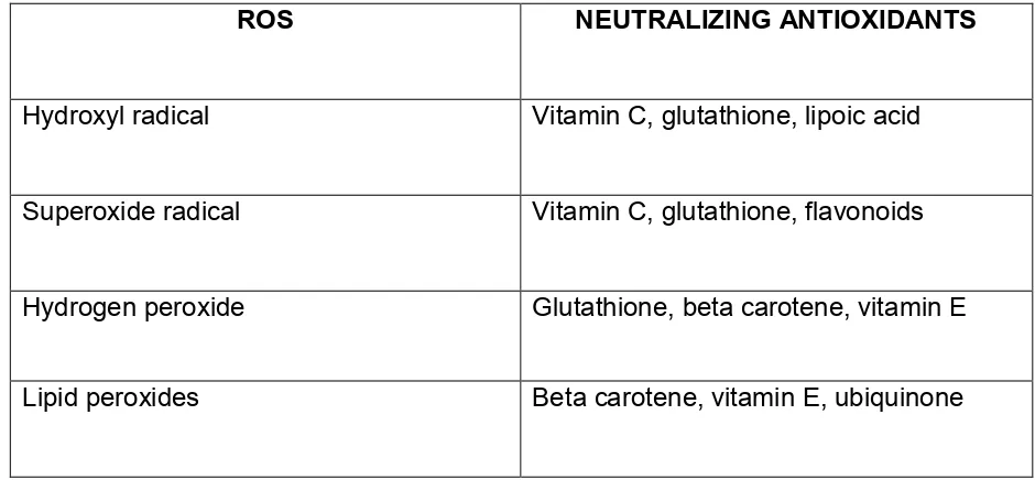
Table No.1: Various reactive oxygen species (ROS) and corresponding neutralizing
antioxidants
ROS NEUTRALIZING ANTIOXIDANTS
Hydroxyl radical Vitamin C, glutathione, lipoic acid
Superoxide radical Vitamin C, glutathione, flavonoids
Hydrogen peroxide Glutathione, beta carotene, vitamin E
[image:27.595.75.545.512.730.2]1.3.3. Dietary antioxidants
Vitamin C, vitamin E, and beta carotene are among the most widely studied
dietary antioxidants. Vitamin C is considered the most important water-soluble
antioxidant in extracellular fluids. It is capable of neutralizing ROS in the aqueous
phase before lipid peroxidation is initiated. Vitamin E, a major lipid-soluble
antioxidant, is the most effective chain-breaking antioxidant within the cell membrane
where it protects membrane fatty acids from lipid peroxidation. Vitamin C has been
cited as being capable of regenerating vitamin E.21,22
1.3.4. Phytonutrients
A number of other dietary antioxidant substances exist beyond the traditional
vitamins discussed above. Many plant-derived substances, collectively termed
“phytonutrients,” or “phytochemicals,” are becoming increasingly known for their
antioxidant activity. Phenolic compounds such as flavonoids are ubiquitous within the
plant kingdom: approximately 3,000 flavonoid substances have been described. In
plants, flavonoids serve as protectors against a wide variety of environmental
stresses while, in humans, flavonoids appear to function as “biological response
modifiers.23,24
1.3.5. Endogenous antioxidants
In addition to dietary antioxidants, the body relies on several endogenous
defense mechanisms to help protect against free radical-induced cell damage. The
antioxidant enzymes – glutathione peroxidase, catalase, and superoxide dismutase
(SOD) – metabolize oxidative toxic intermediates and require micronutrient cofactors
such as selenium, iron, copper, zinc, and manganese for optimum catalytic
activity.25,26
1.3.6. Antioxidant system in our body
The body has developed several endogenous antioxidant systems to deal with
Water soluble vitamins-ascorbic acid.
Enzymatic systems
E.g.: Super oxide dismutase, catalase, peroxidase.27,28
Herbs are found out to be a source of various phytochemicals which possess
antioxidant property.
1.4. ADVANTAGES OF HERBAL MEDICATION
Herbal medicine exhibit fewer side effects and they are very much safe. Plants
are gifts of natural to mankind for treating different types of diseases. Herbal
medicines are cheaper and easily available. Also for certain diseases like hepatitis,
herbs and herbal drugs are the only remedies. The traditional medicine is largely
getting popularity over allopathic medicine because of their cost, availability and free
from side effects.29
The plant Alpinia calcarata has been traditionally used for various diseases. In
present study rhizomes of the plant Alpinia calcarata have been used which
traditionally indicated in the treatment of asthma.30
1.5. HERBAL DRUGS USED IN ANTI
–
ASTHMATIC STUDY
Table No.2: List of Herbal Drugs used in Anti – asthmatic study
SL.
NO
PLANT FAMILY PART EXTRACT MODEL AUTHORS
1 Aerva lanta Amaranthaceae Aerial
parts
Ethanol
1) 100 µg/mL in the isolated goat tracheal chain
preparation model. 2) 30 and 60 mg/kg doses orally in
clonidine-Kumar D, Prasad DN, Parkash J, Bhatnaga.S. P,
induced catalepsy. 3) Mast cell degranulation in mice possesses anti-asthmatic activity
2 Ageratum conyzoides
Asteraceae Leaves Hydro
alcoholic 1) Inhibiting clonidine induced catalepsy in mice Tote MV, Mahire NB, Jain AP, Bose S, Undale VR, Bhosale 66
3 Asystasia gangetica
4 Bacopa monnieri
Scrophulariaceae Leaves Evaluated
petroleum ether, chloroform, methanol and water extracts 1)Mast cell stabilizing activity in rats.
Samiulla DS, Prashanth.D , Amit A 68
5 Cassia sophera
Caesalpiniaceae Leaves Ethanol 1) Anti-asthmatic activity in carrageenan induced paw edema, 2) Histamine induced bronchoconstr iction, 3) Clonidine and haloperidol induced catalepsy, 4) Milk induced leukocytosis, and eosinophilia, 5)Passive paw anaphylaxis animal-models. Nagore DH, Ghosh VK, Patil MJ.69
6 Casuarina equisetifolia
Casuarinaceae Wood & Bark
Methanol
1)Antihistami-nic activity by
inhibiting the histamine induced contraction of trachea (10-80 mcg/mL), 2) Clonidine induced catalepsy and mast cell degranulation at doses 100 mg/kg.
Pal SC, Pati UK, Yadav SK,
Bhattacharya
S.70
7 Clerodendrum Serratum
Verbenaceae Roots Ethanol
1)Anti- asthmatic activity using isolated goat tracheal chain preparation, 2) Clonidine induced catalepsy, 3) Milk induced leukocytosis and eosinophilia in mice. Bhujbal SS, Kewatkar SM, Kumar.D, Mudgade.SC , Patil MJ.71
8 Cnidium monnieri
Umbelliferae Fruits Ethanol
1) In passive cutaneous
Kubo M.72
9 Curculigo orchioides
Amaryllidaceae Rhizome Alcoholic
10 Euphorbia hirta
Euphorbiaceae Whole aerial part Ethanol 1) Inhibited the passive cutaneous anaphylaxis and paw anaphylaxis reaction, 2) Protection of mast cell from degranulation. Youssouf MS, Kaiser.P, Tahir M, Singh GD, Singh S, Sharma VK75.
11 Hemidesmus indicus
Asclepiadaceae Roots Ethanol
1) Isolated goat tracheal chain preparation, 2) Passive paw anaphylaxis in rat, 3) Clonidine-induced catalepsy in mice. Bhujbal SS, Kumar D, Deoda RS, Deore TK, Patil MJ.76
12 Ficus bengalensis
Moraceae Bark Ethyl acetate, ethanol and aqueous extracts.
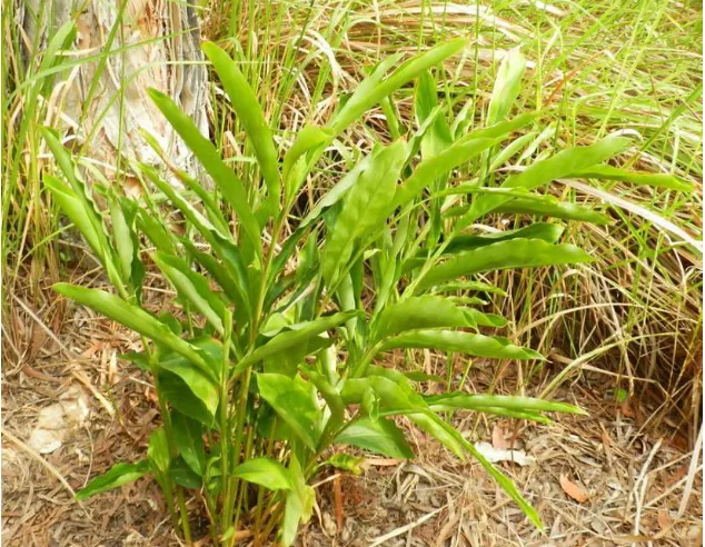
1.5. PLANT PROFILE
[image:35.595.154.471.184.430.2]Figure No.1: Alpinia calcarata – PLANT
1.5.1. PLANT DESCRIPTION
Selected plant : Alpinia calcarata
1.5.1.1. Classification
Kingdom : Plantae
Division : Mangnoliphyta
Class : Liliopsida
Order : Zingiberales
Family : Zingiberaceae
Genus : Alpinia
Species : Alpinia calcarata
1.5.1.2. Synonyms
Alpinia calcarata Rosk., Alpinia erecta Lodd. and Steud., Alpinia bracheata Rosk.,
Alpinia cerrnta Sims., Renealmia calcarata Haw., Globba eracta Retx., Languas
calcarata Mem.31
1.5.1.3. Selected Vernacular Names
Sinhala- Heen aratta, Aratta
English- Galanga, Small galanga
Tamil- Amkolinji
Sanskrit- Rasna32
1.5.1.4. Distribution`
It is native to India. Occurs in Southern Malay Peninsula and Sri Lanka. It is
1.5.1.5. Botanical description
Alpinia calcarata is a rhizomatous perennial herb with a non-tuberous
rootstock, stems slender, about 75 cm tall; leaves simple, alternate, 25 - 32 cm long
and 2.5 - 5 cm broad, lanceolate, acuminate, long-pointed, glabrous on both surfaces
and shining on the upper surface, scantily hairy along the margin, petioles sheathing;
flowers pinkish white, irregular, bisexual, in pendunculate, terminal, dense flowered
panicles 8.5 cm long, two flowers together at each node, one opening earlier than the
other, each bearing a pair of bracteoles, the inner one smaller than the outer,
bracteoles oblong, papery white, each flower about 4 cm long, pedicels short, hairy;
sepals 3, fused into a campanulate tube l cm long, pubescent outside, glabrous
inside, apices rounded; petals 3, fused at base but segments free tinged with pink,
segments oblong-spathulate, pubescent outside, lateral narrow; staminodes 3, fused
at base with the stamen into a tube adnate to corolla, two basal staminodes reduced
to minute filaments, the larger one petaloid, 3 cm by 2.3 cm ovate, yellow with vinous
red streaks, emarginated, apex frilled and darker, glabrous and shining on both
surfaces; stamen, anther tubular, style passing through, filament flat, 1.5 cm long,
anther 0.8 cm long, style 3.5 cm long, tinged pink, hairy towards the apex, stigma
swollen; ovary inferior, 3 mm long, strongly pubescent, 3-locular with ovules in each
Silvy Mathew et al., (2014)36 conducted a study on the antimicrobial activity of the plant Alpinia calcarata rhizome. The present study was designed to investigate
the anti-microbial activity of four solvent extracts (Petroleum ether, Dichloromethane,
Acetone and Methanol) of rhizome of Alpinia calcarata. The rhizome of this plant is
an important antimicrobial agent and a digestive stimulant. The plant extracts showed
considerable activity against ten tested strains viz., Pseudomonas aeroginosa,
Escherichia coli, Enterobacter aerogens, Bacillus subtilis, Staphylococcus aureus,
Streptococcus faecalis, Vibrio cholerae, Salmonella paratyphi, Klebsiella pneumoniae
and Proteus vulgaris by using agar disc diffusion assay. This study reveals that
Alpinia calcarata has antimicrobial activity against gram positive and gram negative
bacteria.
Arambewela L et al., (2004)37 conducted a study on the antinociceptive activities of aqueous and ethanolic extracts of Alpinia calcarata rhizomes in rats.
Rhizomes of Alpinia calcarata (Zingiberaceae) possess several bio-activities and are
used in traditional medicine of Sri Lanka. However, their antinociceptive activity has
not been investigated so far. The aim of this study therefore, was to examine the
antinociceptive activity of hot water extract (HWE) and hot ethanol extract (HEE) of
Alpinia calcarata rhizomes using rats and three models of nociception (tail flick, hot
plate and formalin tests). Different concentrations of HWE (100, 250, 500, 750,
1000 mg/kg) and HEE (100, 250, 500, 750, 1000 mg/kg) were made and orally
administrated to rats and the reaction times determined. The results showed that the
extracts have marked dose-dependent antinociceptive activity, when evaluated in the
hot plate and the formalin tests but not in the tail flick test. The antinociceptive effect
was slightly higher in HEE than that of HWE. The antinociceptive effect was mediated
via opioid mechanisms.
Ramya Rajasekar et al., (2014)38 proposed a study on the antidiabetic activity
of the Alpinia calcarata rhizomes. The study was to determine effect of
Alpinia calcarata on glucose uptake in diabetic rats-an in vitro and in vivo model.
Diabetes mellitus is a heterogeneous metabolic disorders characterized by
The diabetes was induced by single dose of STZ (45 mg/kg) in citrate buffer,
while the normal control group was given the vehicle (citrate buffer) only. After
induction of diabetes, the diabetic animals were treated with ethanolic extract of
Alpinia calcarata (200 mg/kg) and glibenclamide (2 mg/kg) for 30 days. Blood
glucose estimation was performed every week of the study. At the end of study
period, animals were sacrificed for biochemical studies.
Streptozotocin induced diabetic rats shows the altered levels of various
biochemical profiles. Those levels were brought back to near normal upon treatment
with ethanolic extract of Alpinia calcarata and standard drug glibanclamide. No
significant changes were observed on treatment with plant.
Lakshmi Arambewela et al., (2011)39 conducted a study on the safety profile of the plant Alpinia calcarata rhizomes. The aim of the present study was to
investigate whether Alpinia calcarata (Family: Zingiberaceae) rhizomes have any
toxic effects in rats. Albino wistar rats were used as the experimental model and
orally administered hot water extract (HWE) and hot ethanolic extract (HEE) of
Alpinia calcarata rhizomes at a dose of 1500 mg/kg respectively for 42 consecutive
days. Administration of the HWE or HEE to rats did not result in any chronic toxic
effects as evident from their effects on (a) liver function (b) kidney function (c)
hematological parameters such as red blood cell (RBC) count, white blood cell
(WBC) count and haemoglobin (Hb) concentration (d) external morphology and wet
weights of selected organs. Further, the HWE and the HEE did not appear to mediate
any unacceptable effects on food and water intake, % weight gain, consistency of
faeces and color of urine. In conclusion, the results of this study have revealed that
the HWE and the HEE of Alpinia calcarata at the doses tested do not produce any
serious toxic side effects in rats.
Mizanur Rahman etal., (2012)40 conducted a study on the anti-inflammatory, analgesic and gc – ms (gas chromatographic and mass spectrometric) analysis of
essential oil of Alpinia calcarata rhizome. In the present study, essential oil isolated
from Alpinia calcarata was analyzed and also assessed for acute toxicity,
anti-inflammatory and analgesic activities in animals. The essential oil isolated from
Alpinia calcarata was analyzed by using GC-MS on a combined GC-MS instrument.
model” served as acute model. “Acetic acid induced writhing response model” was
used to assess analgesic activity in mice. The major components of essential oils
isolated from Alpinia calcarata were Camphene (3.86%), Betamyrcene (4.39%),
Eucalyptol (14.05%), Linalol (2.48%), Pyrazine (1.72%), L-camphor (7.90%) and
Berneol (5.67%). Intraperitoneal injection of essential oil isolated from Alpinia
calcarata significantly suppressedthe paw edema induced by carrageenan in two
different dose levels studies namely 400 mg/kg and 380 mg/kg. Intraperitoneal
injection of essential oil also significantly attenuated the acetic acid induced writhing
response in three different dose levels studies namely 200 mg/kg, 400 mg/kg and
600 mg/kg. These studies suggest that essential oil isolated from Alpinia calcarata
might possess significant anti-inflammatory activity and analgesic effect and could be
a potential source for treatment of different inflammatory diseases.
Ratnasooriyaetal., (2006)41 conducted a study on the effect of aqueous extract of the plant Alpinia calcarata rhizomes on reproductive competence of male
rats. This study examined the effects of rhizomes of Alpinia calcarata (Zingiberaceae)
on male sexual competence and fertility, using a hot water extract (HWE) and rats.
Different doses of HWE (150, 250 and 500 mg/kg) were orally administrated to male
rats and their sexual behaviour was monitored 3 hours later using receptive females.
Fertility was determined in a separate group (with the highest dose) using a
noncompetitive copulation test. In the sexual behaviour study, the HWE impaired the
number of rats ejaculating and markedly prolonged the latency for ejaculation.
Further, the number of rats mounting and intromitting, and the latencies for mounting
and intromission were inhibited. Collectively, these observations indicate a strong
aphrodisiac action. The other parameters remained unchanged indicating
non-impairment in libido, sexual arousability, sexual vigour and sexual performance or
penile erectile ability. However, a slight impairment was evident in sexual motivation
(with the highest dose) in a partner preference test. In the fertility test, HWE induced
profound oligozoospermia but fertility was uninhibited. The highest dose of HWE also
elevated the serum testosterone level and the number of spontaneous penile
3.1. AIM & OBJECTIVES
In recent year there has been tremendous increase in demand for herbal drugs
because of its safety, efficacy and better therapeutic results. Due to its economic
pricing as compared to synthetic or allopathic drugs, which have several therapeutic
compilations.
Alpinia calcarata is also considered to be therapeutically important in traditional
system of medicine.
Aim of the study to evaluate the anti-asthmatic, antioxidant and anti-inflammatory
activities of ethanolic extract of Alpinia calcarata rhizome.
The objective study includes
To study the anti-asthmatic effect of ethanolic extract of Alpinia calcarata rhizomes.
To study the in vitro antioxidant activity of the ethanolic extract of Alpinia calcarata rhizomes.
4.1. PLAN OF WORK
1. Collection and authentification of plant material (rhizomes of
Alpinia calcarata
2. Extraction of the plant material (Rhizomes of the plant Alpinia calcarata).
3. Conduction of the preliminary phytochemical screening and finding out the
phytoconstituents present in the ethanolic extract of Alpinia calcarata
rhizomes.
4. Performing of acute toxicity studies of ethanolic extract of Alpinia calcarata
rhizomes (OECD 423).
5. Evaluation of the anti-asthmatic effect of Alpinia calcarata rhizomes In vivo methods
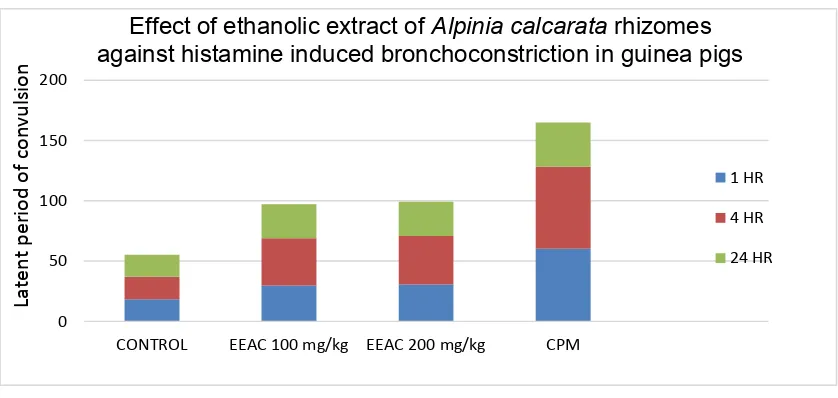
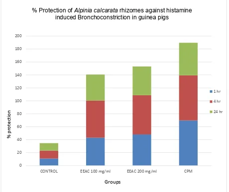
1. Histamine aerosol induced bronchoconstriction in guinea pigs.
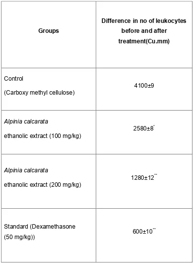
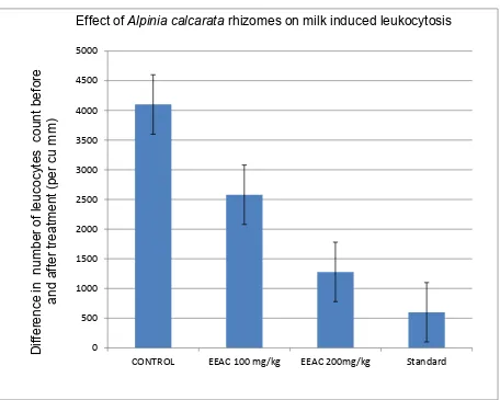
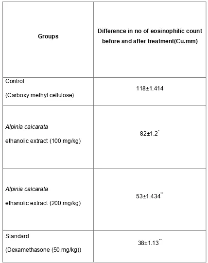
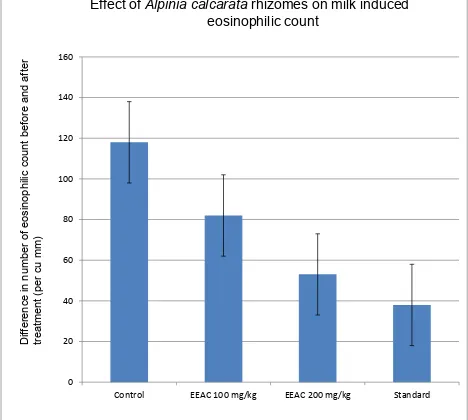
2. Milk induced leukocytosis and eosinophil count Ex vivo methods
1. Isolated guinea pig tracheal preparation.
6. Evaluation of the antioxidant study of Alpinia calcarata rhizomes In vitro methods
1. Hydrogen peroxide scavenging assay
2. Reducing power assay
7. Evaluation of the anti-inflammatory study of Alpinia calcarata rhizomes In vitro methods
1. Protein denaturation method
2. The rabbit red blood cell (RRBC) membrane stabilization
5.1. MATERIALS
5.1.1. Plant selected
In the present study, Alpinia calcarata was selected because of its traditional
uses. The part used was rhizomes.
5.1.2. Chemicals and reagents used
Carboxy methyl cellulose (Spectrum reagents and chemicals pvt. Ltd.) Ascorbic acid (Spectrum reagents and chemicals pvt. Ltd.)
Histamine (NICE chemicals pvt.Ltd.)
Hydrogen peroxide (Spectrum reagents and chemicals pvt. Ltd.) Glacial acetic acid (Ozone international, Mumbai)
Trichloro acetic acid (NICE chemicals pvt. Ltd.) Diclofenac sodium (Rajesh chemicals, Mumbai)
5.1.3. Drugs used
Chlorpheniramine maleate (Abbott Laboratories pvt. Ltd.) Dexamethasone (Zydusbiogem, cadila health care Ltd.)
5.1.4. Instruments used for the study
UV- Visible spectrophotometer- Jasco international
Incubator- Rotek Instruments, B&C Industries, W. Vengola. Centrifuge- Rotek Instruments, B&C Industries, W. Vengola. Histamine chamber- Orchid Scientific Innovations India pvt. Ltd
5.1.5. Animals
Swiss albino mice (25-40 gm) and Guinea pig (400-600 gm) were used to
carry out the activities. The animals had free access to standard commercial diet and
water. Animals were housed in cages under standard conditions i.e., 12:12 hour light
or dark cycle at 25±20 C. The experiments were carried out as per the guideline of
5.2. METHODS
5.2.1. Collection and authentication of Alpinia calcarata
The dried rhizomes of the Alpinia calcarata were collected. The rhizomes were
cleaned and shade dried and milled into coarse powder by a mechanical grinder.
5.2.2. Preparation of plant extract
The powdered rhizomes were extracted using ethanol by soxhlet extractor. In
this process the powdered drug is placed into the extractor with ethanol as solvent.
After extraction the extract was concentrated by evaporation then it was kept in a
refrigerator for further use.42, 43
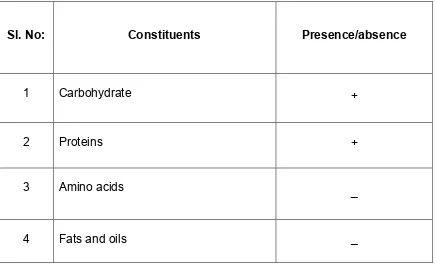
5.2.3. Preliminary phytochemical screening
The ethanolic extract of Alpinia calcarata rhizomes were subjected for the
following chemical tests for the identification of various active constituents.44, 45
5.2.3.1. Detection of carbohydrates: -
Molish test (General test)
To 2-3 ml of aqueous extract, add few drops of α-naphthol solution
in alcohol, shake and add conc. H2SO4 from a test tube. Violet ring
is formed at the junction of two liquids. Test for reducing sugar
(a)Fehling’s test
Mix 1 ml of fehling’s A and 1 ml of fehling’s B solution Boil for 1 min.
Add equal volume of test solution. Heat in boiling water bath for5-10
min. First yellow, then brick red precipitate observed.
(b) Benedict’s test
Mix equal volume of benedict’s reagent and test solution in a test
tube. Heat in boiling water bath for 5 min. Solution appears green,
yellow or red depending on amount of reducing sugar present in test
5.2.3.2. Detection of protein:
- Biuret test (General test)
3 ml of test solution add 4% NaOH and few drops of 1% CUSO4
solution. Violet or pink color appear. Million’s test (for proteins)
Mix 2 ml of extract with million’s reagent. white precipitate formed.
warm precipitate turns brick red or the precipitate dissolves giving red
colored solution.
5.2.3.3. Detection of proteins and amino acids: -
About 100 mg of extract was dissolved in 10 ml of distilled water and filtered through
Whatman No.1 filter paper and the filtrate was subjected to tests for proteins and
amino acids.
Biuret test (General test)
3ml of test solution add 4% NaOH and few drops of 1% CUSO4
solution. Violet or pink color appear.
Million’s test (for proteins)
Mix 2 ml of extract with millions reagent. White precipitate formed.
Warm precipitate turns brick red or the precipitate dissolves giving red
colored solution
Ninhydrin test (for amino acid)
About 2 drops of ninhydrin solution were added to 2 ml of test solution.
Purple or bluish color appears
5.2.3.4. Detection for Fats and oils:
- Solubility test
Oils are soluble ether, benzene, chloroform but insoluble in 90%
ethanol and in water. Hence filter paper get permanently stained with
5.2.3.5. Detection of steroid: -
Salkowski reaction
To 2 ml of extract, add 2 ml chloroform and 2 ml con H2SO4 shake well.
Chloroform layer appear red and acid layer shows greenish yellow
fluorescence.
Liebermann – burchard reaction
Mix 2 ml extract with chloroform. Add 1-2 ml of acetic anhydride and
2 drops of con H2SO4 from the side of test tube. First red, then blue
finally green color appear.
5.2.3.6. Detection of glycoside: -
1) Cardiac glycoside
Legal ‘s test (for cardiac glycoside)
To aqueous or alcoholic extract add 1 ml pyridine and 1 ml sodium
nitroprusside. pink to red color appears. Test for reducing sugar (for cardiac glycoside)
To 2 ml extract add glacial acetic acid one drop 5% Fecl3 and
con H2SO4. Reddish brown color appears at junction of two liquid layer
and upper layer appears bluish green.
2) Anthraquinone glycoside
Bontragers test
To 3 ml extract, dil. H2SO4 was added, boiled and filtered. To cold
filtrate, equal volume of benzene or chloroform was added and shaken
well. The organic solvent was separated and ammonia was added. The
ammonical layer turns pink to red.
3) Saponin glycoside
Foam test
Heamolytic test
Add drug extract or dry powder to one drop of blood placed on glass
slide. Heamolytic zone appears
4) Cyanogenic glycoside
Grignard reaction or sodium picrate test
Soak a filter paper strip first in 10% picric acid, Then in 10%
sodium carbonate, dry. In conical flask place moistened powdered
drug. Cork it, place the above filter paper strip in the slit in cork. Filter
paper turn brick red or maroon.
5) Coumarin glycoside
Coumarin glycoside have aromatic odour
Alcoholic extract when made alkaline, shows blue or green fluorescence
5.2.3.7. Detection for flavonoids
Shinoda test
To dry powder or extract, add 5ml 95% ethanol, few drops con. Hcl and
0.5 g Magnesium turnings. Pink color observed.
To small quantity of residue, add lead acetate solution. Yellow color precipitate is formed.
Addition of increasing amount of NaOH to residue shows yellow coloration, which decolorizes after addition of acid.
5.2.3.8. Detection of alkaloid
Aqueous alcoholic and chloroform extract was evaporated separately. To
residue dilute Hcl was added. Shaken well and filtered. With the filtrate the following
test was performed.
Dragondorff ‘s test
To 2-3 ml. Filtrate, add few drops of dragondorff‘s reagent. Orange
Mayers test
To 2-3 ml of filtrate with few drops of mayer’s reagent gives precipitate Hager’s test
2-3 ml filtrate with hager’s reagent gives yellow precipitate Wagner’s test
2-3 ml of filtrate with wagner’s reagent gives reddish brown precipitate
5.3. ACUTE TOXICITY STUDIES
Acute toxicity of Alpinia calcarata was done as per OECD guidelines 423. The
substance was administered in a single dose by gavage using specially designed
mice oral tube. Animals were fasted prior to dosing with food but not water withheld
overnight. Following the period of fasting, the animals were weighed and the test
substance was orally at a dose of 5, 50, 300 and 2000 mg/kg. The animals are
observed continuously for first three hours, four any toxic manifestations like
increased motor activity, salivation, acute convulsion, coma and death. Changes in
the animal behavior should be noted before and