A Comparative study of Epidural 0.5% Isobaric Levobupivacaine
and Epidural 0.5% Isobaric Levobupivacaine with Dexmedetomidine
for patients undergoing elective infraumbilical and lower limb
surgeries
Dissertation submitted
in partial fulfillment of the requirements
for the award of the Degree
M.D. (Anaesthesiology)
Branch X
GOVT. KILPAUK MEDICAL COLLEGE
THE TAMIL NADU DR. M.G.R. MEDICAL UNIVERSITY
CHENNAI, TAMILNADU
CERTIFICATE
This is to certify that this dissertation entitled “
A Comparative
study of Epidural 0.5% Isobaric Levobupivacaine and Epidural
0.5% Isobaric Levobupivacaine with Dexmedetomidine for patients
undergoing elective infraumbilical and lower limb surgeries
submitted by Dr.MANGAL SWATHI.V in partial fulfillment for the
award of the Degree Doctor of Medicine in Anaesthesiology by The
Tamil Nadu Dr. M.G.R. Medical University, Chennai is a bonafide work
done by her at GOVERNMENT KILPAUK MEDICAL COLLEGE,
during the academic year 2012-2015.
PROF.Dr. T.MURUGAN, M.D.,D.A., Professor & HOD and Guide
Department of Anaesthesiology
Government Kilpauk Medical College,
Chennai -10 PROF. Dr.N.GUNASEKARAN,
M.D,D.T.C.D
Dean Government Kilpauk Medical College,
Chennai-10
DECLARATION
I, Dr. MANGAL SWATHI.V, solemnly declare that this
dissertation, entitled “
A Comparative study of Epidural 0.5% Isobaric
Levobupivacaine and Epidural 0.5% Isobaric Levobupivacaine with
Dexmedetomidine for patients undergoing elective infraumbilical and
lower limb surgeries”
has been prepared by me, under the expert
guidance and supervision of
PROF.Dr.T.MURUGAN,M.D.,D.A.,
Professor & HOD, Department of Anaesthesiology, Government Kilpauk
Medical College ,Chennai and submitted in partial fulfillment of the
regulations for the award of the Degree M.D.(Anaesthesiology) by The
Tamil Nadu Dr. M.G.R. Medical University and the examination to be
held in April 2015.
This study was conducted at Government Kilpauk Medical College
Hospital and Government Royapettah Hospital, Chennai. I have not
submitted this dissertation previously to any university for the award of
any Degree or Diploma.
Place: Chennai (Dr.
MANGAL SWATHI.V
)
ACKNOWLEDGEMENT
I wish to express my sincere thanks to
PROF. Dr.N.GUNASEKARAN,M.D.,D.T.C.D Dean, Government Kilpauk
Medical College, Chennai for having kindly permitted me to utilize the
facilities of the college for the conduct of the study.
I indeed express my heartful gratitude to my Guide
PROF. Dr. T.MURUGAN., M.D.,D.A.,Professor and Head of the Department
of Anaesthesiology, Government Kilpauk Medical College,
for his constant motivation, valuable guidance, and for providing all necessary
arrangements for conducting this study at both hospitals.
I am extremely grateful and indebted to Prof.DR.G.R.Rajashree, M.D.,
Professor, Department of Anaesthesiology, Government Kilpauk Medical
College, Chennai for her concern, inspiration, meticulous guidance, expert
advice and constant encouragement in preparing this dissertation.
I also express my sincere gratitude to all other Professors
of Anaesthesiology, KMC, Prof. Dr. S. Selvamani, MD., DA.,
Prof. Dr. M. Vellingiri, M.D.,D.A.,Prof. Dr. M. Bhavani, M.D., for their
constant motivation, encouragement and valuable suggestions.
I thank all the Assistant Professors and tutors of Anaesthesiology
KMCH and GRH for their keen interest and support without which this study
I am thankful to the Institutional Ethical Committee for their guidance
and approval of the study.
I also thank my entire colleague Postgraduates for supporting me
throughout the study.
I thank the Department of Surgery, KMCH and GRH and their faculty
members for their kind cooperation and permitting me to use the hospital
facilities for the study.
I also thank the theatre personnel for their co-operation and assistance.
I wish to thank all the patients whose willingness and patience made this
study possible.
I am really indebted to my beloved parents and my sister for their
encouragement throughout the study.
I finally thank God Almighty for His blessings in successfully
CONTENTS
S.NO
TITLE
PAGE
NO.
1.
INTRODUCTION
1
2.
AIM OF THE STUDY
6
3.
ANATOMY OF VERTEBRAL COLUMN
7
4.
ANATOMY OF EPIDURAL SPACE
16
5.
HISTORY OF EPIDURAL ANAESTHESIA
23
6.
PHYSIOLOGY OF EPIDURAL ANAESTHESIA
25
7.
PHYSIOLOGICAL EFFECTS OF EPIDURAL
BLOCKADE
31
8.
PHARMACOLOGY-LEVOBUPIVACAINE
38
9.
PHARMACOLOGY-DEXMEDETOMIDINE
49
10.
REVIEW OF LITERATURE
53
11.
MATERIALS & METHODS
58
12.
OBSERVATION & RESULTS
65
13.
DISCUSSION
93
14.
SUMMARY
99
15.
CONCLUSION
101
16.
BIBLIOGRAPHY
17.
ANNEXURES
Ethical Committee Clearance Form
Proforma
ABSTRACT
BACKGROUND
The quest for searching newer and safer anaesthetic agents has always been one of the primary needs in anaesthesiology practice. Regional anaesthesia techniques have seen numerous modifications over the last two decades with the advent of many newer and safer local anaesthetics
Keeping these factors in mind, S (−)-enantiomer of bupivacaine, levobupivacaine has been developed. The advantages of levobupivacaine over bupivacaine are decreased cardiovascular toxicity and there is also a relatively decreased motor nerve fiber penetration and block, thereby a decreased post operative motor blockade and thus early ambulation of the patients can be achieved.
The present study compared the effects of addition of epidural dexmedetomidine 50 micrograms to epidural 0.5% levobupivacaine for infraumbilical and lower limb surgeries.
METHODS
Sixty patients of either sex belonging to ASA I & II in the age group of 25-45 years scheduled for infraumbilical and lower limb surgeries were randomly divided into 2 groups (30 each)to receive 0.5% isobaric levobupivacaine 20 ml epidurally with 0.5 ml distilled water(Group A) and 0.5% isobaric levobupivacaine 20 ml plus 0.5 ml dexmedetomidine containing 50 micrograms(Group B).
This study evaluated the following parameters like time of onset of sensory blockade at T10 level, maximum sensory blockade achieved and time taken to achieve the same, onset time of motor blockade, degree of motor blockade, time taken to achieve maximal motor blockade, hemodynamic changes in pulse rate, blood pressure and oxygen saturation, side effects and complications, intraoperative sedation scores, duration of analgesia, sensory & motor blockade, and any postoperative adverse reactions.
RESULTS
The data obtained from the above parameters were statistically analysed using SSPS version 16 software. Student t test was used for parametric data and Chi-square test for non parametric data.P<0.05 was considered as statistically significant.
minimal in the dexmedetomidine group. Adverse effects experienced in general were statistically insignificant in both the groups. Mean sedation score in group B (Dexmedetomidine group) was predominantly found to be 2 as per Ramsay sedation score. None of the patients in group B had deep sedation or profound respiratory depression.
1
INTRODUCTION
Regional anaesthesia came in vogue from the time of Sir August Bier in 1898. Intrathecal anaesthesia and epidural anaesthesia are the most popular regional anaesthetic techniques used for lower abdominal & lower limb surgeries. Regional anaesthesia forms an excellent alternative due to its various advantages over general anaesthesia including(1)
1. Awake patient
2. Polypharmacy avoided
3. No airway manipulation
4. Good motor & sensory blockade
5. Good intra operative and post operative analgesia
6. Less incidence of post operative nausea and vomiting
7. Early ambulation and food intake by the patient.
8. Less incidence of deep vein thrombosis and thromboembolism
2
response to surgery and improves surgical outcome.Epidural anaesthesia and analgesia has become one among the best accepted techniques for lower abdominal & lower limb surgeries as it provides good sensory and motor blockade with contracted bowels retaining adequate spontaneous respiration, hemodynamic stability and also an indwelling epidural catheter facilitates further administration of analgesic doses for the postoperative period(2).
Advantages of epidural over spinal anaesthesia being that it (3)
1. Reduces the incidence of hemodynamic changes due to sympathetic blockade as it produces segmental anaesthesia unlike subarachnoid block.
2. Provides effective surgical anaesthesia for prolonged procedures.
3. Incidence of postdural puncture headache is not encountered as dura is not pierced.
4. Provides prolonged postoperative analgesia.
Lignocaine, a tertiary amide local anaesthetic was synthesised by Nils Lofgren in 1943(3).Use of lignocaine for extradural blockade was found to have a greater speed of onset and greater degree of both sensory and motor blockade (4).
But it is also observed that bupivacaine is four times more potent than
lignocaine and duration of anaesthesia is 2 or 3 times longer than Lignocaine. It has
excellent prolonged duration of sensory blockade, but muscle relaxation was not
3
Bupivacaine was synthesised by Ekanstam in 1956 and introduced into clinical practice in 1963 by Telivuo. Bupivacaine, the widely used local anaesthetic in regional anaesthesia is available in a commercial preparation as a racemic mixture (50:50) of its two enantiomers levobupivacaine, S (-) isomer and dextrobupivacaine, R (+) isomer. Among the amide local anaesthetics used in regional anaesthetic techniques bupivacaine has emerged as the most commonly used drug for central neuraxial blockade. Since then bupivacaine is extensively used and has become very popular for epidural anaesthesia as well as analgesia because of its long duration of action. Despite its undoubted efficacy, bupivacaine is associated with cardiotoxicity and neurotoxicity(7,8).
Severe neurotoxic and cardiovascular adverse reactions reported in the literature after inadvertent intravascular injection or intravenous regional anaesthesia have been linked to the R(+) isomer of bupivacaine. The levorotatory isomers were shown to have a safer pharmacological profile with less cardiac and neurotoxic adverse effects.(7,8)Apart from cardiotoxicity, it also carries undesirable effects like prolonged post operative motor blockade. Hence there was a need for introduction of drugs with all the features of bupivacaine but having less toxicity profile.(9)
The quest for searching newer and safer anaesthetic agents has always been one of the primary needs in anaesthesiology practice. Regional anaesthesia techniques have seen numerous modifications over the last two decades with the advent of many newer and safer local anaesthetics (10,11).
4
bupivacaine are decreased cardiovascular toxicity and there is also a relatively decreased motor nerve fiber penetration and block, thereby a decreased post operative motor blockade and thus early ambulation of the patients can be achieved(9).
It demonstrated less affinity and strength of depressant effects onto myocardial and central nervous vital centres in pharmacodynamic studies and a superior pharmacokinetic profile. Reports of toxicity with levobupivacaine are scarce and occasional toxic symptoms are usually reversible with minimal treatment with no fatal outcome. In anaesthesia and analgesia practice, levobupivacaine and bupivacaine produce comparable surgical sensory block but levobupivacaine has the advantage of producing less motor blockade. (9)
The low cardiovascular and neurological toxicity of levobupivacaine has led to its varied utility in regional anaesthetic techniques including subarachnoid block, epidural anaesthesia and analgesia, brachial plexus blocks as well as local infiltration. It is also being used for intraoperative anaesthesia, labour analgesia, and postoperative pain management. Both levobupivacaine and ropivacaine are being favoured in labour analgesia because of comparable long lasting analgesia, less motor block and less toxicity compared to bupivacaine.(12)
Efforts to find a better adjuvant in regional anaesthesia are underway since long. The addition of adjunctive agents (opioids & alpha 2 agonists) to local anaesthetics via epidural and intrathecal routes may provide a dose sparing effect and increase the duration and quality of analgesia. (9)
5
anaesthetic effect. The addition of opioid does provide a dose sparing effect of local anaesthetic and superior analgesia but there is always a possibility of an increased incidence of pruritis, urinary retention, nausea, vomiting and respiratory depression. (9)
The pharmacologic properties of alpha 2 agonist have been extensively studied and have been employed clinically to achieve desired effects in regional anaesthesia. Clonidine is an alpha-2 adrenergic agonist which, when administered by epidural route, has analgesic properties and potentiates the effect of local anaesthetics. Clonidine is 200-folds more selective for alpha-2 as compared to alpha-1.
Dexmedetomidine is a new addition to the class of alpha 2 agonists which has got numerous beneficial effects when used through epidural route. It acts on both pre and post synaptic sympathetic nerve terminals and central nervous system, thereby decreasing the sympathetic outflow and nor epinephrine release causing sedative, anti-anxiety, analgesic, sympatholytic and hemodynamic effects.(13)
Dexmedetomidine, made up of medetomidine’s dextrogyrous enantiomer,is currently considered a super selective alpha-2 adrenergic agonist prototype and is 1600-folds more selective for alpha-2 receptor. Dexmedetomidine is a highly selective alpha 2 adrenergic agonist with greater receptor affinity than clonidine.(9) In addition it also has hemodynamic stabilising effects and reduction of anaesthetic drug requirements. Epidural dexmedetomidine does cause a manageable hypotension & bradycardia but the striking feature of this drug is its lack of opioid related side effects like respiratory depression, pruritis, nausea and vomiting. (14)
6
AIM OF THE STUDY
The aim of the present study is to evaluate the effect of addition of Dexmedetomidine to epidural 0.5% Levobupivacaine solution on the
1. Time of onset of sensory blockade at T10 level
2. Maximum sensory blockade and time taken to achieve that level
3. Onset of motor blockade
4. Degree of motor blockade achieved
5. Haemodynamic changes in the pulse rate & blood pressure
6. Side effects and complications
7. Intraoperative sedation scores
8. Duration of analgesia
9. Duration of sensory blockade
10. Duration of motor blockade
7
ANATOMY OF THE VERTEBRAL COLUMN
(15)Knowledge of the anatomy of vertebral column is of particular importance to anaesthesiologists.There are
7 Cervical vertebrae 12 Thoracic vertebrae 5 Lumbar vertebrae 5 Sacral vertebrae (Fused) 4 Coccygeal vertebrae (Fused)
[image:17.595.239.455.381.605.2]The cervical and lumbar curvatures are convex anteriorly while, thoracic and sacral curvatures are convex posteriorly.
FIGURE 1: ANATOMY OF VERTEBRAL COLUMN
A typical vertebra has two parts:
1. anteriorly, BODY or the base which bears the weight.
8 pedicles.
3. There are seven processes or projections:
a. Three muscular processes - two transverse and one spinous. b. There are four articular processes - two upper and two lower. The typical thoracic vertebra:
It has a heart-shaped body bearing one or two facets for articulating with the head of a rib. Its vertebral foramen is smaller and circular than those of the cervical and lumbar regions.
[image:18.595.101.535.449.662.2]The two pedicles bear long and strong transverse processes. It articulates with the neighbouring vertebra by means of articular processes that bear vertical facets superior facing posteriorly and inferior facing anteriorly. Its spinous process is long and slopes posteroinferiorly so that the tip overlies the level of the vertebral body below.
9 The typical lumbar vertebra
[image:19.595.151.476.252.421.2]It has a larger kidney-shaped body and its vertebral foramen is larger when compared to that of thoracic vertebra. Its transverse processes are long and slender. Its articular processes are directed (superior) posteromedially and (inferior) anterolaterally. Its spinous processes are shorter, broader and more horizontal than those of thoracic vertebrae.
FIGURE 3: LUMBAR VERTEBRA
Intervertebral disc
These are the connecting links between the vertebral bodies and they account 25% of the length of spine. Each disc adheres above and below to the hyaline cartilage which covers the facet of adjacent vertebral body in front and behind and the disc also gets attached to the anterior and posterior longitudinal ligaments.
Joints of the vertebral column
10
attachment between vertebrae. The facet joints lie between superior and inferior articular processes.
Contents of vertebral canal: 1. Spinal cord and meninges. 2. Cerebrospinal fluid. 3. Spinal nerve roots. 4. Vessels, areolar tissue.
SPINAL CORD:
Medulla oblongata continues as spinal cord below the level of foramen magnum tapering off into conical extremity known as conus medullaris. From the apex of conus medullaris, delicate fibrous filaments descend into the back of first segment of coccyx, which is called as filum terminale. At birth, the spinal cord ends at the level of lower border of third lumbar vertebra. In adults the level of termination of spinal cord may be as follows:
• Lower border of L1 vertebra 50% • Upper border of L2 vertebra 40% • Lower border of L3 vertebra 10%
THE MENINGES
11
terminale. It extends laterally along the spinal nerve roots to become continuous with the connective tissues of the epineurium at the level of intervertebral foramina. The dura mater is composed of randomly arranged collagen fibres along with longitudinally and circumferentially arranged elastin fibres. It is largely acellular except for a single layer of cells that forms the border between the dura and arachnoid mater. Despite the lack of cellular elements, the inner edge is highly vascular, due to which it forms an important route for drug clearance from epidural and subarachnoid space. The inner surface of the dura mater abuts the arachnoid mater. There is a potential space between these two membranes called subdural space. Occasionally it is possible to inadvertently insert an epidural catheter into the subdural space.
Lower extent of dural sac can be as follows: S2 vertebra -35%
Below S2- 40%
Above S2- 25%
Below this the dura continues as the covering of filum terminale.
Arachnoid mater: It is a delicate, avascular membrane composed of overlapping layers of flattened cells with connective tissue fibres running between the cellular layers. These cells are interconnected by frequent tight junctions and occluding junctions, which account for the fact that arachnoid mater acts as the main barrier for drugs moving between the epidural space and spinal cord. Superiorly the spinal arachnoid continues with the cranial arachnoid and inferiorly it ends along with dura at the lower border of S2. Arachnoidmater herniates through the dura into the epidural
12
serve as exit for the drugs from subarachnoid space. The subarachnoid space lies between arachnoid and pia mater and contains CSF.It is in continuity with cranial CSF and provides route for the drugs to reach brain.
[image:22.595.165.469.498.694.2]Pia mater: It is also a delicate, highly vascular membrane which adheres intimately to the spinal cord and dips into the long sulci along its surface. It consists of a thin layer of connective tissue cells interposed with collagen. There are trabeculae which connect the piamater with arachnoid mater and the cells of these two meninges blend together along the trabeculae.Fenestrations in pia mater help in communication with the subarachnoid space. Pia forms a separate sheath for each nerve rootlet as far as the intervertebral foramina, where it binds with the epineurium. At the lower end of the cord, pia continues as a thin thread the filum terminale which becomes invested by the dural (external) filum terminale and continues to the coccyx. Considered an extension of piamater, the delicate ligament is much tougher and less vascular than that of pia. These are attached to the dura by a series of 20-21 meticulous processes (tooth like projections) which aid in the supporting the spinal cord.
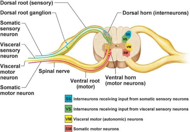
13 Spinal segments
The spinal cord is divided into segments by means of spinal nerves arising from it. The spinal nerves are 31 in number and are as follows:-
8 Cervical 12 Thoracic 5 Lumbar 5 Sacral
1 Coccygeal (rudimentary)
The nerve roots within the dura have no epidural sheath. Therefore they are easily affected by doses of analgesic drugs brought into contact with them.
Spinal nerves
14
FIGURE 5: CROSS SECTION OF SPINAL NERVE
Segmental levels
1. Perineum S1- S4 2. Inguinal region L1 3. Umbilicus T10 4. Subcostal T6-9 5. Nipple line T4-5
6. Second intercostal space T2 7. Clavicle C3-4
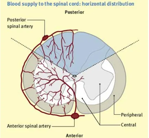
15 Blood supply of spinal cord
[image:25.595.190.443.439.673.2]The principle arterial supply of the spinal cord is derived from one anterior and two posterior spinal arteries which descend from the level of foramen magnum. The anterior spinal artery is formed by the union of branches from each vertebral artery at the foramen magnum. It supplies anterior 2/3 of the spinal cord. There are two posterior spinal arteries, one on each side. They are derived either directly from the vertebral artery or more often from a primary branch of each vertebral artery. They supply the posterior 1/3rd of the spinal cord. This supply is augmented by spinal branches of the vertebral, ascending cervical, posterior intercostals, lumbar and lateral sacral arteries, which also pass through the intervertebral foramina. Venous drainage is via a plexus of anterior and posterior spinal veins in the neck, the azygous vein in thorax, lumbar veins in the abdomen and lateral sacral veins in the pelvis.
16 ANATOMY OF EPIDURAL SPACE(16,17)
The epidural space is a circular space surrounding the dura which extends from foramen magnum to the coccyx. The lower limit is sacrococcygeal membrane. The space is potential and exists between the lining of the vertebral canal and the dural sac. Structures encountered from skin to the epidural space by the advancing epidural needle in midline approach are:
1. Skin
2. Subcutaneous tissue 3. Supraspinous ligament 4. Interspinous ligament 5. Ligamentum flavum
Ligaments
a. Supraspinous ligament runs superficial to the spinous processes b. Posterior spinous processes are connected by Interspinous ligament c. Laminae are connected by Ligamentum flavum
Supraspinous ligament
17 Interspinous ligament
It is a thin membranous ligament that connects the spinous processes blending anteriorly with the ligamentum flavum and posteriorly with the supraspinous ligaments. Like supraspinous ligaments, the interspinous ligaments are thickest and broadest in the lumbar region.
FIGURE7:LIGAMENTS Ligamentum flavum
18
[image:28.595.101.530.447.649.2]FIGURE 8: LIGAMENTUM FLAVUM
Table: 1 Thickness of ligamentum flavum at different levels(18)
Level Thickness
Cervical 1-2 mm
Thoracic 3-5 mm
Lumbar 5-6 mm
Caudal 2-6 mm
19 BOUNDARIES OF THE EPIDURAL SPACE
Superior: The foramen magnum. The two layers of the dura mater are attached to the margins of the foramen magnum.
Inferior: Sacral hiatus and the sacrococcygeal membrane.
Lateral: Periosteum of the pedicles and the inter-vertebral foramina.
Anterior: Posterior longitudinal ligament covers the vertebral bodies and the intervertebral discs.
Posterior: The anterior surfaces of the laminae and their connecting ligaments, roots of the vertebral spines and the ligamentum flavum.
CONTENTS OF THE EPIDURAL SPACE
Dural sac and the spinal nerve roots.
Epidural plexus of veins.
Epidural fat.
Lymphatics.
Spinal arteries.
Epidural fat
20
present elsewhere in the body, so that obese patients may have epidural spaces that are occupied by generous amount of fat. The fat itself has a great affinity for drugs with high lipid solubility, which may remain in epidural fat for longer periods. Uptake of local anaesthetics into epidural fat competes with vascular and neural uptake.
Epidural veins
The large valveless epidural veins are part of the internal vertebral venous plexus, which drains the neural tissue of the cord, the CSF and the bony spinal canal. The major portion of this plexus lies in the anterolateral part of the epidural space. The plexus has rich segmental connections at all levels within the intervertebral foramina, epidural space and within the body of the vertebrae. Superiorly, the plexus communicates with the occipital, sigmoid and basilar venous sinuses within the cranium. Inferiorly, anastomosis by way of the sacral venous plexus links the vertebral plexus to uterine and iliac veins.
21
increased thoracic and abdominal pressure, will also diminish the effective volume of the epidural space, with the result that injected local anaesthetic spread more widely up and down the epidural space.
Three important aspects of safety Include:
1. The epidural needle should pierce the ligamentum flavum in the midline to avoid the laterally placed epidural veins.
2. Insertion of epidural needles or catheters or injections of local anaesthetics should be avoided during episodes of marked increase in size of epidural veins, such as during straining in labour.
3. The presence of venacaval obstruction calls for a reduction in dose, a decreased rate of injection and increased care in aspirating blood before epidural injection.
Spinal arteries
It is of significance to epidural block that the spinal branches of the subclavian, aortic and iliac arteries enters the epidural space in the region of the dural cuffs. The anterior spinal artery territory supplying the anterior horn or motor area of the spinal cord is the most vulnerable as it is a single artery and does not anastomose with the two posterior spinal arteries.
Epidural lymphatics
22 Dural sac
Encases dura, arachnoid, spinal fluid, pia, spinal nerves and spinal cord.
Epidural pressure
In the lumbar region, the major cause of generation of a negative pressure lies in coning of the dura by the advancing needle point. Negative pressure increases as the needle advances across the epidural space towards the dura. Blunt needles with side openings produce the greatest negative pressure; they produce a good coning effect on the dura without puncturing it and transmit the negative pressure well because of their side opening.
[image:32.595.165.470.611.760.2]Slow introduction of the needle produces the greatest negative pressure. Greatest negative pressure can be obtained if the dura is not distended [eg. By gravity in sitting position or by high abdominal or thoracic pressure]. In pregnancy, the epidural space may well have a positive pressure. Hence hanging drop technique may not be reliable in pregnant women to identify the epidural space.
Table: 2 Thickness of epidural space at different levels(18)
Levels Thickness(mm)
Cervical 2-3
Thoracic 3-5
Lumbar 5-6
23
HISTORY OF EPIDURAL ANAESTHESIA
History of epidural and intrathecal anaesthesia and analgesia has been in parallel with the development of general anaesthesia. As ether anaesthesia (1846) is considered the first modern anaesthetic since its use by Morton 162 years ago, so also was August Bier, the first to introduce intrathecal anaesthesia by using cocaine in 1898. The discovery of the local analgesic effect of cocaine by Carl Koller in 1884 made possible the vast array of regional analgesic therapy. (19)
Interspinous approach to the epidural space was first introduced by Pages in
1921(20) and further popularized by Dogliotti in 1951(21).Next important development was the adaptation of Tuohy’s catheter technique in 1945 for continuous spinal anaesthesia by Tuohy (22) and epidural anaesthesia by Curbello (23) in 1949.Advantages of epidural anaesthesia is that a single injection will provide perioperative analgesia, muscular relaxation, graded hypotension and decreased blood loss.
The first epidural injection was introduced by Jean Athanase Sicard, a French neurologist and Cathelin FC in 1901.Sicard also described “loss of resistance”technique for locating the epidural space in 1921.Lumbar epidural technique was first performed by Fidel Pages of Spain in1921 for abdominal surgeries.(20)
24 EPIDURAL NEEDLES
In 1944, Tuohy used 15 G Barker needle through which no.4 ureteric silk catheter was passed into subarachnoid space. (22).A Seattle dentist Huber RL, invented hypodermic needle with a long, sharp and curved tip to lessen the pain on injection. Tuohy recognised that this curved tip (Huber point) would facilitate placement of epidural catheter and applied this design to his needle in 1945.He also added a stylet to decrease the risk of skin plugging.(22,24)
In 1954 Hustead introduced epidural needle with a rounded heel to reduce the danger of trapping the catheter. Weiss was practising hanging drop method for locating epidural space. He introduced metal wings to the hub of the epidural needle. In 1987, Sprotte introduced pencil point epidural needle, to minimise tissue trauma. (25)
EPIDURAL CATHETERS
The first indwelling catheter to be used for continuous epidural anaesthesia was silk 3.5 to 4F ureteric catheters. Flowers in 1949 described the use of plastic catheters. First material to be used was polyethylene, which was replaced by polyvinylchloride (PVC).Recently nylon, Teflon, polyurethane and silicone materials are being used to produce thin, yet kink resistant catheter with good stiffness and tensile strength.
25 PHYSIOLOGY OF EPIDURAL ANAESTHESIA (1)
Originally negative extradural pressure was described in 1928 by Heldt and Moloney. This so called negative pressure in the peridural space is greatest at points of firm attachments. It is also greatest in the thoracic region, less in the lumbar region and least in the sacral area.
Possible causes of negative pressure in the epidural space include:
1. CONE THEORY: Dimpling / coning of dura by the advancing needle.
2.TRANSMISSION THEORY: Transfer of negative pressure from thorax (transmission of negative intrapleural pressure into the peridural space).
[image:35.595.157.450.450.678.2]3. The initial bulge of the yellow ligament, in front of the advancing needle followed by its rapid return to the resting position once the needle has perforated the ligament.
26 Identification of Epidural space
Loss of resistance method:
1. Syringe technique 2. Spring loaded technique
3. Balloon technique (Macintosh's epidural space indicator) 4. Brooks device
5. Vertical tube of Dawkin
Negative pressure technique
1. Hanging drop sign 2. Capillary tube method 3. Manometer technique
Others
1) Ultrasonic localization
2) Oxford epidural space indicator
Newer techniques
1) Auditory amplification method
2) Doppler guidance
27 Factors affecting epidural anaesthesia
1. Site of injection
After lumbar injection, analgesia spreads to a greater extent, cranially, with a delay at the L5 and S1 segments, due to larger size of these nerve roots.
After thoracic injection, analgesia spreads evenly from the site of injection. The upper thoracic and lower cervical roots are resistant to blockade due to their larger size. The epidural space in the thoracic region is usually smaller and a lower volume of local anaesthetic is needed.
2. Age, height & weight
There is an age related decrease in the size and compliance of the epidural space due to which there is a decrease in the volume of local anaesthetic needed to achieve a given level of block. The patient's height correlates to some extent with the volume of local anaesthetic needed, so that shorter individuals need lower volume of local anaesthetic i.e. 1 ml per segment to be blocked, while volumes up to 2 ml per segment may be required in case of taller individuals. The safest approach is to inject incremental doses and monitor the effect carefully.There is little correlation between the weight of the patient and the volume of local anaesthetic needed, but in morbidly obese patients the epidural space may be narrowed due to the increase in intra-abdominal pressure, and hence a smaller volume of local anaesthetic is needed.
28 3. Dosage
The dosage required for analgesia or anaesthesia is determined by several factors but usually, 1-2ml of local anaesthetic is needed per segment to be blocked. The spread of local anaesthetic inside the epidural space is unpredictable as its size is variable, as is the amount of local anaesthetic that leaks into the paravertebral space. The dosage is a function of the volume injected and the concentration of the solution used and so the response need not be the same if similar dose is used in a different volume and concentration. A higher volume of local anaesthetic with low concentration will result in a large number of segments to be blocked but with less dense sensory and motor blockade.
It is important to remember that sympathetic fibres having the smallest diameter are the most easily blocked. Even with low concentrations of local anaesthetic the degree of sympathetic blockade is related to the number of segments blocked.
29 4. Vasoconstrictors
• Although the addition of vasoconstrictors to local anaesthetics prolongs anaesthesia with other regional techniques, their effects on epidural anaesthesia is less consistent.
• With bupivacaine, the addition of epinephrine has not been shown to prolong anaesthesia while with lignocaine the addition of adrenaline (usually 1:200000) prolongs the duration of action of lignocaine.
• However, vasoconstriction does reduce the amount of systemic absorption of local anaesthetics, and reduces the risk of toxicity.
5. Posture
The effect of gravity during placement of the epidural technique has an effect on the spread of local anaesthetic with the caudad area getting blocked with more intensity.
6. Alkalinisation of local anaesthetics
30
7. Number and frequency of local anaesthetic injections
31
PHYSIOLOGICAL EFFECTS OF EPIDURAL BLOCKADE
(26)Epidural blockade implies sympathetic blockade accompanied by somatic blockade, which may involve sensory and motor blockade alone or in combination. Some of the most important physiological effects of epidural blockade can be discussed in relation to either sympathetic blockade of vasoconstrictor fibers below T4 and or of cardiac sympathetic fibers.
Zone of differential blockade
Sensory
In intradural block sympathetic fibers are blocked two or three segments higher than sensory fibers. In extradural block, the relationship is complex. Level of sympathetic block is the same as (or lower than) sensory with epidural blockade. Sympathetic block will be greater when more concentrated solutions are used or when Adrenaline is added..
Motor
32 slower non- myelinated post ganglionic C fibers.
Sensory A fibers appear to be more sensitive to blockade than motor A
fibers, although of the same conduction velocity, this may be because sensory fibers conduct at a higher frequency. It has been suggested that this selectivity for sensory fibers exhibited by Bupivacaine and Ropivacaine is a function of frequency dependent block, a property not shared by Etidocaine and Amethocaine.
Cardiovascular System
These are the different ways in which extradural block can influence the cardiovascular system. .
1.Vasodilatation of resistance and capacitance vessels. Block of cardiac efferent sympathetic fibers from T1 to T4 resulting in loss of chronotropic and Inotropic drive and fall in cardiac output.
2. The arterial or Bainbridge reflex causing-bradycardia. 3. The operation of Marey's law causing tachycardia.
4. Depression of vascular smooth muscle and adrenergic blockade of myocardium with fall in cardiac output.
33 (1874-1921) effect].
On the other hand, Tachycardia during spinal analgesia may result from the operation of Marey's Law (a pulse of low tension is fast). Bradycardia is the more frequent effect.
Theories of causation of fall in blood pressure
1. Diminished cardiac output consequent to reduction of venous return to heart, and lack of muscular propulsive force on veins.
2. Dilatation of post arteriolar capillaries and small venules due to paralysis of Vasoconstrictor fibres. Compensatory vasoconstriction takes place in areas not anaesthetized via carotid sinus reflexes.
3. Paralysis of sympathetic nerve supply to heart T1-T4. Bradycardia may give rise to fall in cardiac output.
4. Paralysis of sympathetic nerve supply to the adrenal glands, with consequent catecholamine depletion
5. Absorption of drug into circulation. This is more likely to be a cause of hypotension after extradural than after intradural analgesia because of the large amount of analgesic drug injected.
6. Ischemia and hypoxia of vital centers
7. Hypovolemia, if present, may give rise to fall in blood pressure if central neural blockade is employed.
34 Respiratory system
The phrenic nerve supplying diaphragm arises from the anterior roots of C3, C4, C5 and should not be encroached on in spinal anaesthesia, but phrenic nerve paralysis can occur.
Apnoea may be due to medullary ischemia or due to toxic effect of the drug in extradural blocks. During spinal analgesia breathing becomes quite and tranquil. This is not only due to motor blockade, but also due to reduction of sensory input to respiratory centre. Lowered arterial and venous tone also lessens the work of heart and tends to relieve any pre existing pulmonary congestion. The ventilation perfusion during extradural block is not greatly altered and effects on respiratory functions are relatively small with no evidence of changes in FRC or V/Q ratio. The pulmonary gas exchange is preserved.
Vital capacity and forced expiratory volume may be reduced, especially in cigarette smokers. Intercostal muscle paralysis is compensated for by descent of diaphragm, which is made easier by the lax abdominal walls. This is not accompanied by hypoxia and hypercapnia although the ability to cough forcibly to expel secretion is impaired. The patient may become apnoeic so that tracheal intubation is required. Causes may be:
Inadequate medullary blood flow due to inadequate cardiac output-a serious situation demanding immediate cardiorespiratory support.
35 region.
Accidental subdural injection
Toxic effects of local analgesic drug.
Injecting narcotic analgesic drugsGastrointestinal system
Preganglionic sympathetic fibers from T5 to L1 are inhibitory to gut. There is no effect on oesophagus, the innervation of which is vagus. The small gut is contracted as the sympathetic inhibitory impulses are removed, the vagus being all powerful, sphincters are relaxed and peristalsis is active. Pressure within the bowel lumen is increased.
Nausea and vomiting due to hypotension may occur and usually come on in waves-lasting a minute or so and then passing away spontaneously. Stimuli arising in the upper abdomen may ascend along the unblocked vagi and perhaps the phrenic nerve, and cause discomfort, if the patient is conscious. Infiltration of local anesthetic solutions may prevent this by blocking vagal afferents. Colonic blood supply and oxygen availability are increased, perhaps an important factor in the prevention of anastomotic breakdown following gut resection.
Theories of causation of nausea and vomiting:
1. Hypotension-correction using a pressor drug may relieve nausea
2. Increased peristalsis
36
4. Presence of bile in stomach due to relaxation of pyloric and bile-duct sphincters
5. Narcotic analgesics (premedication)
6. Psychological factors
7. Hypoxia
Gastric emptying time is quicker when extradural block is employed for postoperative pain relief than when narcotic analgesics are used.
Liver
There are no specific effects of significance. The degree of hypotension that compromises liver function is not known. Liver disease may interfere with the metabolism of local anaesthetic drugs.
Endocrine system
The usual increase of anti diuretic hormone (ADH) during surgery is suppressed. Spinal block delays adrenal response to trauma, whereas surgeries under general anaesthesia cause a rise in steroids. In any case, either regional or general, there is no difference in the postoperative period once the effect of the block wears off. Spinal block suppresses the hyperglycemic response to surgery and stress and so is useful in diabetic patients but this does not extend into postoperative period. The response to insulin is augmented and anaesthetist should be aware of the possibility of hypoglycemia.
37 Genito urinary system
Sympathetic supply of kidney is from T11 to L1 via the lowest splanchnic nerves. Any effects on renal function are due to hypotension. Auto regulation of renal blood flow is impaired if mean arterial pressure falls below 50 mmHg. These changes are transient and disappear when blood pressure rises again.
Sphincters of bladder are not relaxed and tone of ureters is not greatly altered. The penis is often engorged and flaccid due to paralysis of the Nervi erigentes [S2 and S3]. This is a useful positive sign of successful block. Post spinal retention of urine may be moderately prolonged as L2 and L3 contain small autonomic fibers and their paralysis lasts longer than that of the larger sensory and motor fibers.
Body temperature
Vasodilatation favours heat loss. Absence of sweating favors hyperpyrexia in hot environments. Catecholamine secretion is depressed, hence less heat is produced by metabolism.
Extradural space is a temperature sensitive zone, whereas intradural space is not. Cold solutions injected into extradural space may induce shivering
1. Because the large veins act as exchangers. 2. As a result of sensory input.
38
PHARMACOLOGY- LEVOBUPIVACAINE Classification of Local anaesthetics
Local anaesthetic agents consist of a lipophilic group(usually an aromatic benzene ring) separated from a hydrophilic group(usually a tertiary amine) by an intermediate chain that includes an ester or amide linkage.The amino ester group have an ester link and include cocaine, procaine, chloroprocaine and tetracaine. The amino amides have an amide link between the aromatic head and the intermediate chain and include lignocaine, bupivacaine, dibucaine,mepivacaine, prilocaine, etidocaine, ropivacaine and levobupivacaine. (26)
LEVOBUPIVACAINE (12) CHEMICAL STRUCTURE
[image:48.595.169.414.519.687.2]Levobupivacaine [(S)-1-butyl-N-(2,6-dimethylphenyl) piperidine-2-carboxamide] is an amino-amide local anaesthetic drug belonging to the family of n-alkyl substitute pipecoloxylidide. Its chemical formula is C18 H28 N2 O.
39 PHYSIOCHEMICAL PROPERTIES
Levobupivacaine is a white crystalline powder freely soluble in water and alcohol, slightly soluble in chloroform and acetone. It is commercially available as Chirocaine.
MECHANISM OF ACTION
Levobupivacaine exerts its pharmacological action through reversible blockade of neuronal sodium channels. Myelinated nerves are blocked through exposure at the nodes of Ranvier more readily than unmyelinated nerves; and smaller nerves are blocked more easily than larger ones. In general, the progression of anaesthesia is related to the diameter, myelination and conduction velocity of the affected nerve fibres. Specifically, the drug binds to the intracellular portion(cell membrane) of voltage sensitive sodium channels and blocks sodium influx into nerve cells, which prevents depolarization. It also interferes with impulse transmission and conduction in other tissues.
PHARMACOKINETICS
1. ABSORPTION
40
30 min after epidural administration and doses up to 150 mg had resulted in mean Cmax levels up to 1.2 g/mL. The epidural absorption gets affected by age as the fraction absorbed decreases and the fast absorption phase is shorter in older (aged < 70 years) compared with the younger (aged 18-44 years) patients. The older patients also have a higher spread of analgesia by ~ 3 dermatomes. Therefore, in the elderly patients a lower dose of levobupivacaine, according to their physical status is recommended.
2. DISTRIBUTION: The volume of distribution is estimated at 66.91 ± 18.23 L (after intravenous administration of 40 mg in healthy volunteers). The pKa of levobupivacaine is 8.1, similar to the pKa of the racemic bupivacaine. The half-life is 3.3h. The rate of clearance is 39.06 ± 13.29 L/h (after intravenous administration of 40 mg in healthy volunteers).
Alpha1-glycoprotein is the main binding site for levobupivacaine. Protein binding of levobupivacaine is more (97%) than that of racemic bupivacaine (95%). Less than 3% of the drug circulates free in plasma. The free proportion of the drug can have an action on the other tissues, causing unwanted side-effects and toxic manifestations. In newborns and in protein-deficient states like under nutrition and nephrotic syndrome, lesser amount of protein is available for binding, causing higher levels of free drug, resulting in toxic effects at lower doses.
41
levobupivacaine and 3-hydroxy levobupivacaine, respectively. In vivo, the 3-hydroxy levobupivacaine appears to undergo further transformation to glucuronide and sulphate conjugates, which are excreted in urine. Metabolic inversion of levobupivacaine to R (+)-bupivacaine was not evident both in vitro and in vivo.
4. EXCRETION: Following intravenous administration, recovery of the radio-labelled dose of levobupivacaine was essentially quantitative with a mean total of about 95% being recovered in urine and faeces in 48 h. Of this 95%, about 71% was in urine while 24% was in faeces.
Clinical Utility
42 Subarachnoid block
Levobupivacaine is an interesting alternative to bupivacaine for spinal anaesthesia. Levobupivacaine produces subarachnoid block with similar sensory and motor characteristics and recovery like bupivacaine. The onset of sensory and motor block is hastened with the use of hyperbaric levobupivacaine as compared to isobaric levobupivacaine. The regression of motor block occurs earlier with levobupivacaine and ropivacaine as compared with bupivacaine. Intrathecal administration of 15 mg of levobupivacaine provides an adequate sensory and motor block lasting for approximately 6.5 h. Smaller doses (i.e., 5-10 mg) are used in day-care surgeries. At low concentrations, levobupivacaine produces a differential neuraxial block with preservation of motor function, which may be favourable for ambulatory surgery. Minimum effective local anaesthetic dose of levobupivacaine as recommended by an up- and-down sequential design study is 11.7 mg.The literary evidence has established that addition of opioids improves the quality of the block with less hemodynamic variations during peri-operative period.
Epidural anaesthesia
43
provides a longer duration of sensory and motor block without any increase in the incidence of adverse side effects. An increase in both volume and concentration of levobupivacaine is however associated with a higher incidence of hypotension (82%) and delayed block regression. The incidence of hypotension is similar when either levobupivacaine or bupivacaine is used for epidural anaesthesia for caesarean section. Levobupivacaine and bupivacaine when used in thoracic epidural anaesthesia provide comparable sensory block and intraoperative hemodynamics as well as similar duration of post-operative analgesia after thoracic surgery.
Post-operative analgesia
Epidural analgesia
A continuous epidural infusion of low concentration of local anaesthetics with or without adjuvants provides excellent post-operative analgesia. Equipotent doses of levobupivacaine, bupivacaine and ropivacaine provide comparable post-operative pain relief and recovery of sensory and motor function. A continuous infusion of 15 mg/h of levobupivacaine provides effective pain relief in the post-operative period. The quality of analgesia is also determined by the concentration of levobupivacaine, i.e., 0.25% solution provides better analgesia as compared to 0.125% or 0.0625% solutions. Levobupivacaine, self-administered via post-operative patient-controlled epidural analgesia also provides good post-operative pain control, similar to ropivacaine, but ambulation occurs earlier in ropivacaine-receiving patients.
44
influence the onset, spread and duration of sensory and motor epidural block or the systemic absorption of levobupivacaine. Clonidine added to levobupivacaine also enhances the quality of analgesia and provides a local anaesthetic sparing effect. The motor block tends to be denser with clonidine and some degree of arterial hypotension occurs.
Wound infiltration
Local anaesthetic infiltration along the incision line is used frequently to provide postoperative analgesia. Post-incisional wound infiltration with 0.125% levobupivacaine provides more effective and longer duration of analgesia and early mobilization as compared to rectal paracetamol, in children after unilateral inguinal hernia surgery. Wound infiltration with levobupivacaine with or without tramadol provides good post-operative analgesia following a caesarean section or lumbar disc surgery. It also increased the inflammatory response and collagen synthesis on both the 8th and 21st days.
Peripheral Nerve Blocks
45
surgery. The use of a single dose of 0.5% levobupivacaine to block the tibial and peroneal nerves for hallux valgus surgery using popliteal approach is preferable over 0.5% ropivacaine for good anaesthesia and better control of post-operative pain. Levobupivacaine 0.5% is as effective as bupivacaine 0.5% and is recommended for the 3-in-1 block.
The quality and duration of peripheral nerve block is improved with the use of higher concentrations of levobupivacaine, (0.5-0.75%).Levobupivacaine administered via a peripheral nerve block continuous catheter provides excellent post-operative analgesia and decreases the post-operative systemic opioid requirements. The addition of adjuvants to the local anesthetics in peripheral nerve blocks such as epinephrine, clonidine or opioids improve the quality of analgesia and provide a dose-sparing effect, thereby decreasing the potential for systemic toxicity. Epinephrine does not add to the inherent long duration of sensory and motor block with levobupivacaine in peripheral nerve blocks but may help to decrease the potential for systemic toxicity. The addition of clonidine and fentanyl to levobupivacaine in paravertebral nerve block provides excellent analgesia and decreases post-operative systemic opioid requirement. Similarly, the addition of tramadol to levobupivacaine in middle interscalene block significantly increases the duration of sensory block.
Obstetric Anaesthesia and Analgesia
Subarachnoid block for caesarean delivery
46
levobupivacaine < ropivacaine in caesarean section patients has been confirmed in clinical studies. The accidental intrathecal placement of an epidural-intended catheter can be confirmed with a test dose of 10 mg levobupivacaine.
Labour analgesia
Combined spinal-epidural labour analgesia
Combined spinal-epidural (CSE) technique is widely used in obstetric practice to provide optimal analgesia. It offers effective, rapid-onset analgesia with minimal risk of toxicity. Minimum effective local anaesthetic concentration studies using a CSE analgesia technique (CSE) for labour confirm the potency hierarchy in the following order bupivacaine < levobupivacaine < ropivacaine. The intrathecal minimum local analgesic doses were 2.73-3.16 mg for levobupivacaine and 3.33-3.96 mg for ropivacaine. The addition of fentanyl to levobupivacaine prolongs the duration and increases the success rate of the sensory block after intrathecal administration in a CSE analgesia technique.The addition of epinephrine to a mixture of levobupivacaine and opioid increases the success rate of sensory block, but also increases the frequency of motor blockade.
Ophthalmic Surgery
The low cardiovascular and neurological toxicity of levobupivacaine has led to its application as a preferred local anaesthetic in various ocular blocks including peribulbar block for cataract surgery and retro bulbar block for vitreo-retinal surgery.
47
vitreo-retinal surgery compared with 0.75% ropivacaine. Topical anaesthesia with levobupivacaine 0.75% has been found to be more effective than lidocaine 2% in preventing pain and improving patient and surgeon comfort during cataract surgery, with similar toxicity. Levobupivacaine (0.5%) has better anaesthetic properties with respect to 0.75% ropivacaine and is well-suited for peribulbar block in cataract surgery.
Paediatric Anaesthesia
Levobupivacaine is also increasingly being used in pediatric anaesthesia for subarachnoid block, caudal block, epidural anaesthesia and as a continuous epidural infusion for post-operative analgesia.
Subarachnoid block
The dose of levobupivacaine for spinal anaesthesia in neonates is slightly higher than for bupivacaine or ropivacaine. Appropriate doses for infant spinal anaesthesia are 1 mg/kg of isobaric 0.5% bupivacaine and ropivacaine and 1.2 mg/kg of isobaric 0.5% levobupivacaine.
Caudal block
48 Geriatric Anaesthesia
Elderly patients coming up for various surgeries including transurethral resection of the prostate or bladder tumour, orthopaedic trauma or joint replacement, cataract surgery, usually have some coexisting cardiac or pulmonary disease. Owing to its safer pharmacological profile, levobupivacaine is considered to be a better local anaesthetic than bupivacaine when used for subarachnoid block in the geriatric population having co-morbid systemic diseases.
ADVERSE DRUG REACTIONS
1. Hypotension
2. Nausea, vomiting
3. Dizziness, headache
4. Tachycardia or bradycardia
5. Effects due to over dosage or unintentional intravascular injection
6. Neurological damage-rare complication.
DRUG INTERACTIONS
Metabolism of levobupivacaine may be prolonged by CYP3A4 inhibitors ketoconazole and CYP1A2 inhibitors methylxanthines.
49
PHARMACOLOGY OF DEXMEDETOMIDINE :( 28-31)
FIGURE 11: CHEMICAL STRUCTURE OF DEXMEDETOMIDINE
Dexmedetomidine is a potent and highly selective alpha 2 adrenoreceptor agonist. It exerts analgesic, sedative and anxiolytic effects after intravenous administration. It was introduced as a sedative especially useful when spontaneous breathing is essential. It is a valuable adjunct during surgery because of its anaesthetic and analgesic sparing effects. It has emerged as an alternative to propofol, opioids and benzodiazepines.
MECHANISM OF ACTION :(29, 30)
50
Dexmedetomidine produces analgesic effect by action on alpha 2 receptors in locus ceruleus and spinal cord. Stimulation of alpha 2 receptors reduces central sympathetic output, results in increased firing of inhibitory neurons and alpha 2 receptors in the dorsal horn of spinal cord modulates release of substance p to produce analgesic effects.
EFFECTS ON CVS
1. Decreased heart rate
2. Decreased systemic vascular resistance
3. Decreased myocardial contractility
4. Decreased cardiac output
5. Decreased systemic blood pressure.
Beneficial effect on myocardial oxygen balance decreases perioperative myocardial ischemia in cardiac as well as non cardiac surgery.
PHARMACOKINETICS :(30, 31)
1. ABSORPTION & DISTRIBUTION
51 2. METABOLISM
Undergoes complete biotransformation in liver via glucuronidase and cytochrome p450 (CYP2A6)-mediated metabolism.The major metabolic pathways of dexmedetomidine are:direct N-glucuronidation to inactive metabolites; aliphatic hydroxylation(CYP2A6) to generate 3-hydroxy dexmedetomidine,glucuronide of 3 hydroxydexmedetomidine and 3 carboxy dexmedetomidine; N-methylation of dexmedetomidine to generate 3 hydroxy methyl-dexmedetomidine,3 carboxy N-methyl dexmedetomidine,and dexmedetomidine N-N-methyl O-glucuronide.
3. EXCRETION
Metabolites are eliminated via kidneys in urine (95%) and in faeces (4%) Elimination half life is 2 hours. Clearance rate 39 l/hr.
DOSAGE: 1 Microgram/kg followed by 0.2 to 0.7 microgram/kg/hr.
CLINICAL APPLICATIONS
1. Attenuates hemodynamic response to laryngoscopy and intubation and reduces the dose requirement of other anaesthetic drugs.
2. Improves perioperative hemodynamic stability in laparoscopic surgeries, craniotomies.
3. Reduces myocardial oxygen demand and hence perioperative myocardial ischemia.
52 5. Used for induced hypotension in ENT surgeries
6. For ICU and short procedural sedation.
DRUG INTERACTIONS
Administration with other sedatives results in enhanced sedative effects. Although dexmedetomidine undergoes metabolism by CYP450, no drug interactions involving this pathway have been identified.
Concurrent administration of dexmedetomidine and digoxin results in severe bradycardia.
ADVERSE REACTIONS
1. Hypotension, bradycardia, sedation
2. Nausea, vomiting
3. Dry mouth
4. Cardiac arrest
53
REVIEW OF LITERATURE
Jung SM et al(32) conducted a randomized double blind study to compare the efficacy and safety of epidural anaesthesia produced by levobupivacaine,bupivacaine and ropivacaine for cesarean section.90 parturients undergoing elective caesarean received epidural block with 20 ml of 0.5% bupivcaine(group B),0.5% ropivacaine(group R) or 0.5% levobupivacaine (group L).Onset time to T6 was 15.7+/-9.8 mins in group L,11.1+/-5.9 mins in group B,18.1+/-13.1 mins in group R.Maximal block height achieved was T4 in all three groups.They also concluded that levobupivacaine produced longer duration of sensory block and shorter duration of motor blockade than bupivacaine and ropivacaine.Bupivacaine produced longer duration and higher incidence of Bromage 3 motor block.Degrees of abdominal muscle relaxation were comparable in all groups.
Peduto VA et al(33) compared the onset time and duration of epidural anaesthesia produced by levobupivacaine and ropivacaine.ASA PS I-III adults undergoing elective lower limb procedures were randomized to receive epidural levobupivacaine 0.5% 15 ml or epidural ropivacaine 0.75% 15 ml.With levobupivacaine onset time was 29+/-24 min and with ropivacaine it was 25+/-22 min.Complete resolution of motor block was 105+/63 min with levobupivacaine and 95+/-48 min ropivacaine.
54
Cox CR et al(35) compared the clinical efficacy and safety of levobupivacaine with bupivacaine for epidural anaesthesia.88 patients undergoing elective lower limb surgery under lumbar epidural anaesthesia received 15 ml of 0.5% levobupivacaine or 0.5% bupivacaine in a double blind manner.There was no difference in onset time,maximum spread of sensory block or intensity of motor block between the two groups.
A randomized double blind study conducted by Tanakka & Ogleari et al(36) included 87 patients undergoing lower abdominal procedures in epidural anaesthesia. Group 1 given 0.5% levobupivacaine, group 2 given 50% enantiomeric excess 0.5% bupivacaine and group 3 given 0.5% bupivacaine. It was concluded that levobupivacaine produced less motor block as compared to other compounds.
In a study conducted by Cox et al 1998(35) ,onset of sensory block ranged between 8 to 30 minutes with maximum upper spread T7-T8 after L2-L3 or L3-L4 lumbar epidural injection and duration 4-6 hours are similar after equal doses of levobupivacaine and bupivacaine(15 ml of 0.5%)
A study conducted by Kopacz et al 2000(37) found that the onset of motor block is slower with levobupivacaine and the quality of motor blockade follows the rank of order bupivacaine>levobupivacaine>ropivacaine. However increasing both volume and concentration of levobupivacaine prolong the duration of both sensory and motor blockade which is associated with high incidence of hypotension(82%) and delayed block regression.
55
associated with excellent postoperative analgesia and similar recovery of sensory and motor function after equipotent doses of levobupivacaine, bupivacaine and ropivacaine.
A study conducted by Murdoch et al(2002)(39)compared the continuous epidural infusion of 0.0625% levobupivacaine with 0.125% levobupivacaine at 6 ml/hr for hip & knee replacement surgeries and found that the effective dose of epidural levobupivacaine for continuous postoperative analgesia approached to 15 mg/hr.
Dernedde et al 2006(40) in their study found that the use of large concentration, small volume of epidural infusion (i.e) 3ml/hr of levobupivacaine 0.5% provides better analgesia with significant hemodynamic stability
56
Elhakim M, Abdelhamid D, Abdelfattach H, Magdy H.et.al (2010)(42) conducted study on 50 adult male patients in thoracic surgery with open lung ventilation, where after induction of general anaesthesia group D received epidural dexmedetomidine 1 mcg/kg with bupivacaine 0.5% & group B received epidural bupivacaine 0.5% alone. They found that use of epidural dexmedetomidine decreases the anaesthetic requirements significantly, prevents awareness during anaesthesia and improves intraoperative oxygenation and post-operative analgesia. (44)
Lopez et al(43) conducted a study on 40 patients for surgery on abdomen and lower limbs (ASAI and II) in a single group. They were given epidural dexmedetomidine 1µ/kg plus lidocaine and epinephrine .It was concluded that use of dexmedetomidine reduces the doses of local anaesthetics as it potentiates the effect of both drugs, with consequent reduction of their adverse effects.
Kanazi et al(44) compared in their study the effects of supplementation of epidural 0.5%bupivacaine with 50 micrograms dexmedetomidine epidurally on 60 patients of ASA I & II. They concluded that epidural bupivacaine with dexmedetomidine produced significantly longer sensory and motor blocks with shorter onset of motor block in comparison to epidural bupivacaine alone.
57
58
METHODOLOGY
PATIENT SELECTION
GROUP A: 30 patients who received 20 ml of epidural 0.5% isobaric Levobupivacaine with 0.5 ml distilled water.
GROUP B: 30 patients who received 20 ml of epidural 0.5% isobaric Levobupivacaine with 0.5 ml of Dexmedetomidine containing 50µg.
INCLUSION CRITERIA
Adult patients of either sex belonging to the age group 25-45 years weighing between 50-70kg with BMI ranging between 19-24.
Patients undergoing elective infraumbilical and lower limb surgeries with site of incision below T10 level such as:
Inguinal hernia-Hernioplasty/Herniorrhaphy
Appendicitis-Open appendicectomy
Hydrocele sac eversion
Incisional hernia-Anatomical repair/mesh repair
Varicose veins-Trendelenberg procedure
Varicocele
Split skin grafting & flap cover.
ASA I & ASA II
Consent from patient
59 EXCLUSION CRITERIA
Patients not willing for the study
Pregnant women
ASA III & ASA IV
Patients who are known to be allergic to study drugs
Patients in sepsis
Emergency surgeries
Infection at the site of injection
Coagulopathy or other bleeding diathesis
Severe hypovolemia
Orthopaedic surgeries
Gynaecological surgeries
Duration of surgery more than 2 hours DRUGS USED
Inj 0.5% Isobaric Levobupivacaine
Inj Dexmedetomidine
Inj Ephedrine
Inj Atropine
Inj Midazolam
MONITORS USED
60 MATERIALS & METHODS
A randomized controlled double blind study was planned. The study solutions
were prepared by an anaesthesiologist not involved in the patient c