RNA Interference
Daniel Boden, Oliver Pusch, Frederick Lee, Lynne Tucker,
and Bharat Ramratnam*
Laboratory of Retrovirology, Division of Infectious Diseases, Department of Medicine,
Brown Medical School, Providence, Rhode Island 02903
Received 18 April 2003/Accepted 29 July 2003
Sequence-specific degradation of mRNA by short interfering RNA (siRNA) allows the selective inhibition of
viral proteins that are critical for human immunodeficiency virus type 1 (HIV-1) replication. The aim of this
study was to characterize the potency and durability of virus-specific RNA interference (RNAi) in cell lines that
stably express short hairpin RNA (shRNA) targeting the HIV-1 transactivator protein gene
tat
. We found that
the antiviral activity of
tat
shRNA was abolished due to the emergence of viral quasispecies harboring a point
mutation in the shRNA target region. Our results suggest that, in order for RNAi to durably suppress HIV-1
replication, it may be necessary to target highly conserved regions of the viral genome. Alternatively, similar
to present antiviral drug therapy paradigms, DNA constructs expressing multiple siRNAs need to be developed
that target different regions of the viral genome, thereby reducing the probability of generating escape mutants.
Silencing of gene expression by RNA interference (RNAi)
offers a new tool with potential therapeutic applications for the
treatment of chronic viral infections such as those with human
immunodeficiency virus type 1 (HIV-1) (3, 4, 6–8, 10, 11).
Sequence-specific degradation of mRNA by short interfering
RNA (siRNA) allows the selective inhibition of viral proteins
that are critical for HIV-1 replication. Thus far, virus-specific
RNAi has been achieved through transient transfection of
ei-ther synthetic siRNA molecules or vectors containing siRNA
expression cassettes. Development of efficient vector delivery
systems capable of mediating stable siRNA expression in
CD4
⫹T lymphocytes or progenitor stem cells will be required
for RNAi to be used as a therapeutic modality against HIV-1.
Furthermore, siRNA must be capable of sustained inhibition
of the rapid replication kinetics of HIV-1 infection (9). The
aim of this study was to characterize the potency and durability
of HIV-1-specific RNAi.
MATERIALS AND METHODS
Synthetic siRNA transfection.293T cells were trypsinized and plated at 4⫻
105cells per well in six-well plates 24 h prior to siRNA transfection in Dulbecco
modified Eagle medium (DMEM) containing 10% fetal bovine serum (FBS). A 200-pmol amount of the indicated siRNA was cotransfected with 1g of HIV-1NL4.3with 6l of Lipofectamine 2000 (Invitrogen, Carlsbad, Calif.) per
reac-tion. Cell-free supernatant was collected on day 4, and viral p24 antigen level was quantified by enzyme-linked immunosorbent assay (ELISA; Beckman-Coulter, Fullerton, Calif.) according to the manufacturer’s instructions. siRNAs with the indicated sense and antisense sequences were used:tat(sense), 5⬘-CUG CUU GUA CCA AUU GCU AdTdT-3⬘;tat(antisense), 5⬘-UAG CAA UUG GUA CAA GCA GdTdT-3⬘; non-HIV (sense), 5⬘-UGG CCC AGC GUA AGA AUG CdTdT-3⬘; non-HIV (antisense), 5⬘-GCA UUC UUA CGC UGG GCC AdTdT-3⬘. All siRNA molecules were purchased from Dharmacon Research (Lafayette, Colo.) and were deprotected and annealed according to the manufacturer’s instructions.
Construction of H1-driventatshort hairpin RNA (shRNA) expression
cas-sette.The H1 promoter was PCR amplified from human genomic DNA isolated from HeLa cells with primers 5⬘-CCA TGG AAT TCG AAC GCT GA-3⬘and 5⬘-GGG AAA GAG TGG TCT CAT AC-3⬘. The resulting PCR product was subjected to a second PCR with primers 5⬘-GTG ACA GCG GCC GCC CAT GGA ATT CGA ACG CT-3⬘and 5⬘CAA CAG GCG GCC GCA TCG ATG TTA GGA GAT CTA AAA ACT CGA GAT TTC ATC TAG AGG GAA AGA GTG GTC TCA-3⬘. The gel-purified product was digested withNotI and cloned into theNotI site of shuttle vector pCMV-MCS (Stratagene, Valencia, Calif.), resulting in pCMV-H1-MCS. A neomycin resistance cassette including the sim-ian virus 40 (SV40) early promoter and the SV40 early polyadenylation signal was cloned intoBglII andClaI sites of pH1-MCS. The resulting CMV-H1-Neo vector was subsequently digested withNotI, and the released Not-H1-Neo DNA cassette was transferred into pAAV-MCS. Oligonucleotides contain-ing 21-nucleotidetatsense and antisense sequences separated by a 6-nucleotide spacer were annealed and cloned intoXbaI andXhoI sites of pAAV-H1-Neo (Fig. 1). pAAV-tatwas generated by using oligonucleotides 5⬘-CTA GAA CTG CTT GTA CCA ATT GCT ATT AGG ATC AAT AGC AAT TGG TAC AAG CAG TTT TTC-3⬘and 5⬘-TCG AGA AAA ACT GCT TGT ACC AAT TGC TAT TGA TCC TAA TAG CAA TTG GTA CAA GCA GTT-3⬘. pAAV-tatmut
containing a point mutation at position 9 (indicated in boldface) of thetattarget sequence was created with oligonucleotides 5⬘-CTA GAA CTG CTT GTTCCA ATT GCT ATT AGG ATC AAT AGC AAT TGGAAC AAG CAG TTT TTT C-3⬘and 5⬘-TCG AGA AAA AAC TGC TTG TTC CAA TTG CTA TTG ATC CTA ATA GCA ATT GGAACA AGC AGT T-3⬘. A control plasmid
(pAAV-⌬tat) was constructed by deletingtat-specific sequences in the shRNA expression cassette. All constructs were sequence verified.
Transfection with shRNA expression vectors.293T cells were trypsinized and plated at 4⫻105cells per well in six-well plates 24 h prior to transfection in
DMEM containing 10% FBS. One microgram of DNA of pAAV-tat,
pAAV-tatmut, and pAAV-⌬tat(control mock vector) was cotransfected with 1g of
HIV-1NL4.3or HIV-1NL4.3mutwith 6l of Lipofectamine 2000 per reaction. On
day 2 supernatant was collected for p24 ELISA.
Generation of shRNA-expressing cell lines.We used the adeno-associated virus (AAV) Helper-Free system (Stratagene) to produce recombinant AAV particles containing shRNA expression cassettes. The AAV transduction system consists of pAAV expression, pAAV-RC packaging, and pAAV helper plasmids. The original expression plasmid pAAV-MCS contains the inverted terminal repeats (ITRs) necessary for chromosomal integration of AAV type 2 (AAV-2) and a cytomegalovirus promoter-driven cassette for the expression of the desired gene product. The AAV packaging plasmid encodesrepandcapgene products intrans. The AAV helper plasmid contains the adenovirus genes E2A and E4, which are critical for virus particle production. Upon cotransfection into 293T cells, which stably express the adenovirus E1A and E1B proteins, replication-deficient, infective viral particles are produced. The AAV expression vector was modified by removing the cytomegalovirus promoter-controlled expression
cas-* Corresponding author. Mailing address: Laboratory of
Retrovirol-ogy, 55 Claverick St., 4th Floor, Providence, RI 02903. Phone: (401)
444-5219. Fax: (401) 444-2939. E-mail: [email protected].
11531
on November 8, 2019 by guest
http://jvi.asm.org/
sette flanked by the ITRs. This region was replaced with an H1-driventatshRNA expression cassette followed by the neomycin selection gene (Fig. 1). 293T cells were plated at 5⫻105cells per well in 2 ml of DMEM containing 10% FBS in
six-well plates 24 h prior to transfection. A 1.5-g amount of each plasmid (recombinant pAAV expression plasmid, pAAV-RC, and pAAV-Helper) was combined, precipitated, and eluted in 20l of elution buffer and used to transfect 293T cells. Cells were harvested 72 h posttransfection and subjected to four rounds of freeze-thaw in dry ice-ethanol and a 37°C water bath, respectively. Cell debris was removed by centrifugation at 5,000⫻gfor 10 min, and viral particles from the supernatant were used for the transduction of target cells.
H9 cells were collected and resuspended in 1 ml of RPMI medium supple-mented with 10% FBS and chemically induced with 100 mM hydroxyurea and 3 mM sodium butyrate for 6 h at 37°C. Cells were washed and resuspended in 500
l of RPMI medium containing 5g of Polybrene/ml and transduced with recombinant AAV at a multiplicity of infection of 0.3 at 37°C for 2 h. Two days later, cells were washed and resuspended in RPMI medium containing 600g of neomycin/ml and maintained for 4 weeks. Subsequently, cells were challenged with 100 50% tissue culture infective doses (TCID50) of HIV-1NL4.3, and p24
antigen levels were assessed over a 2-month period.
Northern blot analysis.Northern blot analysis was performed with 10 to 50g of total RNA isolated from H9 cells with Trizol reagent (Invitrogen). RNA was denatured in glyoxal sample loading dye (Ambion, Austin, Tex.) according to the manufacturer’s protocol and loaded onto a 1.2% agarose gel. RNA was blotted on a Zeta-Probe GT membrane (Bio-Rad Laboratories, Hercules, Calif.) using the Turboblotter system (Schleicher & Schuell, Dassel, Germany) and
immobi-lized by UV fixation. The hybridization was carried out at 42°C with Ultrahyb-Oligo hybridization buffer (Ambion) and a 32P-labeled tat-specific antisense
oligonucleotide probe, 5⬘-AAT AGC AAT TGG TAC AAG CAG T-3⬘. The membranes were washed twice in 2⫻SSC (1⫻SSC is 0.15 M NaCl plus 0.015 M sodium citrate)–0.1% sodium dodecyl sulfate and 0.2⫻SSC–0.1% sodium do-decyl sulfate at 37°C. HIVtatRNA in vitro transcripts of various sizes were used as markers.
Genotypic analysis of thetatsiRNA target region of HIV-1NL4.3.Viral RNA
[image:2.603.321.515.67.197.2]was isolated from cell-free culture supernatant with the QIAamp viral RNA kit (Qiagen) according to the manufacturer’s instructions. Five microliters of RNA was used in a reverse transcriptase reaction including Powerscript reverse tran-scriptase (Clontech, Palo Alto, Calif.), 1 mM (each) deoxynucleoside triphos-phates, 1⫻First-Strand buffer (Clontech), 200 ng of random hexamers (Pro-mega, Madison, Wis.), and 10 U of RNasin (Promega). Reverse transcription was performed at 42°C for 1 h followed by heat inactivation of the reverse transcriptase enzyme at 70°C for 15 min. Two microliters of cDNA was added to 48l of PCR mix including 1⫻QiagenTaqPCR buffer including 1.5 mM MgCl2,
FIG. 1. Schematic representation of a short hairpin siRNA
expres-sion cassette introduced within the ITRs of a recombinant AAV-2
DNA vector. The H1 promoter was used to express
tat
siRNA in the
form of a hairpin transcript consisting of a 21-nucleotide (nt) sense and
21-nucleotide antisense sequence separated by a hexaloop (5
⬘
-CTG
CTT GTA CCA ATT GCT ATT
AGG ATC
AAT AGC AAT TGG
TAC AAG CAG TTT TT-3
⬘
). A five-thymidine transcription
termi-nation signal was placed downstream of the hairpin sequence. To allow
the generation of shRNA-expressing cell lines, an SV40 early
promot-er-driven neomycin resistance gene was introduced downstream of the
hairpin expression cassette.
FIG. 2. Cotransfection of various synthetic RNA species including
tat-specific sense (s), antisense (as), and short interfering (si)
mole-cules with HIV-1
NL4.3in 293T cells. An siRNA with no sequence
[image:2.603.347.490.481.685.2]homology to HIV-1 was included as a control. Cell-free supernatant
was assayed for HIV-1 p24 antigen (Ag) levels on day 4
posttransfec-tion and demonstrated 91% reducposttransfec-tion of p24 in cells transfected with
tat
siRNA compared to controls. Values represent averages of three
independent experiments, with the ranges indicated.
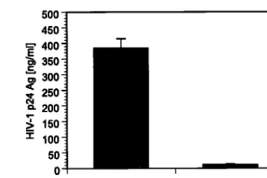
FIG. 3. To verify the antiviral activity of H1 promoter-driven
tat
siRNA, 293T cells were cotransfected with HIV-1
NL4.3and pAAV
DNA constructs. Quantification of viral p24 antigen levels 48 h
post-transfection in cell-free supernatant revealed a 97% decrease in cells
expressing
tat
shRNA (pAAV-tat) compared to cells that were
trans-fected with HIV-1
NL4.3and control mock vector lacking
tat-specific
[image:2.603.48.282.500.649.2]sequences (pAAV-
⌬
tat). Values represent averages of three
indepen-dent experiments, with the ranges indicated.
FIG. 4. Northern blot analysis of
tat
shRNA expression in
HIV-1
NL4.3-infected H9 cells transduced with AAV-tat
or AAV-
⌬
tat.
Con-tinuous expression of
tat
shRNA was observed on days 14 and 35 after
HIV-1
NL4.3infection. rRNA expression was used as a loading control.
11532
BODEN ET AL.
J. V
IROL.
on November 8, 2019 by guest
http://jvi.asm.org/
20 pmol of sense primer Tat-A (5⬘-ATG GAG CCA GTA GAT CCT A-3⬘) and antisense primer Tat-B (5⬘-TGC TTT GAT AGA GAA ACT TGA TG-3⬘), 1 mM deoxynucleoside triphosphates, and 2.5 U ofTaqpolymerase (Qiagen). PCR was carried out in a Mastercycler gradient PCR cycler (Eppendorf, Hamburg, Germany) with a thermal program of 95°C for 1 min; 35 cycles of 95°C for 15 s, 58°C for 30 s, and 72°C for 30 s; and 72°C for 5 min. The PCR product representing the 215-bptatexon 1 was analyzed on a 1% agarose gel and column purified with the QIAquick PCR kit (Qiagen). Cycle sequencing was performed using dye-labeled terminator chemistry.
Generation of escape mutant HIV-1NL4.3mutand rechallenge of wild-typetat shRNA-expressing cells.The introduction of a point mutation (A3T) at position 9 of the tattarget sequence in the infectious molecular clone pNL4.3 was achieved by recombinational overlap extension PCR. Two separate PCRs were performed on pNL4.3 DNA involving primer pair 5⬘-GGC AGG AGT GGA AGC CAT A-3⬘and 5⬘-GCA ATT GGA ACA AGC AGT TTT AGG C-3⬘and primer pair 5⬘-ACT GCT TGT TCC AAT TGC TAT TGT AAA AAG TG-3⬘
and 5⬘-CCC TCC TGA GGA TTG CTT AAA GA-3⬘. The DNA of the gel-purified PCR products was amplified with primers 5⬘-GGC AGG AGT GGA AGC CAT A-3⬘and 5⬘-CCC TCC TGA GGA TTG CTT AAA GA-3⬘. The column-purified PCR product was digested withEcoRI andSalI and introduced in the equally digested pNL4.3. The mutant molecular clone was sequence verified.
Stable cell lines H9-tatand H9-mock (⌬tat) were challenged with either wild-type virus HIV-1NL4.3or mutant virus HIV-1NL4.3mut.Two million cells were
infected with 20 ng of p24 of each virus for 3 h at 37°C. Infected cells were washed once with phosphate-buffered saline and resuspended in RPMI medium. The course of the infection was monitored over a 10-day period by p24 ELISA measurements.
RESULTS AND DISCUSSION
Our initial investigations used a synthetic siRNA molecule
that specifically targets exon 1 of the HIV-1
tat
gene.
tat
en-codes a transactivator protein that is essential for viral
repli-cation. The antiviral potency of
tat-
specific siRNA was assessed
by transient cotransfection of 293T cells with
tat
siRNA and an
infectious molecular clone of HIV-1 (HIV-1
NL4.3). The levels
of HIV-1 p24 antigen were measured in the culture
superna-tant by ELISA 4 days after transfection, and
tat
siRNA
sup-pressed HIV-1 replication by 91% compared to relevant
con-trols (Fig. 2).
To achieve cellular expression of
tat
siRNA, a recombinant
AAV DNA vector (pAAV-
tat
) was created that contained an
H1 promoter-driven shRNA expression cassette (Fig. 1). The
H1 promoter was chosen based on recent evidence that it can
effectively drive shRNA expression with retroviral vectors (2).
A second vector, devoid of
tat
-specific sequences in the shRNA
expression cassette (pAAV-
⌬
tat
), served as a control. Each
vector was cotransfected with HIV-1
NL4.3in 293T cells, and
HIV-1 p24 antigen levels were quantified in cell-free
superna-tant 48 h after transfection. HIV-1 p24 antigen level was
re-duced by 97% in cells expressing
tat
shRNA compared to
control cells transfected with a mock construct, pAAV-
⌬
tat
(Fig. 3). Analysis of total intracellular HIV-1 RNA levels by
Northern blotting and quantitative real-time PCR confirmed a
similar reduction in virus replication (data not shown).
To create a cell line that stably expresses shRNA, AAV-
tat
and AAV
-
⌬
tat
vectors were introduced into an AAV-2
trans-duction system. Recombinant viral particles were generated
and used to infect H9 cells, which are derived from a human
cutaneous T-cell lymphoma. H9 cells are permissive for HIV-1
replication, thereby facilitating determinations of the
magni-tude and durability of antiviral RNAi. Stable expression of
tat
shRNA in H9 cells was confirmed by Northern blot analysis
(Fig. 4). Genomic integration of AAV
-tat
was verified by
Southern blot analysis (data not shown). The cells were then
infected with 100 TCID
50of the infectious molecular clone
HIV-1
NL4.3, and p24 antigen levels were quantified in cell-free
supernatant at weekly intervals over a 2-month period. During
the first 3 weeks, HIV-1 replication was reduced by 95% in
cultures of cells expressing
tat
shRNA compared to control
cells (Fig. 5A). By day 25, however, p24 levels had increased,
indicating loss of
tat
shRNA-mediated antiviral activity.
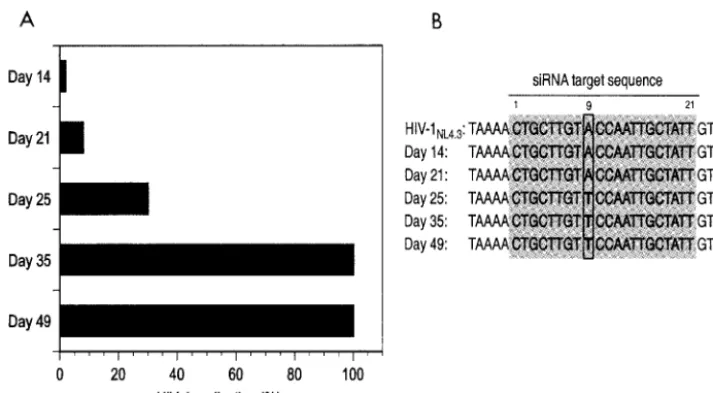
North-FIG. 5. (A) Inhibition of HIV-1 replication in H9 cells stably expressing
tat
shRNA. Cells were challenged with 100 TCID
50of HIV-1
NL4.3, and
p24 antigen levels were assessed over a 2-month period. Viral replication was suppressed by 95% until day 21 compared to control cells. Viral
escape started to emerge on day 25, and viral replication was no longer suppressed by day 35, with observed p24 antigen levels being similar to
those in control cells. (B) Sequence analysis of HIV-1
NL4.3in culture supernatant at indicated time points. A mutation in position 9 of the
tat
target
sequence emerged at day 25, coincident with the loss of
tat
shRNA antiviral activity.
on November 8, 2019 by guest
http://jvi.asm.org/
[image:3.603.114.471.68.264.2]ern blot analysis revealed continued intracellular production of
shRNA (Fig. 4). To determine whether HIV-1 escape from
RNAi was due to the emergence of mutations within the
tat
gene, viral RNA was extracted from sequential samples of
culture supernatant and the
tat
siRNA target region was PCR
amplified and sequenced. During the first 3 weeks, the
se-quence of the
tat
target region was identical to the expressed
shRNA. On day 25, a viral species emerged with a
nonsynony-mous mutation at nucleotide position 9 of the targeted
se-quence leading to an amino acid change from threonine
(ACC) to serine (TCC) (Fig. 5B).
To determine whether the viral escape mutant was indeed
resistant to
tat
shRNA, we created virus HIV-1
NL4.3mutand
shRNA pAAV-
tat
mutwith a point mutation corresponding to
position 9 of the
tat
target sequence. Cotransfection of 293T
cells with HIV-1
NL4.3mutand pAAV-
tat
wtor pAAV-
tat
mut,
re-spectively, resulted in well-suppressed viral p24 production
with pAAV-
tat
mut(92%) opposed to only moderately reduced
virus production with pAAV-
tat
wt(50%) (Fig. 6A). Next, we
infected
tat
shRNA-expressing (H9-
tat
) and control
(H9-mock) cells with HIV-1
NL4.3mutand HIV-1
NL4.3wt.Viral
chal-lenge of the
tat
shRNA-expressing cell line with mutant virus
HIV-1
NL4.3mutdid not result in any suppression of viral
repli-cation whereas challenge of the same cell line (H9-
tat
) with
wild-type virus HIV-1
NL4.3wtshowed inhibition of HIV-1
rep-lication by day 10 (Fig. 6B).
Our results demonstrate a critical obstacle to using RNAi as
an antiretroviral agent. The rapid replication kinetics of HIV-1
and the error-prone nature of viral reverse transcriptase led to
the emergence and selection of viral quasispecies that were
genotypically distinct in the
tat
shRNA region, thereby
elimi-nating the antiviral effect. The point mutation observed in this
study occurred in the middle of the
tat
target sequence.
Pre-vious work has demonstrated that transfection with siRNA
containing mismatches to the target sequence in the middle of
the siRNA molecule reduces the efficiency of gene silencing (1,
2, 5). Our findings suggest that, in order for RNAi to durably
suppress HIV-1 replication, more potent shRNAs need to be
designed which target highly conserved regions of the viral
genome (e.g.,
gag
and
pol
) essential for the viral life cycle.
Alternatively, similar to present antiviral drug therapy
para-digms, RNAi constructs coexpressing multiple shRNAs need
to be developed that simultaneously target different regions of
the viral genome, thereby reducing the probability of
generat-ing shRNA escape mutants.
ACKNOWLEDGMENTS
D. Boden and O. Pusch contributed equally to the preparation of the
manuscript.
This work was supported by a Clinical Scientist Development Award
from the Doris Duke Charitable Foundation, a Daland Fellowship in
Clinical Investigation from the American Philosophical Society, and a
Career Development Grant from NIAID/NIH (B.R.).
We thank the Brown/Tufts/Lifespan CFAR Retrovirology Core
Laboratory for assay support. We acknowledge the AIDS Research
and Reference Reagent Program, Division of AIDS, NIAID, NIH, for
supplying H9 cells (contributed by Robert Gallo) and HIV-1
NL4.3(contributed by Malcolm Martin).
REFERENCES
1. Amarzguioui, M., T. Holen, E. Babaie, and H. Prydz.2003. Tolerance for mutations and chemical modifications in a siRNA. Nucleic Acids Res.31:
589–595.
2. Brummelkamp, T. R., R. Bernards, and R. Agami.2002. Stable suppression of tumorigenicity by virus-mediated RNA interference. Cancer Cell2:243– 247.
3. Capodici, J., K. Kariko, and D. Weissman.2002. Inhibition of HIV-1 infec-tion by small interfering RNA-mediated RNA interference. J. Immunol.
169:5196–5201.
4. Coburn, G. A., and B. R. Cullen.2002. Potent and specific inhibition of human immunodeficiency virus type 1 replication by RNA interference. J. Virol.76:9225–9231.
5. Gitlin, L., S. Karelsky, and R. Andino.2002. Short interfering RNA confers intracellular antiviral immunity in human cells. Nature418:430–434. 6. Jacque, J. M., K. Triques, and M. Stevenson.2002. Modulation of HIV-1
replication by RNA interference. Nature418:435–438.
FIG. 6. (A) Cotransfection of HIV-1
NL4.3mutwith pAAV-tat
wt, pAAV-tat
mut, and mock vector (pAAV-
⌬
tat). When the targeted
tat
sequence
and shRNA were homologous (HIV-1
NL4.3-mut/tat-mut), virus production was reduced by 92%. When the targeted
tat
sequence and shRNA were
not homologous (HIV-1
NL4.3-mut/tat-wt), virus production was suppressed by only 50%. (B) H9 cells stably expressing
tat
shRNA (H9-tat) or
⌬
tat
(H9-mock) were infected with 20 ng of p24 of HIV-1
NL4.3or HIV-1
NL4.3mut. On day 10 viral replication was reduced with wild-type virus
(H9-tat/NL4.3-wt) but not with mutant virus (H9-tat/NL4.3-mut), demonstrating the importance of complete sequence homology between the viral
target sequence and the expressed shRNA.
11534
BODEN ET AL.
J. V
IROL.
on November 8, 2019 by guest
http://jvi.asm.org/
[image:4.603.83.502.69.248.2]S. K. Lee, R. G. Collman, J. Lieberman, P. Shankar, and P. A. Sharp.2002. siRNA-directed inhibition of HIV-1 infection. Nat. Med.8:681–686. 9. Ramratnam, B., S. Bonhoeffer, J. Binley, A. Hurley, L. Zhang, J. E. Mittler,
M. Markowitz, J. P. Moore, A. S. Perelson, and D. D. Ho.1999. Rapid
11. Yamamoto, T., S. Omoto, M. Mizuguchi, H. Mizukami, H. Okuyama, N. Okada, N. K. Saksena, E. A. Brisibe, K. Otake, and Y. R. Fuji.2002. Double-strandednefRNA interferes with human immunodeficiency virus type 1 replication. Microbiol. Immunol.46:809–817.
on November 8, 2019 by guest
http://jvi.asm.org/