A CLINICAL COMPARISON BETWEEN BUPIVACAINE
(PLAIN) AND BUPIVACAINE-MIDAZOLAM IN BRACHIAL
PLEXUS BLOCK VIA SUPRACLAVICULAR APPROACH
A STUDY OF 100 CASES
Dissertation submitted for the degree of
DOCTOR OF MEDICINE
Branch – X (ANAESTHESIOLOGY)
APRIL
–
2015
TAMIL
NADU
DR. M.G.R.
MEDICAL
UNIVERSITY
CHENNAI,
BONAFIDE CERTIFICATE BY THE GUIDE
This is certify that this dissertation entitled A CLINICAL COMPARISON
BETWEEN BUPIVACAINE (PLAIN) AND BUPIVACAINE-MIDAZOLAM IN BRACHIAL PLEXUS BLOCK VIA SUPRACLAVICULAR APPROACH a bonafide record work done by Dr. ROSHIN ANN JAMES under my direct supervision and guidance, submitted to the Tamil Nadu Dr. M.G.R. Medical University in partial fulfillment of University regulation for MD, Branch X – Anaesthesiology
DR.SIVAKUMAR,M.D,DA.,
CHIEF AND PROFESSOR,
DEPARTMENT OFANAESTHESIOLOGY,
THANJAVUR MEDICAL COLLEGE,
THANJAVUR.
ENDORSEMENT BY THE HOD AND DEAN OF THE INSTITUTE
This is to certify that this dissertation entitled A CLINICAL COMPARISON BETWEEN BUPIVACAINE (PLAIN) AND BUPIVACAINE-MIDAZOLAM IN BRACHIAL PLEXUS BLOCK VIA SUPRACLAVICULAR APPROACH is bonafide research work done by Dr.ROSHIN ANN JAMES , Resident in Anaesthesiology, Thanjavur Medical College,Thanjavur
Professor and Head Dean
Department of Anaesthesiology Thanjavur Medical College Thanjavur Medical College Thanjavur
Thanjavur Tamil Nadu Tamil Nadu .
. Date:
Place:
DECLARATION BY THE CANDIDATE
I hereby declare that this dissertation entitled A CLINICAL COMPARISON BETWEEN BUPIVACAINE (PLAIN) AND BUPIVACAINE-MIDAZOLAM IN BRACHIAL PLEXUS BLOCK VIA SUPRACLAVICULAR APPROACH is a bonafide and genuine research work carried out by me in the Department of Anaesthesiology, Thanjavur Medical College.
Date: Signature of the candidate Place: Thanjavur [ Dr.ROSHIN ANN JAMES ] Resident
ACKNOWLEDGEMENTS
First and foremost I would like to express my deepest gratitude to
I am today.
It gives me great pleasure in preparing this dissertation and I take
this opportunity to thank everyone who has made this possible.
I would like to express my deep gratitude and sincere thanks to my
guide Dr. SIVAKUMAR., M.D., D.A., Chief, Department of
Anaesthesiology, Thanjavur Medical College for preparing me for this task, guiding me with his superb talent and professional expertise,showing great care and attention to details and without his supervision and guidance this dissertation would have been impossible.
I am highly indebted to Dr. R. MUTHUKUMARAN M.D., D.A.,
Professor and Head, Department of Anaesthesiology, Thanjavur Medical college for his invaluable guidance, constant encouragement, immense patience and great care and attention to details that he has so willingly shown in helping me to prepare this dissertation. His stature and knowledge has been a constant source of inspiration for the whole of my post graduation period.
I take this opportunity to convey my heart felt gratitude to
constant source of inspiration, encouragement and for her kindness, invaluable
guidance, exhaustive knowledge, professional expertise and emotional
support given willingly and expertly during the course of my study.
It gives me immense pleasure to extend my sincere thanks to all the Asst. Professors of our Department whose authoritative knowledge of practical skills has guided and inculcated in me a sense of confidence. I am thankful to them for their valuable guidance and for understanding and accommodating me during difficult periods of this dissertation.
I owe a great sense of indebtedness to
Dr.K.MAHADEVAN,M.S, DEAN for allowing me to use the institutional facilities.
I owe my gratitude to my husband and parents for their constant help
and encouragement.
I would also like to thank the Superintendent, OT staff of Th a n ja v u r
M e d i c a l C o ll e g e for their help and assistance.
I owe my sincere thanks to the statistician Mr.Jayakumar for helping me with statistical analysis.
who have helped me in preparing this dissertation.
My special thanks to S . J c o m p u t e r s for their meticulous typing and styling of this script.
Last but not the least, I express my special thanks to all my patients and their families, who in the final conclusion are the best teachers
LIST OFABBREVIATIONS USED
g -
ASA -
BT -
BZD -
cm -
CT -
DBP -
dl -
ECG -
GABA -
gm -
HBsAg -
HIV -
HS -
IM -
IV -
Kg -
LA -
MAP -
Min -
ml -
mm of Hg -
NS -
pKa -
RA -
S -
S.D. -
SBP
-microgram
American Society of Anaesthesiologists (classification) Bleeding time
Benzodiazepine centimetre Clotting Time
Diastolic Blood Pressure Decilitre
Electro Cardio Gram
Gamma Amino Butyric Acid gram
Hepatitis B Antigen
Human Immunodeficiency Virus Highly significant
Intramuscular Intravenous Kilogram
Local Anaesthetic Mean Arterial Pressure Minutes
Millilitre
Millimetre of mercury Not significant
Dissociation constant Rescue analgesics Significant
ABSTRACT
Background and objectives : Adjuvants to local anaesthetics for brachial plexus block enhances the quality and duration of analgesia. Midazolam , a water-soluble benzodiazepine, is known to produce antinociception and enhance the effect of local anaesthetics when given epidurally or intrathecally. The purpose of this study was to assess the effect of Midazolam added to brachial plexus block by supraclavicular approach.
Methods : A prospective, randomized, single blinded study was conducted on 100 ASA I or II adult patients undergoing upper limb surgeries under supraclavicular brachial plexus block. Patients were randomly divided into two groups. Patients in Group B (n = 50) were administered 30mL of 0.375% Bupivacaine and Group BM (n = 50) were given 30mL of 0.375% Bupivacaine with Midazolam 2.5 mg. The onset time and duration of sensory and motor blockade were recorded. Hemodynamic variables (i.e., heart rate, noninvasive blood pressure, oxygen saturation), sedation scores and rescue analgesic requirements were recorded for 24 hr postoperatively.
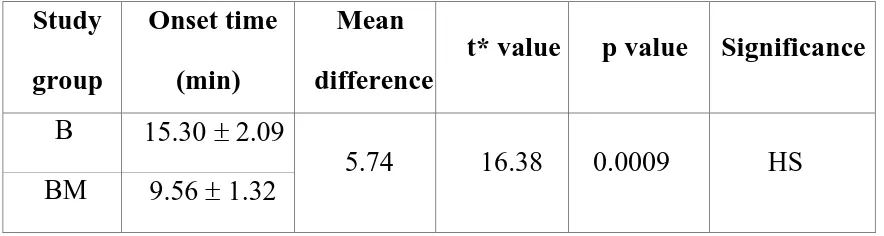
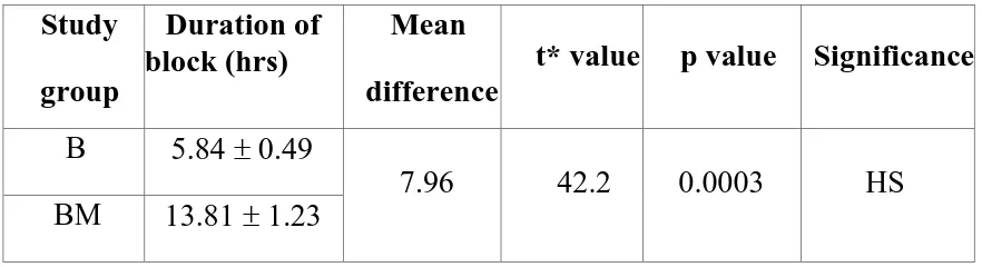
Results : The onset of sensory and motor block was significantly faster in Group BM compared to Group B (P < 0.05). Rescue analgesic requirements were significantly less in Group BM compared to Group B (P < 0.05). Haemodynamics and sedation scores did not differ between the two groups in the post-operative period.
Conclusion : Midazolam (2.5mg) in combination with 30mL of Bupivacaine (0.375%) hastened onset of sensory and motor block, and prolonged postoperative analgesia when used in brachial plexus block, without
producing any adverse effects.
TABLEOF CONTENTS
PageNo.
1. Introduction 1
2. Aim of the study 4
3. Brachial plexus block 6
4. Review ofliterature 48
5. Methodology 67
6. Observations and Results 76
7. Discussion 95
8. Summary 115
9. Conclusion 119
10. Bibliography 121
11. Annexures
Proforma 130
Masterchart 132
Keytothe masterchart 137
Statisticalformulae used 138
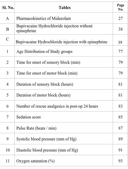
LIST OF TABLES
Sl.No. Tables PageNo.
A Pharmacokinetics of Midazolam 27
B Bupivacaine Hydrochloride injection without epinephrine 38
C
Bupivacaine Hydrochloride injection with epinephrine
39
1 Age Distribution of Study groups 77
2 Time for onset of sensory block (min) 79
3 Time for onset of motor block (min) 79
4 Duration of sensory block (hours) 81
5 Duration of motor block (hours) 81
6 Number of rescue analgesics in post-op 24 hours 83
7 Sedation score 85
8 Pulse Rate (beats / min) 87
9 Systolic blood pressure (mm of Hg) 89
10 Diastolic blood pressure (mm of Hg) 91
11 Oxygen saturation (%) 93
[image:14.595.103.530.122.684.2]LIST OFGRAPHS
Sl.No. Graphs Pg.No.
1 Age Distribution of Study groups 78
2 Onset of block 80
3 Duration of block 82
4 Number of rescue analgesics in post-op 24 hours 84
5 Sedation score 86
6 Pulse Rate (beats / min) 88
7 Systolic blood pressure (mm of Hg) 90
8 Diastolic blood pressure (mm of Hg) 92
9 Oxygen saturation (%) 94
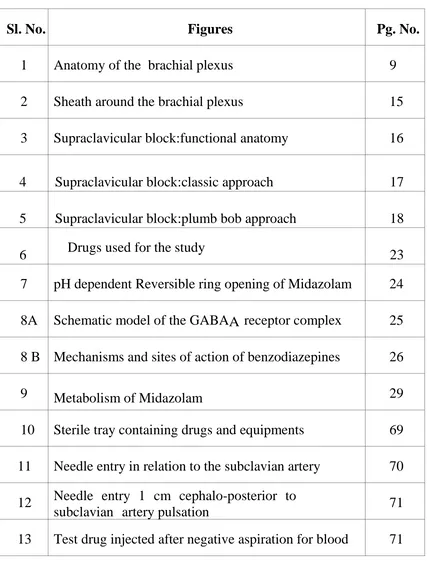
LIST OF FIGURES
Sl.No. Figures Pg.No.
1 Anatomy of the brachial plexus 9
2 Sheath around the brachial plexus 15
3 Supraclavicular block:functional anatomy 16
4 Supraclavicular block:classic approach 17
5
Supraclavicular block:plumb bob approach
18
6 Drugs used for the study 23
7 pH dependent Reversible ring opening of Midazolam 24
8A Schematic model of the GABAA receptor complex 25
8 B Mechanisms and sites of action of benzodiazepines 26
9 Metabolism of Midazolam 29
10 Sterile tray containing drugs and equipments 69
11 Needle entry in relation to the subclavian artery 70
12 Needle entry 1 cm cephalo-posterior to
subclavian artery pulsation 71
[image:16.595.104.531.96.667.2]1
2 INTRODUCTION
“ Pain, like pleasure is passion of the soul,
That is an emotion and not one of the senses”
- PLATO and ARISTOTLE (375 B.C)
Pain is a fundamental biological phenomenon. The International Association for the Study of pain has defined “pain as an unpleasant sensory and emotional experience associated with actual or potential tissue damage." Pain is always under estimated and under treated. The relief of pain during surgery is the main part of anaesthesia.
Regional nerve blocks provide unwanted side effects of anaesthetic drugs used during general anaesthesia and the stress of laryngoscopy and tracheal intubation .It provides better intraoperative and prolonged pain relief during postoperative period. Minimising the stress response and minimising anaesthetic drug requirements are beneficial to the patients with various cardiorespiratory comorbidities.
Brachial plexus blocks provide a wonderful alternative to general anaesthesia
for upper limb surgeries. They achieve near-ideal operative conditions by providing complete and prolonged pain relief, muscle relaxation, maintaining stable intra-operative hemodynamics and adequate sympathetic block. The
3
Of various local anaesthetics, Bupivacaine is used most frequently, as it has a long duration of action varying from 3 to 8 hours. However there are many limiting factors like delayed onset, patchy or incomplete analgesia, sometimes of
short duration etc. Various drugs like Dexmedetomidine,
Opioids2,Hyaluronidase3, Clonidine etc4.have been added to local anaesthetics in
order to modify the block in terms of quicker onset, good quality, prolonged duration and post operative analgesia. But these presented with adverse systemic effects or doubtful efficacy.4
Midazolam, a water-soluble benzodiazepine is known to produce
antinociception and enhance the effect of local anaesthetic when given
epidurally or intrathecally5. Midazolam produces this effect by its action on
gamma aminobutyric acid-A (GABA-A) receptors. GABA receptors have also been found in peripheral nerves5.
4
5
AIM OF THE STUDY
The present study was undertaken to compare the efficacy of adding midazolam (2.5mg) to bupivacaine (0.375%) in supraclavicular
technique of brachial plexus block for upper limb surgeries with respect to
1) Onset time and duration of sensory and motor blockade.
2) Hemodynamic changes
3) Sedation score intra and post-operatively
4) Number of rescue analgesics required in the 24 hour post-operative period.
6
Brachial Plexus Block
7 HISTORY
1. 1858 – theory of pain was a separate and distinct sense was definitely
formulated by Mortiz S.Schiff
2. 1884 – William Halsted and Alfred Hall – idea of injecting cocaine into
nerve trunk
3. 1911 – G. Hirschel performed first percutaneous axillary brachial plexus
block
4. 1911 – D. Kulenkampff performed supraclavicular brachial plexus block
5. 1943 - Lidocaine was synthesized by Lofgreen and Lundquvisit
6. 1956 – Bupivacaine synthesized by Ekenstam
7. 1963 – Bupivacaine introduced clinical practice by Telivuo
8
ANATOMYOF BRACHIALPLEXUS 6,7
Knowledge of formation of brachial plexus and its ultimate cutaneous and muscular distribution is absolutely essential to the intelligent and effective use of brachial plexus anaesthesia for upper limb surgeries. Close familiarity with the vascular, muscular and fascial relationships of the plexus is equally essential to the mastery of various techniques , for it is these perineural structures which serve as the landmark by which needle may accurately locate the plexus percutaneously. In its course from intervertebral foramina to the upper arm, the fibres are
composed consecutively of roots, trunks, divisions, cords and terminal nerves.
FORMATION OFBRACHIALPLEXUS :
Brachial plexus is formed by the union of ventral rami of lower four cervical nerves (C5, 6,7,8) and first thoracic nerve (T1) with frequent contributions from C4 or T2.When contribution from C4 is large and from T2 is lacking, the plexus appears to have a more cephaloid position and is termed “Prefixed”. When contribution from T2 is large and from C4 is lacking, the plexus appears to have a caudal position and is termed “postfixed”. Usually prefixed or postfixed positions are associated with
9 ROOTS:
Represent the anterior primary divisions of lower four cervical and first thoracic nerves. They emerge from the intervertebral foramina and fuse above the first rib to form the trunks.
TRUNKS:
The roots combine above the first rib to form the three trunks of the plexus. C5 and C6 unite at the lateral border of the scalenus medius and form the “Upper trunk”.C8 and T1 unite behind the scalenus anterior to form “lower trunk” and C7 continues as a sole contributor to the “middle trunk”.
DIVISIONS :
As the trunks pass over the first rib and under the clavicle, each one of them divides into anterior and posterior divisions.
CORDS:
10
“posterior cord”, at first above and then behind the axillary artery. The
medial and lateral cords give rise to nerves that supply the flexor surface of
upper extremity, while nerves arising from posterior cord supply extensors7
MAJORTERMINAL NERVES :
Each of these cords gives off a branch that contributes to or become one of the major nerves to the upper extremity and then terminates as a major nerve. The lateral and median cords give off lateral and medial heads of the medial nerve and continue as major terminal nerves, the lateral cord terminating as musculocutaneous nerve and medial cord as ulnar nerve. Posterior cord gives off, axillary nerve as its major branch and then continues as the radial nerve.
DISTRIBUTIONOF BRACHIAL PLEXUS:
These are divided into those that arise above the clavicle – the supraclavicular branches and those that arise below it, the infraclavicular branches.
11 Supraclavicularbranches :
From roots :
1. Nerves to scaleni and longus colli – C5,6,7,8
2. Branch to phrenic nerve – C5
3. Dorsal scapular nerve – C5
4. Long thoracic nerve – C5,6,(7)
From trunks :
1. Nerve to subclavius – C5,6
2. suprascapular nerve – C5,6,
Infraclavicular branches : They branch from cords but their fibres may be tracked back to spinal nerves.
Lateral cord :
1. Lateral pectoral nerve – C5,6,7
2. Musculocutaneous nerve – C5,6,7
3. Lateral root of median nerve – C5,6,7
Medial cord :
1. Medial pectoral nerve – C8, T1
2. Medial cutaneous nerve of forearm – C8, T1
3. Ulnar nerve – C8, T1
4. Medial root of median nerve – C8, T1
12
Posterior cord :
1. Upper subscapular nerve – C5, 6
2. Thoracodorsal nerve – C 6, 7, 8
3. Lower subscapular nerve – C5, 6
4. Axillary nerve – C5, 6
5. Radial nerve - – C5, 6, 7, 8, T1
SYMPATHETICCONTRIBUTIONTO BRACHIALPLEXUS :
The segmental preganglionic sympathetic contributions are
variable, but generally extend more caudal. The highest contribution is usually T2 with T1 contributing only rarely, while lowest may be as far as T8, T9 or even T10. The post ganglionic contributions are from grey rami communicants from the sympathetic chain.
RELATIONSOF BRACHIALPLEXUS :8
13
scalenus anterior. The fascia covering the muscles is derived from the
perivertebral fascia, which splits to invest these muscles and rejoins again at their lateral margins to form an enclosed space, the interscalene space.
As the plexus crosses the first rib, the three trunks are 'stacked' one on top of the other vertically. Not infrequently, the inferior trunk gets trapped behind and even beneath the subclavian artery above the rib, during embryologic development. This may be reason why local anaesthetics injected via the interscalene technique sometimes fail to provide anaesthesia in the distribution of the ulnar nerve, which may be buried deep within inferior trunk behind or beneath the subclavian artery.
14 scalenus anterior.
As it passes over the first rib, becoming the axillary vein it joins the neurovascular bundle so that parts of the plexus are sandwiched between artery and vein. As all the three enter the axilla, they invaginate the perivertebral fascia at the lateral margins of the anterior and medial scalene muscles, carrying this fascial investment of the neurovascular bundle into the axilla as the axillary fascia, an extension of the perivertebral or scalene fascia forming the axillary perivascular space, a tubular
extension of the interscalene space.
In its course through the axilla and upper arm the fascia of the surrounding muscles contribute to the axillary sheath, making it thick and tough, providing the 'fascial click' to the anaesthetic while entering the sheath. It is important to note that major terminal nerves leave the sheath high in the axilla undercover of perctoralis minor muscle.
The musculocutaneous nerve enters the substance of
15
THEBRACHIALPLEXUS SHEATH
Volume of the sheath : 42ml.
Shape of the sheath : Cylindrical to conical – Wide proximally and narrow distally.
Length : 8-10cms long.
The connective tissue of the prevertebral fascia and the anterior and middle scalenes envelops the brachial plexus as well as the subclavian and
axillary artery in a neurovascular “sheath”.The tissue is densely organized as it leaves the deep cervical fascia proximally but becomes more loosely arranged distally. The sheath blends with the fascia of the biceps and brachialis muscle distally.
Anatomic dissection, histologic examination and CT scanning after
injection of radio contrast into the sheath demonstrate the existence of
connective tissue septae which extend inward from the fascia
16 AnaestheticImplications:
Because of these connective tissue septae, anaesthesia might be complete and rapid in onset in some nerves, but delayed and incomplete or completely absent in others. The incidence of partial block is an exception rather than the rule, so septa apparently are of little clinical significance as the local anaesthetic can percolate through them.
TECHNIQUE OF BRACHIAL PLEXUS BLOCK9
Surgical anaesthesia of the upper extremity and shoulder can be obtained following neural blockade of the brachial plexus at several sites. The various approaches that can be used for this blockade are as follows
1.Interscalene approach 2. Supraclavicular approach a. Classic approach
b. Plumb –bob technique
c. Subclavian perivascular technique 3. Axillary approach
17
Supraclavicular approach
1 0a. Classic approach
Patient Position: Supine, with the head turned to the opposite side and
the arm adducted and the shoulder dropped.
Indications: Anesthesia and immediate postoperative analgesia for
surgery above the elbow, or at the elbow, forearm, wrist, or hand.
Needle Size: 22-gauge , 5-cm insulated needle.
Volume: 30 to 40 mL.
Anatomic Landmarks: The lateral border of the clavicular head of the
sternocleidomastoid at its insertion into the clavicle, the subclavian
artery, and the anterior and middle scalene muscles.
Approach and Technique: First, the lateral border of the
sternocleidomastoid, particularly at the level of its insertion into the
clavicle, is marked. Next, the interscalene groove is identified and
marked. The palpating finger is placed parallel and immediately
superior to the clavicle at the level of the subclavian artery and the
18
stimulator (1.0 mA, 2 Hz, 0.1 ms) is inserted posterior to the palpating
finger (Fig. 4). The needle is directed caudad and lateral, so as to cross
the clavicle almost perpendicularly. A muscle response involving the
arm and fingers either in flexion or extension is elicited within 2 to 3
cm. The position of the needle is adjusted to maintain the same motor
response with a current of 0.5 to 0.7 mA. After negative aspiration for
blood, the local anesthetic is slowly injected with repeated aspiration
for blood every 5 mL to be distributed around the brachial plexus
B) Plumb bob approach10
Patient Position: Supine, with the head turned to the opposite side and
the arm adducted and the shoulder dropped.
Approach and Technique Patient’s head is slightly off the back table
so that lateral border of sternocleidomastoid is marked as it inserts to
the clavicle.From that point,a mental plane is visualised that runs parasagittaly through that site. “Plumb blob” was chosen,since if one suspends a “plumb-blob”over the entry site,needle insertion through the point will result in contact
19
sternocleidomastoid ,needle is inserted in parasagittal plane at 900 angle to the table
top.Once paraesthesia has been elicited, after negative aspiration for blood,
the local anesthetic is slowly injected with repeated aspiration for
blood every 5 mL to be distributed around the brachial plexus .
LOCAL ANESTHETIC MECHANISMS IN NERVE BLOCKADE11
Impulse blockade by local anesthetics may be summarized by the following chronolog:
• Solutions of local anesthetic are deposited near the nerve. Removal of free
drug molecules away from this locus is a function of tissue binding, removal by the circulation, and local hydrolysis of amino-ester anaesthetics. The net result is penetration of the nerve sheath by the remaining free drug molecules. • Local anesthetic molecules then permeate the nerve‘s axon membranes and
reside there and in the axoplasm. The speed and extent of these processes
depend on a particular drug‘s pĸa and on the lipophilicity of its base and cation
species.
• Binding of local anesthetic to sites on voltage-gated Na+ channels prevents
opening of the channels by inhibiting the conformational changes that underlie channel activation. Local anesthetics bind in the channel’s pore and also
20
• During onset or recovery from local anesthesia, impulse blockade is incomplete
and partially blocked fibres are further inhibited by repetitive stimulation, which produces an additional use-dependent blinding to Na+ channels.
• One local anesthetic binding site on the Na+ channel may be sufficient to account
for the drug's resting (tonic) and use-dependent (phasic) actions Access to this site may potentially involve multiple pathways, but for clinical local anesthetics, the primary route is the hydrophobic approach from within the axon membrane.
• The clinically observed rates of onset and recovery from blockade are governed
by the relatively slow diffusion of local anesthetic molecule into and out of the whole nerve, not by their much faster binding and dissociation from ion channels. A clinically effective block that may last for hours can be accomplished with local
anesthetic drugs that dissociate from Na+ channels in a few seconds11
.
COMPLICATIONS12 Vascular puncture
21 Pleural puncture
The most significant complication of supraclavicular approach for blocking brachial plexus is development of pneumothorax. The incidence of pneumothorax is 1 percent with this technique and much higher in inexperienced hands.A pneumothorax must be suspected when there is dyspnoea, cough or pleuritic chest pain but the diagnosis can be confirmed only by chest x-ray.
Phrenic nerve block
Phrenic nerve block occurs in 40-60% of patients because of spread of local anaesthetic to the anterior surface of anterior scalene muscle. The effect is avoided if anaesthetic is deposited deep on the middle trunk on division or cord. This is rarely symptomatic. Radiographic confirmation may be obtained.
Recurrent laryngeal nerve block
It causes transient dysphonia, occurs in 1% of case and only on the right side because recurrent laryngeal nerve loops around the subclavian artery on the right side and arch of aorta on the left.
Nerve damage or neuritis
22
preoperatively. Other remote causes include excessive tourniquet time, concentrated solution with vasoconstrictor and susceptible host tissue.
Horner's syndrome
It consists of ptosis, miosis, anhydrosis and enophthalmos. It usually follows stellate ganglion block. It is found in 10% of cases, after interscalene block.
Toxic reaction to drug
It is likely to occur if there is over dosage of drug or inadventent intravascular injection is made, but can be avoided with proper negative aspiration test before drug injection
23
PHARMACOLOGYOF MIDAZOLAM13,14,15,16
Midazolam is a water soluble benzodiazepine. It was first synthesized by Fryer and Walser in 1976. It was the first benzodiazepine that was produced primarily for use in anaesthesia.It is associated with a low incidence of injection pain and post injection phlebitis and thrombosis. It has replaced diazepam as the most commonly administered benzodiazepine in the perioperative period for preoperative medication and intravenous “conscious” sedation. The five principal pharmacologic effects are:
sedation, anxiolysis, anticonvulsant actions, spinal cord-mediated skeletal muscle relaxation and anterograde amnesia.
Chemistry:
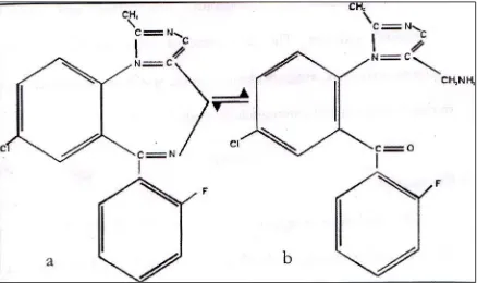
24 Chemicalstructure :
Midazolam has a fused imidazole ring that is different from classic benzodiazepines. The imidazole ring accounts for the basicity, stability of an aqueous solution and rapid metabolism. It is named
8-chloro-6(2-flurophenyl)-1-methyl-4H- imidazo-(1, 5-) (1, 4) benzodiazepine
maleate. Reversible ring opening of midazolam above and below a pH of 4; the ring closes at a pH> 4, converting midazolam from a water soluble to a lipid soluble drug.
pH DEPENDENT RING OPENING OF MIDAZOLAM
Fig. 7 : Reversible ring opening of Midazolam above and below a pH of 4; the ring closes at a pH> 4, converting Midazolam from a watersolubleto alipid solubledrug.
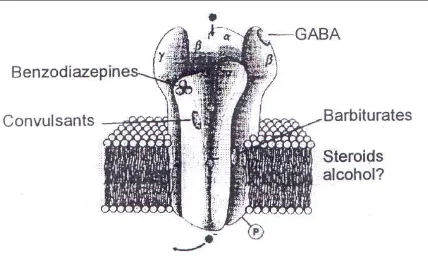
[image:40.595.85.523.384.644.2]25 Mechanismofaction:
Midazolam and benzodiazepines in general appear to produce all their pharmacologic effects by facilitating the actions of gamma amino butyric acid (GABA), the principal inhibitory neurotransmitter in the CNS. GABA-adrenergic neurotransmission counterbalances the influence of excitatory neurotransmitters. The benzodiazepine receptors are found in highest densities in the olfactory bulb, cerebral cortex, cerebellum, hippocampus, substantia nigra and inferior colliculus. Current data suggests
a pentameric protein composed of , , and subunits; the proposed
arrangement of subunits is arbitrary. (Fig. 8 A)
[image:41.595.97.525.408.668.2]26
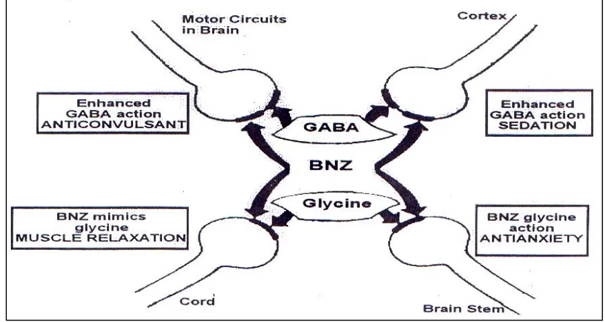
The GABA type A (GABAA) is a receptor complex consisting of up to five glycoprotein subunits. When the GABAA receptor is
activated, transmembrane chloride conductance increases, resulting in hyperpolarization of the postsynaptic cell membrane and functional inhibition of the postsynaptic neuron. Midazolam binds to a specific receptor site that is a part of the GABAA receptor complex. The
binding increases the efficiency of the coupling between the GABA receptor and the chloride ion channel. (Fig. 8 B)
BNZ FACILITATES INHIBITORY ACTIONS OF GABA
BNZ MIMICS INHIBITORY ACTIONS OF GLYCINE
Fig.8B:Mechanisms andsitesof actionofbenzodiazepines
[image:42.595.91.530.432.666.2]27 Pharmacokinetics :
Midazolam blood levels decrease rapidly because of its high hepatic
clearance, relatively shorter elimination half life (t1/2 ) and rapid
redistribution from the brain to inactive tissue sites. The termination of action after single doses is caused both by distribution into peripheral tissues and by metabolic biotransformation. The context- sensitive half time is shorter when compared to other benzodiazepines with a slow effect-site equilibrium time. It undergoes extensive hydroxylation by hepatic microsomal oxidative mechanisms and the water soluble metabolites are excreted in urine as glucoronide conjugates. Less than 0.5% is excreted unchanged in the urine. First pass metabolism is high. Clearance is also sensitive to hepatic blood flow.
TableA:Pharmacokineticsof Midazolam
Distribution half life (t½ ) 7 – 15 minutes
Elimination half life (t½ ) 1.7 – 2.6 hours
Protein binding 94%
Clearance 6.4 – 11 ml.kg-1. min-1
28
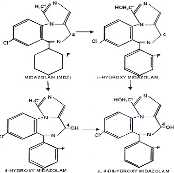
Metabolism:
Midazolam undergoes extensive hydroxylation by hepatic
microsomaloxidative mechanisms (cytochrome p-4503A4, p4503A3 and
p4503A5) to form - hydroxymidazolam, the principal metabolite
and small amounts of 4-hydroxymidazolam and even smaller amounts of
4-dihydroxymidazolam (Fig. 4).These water soluble metabolites are
excreted in urine as glucoronide conjugates.Very little (< 0.5%) unchanged midazolam is excreted by the kidneys. Plasma concentrations accumulate over the first 30 minutes after administration and reach the highest concentration in the first 2 hours.
Excretion :
Less than 1% of midazolam is excreted unchanged by the kidney. The metabolities are conjugated with glucoronic acid and all are excreted as
glucoronides.The principal excretory product is the
-hydroxymethylmidazolam glucoronides.
29
Fig. 9 : Metabolism of Midazolam, with -hydroxy midazolam occurring in the greatest quantity. All the hydroxylated metabolites are quickly excreted via the kidneys.
Pharmacodynamics:
[image:45.595.130.488.39.396.2]30 Effectsonorgansystems :
Central nervous system : Midazolam like other benzodiazepines is a sedative-hypnotic and anxiolytic. It has been suggested that a BZD receptor occupancy of 20% provides anxiolysis, while 30-50% receptor occupancy is associated with sedation and greater than 60% receptor occupancy is required for hypnosis (unconsciousness).It decreases in a dose related manner both cerebral metabolic rate for oxygen(CMRO2) and cerebral blood flow (CBF) maintaining a relatively normal ratio of CBF to CMRO2. In patients with
intracranial pathology, it decreases cerebral perfusion pressure (CPP) with little
effect on intracranial pressure (ICP). It is also a potent
anticonvulsant.Midazolam when administered orally, causes anterograde amnesia. Anterograde amnesia generally persists for 20-40 minutes after IV injection of a single dose. The ability to produce a short period of anterograde amnesia is a useful feature of the efficacy of midazolam when used as a sedative for endoscopy and in dentistry. The mild muscle-relaxant property of these drugs is mediated at the spinal cord level, not at the
neuromuscular junction.
Respiratory system : Midazolam produces dose dependent respiratory depression. In a dosage of 0.15 mg/kg,Midazolam significantly reduces the ventilatory response to CO2. In healthy patients,with small iv doses of
31
premedication is insignificant. However, the depressant effect is enhanced with chronic respiratory disease, and synergistic depressant effects occur when it is co-administered with opioid analgesics. Benzodiazepines also depress the swallowing reflex and decrease upper airway reflex activity. Transient apnea may occur after rapid injection of large doses of midazolam (> 0.15 mg/kg IV).
Cardiovascular system : The predominant hemodynamic change is an
32
Other effects:
o Attenuates stress related epinephrine increase, which is minimal.
o Plasma cortisol levels decrease from approximately 12.5 to 7.5 g/ml
o Adrenocorticotropic hormone (ACTH) changes are minimal.
o Decreases intraocular pressure
Clinicaluses:
Midazolam is the most commonly used benzodiazepine for preoperative medication in pediatric patients, IV (“conscious”) sedation and induction of anaesthesia. It is also used for maintenance of anaesthesia along with other drugs, and as an anticonvulsant.
DOSAGE ROUTE
Induction 0.15-0.40mg/kg i.v
Maintainence 0.25-1mcg/kg/min i.v
Premedication 0.07-0.10 mg/kg
i.m
0.25-0.5 mg/kg
oral
0.2-0.3 mg/kg nasal
I.v sedation 0.35mg/kg Rectal
33 1. Preoperativemedication:
Oral midazolam 0.5 mg/kg 30 minutes before induction provides reliable
sedation and anxiolysis in children without producing delayed recovery.
Intramuscular – 0.05 – 0.1 mg/kg is also effective but less well
accepted by children.
Transmucosal (sublingual) midazolam is as effective as and better
accepted than intranasal route in dose of 0.2 mg/kg.
Jet injection (i.e. using compressed gas instead of a needle) 0.10-0.15
mg/kg produces effective and rapid sedation in children without emotional trauma associated with needle injections.
2. Intravenous sedation : Midazolam in doses of 0.05mg/kg IV is effective for sedation during regional anaesthesia as well as for brief therapeutic procedures. Compared with diazepam, it produces a more rapid onset, with greater amnesia and less post-operative sedation. Pain on injection and subsequent venous thrombosis are less likely with midazolam as it is water soluble.
34
induction dose is 0.1 to 0.4mg/kg or lesser (0.05 – 0.15 mg/kg) in premedicated patients or when co-induced with other agents such as opioids, thiopental or propofol.
4. Maintenance of anaesthesia : It is a useful hypnotic-amnesic during maintenance of general anaesthesia but cannot be used alone for the same. It is used with opioids, propofol and or inhaled anaesthetics. MAC of volatile anaesthetics are decreased in a dose dependent manner. It can also be
used as an infusion in a dose of 0.25-1 g/kg/min.
5. As an anticonvulsant for the treatment of grandmal seizures which may occur with systemic toxicity produced by local anaesthetics.
6. Conscious sedation : Midazolam is probably the only sedative to produce
a true state of “conscious sedation”. It provides relief of anxiety and anterograde amnesia when administered prior to
o Dental or minor surgical procedures
o Upper GI endoscopy
o Bronchoscopy
o Cardiac surgery
35 Adverse effects:
Benzodiazepines are remarkably safe drugs in doses routinely used
Most significant problem with midazolam is respiratory depression
when the drug is given for conscious sedation.
When used as sedatives or for induction and maintenance of
anaesthesia, they can produce an undesirable degree or prolonged interval of post operative amnesia, sedation and rarely respiratory depression. The residual effects can be reversed with flumazenil.
Rarely loss of head control and balance, blurred vision and dysphoria
may be seen.
Drug interactions:
Alcohol, narcotics, sedatives and volatile anaesthetic agents
potentiate CNS and circulatory depressant effects.
Erythromycin, ranitidine, diltiazem, fluconazole, grape fruit juice,
verapamil and roxithromycin, increase serum levels and toxic effects.
Serum levels are decreased by carbamazepine, phenobarbitone,
phenytoin and rifampicin.
It decreases MAC for volatile agents
36 Precautions:
Reduce the dose in elderly, hypovolemic, high risk patients, and with
concomitant use of other sedatives or narcotics.
COPD patients are unusually sensitive to the respiratory depressant
effect.
It is contraindicated in acute narrow angle or open angle glaucoma.
It is excreted in human milk, therefore caution should be exercised
37
PHARMACOLOGYOFBUPIVACAINE16,17,18
Source : Bupivacaine was synthesised by A.F. Ekenstam and his collegues in Sweden in 1957.
Chemistry : The molecular weight of the chloride salt is 325 and that of
the base form is 288. It has a melting point of 2580C. Solutions containing epinephrine have a pH of about 3.5.The chemical name is
1-n-butyl-DL-piperidine-2 carboxylic acid-2, 6 dimethylamilide hydrochloride.
The molecular formula is C18N2OH28HCl.
CH
3-NHCO
N |
C4H9
CH3
Chemical structure
Derived from mepivacaine which has a methyl group on the piperidine N2 atom of
the molecule.Addition of a butyl group to piperidine N2 atom of mepivacaine forms
38
Bupivacaine Hydrochloride is available in sterile isotonic solutions with and
without epinephrine (as bitartrate) 1:200,000 for injection via local infiltration,
peripheral nerve block, and caudal and lumbar epidural blocks. Solutions of
bupivacaine hydrochloride may be autoclaved if they do not contain epinephrine.
Solutions are clear and colorless.
Bupivacaine is related chemically and pharmacologically to the aminoacyl
local anesthetics. It is a homologue of mepivacaine and is chemically related to
lidocaine. All three of these anaesthetics contain an amide linkage between the
aromatic nucleus and the amino, or piperidine group. They differ in this respect
from the procaine-type local anesthetics, which have an ester linkage.Bupivacaine
Hydrochloride Injection USP is available in sterile, isotonic solutions containing
[image:54.595.76.553.490.620.2]bupivacaine hydrochloride in water for injection with characteristics as follows:
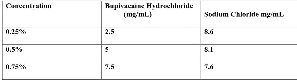
Table-B :Bupivacaine Hydrochloride Injection, USP (without epinephrine)
Concentration Bupivacaine Hydrochloride
(mg/mL) Sodium Chloride mg/mL
0.25% 2.5 8.6
0.5% 5 8.1
0.75% 7.5 7.6
May contain sodium hydroxide and/or hydrochloric acid for pH adjustment.
Multiple-dose vials contain methylparaben 1 mg/mL added as a preservative.
Bupivacaine and Epinephrine Injection, USP is available in sterile, isotonic
solutions containing Bupivacaine hydrochloride and epinephrine 1:200,000 with
39
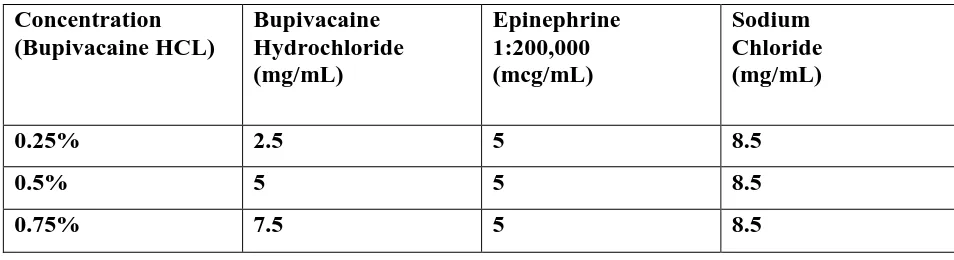
Table-C : Bupivacaine and Epinephrine Injection, USP
Concentration (Bupivacaine HCL)
Bupivacaine Hydrochloride (mg/mL)
Epinephrine 1:200,000 (mcg/mL)
Sodium Chloride (mg/mL)
0.25% 2.5 5 8.5
0.5% 5 5 8.5
0.75% 7.5 5 8.5
Sodium metabisulfite 0.1 mg/mL added as antioxidant and edetate calcium
disodium,anhydrous 0.1 mg/mL added as stabilizer. May contain sodium
hydroxide and/or hydrochloric acid for pH adjustment .Multiple dose vials contain
methylparaben 1 mg/mL added as a preservative. Single-dose solutions contain no
added bacteriostat or anti-microbial agent and unused portions should be discarded
after use
Physiochemicalproperties:
1) Solubility : The base is sparingly soluble, but the hydrochloride is readily soluble in water.
2) Stability and sterilization : Bupivacaine is highly stable and can withstand repeated autoclaving.
40 6) Protein Binding : 95%
7) Volume of Distribution : 73 litres 8) Clearance : 0.47 litres
9) Half – life : 210 minutes
10) Toxic plasma concentration > 3mcg/ml
USES
1)Spinal anaesthesia 2) Epidural anaesthesia 3)Caudal anaesthesia
4)Combined Spinal Epidural anaesthesia 5)Peripheral Nerve Block
Anaestheticproperties:
Potency:
Bupivacaine is approximately three to four times more potent than lidocaine.The duration of action of its motor blockade is two to three times longer than lidocaine.
Pk :
Weak base pKa > physiological pH. At a pH of 7.4, 17% of bupivacaine exists
41 Placental Transfer:
Plasma protein binding influences the rate and degree of diffusion of local anesthetics across the placenta . bupivacaine, which is highly protein bound (approximately 95%), has an umbilical vein-maternal arterial concentration
ratio of about 0.32.Acidosis in the fetus, which may occur during prolonged
labor, can result in accumulation of local anesthetic molecules in the fetus (ion trapping).
Distribution:
Rapid distribution phase (n this phase,the drug gets distributed to highly
vascular region.t1/2 of mins
Slow disapparence phase(Drug distributes to slowly equilibrating
tissues.t1/2 of mins Clearance : 0.47 mmol/min
Dosageandpreparationavailable:
The dosage of Bupivacaine depends on :
Area to be anaesthetized
The vascularity of the tissue to be blocked
The number of neuronal segments to be blocked
Individual tolerance
42
These doses may be repeated in 3-4 hours. 3 mg/kgmg is the maximum dose. The addition of vasoconstrictor produces a very slight increase in the duration of action. However the peak blood level is significantly reduced, there by minimizing the systemic toxicity.
ACTIONS :
Central nervoussystem:
Overdose of bupivacaine produces light headedness and dizziness followed by visual and auditory disturbances such as difficult to focus and tinnitus. Disorientation and drowsiness can also occur. Shivering, muscular tremors and tremors of muscles of face and distal part of extremities can occur. Ultimately generalized tonic clonic convulsions occurs. Further increase in doses causes respiratory arrest. Since bupivacaine is a potent drug, smaller doses can cause rapid onset of toxic symptoms when compared to other drugs.
Autonomicnervoussystem :
Bupivacaine does not inhibit the Noradrenaline uptake and hence has no sympathetic potentiating effect. Myelinated preganglionic beta
43
When used for conduction blockade, all local anaesthetics particularly bupivacaine produce higher incidence of sensory than motor fibres blockade.
Neuro-muscular junctions:
Bupivacaine like other local anaesthetics can block motor nerves if
present in sufficient concentration but has no effect on the neuromuscular junction as such.
Cardiovascularsystem:
The primary cardiac electrophysiologic effect of local anaesthetic is a decrease in the maximum rate of depolarization in the purkinje fibres and ventricular muscle. This is due to a decrease in the availability of sodium channels.Bupivacaine decreases cardiac output by decreasing sympathetic tone,decreasing heart rate and decreasing venous return.It also decreases central venous pressure.There is an increase in blood flow to lower limb with decrease in DVT.
Bupivacaine is highly arrythmogenic. The cardiac contractility is reduced, this is by blocking the calcium transport. Low concentration of
bupivacaine produces vasoconstriction while higher doses causes
vasodilatation.
44
Respiratory system:
Respiratory depression may be caused if excessive plasma level is reached. Respiratory depression may also be caused by paralysis of respiratory muscles as may occur in high spinal or total spinal anaesthesia.
Pharmacodynamics:
The onset of action of bupivacaine is between 4 and 6 minutes maximum anaesthesia is obtained between 15 and 20 minutes. The duration of anaesthesia varies according to the type of block, the average duration for peridural block is about 3.5-5 hours, for nerve blocks, it is about 5 to 6 hours.
Toxicity:
The toxic plasma concentration is set at 4-5g/ml. Maximum
plasma concentration rarely approach toxic levels. Non specific local irritant effects on nerve tissue have been noted in human subjects. No evidence of permanent damage has been found in clinical dosage.
Pharmacokinetics:
Bupivacaine can be detected in the blood within 5 minutes of infiltration or following either epidural or intercostal nerve blocks. Plasma levels are related to the total dose administered. Peak levels of 0.14 to 1.18
g/ml were found within 5 minutes to 2 hours after the administration of
45
Plasmabinding:
In plasma, drug binds avidly with protein (1- acid glycoprotein) to the extent of 70-95%. The rank order of protein binding for this and its
homologues is- bupivacaine<mepivacaine<lidocaine. Conversely, the
unbound active fraction is one seventh that of lidocaine and one fifth that of mepivacaine.
Metabolism:
Because bupivacaine is an amide, the liver is the primary site of metabolism. The drug is metabolized partly by N-dealkylation primarily to pipecolyloxylidine. N-disbutyl-bupivacaine and 4-hydroxy-bupivacaine are also formed. It crosses the placental barrier as any other local anaesthetic by passive diffusion, but the lowest level of placental diffusion is reported for this drug (umbilical vein/maternal ratio is 0.31 to 0.44). The high protein binding capacity of the agent is probably the reason why less diffusion occurs across the placenta. No effects on fetus have been noted.
Excretion:
About 10% of drug is excreted unchanged in urine within 24 hours; 5% excreted as pipecolyloxylidine. Glucoronide conjugate is also excreted.
46 Adversereactions:
Adverse reactions occur with excessive plasma levels which may be due to overdose, inadvertent IV injections or slow metabolic degradation. These manifest by effects on CNS and CVS. The CNS effects are characterized by excitation or depression. The first manifestation may be nervousness, dizziness, blurring of vision or tremors following drowsiness, convulsions unconsciousness and probably respiratory arrest.
Other effects may be nausea, vomiting, chills, constriction of pupils and tinnitus. The CVS manifestations include myocardial depression, hypotension and cardiac arrest, in obstetrics fetal bradycardia may occur. Allergic reactions include urticaria, bronchospasm and hypotension.
Treatmentof adversereaction :
Treatment is mainly symptomatic. After initiation of basic life support and the ACLS protocol ,a rapid bolus of Intralipid 20%, 1.5 mL/kg (or roughly 100 mL in adults), be administered without delay, followed if necessary by an infusion of 0.25 mL/kg/min for the next 10 minutes. (recommendation of
47
ventricular fibrillation and tachycardia by amiodarone (5mg/kg iv) or defibrillation (2-6 joule/kg).
Cardiovascularcollapse/CNS ratio:
The CC/CNS dose ratio for Bupivacaine is 3.7 0.5 or findings
indicating that 3 times drug was required to induce irreversible cardiovascular collapse as was needed to produce convulsions. It has also been suggested that some of the enhanced cardiac toxicity of Bupivacaine is due to greater myocardial uptake.
48
49
REVIEWOFLITERATURE:
1. Edwards et al19 (Anaesthesiol 1990;73:273-7) conducted a randomised
control study in rats to find out the mechanism by which midazolam causes spinally mediated analgesia.In this study the electrical current thresholds for pain (ECTP) in the skin of the neck and tail were measured with chronically implanted subarachnoid catheters. Segmental analgesia following midazolam was attenuated (P<0.05) when the selective GABA antagonist bicuculline was given intrathecally at the same time as midazolam. The highest dose of bicuculline used, caused no significant attenuation of the segmental analgesic effects of either fentanyl or ketocyclazocine. They concluded that segmental analgesia produced by intrathecal midazolam is mediated by benzodiazepine–GABA receptor complex that is involved in other benzodiazepine actions.
2. Batra YK et al20 (Int. Journal Clinical Pharmacol 1999;37:519-23)
conducted a randomised double blinded control study to find out the effect of intrathecal midazolam added to bupivacaine for knee arthroscopy.In this study, 30 healthy patients of ASA-I and ASA-II scheduled for knee arthroscopy were divided into two groups of 15 each.Group M received
Inj.midazolam-bupivacaine mixture (midazolam 50mcg/kg+bupivacaine
50
block, sedation score, assessment of pain using visual analogue score in both groups at regular time intervals. Time to block regression, recovery to ambulation and ability to void were recorded and noted before discharged.
A significantly higher Visual Analogue score (P < 0.05) was seen in group B. All patients in group B received rescue analgesia at a mean
duration of 258 4.68 minutes where as only one patient in group M
required supplemental analgesia within this period. Time to regression of sensory analgesia was longer in group M (P< 0.05). Neither motor block nor time to void were prolonged with the addition of midazolam to
bupivacaine. They concluded that addition of intrathecal midazolam to
bupivacaine provides better postoperative analgesia without prolonging motor blockade.
3. Kim M, Lee Y et al 21 – British Journal 2001:86:77-79
A double blind study was conducted to evaluate the post-operative analgesic
effects of intrathecal midazolam with bupivacaine following
51
placebo and significantly less in BM1 group than in BM2 group. They concluded that intrathecal midazolam increased the analgesic effects of spinal blockade with bupivacaine.
4. Tucker A P et al 22 (Anaesthesia Analgesia 2004; 98:1521-7) conducted a
p r o s p e c t i v e d o u b l e b l i n d study to evaluate the ability of intrathecal midazolam to increase the potency and duration of the analgesic effects of intrathecal fentanyl, without producing adverse effects. 30 parturients of ASA I and ASA II with cervical dilatation 2-6cm were randomized to
receive either intrathecal midazolam 2mg, fentanyl 10g or both combined
to initiate analgesia. Pain scores were recorded before and at 5-min intervals for 30 min after the injection and then every 30 min until the patient requested further analgesia. The presence and severity of nausea, emesis, pruiritus, headache and sedation, in addition to arterial blood pressure, heart rate, respiratory rate, sensory changes to ice, motor impairment, cardiotocograph and apgar score were also recorded.They concluded that intrathecal midazolam potentiated the analgesic effect of fentanyl. It did not increase the occurence of any maternal adverse events or abnormalities on the cardiotocography.
5. Yokoyama et al 23 in 1998 (Canadian journal of Anaesthesia
52
conducted to investigate the effect of midazolam on epidural infusion of bupivacaine. 60 patients of ASA I and ASA II scheduled for gastrectomy were divided into 3 groups of 20. The following mixtures, in 40 ml, were infused continuously over 12 hours after surgery; 40ml bupivacaine 0.5% in group C, bupivacaine 0.5% 38ml + 10mg midazolam in group M10 and bupivacaine 0.5% 36 ml + 20mg of midazolam in group M20. Better analgesia was obtained in patients receiving midazolam than in group C (P < 0.05). Frequency of rescue analgesics administration was greater in group C (P < 0.05). Greater sedation was seen in groups M10 and M 20 during first 120 minutes without any respiratory depression, which was desirable in the postoperative period. They concluded that adding midazolam to a continuous epidural infusion of bupivacaine provides better analgesia and sedation than bupivacaine alone without side effects in patients undergoing laparatomy.
6. Nishiyama et al 24 (J Clinical Anaesthesia 2002;14(2):92-97) conducted
53
The contents of the infuser (40ml) were bupivacaine, 180mg with midazolam 20mg (HM group), bupivacaine, 90mg with midazolam 20mg (LM group), bupivacaine, 180mg without midazolam (HC group) and bupivacaine, 90mg without midazolam (LC group). It was found that HM group had significantly better analgesia and sedation. The time to the first
rescue medication was longest in HM group followed by LM, then HC and
finally LC groups. The number of patients with amnesia were greater in HM and LM groups. They concluded that adding midazolam increased not only the analgesic but also the sedative effect with increasing dose and bupivacaine in a post operative continuous epidural administration.
7.Mahajan R,Batra YK, 25Int J Clin Pharmacol Ther2001;39(3):116-20
A double-blinded study was designed to evaluate the analgesic efficacy of
caudal midazolam- bupivacaine combination in providing post-operative pain relief in children undergoing genito-urinary surgery and to study the occurence of side effects . Thirty children, aged 2 to 8 years, scheduled for genito-urinary surgery were allocated randomly to receive either 0.25% bupivacaine 0.5
ml/kg (group B; n = 15) or 0.25% bupivacaine 0.5ml/kg with 50 g/kg
54
in group BM (11 0.5h) as compared to group B (7.4 2.1h) (p < 0.05). No significant changes in heart rate, blood pressure and oxygen saturation was observed. The authors concluded that caudal administration of midazolam –
bupivacaine mixture prolongs post-operative analgesia compared to
bupivacaine alone without any adverse effects.
8. Culebras X, Van Gessel4(Anaesth Analg 2001;92:199-204)
A prospective randomized double blinded study was conducted to determine the efficacy and adverse effects of clonidine,when mixed with a long acting local anaesthetic on postoperative analgesia. 60 adult patients o f A S A I a n d I I undergoing rotator cuff repair under interscalene brachial plexus block were included in the study. The study group received
150g of clonidine with 40ml of 0.5% heavy bupivacaine. It was found
that duration of analgesia was unaltered but the mean arterial pressure and heart rate were significantly decreased in clonidine group. They concluded that clonidine,as an adjuvant in brachial plexus block does not improve post operative analgesia but does induce haemodynamic changes.
55
intravenous injections of GABA caused an increase in excitability of the low threshold fast conducting fibres of the superficial radial and median nerves of cat. Similar, graded, reversible effects were confirmed (using changes in the amplitude / integral of the stimulus evoked A fiber submaximal compound action potential to assess excitability) in in-vitro studies with the isolated, desheathed frog sciatic nerve. GABA caused a mean maximal increase in half-maximal action
potential of 29.8% (S.E. 2.7), with an ED50 value of 0.09 mM and hill
coefficient of 0.70.. They concluded that extrasynaptic receptors of GABA are present on myelinated axons of peripheral nerves
10. Jarbo K et al 28 (Canadian journal of anaesthesia 2005 :52:822-6)
conducted a prospective randomized double blind study to assess the effect of midazolam added to brachial plexus block.This study was conducted on 40 ASA I and II adult patients undergoing upper limb surgeries under supraclavicular brachial plexus block.. Patients in group B (n = 20) were administered 30ml of 0.5% bupivacaine and group BM (n = 20) were given
30ml of 0.5% bupivacaine with midazolam /kg.Haemodynamic variables
(i.e., heart rate, noninvasive blood pressure), pain scores and rescue analgesic requirements were recorded for 24 hr postoperatively. The onset time and duration of sensory and motor blockade were recorded. The onset of sensory
block was significantly faster in group BM (12±2.9 mins) when compared to
56
The onset of motor block was significantly faster in group BM (9.2 ± 2.38 mins)
when compared to group B (17.1+3.83). T h i s r e s u l t w a s s t a t i s t i c a l l y s i g n i f i c a n t (P < 0.05). T h e d u r a t i o n o f m o t o r b l o c k a d e w a s n o t d i f f e r e n t i n t w o g r o u p s . I n G r o u p B , t h e d u r a t i o n o f m o t o r
b l o c k a d e w a s 5 . 1 + 1 . 1 4 w h i l e i n G r o u p B M , t h e d u r a t i o n o f m o t o r b l o c k a d e w a s 5 . 6 + 3.32. T h i s r e s u l t w a s s t a t i s t i c a l l y i n s i g n i f i c a n t (P > 0.05). Pain scores were significantly higher in group B compared to group BM from two hours to 24 hrs postoperatively (P < 0.05). Rescue analgesic requirements were significantly less in group BM compared to group B (P < 0.05). Haemodynamics and sedation scores did not differ between groups in the postoperative period. They concluded that Midazolam (0.05mg/kg) in combination with 30ml of bupivacaine (0.5%) hastened onset of sensory and motor block, and improved postoperative analgesia in brachial plexus block, without producing any adverse effects.
11. Naguib M et. al 29(Can J Anaesthesia 1995;42:758-64) conducted a
randomized double-blinded study,in which they compared the efficacy of midazolam with bupivacaine for caudal analgesia in children undergoing inguinal
herniotomy. They were allocated randomly into three groups (n = 15 in each) to
receive a caudal injection of either 0.25% bupivacaine 1 ml/kg with or without
midazolam 50 μg /kg or midazolam 50 μg/ kg with normal saline 1 ml/kg. There