0022-538X/96/$04.0010
Copyrightq1996, American Society for Microbiology
The HeLa Cell Receptor for Enterovirus 70 Is
Decay-Accelerating Factor (CD55)
TIMOTHY M. KARNAUCHOW,
1DOUGLAS L. TOLSON,
2BLAIR A. HARRISON,
2ELEONORA ALTMAN,
2DOUGLAS M. LUBLIN,
3AND
KENNETH DIMOCK
1*
Department of Microbiology and Immunology, University of Ottawa, Ottawa, Ontario, Canada K1H 8M5
1;
National Research Council of Canada, Institute for Biological Sciences, Ottawa, Ontario, Canada K1A OR6
2;
and Department of Pathology, Washington University School of Medicine, St. Louis, Missouri 63110
3Received 1 March 1996/Accepted 30 April 1996
Enterovirus 70 (EV70) is a recently emerged human pathogen belonging to the family
Picornaviridae
. The
ability of EV70 to infect a wide variety of nonprimate cell lines in vitro is unique among human enteroviruses.
The importance of virus receptors as determinants of viral host range and tropism led us to study the host cell
receptor for this unusual picornavirus. We produced a monoclonal antibody (MAb), EVR1, which bound to the
surface of HeLa cells and protected them against infection by EV70 but not by poliovirus or by coxsackievirus
B3. This antibody also inhibited the binding of [
35S]EV70 to HeLa cells. MAb EVR1 did not bind to monkey
kidney (LLC-MK
2) cells, nor did it protect these cells against virus infection. In Western immunoassays and
in immunoprecipitations, MAb EVR1 identified a HeLa cell glycoprotein of approximately 75 kDa that is
attached to the cell membrane by a glycosyl-phosphatidylinositol (GPI) anchor. Decay-accelerating factor
(DAF, CD55) is a 70- to 75-kDa GPI-anchored membrane protein that is involved in the regulation of
complement and has also been shown to function as a receptor for several enteroviruses. MAb EVR1 bound to
Chinese hamster ovary (CHO) cells constitutively expressing human DAF. Anti-DAF MAbs inhibited EV70
binding to HeLa cells and protected them against EV70 infection. Transient expression of human DAF in
murine NIH 3T3 cells resulted in binding of labelled EV70, and stably transformed NIH 3T3 cells expressing
DAF were able to support virus replication. These data indicate that the HeLa cell receptor for EV70 is DAF.
Viruses belonging to the family Picornaviridae are
responsi-ble for a wide range of illnesses of humans and animals.
En-terovirus 70 (EV70), a human enEn-terovirus, is the etiologic
agent of acute hemorrhagic conjunctivitis, a highly contagious
form of conjunctivitis (55) that is distinct from ocular
infec-tions caused by other human enteroviruses (11). In rare
in-stances, acute hemorrhagic conjunctivitis is followed by
infec-tion of the central nervous system, leading to poliomyelitis-like
paralysis (17, 54). Since its emergence as a human pathogen in
West Africa in 1969 (24), EV70 has been responsible for two
pandemics and has demonstrated the potential to cause
wide-spread outbreaks of acute hemorrhagic conjunctivitis
through-out the world (54).
EV70 possesses biological and pathogenic properties that
are unique among human enteroviruses (55). Among these
properties is the ability to replicate in a wide variety of
non-primate-derived cell lines. Cell lines of porcine, murine,
lepo-rine, crecitine, and bovine origin can support the growth of
EV70 in vitro (56).
Viruses initiate infection by binding to specific receptors on
the surface of susceptible cells (2, 38, 41). Although factors
affecting steps after this interaction may influence infectivity
and replication, the nature and distribution of host cell
recep-tors are recognized as major determinants of viral host range
and of cell and tissue tropism (33).
A growing number of picornavirus receptors have been
char-acterized, and it appears that most are molecules involved in
cell-cell interactions or in the regulation of cell function (4, 53).
Molecules belonging to the immunoglobulin (Ig) superfamily
serve as receptors for the major-group human rhinoviruses
(intercellular cell adhesion molecule 1 [15, 47]), for poliovirus
(32), and for a variant of encephalomyocarditis virus (20).
Echoviruses 1 and 8 attach to integrin VLA-2 (6, 7, 22), and
a
vb
3integrin is reported to be the receptor for coxsackievirus
A9, echovirus 22 (40), and foot-and-mouth disease virus (8).
Minor-group human rhinoviruses bind to cells via members of
the low-density lipoprotein receptor family (18).
Decay-accel-erating factor (DAF, CD55) is recognized as the cellular
re-ceptor for at least six echovirus serotypes (3, 51) and as a
receptor for coxsackieviruses B1, B3, and B5 (5, 42).
Interac-tion of group B coxsackieviruses with permissive cells may also
involve a nucleolin-like membrane protein (39). Human DAF
is a 70- to 75-kDa membrane glycoprotein involved in
protect-ing cells against lysis by homologous complement (37). The
molecule has five extracellular domains (27, 29): four
contig-uous short consensus repeat (SCR) domains of approximately
60 amino acids each, followed by a serine/threonine-rich,
heavily O-glycosylated C-terminal domain. A
glycosyl-phos-phatidylinositol (GPI) anchor attaches the molecule to the
outer leaflet of the cell membrane.
Here, we demonstrate that DAF is also the human (HeLa)
cell receptor for EV70. A monoclonal antibody (MAb), EVR1,
which blocks infection of HeLa cells by EV70, was produced.
MAb EVR1 reacts with a 75-kDa cell surface glycoprotein that
shares a number of properties with DAF, including sensitivity
to phosphatidylinositol-specific phospholipase C (PI-PLC).
Several anti-DAF MAbs also protect HeLa cells against EV70
infection and interfere with virus binding. The ability to bind
virus is conferred to NIH 3T3 cells following transient surface
expression of human DAF in these cells, and stably
trans-formed NIH 3T3 cells expressing human DAF support EV70
replication. We therefore propose that DAF is the HeLa cell
receptor for EV70.
* Corresponding author. Phone: (613) 562-5800, ext. 8311. Fax: (613) 562-5452. Electronic mail address: [email protected].
5143
on November 9, 2019 by guest
http://jvi.asm.org/
MATERIALS AND METHODS
Cells.HeLa cells were obtained from the National Institute of Allergy and Infectious Diseases AIDS Research and Reference Reagent Program, Bethesda, Md. Rhesus monkey (Macaca mulatta) kidney cells, LLC-MK2, were purchased
from Flow Laboratories, Rockville, Md. African green monkey (Cercopithecus aethiops) kidney cells, CV-1, were obtained from the American Type Culture Collection, Rockville, Md., and murine NIH 3T3 cells were provided by E. G. Brown, Department of Microbiology and Immunology, University of Ottawa. Growth medium consisted of Eagle’s minimal essential medium containing Ear-le’s salts (MEM) supplemented with 0.15% (wt/vol) sodium bicarbonate, 2 mM
L-glutamine, 50mg of gentamicin (Roussel Canada, Montreal, Quebec, Canada)
per ml, and either 5% (vol/vol) (for LLC-MK2and NIH 3T3 cells) or 10%
(vol/vol) (for HeLa and CV-1 cells) heat-inactivated fetal bovine serum (FBS). Cells were grown as monolayer cultures at 378C in a 5% CO2atmosphere.
Chinese hamster ovary (CHO) cells and CHO cells stably expressing human DAF (CHO-DAF cells) were grown in Ham’s F12 medium (28). NIH 3T3 cells transfected with the gene for human DAF under the control of the simian virus 40 early promoter (3T3-DAF) and NIH 3T3 cells transfected with the DAF gene in reverse orientation with respect to the promoter (3T3-RDAF) were gifts of J. Atkinson, Washington University School of Medicine, St. Louis, Mo., and were maintained as described previously (52). All media and supplements were from Gibco/BRL Life Technologies Canada, Burlington, Ontario, Canada, unless otherwise stated.
Viruses.EV70 prototype strain J670/71 was obtained from M. Hatch and M. Pallansch, Centers for Disease Control and Prevention, Atlanta, Ga. Poliovirus Sabin 1 and coxsackievirus B3 were provided by S. A. Sattar, Department of Microbiology and Immunology, University of Ottawa. Vaccinia virus vTF7-3 was obtained from the American Type Culture Collection and was propagated and subjected to titer determination in CV-1 cells.
Growth and purification of EV70.Concentrated stocks of EV70 were prepared as follows. LLC-MK2cells were infected at a multiplicity of infection (MOI) of
0.1 PFU per cell for 1 h in serum and antibiotic-free growth medium and then incubated at 338C for 30 h in complete medium. Virus was collected by harvest-ing the medium and cells, performharvest-ing three cycles of freezharvest-ing and thawharvest-ing, and subjecting the mixture to clarification at 4,0003g at 48C. Pooled supernatants were concentrated with a Minitan ultrafiltration apparatus (Millipore Ltd., Mis-sissauga, Ontario, Canada), and the virus was pelleted in a Beckman SW28 rotor at 110,0003g for 4 h at 48C. The virus pellet was resuspended in R buffer without glycerol and placed on a linear density gradient of 10 to 40% (wt/vol) sucrose in R buffer (1). After centrifugation at 154,0003g at 48C in a Beckman SW41 rotor for 3.5 h, gradient fractions were recovered and portions of each were tested by plaque assay. Peak fractions were pooled, virus titers were deter-mined, and aliquots were stored at2808C.
Purification of radiolabelled EV70.LLC-MK2cells were infected with EV70 at
a MOI of 0.1 for 1 h at 338C. The inoculum was removed, and serum-free, methionine-free growth medium (ICN Biomedicals Canada Ltd., Mississauga, Ontario, Canada) was added to the monolayers. After 3 h, 5mCi of Tran35S-label
(ICN) was added per ml of medium. Incubation was continued for a further 30 h, and virus was harvested as described above. Sucrose gradient fractions were recovered and analyzed for the presence of35S by scintillation counting. Peak
fractions were pooled, divided into aliquots, and stored at2808C.
Membrane preparation.Cell monolayers were incubated for 15 min at 378C with PBS (137 mM NaCl, 2.7 mM KCl, 10 mM Na2HPO4, 1.8 mM KH2PO4[pH
7.4]) containing 50 mM EDTA, removed, pooled, washed three times with cold PBS, resuspended in 10 mM sodium phosphate buffer, and swollen on ice for 15 min. The cells were disrupted in a Dounce homogenizer in the presence of protease inhibitors (1 mM benzamidine-HCl, 1 mM phenylmethylsulfonyl fluo-ride, 5 mM EDTA), and centrifuged at 1,4003g for 5 min. The supernatant was transferred to fresh tubes and centrifuged at 200,0003g for 2 h in a TH641 rotor (DuPont Inc., Mississauga, Ontario, Canada). The pellet was resuspended in PBS, the protein concentration was adjusted to 10 mg/ml (Bradford assay; Bio-Rad Laboratories Ltd., Mississauga, Ontario, Canada), and the membranes were stored at2808C.
Antibodies. (i) Production of anti-receptor antibodies.Inbred female BALB/c mice (6 to 8 weeks old) (Charles River Laboratories, St. Constant, Quebec, Canada) were immunized by intraperitoneal injection of HeLa cell membranes at 3-week intervals. The first injection consisted of 50mg of membrane protein emulsified in complete Freund’s adjuvant, the second consisted of 80mg of antigen in incomplete Freund’s adjuvant, and the third consisted of 120mg of antigen in PBS. Antibody production against HeLa cell membranes was dem-onstrated by indirect enzyme-linked immunosorbent assay (ELISA), and 3 days prior to cell fusion, mice were primed by tail vein injection of 175mg of mem-brane protein in PBS. Cells were prepared from the spleens of two mice and fused to SP2/0 myeloma cells (44) by a standard method (23). Supernatants of viable hybridoma cultures were screened for the presence of antibodies that inhibited EV70 infection of HeLa cells as determined in the cell protection assay described below. Hybridomas producing protective antibodies were cloned, and their supernatants were retested. Isotyping was performed by indirect ELISA, using standard methods (14). HeLa cell membrane protein was adsorbed to ELISA microplate wells, and undiluted tissue culture supernatants from cloned cells were used as the primary antibody. Horseradish peroxidase-conjugated goat
anti-mouse isotype-specific antibodies (Caltag, South San Francisco, Calif.) at a dilution of 1:2,000 were used as secondary antibodies. Ascites fluid containing MAb was obtained from retired breeders. Antibodies in culture supernatants and ascites fluids were quantitated by antibody sandwich ELISA (19). Briefly, ELISA plates were coated with 1mg of rat-anti mouse IgG (heavy plus light chains) (Jackson Immunoresearch Laboratories Inc., West Grove, Pa.) capture antibody per ml, incubated with dilutions of hybridoma culture supernatant or ascites fluid, and then reacted with a biotinylated mouse-specific IgG1 MAb. After incubation with peroxidase-conjugated streptavidin (Jackson) and o-phenylene-diamine dihydrochloride substrate (OPD; Sigma Chemical Co., St. Louis, Mo.), the A490 was determined. Antibody concentrations were determined from a
standard curve constructed with known amounts of mouse IgG1 (Coulter Elec-tronics, Burlington, Ontario, Canada) as the primary antibody. EVR1 hybridoma supernatant contained 17.5mg of Ab/ml, and EVR1 ascites contained 11 mg of Ab/ml.
(ii) Sources of other antibodies.DAF-specific MAbs 1H4 (35mg/ml), 8D11 (36mg/ml), 11D7 (40mg/ml) (10), and 1F7 (1.2 mg/ml) (3) were used. 8D11 and 11D7 hybridoma culture supernatants were gifts from W. Rosse, Department of Medicine, Duke University Medical Center, Durham, N.C., and 1F7 ascites fluid was from R. W. Finberg, Dana-Farber Cancer Institute, Boston, Mass. Anti-lymphocytic choriomeningitis virus (LCMV) ascites fluid (IgG1; 4 mg/ml) was provided by K. Wright, Department of Microbiology and Immunology, Univer-sity of Ottawa. Fluorescein isothiocyanate (FITC)-conjugated and nonconju-gated mouse antibodies specific for human HLA ABC class 1 IgG1 (anti-MHC 1) were from Serotec Canada, Toronto, Ontario, Canada.
Cell protection assay. (i) Assessment of antibody EVR1.HeLa cells were grown in 96-well plates (Corning Science Products, Richmond Hill, Ontario, Canada). The growth medium was removed, and the monolayers were incubated with 100ml of undiluted hybridoma cell culture supernatant for 1 h at 378C. The cells were washed with PBS, and EV70 was added at a MOI of 1 in serum-free growth medium. After a 45-min incubation at 338C, the inoculum was removed, fresh medium containing 10% FBS was added, and the cells were placed at 338C. Monolayers were monitored at regular intervals by light microscopy for signs of virus-induced cytopathic effects (CPE). Results of antibody screening were con-firmed on duplicate plates by a colorimetric assay based on the ability of viable cells to metabolize the tetrazolium salt 3-(4,5-dimethylthiazol-2-yl)-2,5-diphenyl tetrazolium bromide (MTT) (16). Cell protection assays with poliovirus and coxsackievirus were performed in an identical manner, except that incubations were performed at 378C.
(ii) Assessment of DAF-specific MAbs.DAF-specific MAbs 1H4, 8D11, 11D7, and 1F7 were tested for their protective ability as described above. Antibodies were diluted appropriately in RPMI 1640 medium containing 2 mM glutamine. For MAb 1F7, 2 mM CaCl2and 2 mM MgCl2were added (3).
Binding and binding inhibition assays.Cells were grown to confluency in 24-well dishes (Corning). The medium was aspirated, the monolayers were washed with PBS, and [35
S]EV70 was added in serum-free MEM. After a 45-min incubation at 338C, the inoculum was removed and the monolayers were washed with PBS. The inoculum and PBS wash were pooled, and they represent the unbound fraction of input virus. The monolayers were then disrupted with PBS containing 1% (wt/vol) sodium dodecyl sulfate (SDS), and the wells were washed. These samples, representing the bound fraction of input virus, were also pooled. The samples were analyzed by scintillation counting. The percentage of input virus bound to cells was determined by dividing the counts per minute (cpm) of labelled EV70 in the bound fraction by the total number of cpm recovered. For binding inhibition studies, after removal of growth medium and the PBS wash, monolayers were treated with 100ml of test sample (MAb EVR1, 1H4, 8D11, or 11D7; anti-major histocompatibility complex class [MHC] I; or growth medium) for 1 h at 378C. The test sample was then removed, monolayers were washed with PBS, and virus-binding assays were performed as described above.
Indirect immunofluorescence. Cells were removed from 100-mm plates by scraping or by treatment with 0.05% trypsin–0.5 mM EDTA (Gibco/BRL) in PBS and washed in PBS–2% (wt/vol) bovine serum albumin (BSA). Approxi-mately 53105cells were dispensed into separate tubes, pelleted, and
resus-pended in 25ml of hybridoma cell culture supernatant or ascites fluid diluted 1:200 in PBS-BSA. After incubation on ice for 1 h, the cells were washed in PBS-BSA and the cell pellets were resuspended in 20ml of PBS-BSA containing a 1:5 dilution of FITC-conjugated sheep anti-mouse Ig (Amersham Canada, Oakville, Ontario, Canada). After 1 h on ice, the cells were washed, resuspended in 90% glycerol–PBS (pH 9.6), deposited on glass slides under coverslips, and examined under a Zeiss Aristoplan epifluorescence microscope. To examine the susceptibility of the EVR1 ligand to PI-PLC, HeLa cells were trypsinized and washed three times prior to resuspension in PI-PLC buffer (RPMI 1640 medium, 0.2% BSA, 50mMb-2-mercaptoethanol, 10 mM N-2-hydroxyethylpiperazine-N9-2-ethanesulfonic acid [HEPES], 0.1% [wt/vol] sodium azide). The cells were then divided into two fractions. One fraction received 0.5 U of Bacillus thurin-giensis PI-PLC (Oxford Glycosystems, Rosedale, N.Y.) per 106
cells, and the other did not. Following incubation at 378C for 1 h, both sets of cells were washed and processed for surface immunofluorescence as described above (EVR1 and 1H4 culture supernatant, neat; anti-MHC 1, 1:10). All photography was per-formed at 403power with Kodak Ektachrome 400 ASA film.
on November 9, 2019 by guest
http://jvi.asm.org/
Radioimmunoprecipitations.Cells grown on 100-mm tissue culture dishes were starved of sugars for 2 h and then incubated in 10 ml of medium containing 15mCi ofD-[6-3H]glucosamine hydrochloride (Amersham) per ml. Labelling
periods of 4 and 16 h gave identical results. The cells were then scraped and resuspended in 600ml of RIPA buffer (50 mM Tris-HCl [pH 7.2], 150 mM NaCl, 1% sodium deoxycholate [Difco Laboratories, Detroit, Mich.], 1% Nonidet P-40) containing 1 mM phenylmethylsulfonyl fluoride and 1 mM benzamidine-HCl and given a short pulse in a benchtop centrifuge to pellet the cellular debris. Protein G-Sepharose 4 Fast Flow beads (Pharmacia Canada, Baie d’Urfe´, Quebec, Can-ada) were washed in RIPA buffer containing 2% ovalbumin, and then 200ml of cell lysate and 10ml of undiluted ascites fluid were added. After an overnight incubation with mixing at 48C, the samples were analyzed by electrophoresis in 8% polyacrylamide gels containing SDS (13, 26). Prestained high-molecular-weight markers were from Amersham.
Western blots (immunoblots).Lysates of unlabelled cells were prepared as described above for radiolabelled lysates. Proteins (25mg per lane) were sepa-rated by electrophoresis at 48C in 8% polyacrylamide gels by using standard protocols (26) with the following modifications. Gels and running buffers con-tained 0.05% SDS, and the samples were prepared, without being heated, in electrophoresis sample buffer containing 0.05% SDS and nob-mercaptoethanol. Proteins were transferred at 48C to Schleicher & Schuell NC nitrocellulose membranes (Mandel Scientific Co. Ltd., Guelph, Ontario, Canada) with a Bio-Rad Mini Trans-Blot apparatus (100 V for 2 h) and the transfer buffer of Towbin et al. (49). The membranes were blocked in PBS–3% (wt/vol) BSA for 30 min at 378C. After three 378C washes in PBS–0.02% Tween 20 (PBS-Tween), the membranes were incubated for 1 h at 378C with MAb as either hybridoma supernatant (1H4, 1:3,000) or ascites fluid (EVR1, 1:1,000; anti-LCMV IgG1, 1:300) diluted in PBS–3% BSA. After three washes in PBS-Tween, the mem-branes were incubated for 1 h at 378C with peroxidase-conjugated goat anti-mouse IgG (heavy plus light chains) (Jackson) diluted 1:20,000 in PBS–3% BSA. The membranes were washed with PBS-Tween, and the color was developed in a solution consisting of 0.01 M (NH4)2SO4, 0.01 M KH2PO4(pH 6.0), 0.03%
(vol/vol) H2O2, and 0.005% (wt/vol) o-dianisidine (Sigma). The reaction was
stopped with repeated washes in distilled water.
Transient expression of DAF.A human DAF cDNA fragment (28) was sub-cloned into the EcoRI site of plasmid pcDNA3 (Invitrogen Corp., San Diego, Calif.). Twenty-four-well tissue culture plates (Corning) seeded with 83104
NIH 3T3 cells were transfected 24 h later with 1mg of plasmid DNA per well, using OptiMEM and Lipofectamine reagent (Gibco/BRL), by using protocols provided by the manufacturer. An equal volume of MEM containing 10% FBS was added to the transfection mix 9 h later, and the cells were cultured for a further 15 h. Transfected cells were then infected with vaccinia virus vTF7-3 at a MOI of 20 for 1 h at 378C. The inoculum was removed, and the cells were incubated for a further 18 h in complete growth medium containing 5% FBS. Monolayers were then used in binding assays or in immunofluorescence studies, as described above.
RESULTS
Identification of a MAb that blocks EV70 infection of HeLa
cells.
To identify a HeLa cell membrane component that acts
as the receptor for EV70, we chose to produce a MAb that
would bind to the surface of HeLa cells and inhibit their
infection by EV70. Mice were immunized with either whole
HeLa cells or membrane preparations of HeLa cells.
Hybri-domas were generated by fusion of spleen cells with SP2/0
cells, and hybridoma culture supernatants were screened for
their ability to protect HeLa cell monolayers from EV70
in-fection, as evidenced by the absence of CPE. Under the
con-ditions of the cell protection assay, EV70 at a MOI of 1
re-sulted in complete destruction of HeLa monolayers in control
cultures (no antibody) within 48 h postinfection. Of more than
900 hybridomas tested, one hybridoma (derived from mice
immunized with HeLa cell membranes) produced Ab that
pre-vented the appearance of CPE for the duration of the
screen-ing period (72 h). Nine clones were isolated from this culture,
all of which were found to secrete IgG1. Ascites fluids
gener-ated from these clones were retested by the cell protection
assay, and all demonstrated concentration-dependent
protec-tion of HeLa cell monolayers. MAb in ascites fluid or in the
culture supernatant of cloned hybridoma cells was designated
EVR1.
Virus and cell specificity of MAb EVR1.
In binding
experi-ments with [
35S]EV70 and unlabelled competitor viruses, we
previously determined that EV70 does not compete with
po-liovirus (strain Sabin 1) for binding sites on LLC-MK
2cells and
that coxsackievirus B3 can partially inhibit the binding of
ra-diolabelled EV70 (data not shown). To rule out the possibility
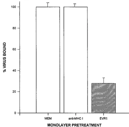
FIG. 1. MAb EVR1 inhibits binding of EV70 to HeLa cells. HeLa cell monolayers in 24-well plates were treated as indicated for 1 h at 378C before incubation with 53103cpm of [35S]EV70 for 45 min at 338C. The amount of
virus bound to cells was determined as described in Materials and Methods. MEM, growth medium alone; anti-MHC I, growth medium containing 10mg of MHC 1-specific IgG1 per ml; EVR1, EVR1 hybridoma culture supernatant (17.5 mg/ml). Results are shown as the mean percentage of virus bound relative to the MEM control6standard deviation for four samples.
FIG. 2. MAb EVR1 immunoprecipitates a HeLa cell glycoprotein. Proteins immunoprecipitated from lysates of cells labelled for 16 h with [3
H]glucosamine were analyzed by SDS-PAGE as described in Materials and Methods. Lanes: 1, LLC-MK2lysate without Ab; 2, LLC-MK2lysate with LCMV-specific IgG1; 3,
LLC-MK2lysate with EVR1; 4, HeLa lysate without antibody; 5, HeLa lysate
[image:3.612.325.540.72.283.2]with LCMV-specific IgG1; 6, HeLa lysate with EVR1. Positions of molecular mass standards are indicated on the left.
TABLE 1. Specificity of MAb EVR1 cell protection
Virus Cell protection
a
HeLa LLC-MK2
EV70 1 2
Poliovirus Sabin 1 2 2
Coxsackievirus B3 2 2
a1
, no observable CPE for a minimum of 96 h postinfection;2, CPE ob-served within 45 h postinfection. EVR1 was used at 17.5mg/ml.
on November 9, 2019 by guest
http://jvi.asm.org/
that MAb EVR1 protects HeLa cells by nonspecific masking of
the cell surface, we tested the ability of the antibody to protect
HeLa and LLC-MK
2monolayers against infection by these two
other enteroviruses (Table 1). Undiluted EVR1 hybridoma
supernatant completely protected HeLa cell monolayers
against EV70 infection, with monolayers remaining free of
CPE for 96 h, at which time monitoring was terminated.
Nei-ther HeLa nor LLC-MK
2cells were protected against
poliovi-rus or coxsackievipoliovi-rus B3. In all cases, the monolayers were
completely destroyed within 18 h following infection. MAb
EVR1 also failed to protect LLC-MK
2monolayers from EV70
infection, as evidenced by the appearance of extensive CPE
within 45 h after infection. To show that MAb EVR1 blocks
the attachment of EV70, HeLa cell monolayers were incubated
with various dilutions of hybridoma culture fluid and then with
[
35S]EV70 in binding inhibition assays. At 10
m
g/ml, inhibition
by MAb EVR1 was maximal, reducing the binding of
radiola-belled EV70 by 74 to 84% compared with controls (Fig. 1).
Lower concentrations of antibody resulted in less inhibition.
The cell line specificity of binding was confirmed by
immuno-fluorescence with live cells. MAb EVR1 recognized an epitope
that was evenly distributed over the surface of HeLa cells (see
Fig. 4A). Dispersal of HeLa cells by trypsin treatment prior to
incubation with antibody had no effect on MAb EVR1 binding,
whereas LLC-MK
2cells, dispersed either manually or by
tryp-sin treatment, did not bind MAb EVR1 (data not shown).
Binding inhibition assays and immunofluorescence confirmed
the specificity observed in cell protection assays and indicated
that MAb EVR1 binds to an epitope that is present on the
surface of human (HeLa) but not monkey (LLC-MK
2) cells.
Immunoprecipitation of a HeLa cell glycoprotein by MAb
EVR1.
To identify the protein recognized by MAb EVR1,
lysates of [
3H]glucosamine-labelled HeLa and LLC-MK
2
cells
[image:4.612.129.223.74.200.2]were incubated with Ab and the immunoprecipitates were
an-alyzed by SDS-polyacrylamide gel electrophoresis (PAGE)
(Fig. 2). MAb EVR1 reacted specifically with a HeLa cell
protein of approximately 70 to 75 kDa that appeared to
mi-grate as a doublet in polyacrylamide gels (Fig. 2, lane 6).
Successful labelling after a 4-h (data not shown) or a 16-h
[image:4.612.59.554.394.691.2]FIG. 3. Immunoblot analysis of HeLa cell lysates with MAbs EVR1 and 1H4. HeLa cell lysates were separated by electrophoresis in 8% polyacrylamide gels containing 0.05% SDS under nonreducing conditions and transferred to nitro-cellulose. Membrane strips were incubated with MAbs as indicated and devel-oped as described in Materials and Methods. Lanes: 1, EVR1, 2, 1H4, 3, LCMV-specific IgG1. Positions of molecular mass standards are indicated on the left.
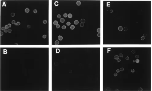
FIG. 4. The ligand of EVR1 is sensitive to PI-PLC. Cell monolayers were dispersed by trypsin treatment, washed, treated, and incubated with Abs as indicated. Ab binding was detected by indirect immunofluorescence with an FITC-conjugated sheep anti-mouse Ab, as described in Materials and Methods. (A) HeLa cells, MAb EVR1; (B) PI-PLC-treated HeLa cells, MAb EVR1; (C) HeLa cells, MAb 1H4; (D) PI-PLC-treated HeLa cells, MAb 1H4; (E) HeLa cells, anti-MHC I IgG1; (F) PI-PLC-treated HeLa cells, anti-MHC I IgG1.
on November 9, 2019 by guest
http://jvi.asm.org/
incubation with [
3H]glucosamine indicated that the protein
was glycosylated. No protein was immunoprecipitated from
lysates of LLC-MK
2cells (lane 3) or from HeLa or LLC-MK
2lysates incubated with mouse LCMV-specific IgG1 or protein
G beads alone (lanes 1, 2, 4, and 5). Preliminary
immunoblot-ting studies demonstrated that the epitope recognized by MAb
EVR1 was both heat labile and sensitive to reducing agents in
electrophoresis sample buffer (
b
-mercaptoethanol) (data not
shown). When membrane proteins were separated on 8%
poly-acrylamide gels containing 0.05% SDS under nonreducing
con-ditions, MAb EVR1 recognized a HeLa cell protein of
approx-imately 75 kDa (Fig. 3, lane 1), but did not react with proteins
from LLC-MK
2cells (data not shown).
The EVR1 ligand on HeLa cells is DAF.
A survey of the
literature suggested that the protein recognized by MAb
EVR1 might be DAF (CD55). Because DAF is anchored to
cells by a GPI moiety, we wished to determine if the ligand of
EVR1 was also attached to HeLa cells by a GPI tail (12, 30).
Therefore, prior to reaction with antibody, HeLa cells were
treated with PI-PLC, an enzyme known to release GPI-linked
proteins from the surface of cells. As shown in Fig. 4, the ability
of MAb EVR1 to bind to HeLa cells treated with PI-PLC was
greatly diminished (Fig. 4A and B), as was binding of the
DAF-specific MAb, 1H4 (Fig. 4C and D). Binding of a MAb
specific for human MHC 1 (a non-GPI-linked integral
mem-brane glycoprotein) was not affected by PI-PLC (Fig. 4E and
F). In Western blots, MAb 1H4, like MAb EVR1, reacted
specifically with a HeLa cell protein of approximately 75 kDa
(Fig. 3, lane 2).
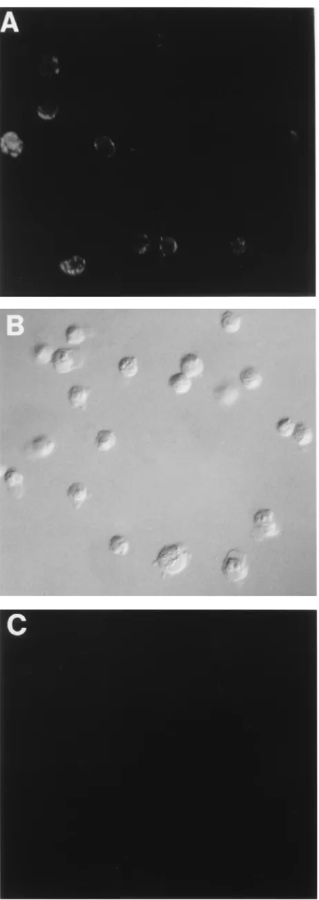
Although we previously observed that CHO cells were able
to bind EV70 and to support EV70 replication (data not
shown), we exploited the availability of CHO cells transfected
with a human DAF cDNA and constitutively expressing DAF
(CHO-DAF [28]) to confirm the specificity of MAb EVR1 for
human DAF. In immunofluorescence studies (Fig. 5), MAb
EVR1 bound to CHO-DAF but not to CHO cells. MAb 1H4
behaved similarly (data not shown).
DAF-specific MAbs block EV70 binding to and infection of
HeLa cells.
To demonstrate that DAF acts as the HeLa cell
receptor for EV70, monolayers of HeLa and LLC-MK
2cells
were treated with MAb 1H4 (10 to 35
m
g/ml), prior to
chal-lenge with EV70, in a cell protection assay. As observed with
MAb EVR1, MAb 1H4 protected HeLa cells (Table 2) but not
LLC-MK
2cells (data not shown) against EV70 infection, in a
concentration-dependent manner (data not shown). We
sub-sequently assessed the ability of other anti-DAF MAbs to
protect HeLa cells (Table 2). Antibodies directed against
SCRs 1 and 3 of DAF (MAbs 11D7 and 1H4, respectively [10])
protected cells, while MAbs specific for SCRs 2 and 4 (1F7 and
8D11, respectively [3, 10]), did not. MAbs EVR1 (17.5
m
g/ml),
1H4 (35
m
g/ml), and 11D7 (4
m
g/ml) prevented the appearance
of CPE for 96 h, whereas cells treated with MAb 8D11 (40
m
g/ml) began showing CPE by 48 h postinfection. The ability of
DAF-specific Abs to prevent EV70 infection of HeLa cells
correlated with their ability to inhibit virus binding (Fig. 6).
The HeLa cell receptor for EV70 is DAF.
NIH 3T3 cells,
[image:5.612.63.294.72.722.2]which do not bind EV70 or support virus replication, were used
in transient-expression experiments to evaluate the ability of
DAF to act as a receptor for EV70. Human DAF cDNA
FIG. 5. MAb EVR1 recognizes DAF. Cell monolayers were dispersed by trypsin treatment, washed, and incubated with MAb EVR1. Ab binding was detected by indirect immunofluorescence with an FITC-conjugated sheep anti-mouse Ab, as described in Materials and Methods. (A) CHO-DAF cells; (B) CHO cells with Nomarski optics; (C) CHO cells, same field as in panel B.
on November 9, 2019 by guest
http://jvi.asm.org/
subcloned into vector pcDNA3 (pcDNA3-DAF) was
intro-duced into NIH 3T3 cells, and transfected cells were infected
with vaccinia virus vTF7-3, to drive DAF expression. As shown
in Fig. 7, cells transfected with pcDNA3-DAF expressed DAF
at their surfaces and reacted with MAbs EVR1 and 1H4 (Fig.
7B and F) whereas cells transfected with pcDNA3 alone did
not (Fig. 7C and G). As shown in Fig. 8, NIH 3T3 monolayers
transfected with pcDNA3-DAF were also able to bind
[
35S]EV70. Although the amount of virus binding to
pcDNA3-DAF-transfected cells varied among experiments, DAF
trans-fectants consistently bound 50 to 100% of the amount of virus
that bound to HeLa cells and bound two to four times as much
virus as did cells transfected with pcDNA3.
Flow cytometric analysis (data not shown) indicated that
transfected monolayers were a mixed population of cells
dis-playing a range of DAF expression levels. For the experiments
represented in Fig. 7 and 8, 10% of cells transfected with
pcDNA3-DAF expressed approximately 10 times as much
DAF on their surfaces as did HeLa cells and approximately
40% of cells expressed low to moderate levels of DAF. The
remaining cells were negative for DAF expression.
NIH 3T3 cells constitutively expressing human DAF
(3T3-DAF) have been described previously (51). In
immunofluores-cence assays, MAbs EVR1 and 1H4 reacted much more weakly
with 3T3-DAF cells than with HeLa cells (data not shown). In
binding assays, we were unable to detect EV70 binding to
DAF cells above the background levels observed in
3T3-RDAF cells (data not shown), which do not express DAF (51).
Nevertheless, this 3T3-DAF cell line was able to support
low-level EV70 replication. Monolayers were infected with EV70
at a MOI of 5 and assayed for the presence of infectious virus
at regular intervals, for a period of 48 h (Fig. 9). The difference
in the amount of virus associated with RDAF and
3T3-DAF cells at time zero may indicate increased binding of EV70
to 3T3-DAF cells. Virus yield from DAF-expressing cells
in-creased to a peak value of 1.6
3
10
3PFU/ml by 24 h,
repre-senting a 2.5-fold increase from time zero, and began to
de-cline thereafter. 3T3-RDAF cells could not be infected. EV70
titers from these cells remained low (approximately 300 PFU/
ml) for 24 h and then, as with 3T3-DAF cells, began to fall.
Experiments in which samples were assayed at 24-h intervals
for 120 h (data not shown) indicated that this decline in virus
titer continued, reflecting a decay of progeny virus following
one cycle of replication (3T3-DAF cells) and decay of input
virus (3T3-RDAF cells). No CPE was observed in
EV70-in-fected 3T3-DAF monolayers. At this time, it is not known if
these cells become persistently infected.
DISCUSSION
The initial interaction of a virus with specific host cell
sur-face components is recognized to be an important determinant
of viral host range and tissue tropism (33, 38). This suggested
to us that the unique in vitro and in vivo replication
charac-teristics of EV70 may be determined by its receptor and would
reflect differences between the receptor for EV70 and those
identified for other picornaviruses. In view of this information,
we undertook studies to identify and characterize the HeLa
cell receptor for EV70.
A MAb that exhibited specific receptor-blocking activity in
both biological and physical assays was isolated. MAb EVR1
prevented infection of HeLa cells by EV70 but not by the
closely related picornaviruses poliovirus and coxsackievirus B3
(46). Binding inhibition assays confirmed that this antibody
acts at the level of virus-receptor interaction.
Immunoprecipi-tations, Western blots, and immunofluorescence assays
dem-onstrated that MAb EVR1 reacted with a GPI-anchored,
70-to 75-kDa protein on the surface of HeLa cells.
[image:6.612.57.298.91.158.2]Our data were consistent with the possibility that MAb
EVR1 identified DAF (CD55). DAF is an important
modula-tor of the complement system and, together with membrane
cofactor protein, complement receptor types 1 and 2, and
C4-binding protein, is a member of the family of regulators of
complement activation (RCA) (27, 34, 37). The human DAF
molecule is typically expressed as a 70- to 75-kDa glycoprotein
on the surface of many cell types, including HeLa cells (27, 30).
The nucleotide sequence of DAF predicts a structure with five
extracellular domains (27, 29). Proximal to the cell membrane
is a serine/tyrosine-rich domain, which is extensively O
glyco-sylated, and distally there are four SCR domains characteristic
of RCA family members. Each SCR contains four cysteine
residues that are predicted to form internal disulfide bonds
(27). The sensitivity of the interaction between MAb EVR1
and its ligand to both heat and reducing agents suggests that
the antibody recognizes a conformational epitope on one of
FIG. 6. DAF-specific MAbs inhibit the binding of EV70 to HeLa cells. HeLa cell monolayers in 24-well plates were treated with MAbs as indicated for 1 h at 378C, before incubation with 53103
cpm of [35
[image:6.612.330.544.434.646.2]S]EV70 for 45 min at 338C. The amount of virus bound to cells was determined as described in Materials and Methods. MEM, growth medium alone; anti-MHC I, 10mg of MHC I-specific MAb per ml; EVR1, 10mg of EVR1 per ml; 11D7, 10mg of 11D7 per ml; 1H4, 10mg of 1H4 per ml; 8D11, 20mg of 8D11 per ml. Results are shown as the mean percentage of virus bound relative to the MEM control6standard deviation for two samples.
TABLE 2. Anti-DAF MAb protection of HeLa cells against EV70 infection
MAba SCR Protectionb
11D7 1 1
1F7 2 2
1H4 3 1
8D11 4 2
EVR1 ? 1
a
11D7, 4mg/ml; 1F7, 240mg/ml; 1H4, 35mg/ml; 8D11, 36mg/ml; EVR1, 17.5 mg/ml.
b1
, no observable CPE for a minimum of 96 h postinfection;2, CPE ob-served within 48 h postinfection.
on November 9, 2019 by guest
http://jvi.asm.org/
FIG. 7. Transfection of NIH 3T3 cells results in surface expression of DAF. Monolayers of transfected NIH 3T3 cells were dispersed by trypsin treatment, washed, and incubated with Abs as indicated. Ab binding was detected by indirect immunofluorescence with an FITC-conjugated sheep anti-mouse Ab, as described in Materials and Methods. (A through D) Incubation with EVR1 ascites fluid (1:200). (A) HeLa cells; (B) NIH 3T3 cells transfected with pcDNA3-DAF; (C) NIH 3T3 cells transfected with pcDNA3; (D) same field as in panel C with Nomarski optics. (E through H) Incubation with MAb 1H4 (1:5). (E) HeLa cells; (F) NIH 3T3 cells transfected with pcDNA3-DAF; (G) NIH 3T3 cells transfected with pcDNA3; (H) same field as in panel G with Nomarski optics.
5149
on November 9, 2019 by guest
the SCR domains. The appearance of a doublet in
immuno-precipitations may reflect variable glycosylation of the protein.
The specificity of MAb EVR1 for DAF was substantiated by
the observation that CHO-DAF (28) but not CHO cells
re-acted with antibody. Demonstration that DAF-specific MAbs
11D7 and 1H4 (directed against SCRs 1 and 3, respectively)
blocked virus attachment to HeLa cells and were
cytoprotec-tive strongly suggested that DAF acts as the HeLa cell receptor
for EV70.
Conclusive evidence of this was provided by expression of
human DAF in NIH 3T3 cells. Transient expression of DAF on
the surface of NIH 3T3 cells resulted in virus binding. We were
unable to detect significant EV70 binding to NIH 3T3 cells
stably expressing human DAF (52). These 3T3-DAF cells also
reacted weakly in immunofluorescence studies with MAbs
EVR1 and 1H4. Since this cell line has been shown to express
approximately seven times less DAF than reported for HeLa
cells (3
3
10
4versus 2
3
10
5DAF molecules per cell [31, 52]),
our results may indicate that the level of DAF expression was
too low for us to detect virus binding above background levels.
Nevertheless, we were able to detect virus replication in these
3T3-DAF cells, indicating that DAF expression is sufficient for
EV70 to productively infect NIH 3T3 cells. Our findings
sug-gest that DAF-expressing cells were able to support one round
of virus replication, after which we observed the gradual decay
of progeny virus. Control 3T3-RDAF cells were refractory to
EV70 infection. An explanation for the low yield of virus
re-covered from 3T3-DAF cells may be that EV70 replication is
poorly supported in NIH 3T3 cells. As yet unidentified cellular
factors required for efficient growth of EV70 in these cells may
be lacking, and their absence may contribute to a partial block
in the viral replication cycle. Furthermore, our EV70 stocks
were propagated on LLC-MK
2monolayers, and the virus
in-ocula used in NIH 3T3 infectivity assays may contain only a
small proportion of variants able to replicate in this murine cell
line. Adaptation of EV70 to this new host by repeated passage
may result in more productive infection.
Human (36), guinea pig (35), rabbit (48), and mouse (45)
DAF molecules have been identified. The presence of
DAF-like molecules in different mammalian species is consistent
with the ability of EV70 to infect a wide range of mammalian
cell lines in vitro. MAb EVR1 did not bind to monkey kidney
(LLC-MK
2) cells or protect them against EV70 infection.
Al-though the predicted amino acid sequence of the DAF
mole-cule of rhesus monkeys shares approximately 95% amino acid
identity with that of human DAF (21a), the sequence
differ-ences might account for our observations. In addition, CHO
cells were able to bind labelled EV70 and to support EV70
replication. This is also consistent with the broad in vitro host
range of this virus, which is known to include cells of hamster
origin (56). It will be of interest to determine if DAF homologs
expressed on other mammalian cell lines function as receptors
for EV70 or if an altogether different receptor molecule is
used.
In addition to its role in the regulation of complement, DAF
has recently been shown to act as a cellular receptor for at least
six echovirus serotypes (3, 51) and for coxsackieviruses B1, B3,
and B5 (5, 42). Regions of DAF involved in binding these
different picornaviruses have been identified. Antibody
block-ade experiments showed that echovirus 7 and related viruses
(3) and RD cell-adapted coxsackievirus B3 (5) interact with
SCRs 2 and 3. Transfection experiments with DAF cDNAs
containing deletions of specific SCRs subsequently determined
that SCRs 2, 3, and 4 are required for echovirus 7 binding (9)
and that SCR 2 is essential for coxsackievirus B3-RD
attach-ment (5). Antibody blockade experiattach-ments also identified a site
within or near SCR 3 as the possible binding region for
cox-sackievirus B5 (42). As reflected in these reports, mapping
virus-binding sites by antibody blockade alone has certain
lim-itations. The large size of antibodies relative to the DAF
mol-ecule and the absence of information regarding both the
se-quences of the epitopes recognized by the antibodies and the
FIG. 8. Transient expression of DAF in NIH 3T3 cells results in binding of EV70. Monolayers of cells in 24-well plates were incubated with 33103cpm of
[35S]EV70 for 45 min at 338C. The amount of virus bound to cells was
deter-mined as described in Materials and Methods. HeLa, HeLa cell control; 3T3/ pcDNA3, NIH 3T3 cells transfected with pcDNA3; 3T3/DAF, NIH 3T3 cells transfected with pcDNA3-DAF. Results are shown as the mean percentage of virus bound relative to the HeLa cell control6standard deviation for three samples.
FIG. 9. DAF-expressing NIH 3T3 cells support EV70 replication. 3T3-DAF cells (F) and control 3T3-RDAF cells (E) were grown in 24-well plates and infected at an MOI of 5 for 1 h at 338C. Following infection, monolayers were incubated at 338C in growth medium containing 10% FBS. At the indicated intervals, cells and supernatants were recovered and frozen and thawed twice to liberate infectious virus. EV70 titers were measured by plaque assay on LLC-MK2monolayers. Results are expressed as means6standard deviation for two
duplicate independent determinations.
on November 9, 2019 by guest
http://jvi.asm.org/
precise location of these epitopes in the folded DAF molecule
make definitive interpretation of data difficult. Our
antibody-blocking data suggest that regions within or proximal to SCRs
1 and 3 are involved in the binding of EV70 and therefore that
the EV70-binding site(s) on DAF may be different from those
used by other enteroviruses.
EV70 and the echoviruses and coxsackieviruses that use
DAF for attachment are all able to agglutinate human
eryth-rocytes (3, 5, 25, 42, 50). An early study of the interaction
between EV70 and human erythrocytes (50) demonstrated
that the hemagglutinating ability of EV70 was sensitive to
neuraminidase. However, treatment of erythrocytes with
neur-aminidase did not eliminate their capacity to adsorb
echovi-ruses 7 and 11. This also would suggest that different regions of
DAF are involved in binding these viruses. Experiments
de-signed to more precisely define the DAF sequences involved in
EV70 binding are under way.
DAF is expressed on conjunctival epithelial cells, and DAF
antigen has been detected in cerebrospinal fluid (31). Thus, the
receptor for EV70 is found at sites of virus replication, in
tissues involved in the pathogenesis of EV70 and the clinical
manifestations of acute hemorrhagic conjunctivitis (17, 21, 55).
The presence of DAF on cells and tissues that are not
recog-nized as sites of EV70 replication and the fact that DAF
functions as a receptor for several other enteroviruses suggest
that as with poliovirus (43), the tropism of EV70 is probably
determined by the concomitant expression of receptor and
additional host cell-specific proteins. Clearly, the disparate
host range, tropism, and pathogenicity of human enteroviruses
cannot simply be explained by differences in the nature and
distribution of their receptors.
ACKNOWLEDGMENTS
We gratefully acknowledge J. Atkinson for providing NIH 3T3 cell lines transfected with human DAF cDNA, R. Finberg for providing MAb 1F7, and W. Rosse for providing MAbs 8D11 and 11D7. We also thank W. Staines, Department of Anatomy and Neurobiology, Uni-versity of Ottawa, for assistance with photomicrographs.
This research was supported by the Natural Sciences and Engineer-ing Research Council of Canada. T.M.K. is the recipient of an Ontario Graduate Scholarship.
REFERENCES
1. Abraham, G., and R. J. Colonno. 1984. Many rhinovirus serotypes share the same cellular receptor. J. Virol. 51:340–345.
2. Bass, D. M., and H. B. Greenberg. 1992. Strategies for the identification of icosahedral virus receptors. J. Clin. Invest. 89:3–9.
3. Bergelson, J. M., M. Chan, K. R. Solomon, N. F. St. John, H. Lin, and R. W. Finberg.1994. Decay-accelerating factor (CD55), a glycosylphosphatidyli-nositol-anchored complement regulatory protein, is a receptor for several echoviruses. Proc. Natl. Acad. Sci. USA 91:6245–6248.
4. Bergelson, J. M., and R. W. Finberg. 1993. Integrins as receptors for virus attachment and cell entry. Trends Microbiol. 1:287–288.
5. Bergelson, J. M., J. G. Mohanty, R. L. Crowell, N. F. St. John, D. M. Lublin, and R. W. Finberg.1995. Coxsackievirus B3 adapted to growth in RD cells binds to decay-accelerating factor (CD55). J. Virol. 69:1903–1906. 6. Bergelson, J. M., M. P. Shepley, B. M. C. Chan, M. E. Hemler, and R. W.
Finberg.1992. Identification of the integrin VLA-2 as a receptor for echo-virus 1. Science 255:1718–1720.
7. Bergelson, J. M., N. St. John, S. Kawaguchi, M. Chan, H. Stubal, J. Modlin, and R. W. Finberg.1993. Infection by echovirus 1 and 8 depends on thea2
subunit of human VLA-2. J. Virol. 67:6847–6852.
8. Berinstein, A., M. Roivainen, T. Hovi, P. W. Mason, and B. Baxt. 1995. Antibodies to the vitronectin receptor (integrinavb3) inhibit binding and
infection of foot-and-mouth disease virus to cultured cells. J. Virol. 69:2664– 2666.
9. Clarkson, N. A., R. Kaufman, D. M. Lublin, T. Ward, P. A. Pipkin, P. D. Minor, D. J. Evans, and J. W. Almond.1995. Characterization of the echo-virus 7 receptor: domains of CD55 critical for echo-virus binding. J. Virol. 69: 5497–5501.
10. Coyne, K. E., S. E. Hall, E. S. Thompson, M. A. Arce, T. Kinoshita, T. Fujita, D. J. Anstee, W. Rosse, and D. M. Lublin.1992. Mapping of epitopes,
glycosylation sites, and complement regulatory domains in human decay accelerating factor. J. Immunol. 149:2906–2913.
11. Darougar, S., M. A. Monnickendam, and R. M. Woodland. 1989. Manage-ment and prevention of ocular viral and chlamydial infections. Crit. Rev. Microbiol. 16:369–418.
12. Davitz, M. A., M. G. Low, and V. Nussenzweig. 1986. Release of decay-accelerating factor (DAF) from the cell membrane by phosphatidylinositol-specific phospholipase C (PIPLC). Selective modification of a complement regulatory protein. J. Exp. Med. 163:1150–1161.
13. Dimock, K., D. G. Harnish, G. Sisson, W.-C. Leung, and W. E. Rawls. 1982. Synthesis of virus-specific polypeptides and genomic RNA during the repli-cative cycle of Pichinde virus. J. Virol. 43:273–283.
14. Engvall, E., and P. Perlmann. 1971. Enzyme-linked immunosorbent assay (ELISA). Quantitative assay of immunoglobulin G. Immunochemistry 8:871–874.
15. Greve, J. M., G. Davis, A. M. Meyer, C. P. Forte, S. C. Yost, C. W. Marlor, M. E. Kamarck, and A. McClelland.1989. The major human rhinovirus receptor is ICAM-1. Cell 56:839–847.
16. Hansen, M. B., S. E. Nielsen, and K. Berg. 1989. Re-examination and further development of a precise and rapid dye method for measuring cell growth/ cell kill. J. Immunol. Methods 119:203–210.
17. Higgins, P. G. 1982. Enteroviral conjunctivitis and its neurological compli-cations. Arch. Virol. 73:91–101.
18. Hofer, F., M. Gruenberger, H. Kowalski, H. Machat, M. Huettinger, E. Kuechler, and D. Blaas.1994. Members of the low density lipoprotein receptor family mediate cell entry of a minor group common cold virus. Proc. Natl. Acad. Sci. USA 91:1839–1842.
19. Hornbeck, P. 1991. Antibody-sandwich ELISA to detect soluble antigens, p. 2.1.9–2.1.11. In J. E. Coligan, A. M. Kruisbeek, D. H. Margulies, E. M. Shevach, and W. Strober (ed.), Current protocols in immunology, vol. 1. John Wiley & Sons, Inc., New York.
20. Huber, S. A. 1994. VCAM-1 is a receptor for encephalomyocarditis virus on murine vascular endothelial cells. J. Virol. 68:3453–3458.
21. Johnson, R. T. 1994. The Soriano Award Lecture. Emerging infections of the nervous system. J. Neurol. Sci. 124:2–14.
21a.Kaufman, R., and D. Lublin. Unpublished observation.
22. King, S. L., J. A. Cunningham, R. W. Finberg, and J. M. Bergelson. 1995. Echovirus 1 interaction with the isolated VLA-2 domain. J. Virol. 69:3237– 3239.
23. Ko¨hler, G., and C. Milstein.1975. Continuous cultures of fused cells secret-ing antibodies of predefined specificity. Nature (London) 256:495–497. 24. Kono, R., A. Sasagawa, K. Ishii, M. Ochi, S. Sugiura, H. Matsumiya, Y.
Uchida, K. Kameyama, M. Kaneko, and N. Sakurai.1972. Pandemic of a new type of conjunctivitis. Lancet i:1191–1194.
25. Kono, R., E. Tajiri, K. Miyamura, A. Sasagawa, and T. Tsuruhara. 1978. Hemagglutination and hemagglutination inhibition tests with enterovirus type 70. J. Clin. Microbiol. 7:595–598.
26. Laemmli, U. K. 1970. Cleavage of structural proteins during the assembly of the head of bacteriophage T4. Nature (London) 227:680–685.
27. Lublin, D. M., and J. P. Atkinson. 1989. Decay-accelerating factor: biochem-istry, molecular biology, and function. Annu. Rev. Immunol. 7:35–58. 28. Lublin, D. M., and K. E. Coyne. 1991. Phospholipid-anchored and
trans-membrane versions of either decay-accelerating factor or trans-membrane cofac-tor protein show equal efficiency in protection from complement-mediated cell damage. J. Exp. Med. 174:35–44.
29. Medof, M. E., D. M. Lublin, V. M. Holers, D. J. Ayers, R. R. Getty, J. F. Leykam, J. P. Atkinson, and M. L. Tykocinski.1987. Cloning and charac-terization of cDNAs encoding the complete sequence of decay-accelerating factor of human complement. Proc. Natl. Acad. Sci. USA 84:2007–2011. 30. Medof, M. E., E. I. Walter, W. L. Roberts, R. Haas, and T. L. Rosenberry.
1986. Decay accelerating factor of complement is anchored to cells by a C-terminal glycolipid. Biochemistry 25:6740–6747.
31. Medof, M. E., E. I. Walter, J. L. Rutgers, D. M. Knowles, and V. Nussenz-weig. 1987. Identification of the complement decay-accelerating factor (DAF) on epithelium and glandular cells and in body fluids. J. Exp. Med. 165:848–864.
32. Mendelsohn, D. L., E. Wimmer, and V. R. Racaniello. 1989. Cellular recep-tor for poliovirus: molecular cloning, nucleotide sequence, and expression of a new member of the immunoglobulin superfamily. Cell 56:855–865. 33. Mims, C. A. 1986. Virus receptors and cell tropisms. J. Infect. 12:199–203. 34. Nicholson-Weller, A. 1992. Decay accelerating factor (CD55). Curr. Top.
Microbiol. Immunol. 178:8–30.
35. Nicholson-Weller, A., J. Burge, and K. F. Austen. 1981. Purification from guinea pig erythrocyte stroma of a decay-accelerating factor for the classical C3 convertase, C4b2a. J. Immunol. 127:2035–2039.
36. Nicholson-Weller, A., J. Burge, D. T. Fearon, P. F. Weller, and K. F. Austen. (1982). Isolation of a human erythrocyte membrane glycoprotein with decay-accelerating activity for C3 convertases of the complement system. J. Immu-nol. 129:184–189.
37. Nicholson-Weller, A., and C. E. Wang. 1994. Structure and function of decay accelerating factor CD55. J. Lab. Clin. Med. 123:485–491.
38. Norkin, L. C. 1995. Virus receptors: implications for pathogenesis and the
on November 9, 2019 by guest
http://jvi.asm.org/
design of antiviral agents. Clin. Microbiol. Rev. 8:293–315.
39. Raab de Verdugo, U., H.-C. Selinka, M. Huber, B. Kramer, J. Kellermann, P. H. Hofschneider, and R. Kandolf.1995. Characterization of a 100-kilo-dalton binding protein for the six serotypes of coxsackie B viruses. J. Virol. 69:6751–6757.
40. Roivainen, M., L. Piirainen, T. Hovi, I. Virtanen, T. Riikonen, J. Heino, and T. Hyypia¨.1994. Entry of coxsackievirus A9 into host cells: specific interac-tions withavb3integrin, the vibronectin receptor. Virology 203:357–365.
41. Rotbart, H. A., and K. Kirkegaard. 1992. Picornavirus pathogenesis: viral access, attachment and entry into susceptible cells. Semin. Virol. 3:483–499. 42. Shafren, D. R., R. C. Bates, M. V. Agrez, R. L. Herd, G. F. Burns, and R. D. Barry.1995. Coxsackieviruses B1, B3, and B5 use decay-accelerating factor as a receptor for cell attachment. J. Virol. 69:3873–3877.
43. Shepley, M. P., and V. R. Racaniello. 1994. A monoclonal antibody that blocks poliovirus attachment recognizes the lymphocyte homing receptor CD44. J. Virol. 68:1301–1308.
44. Schulman, M., C. D. Wilde, and G. Kohler. 1978. A better cell line for making hybridomas secreting specific antibodies. Nature (London) 276:269– 271.
45. Spicer, A. P., M. F. Seldin, and S. J. Gendler. 1995. Molecular cloning and chromosomal localization of the mouse decay-accelerating factor genes. J. Immunol. 155:3079–3091.
46. Stanway, G. 1990. Structure, function and evolution of picornaviruses. J. Gen. Virol. 71:2483–2501.
47. Staunton, D. E., V. J. Merluzzi, R. Rothlein, R. Barton, S. D. Marlin, and T. A. Springer.1989. A cell adhesion molecule, ICAM-1, is the major surface receptor for rhinoviruses. Cell 56:849–853.
48. Sugita, Y., M. Uzawa, and M. Tomita. 1987. Isolation of decay-accelerating
factor (DAF) from rabbit erythrocyte membranes. J. Immunol. Methods 104:123–130.
49. Towbin, J., T. Staehelin, and J. Gordon. 1979. Electrophoretic transfer of proteins from polyacrylamide gels to nitrocellulose sheets: procedure and some applications. Proc. Natl. Acad. Sci. USA 76:4350–4354.
50. Utagawa, E. T., K. Miyamura, A. Mukoyama, and R. Kono. 1982. Neura-minidase-sensitive erythrocyte receptor for enterovirus type 70. J. Gen. Vi-rol. 63:141–148.
51. Ward, T., P. A. Pipkin, N. A. Clarkson, D. M. Stone, P. D. Minor, and J. W. Almond.1994. Decay-accelerating factor CD55 is identified as the receptor for echovirus 7 using CELICS, a rapid immuno-focal cloning method. EMBO J. 13:5070–5074.
52. White, D. J. G., T. Oglesby, M. K. Liszewski, I. Tedja, D. Hourcade, M.-W. Wang, L. Wright, J. Wallwork, and J. P. Atkinson.1992. Expression of human decay accelerating factor or membrane cofactor protein genes on mouse cells inhibits lysis by human complement. Transplant. Proc. 24:474– 476.
53. White, J. M., and D. R. Littman. 1989. Viral receptors of the immunoglob-ulin superfamily. Cell 56:725–728.
54. Wright, P. D., G. H. Strauss, and M. P. Langford. 1992. Acute hemorrhagic conjunctivitis. Am. Fam. Physician 45:173–178.
55. Yamazaki, S., and K. Miyamura. 1989. General characteristics of enterovirus 70, p. 345–357. In Y. Uchida, K. Ishii, K. Miyamura, and S. Yamazaki (ed.), Acute hemorrhagic conjunctivitis. Etiology, epidemiology and clinical man-ifestations. S. Karger AG, New York.
56. Yoshii, T., K. Natori, and R. Kono. 1977. Replication of enterovirus 70 in nonprimate cell cultures. J. Gen. Virol. 36:377–384.