0022-538X/96/$04.0010
Copyrightq1996, American Society for Microbiology
Initiation of Baculovirus DNA Replication: Early Promoter
Regions Can Function as Infection-Dependent Replicating
Sequences in a Plasmid-Based Replication Assay
YUNTAO WUANDERIC B. CARSTENS*
Department of Microbiology and Immunology, Queen’s University, Kingston, Ontario K7L 3N6, Canada
Received 5 February 1996/Accepted 12 July 1996
From the results of transient plasmid-based replication assays, it has been postulated that homologous
regions (hrs) ofAutographa californicanuclear polyhedrosis virus (AcMNPV) function as origins of viral DNA
replication. However, these assays vary in specificity according to the methodology used and may not be
dependent solely on the presence ofhrsequences. To determine the role thathrs and other sequences might
play in the replication process, a series of plasmids containing specific deletions of varioushrs was generated
and tested in a standardized replication assay. Deletion of the AcMNPVhr2 andhr5 sequences abolished the
ability of plasmids to replicate in the standard infection-dependent replication assay, while deletion ofhr1,hr3,
andhr4a sequences decreased but did not eliminate plasmid replication in this assay. Plasmids carrying the
completeie-2andpe38genes, theie-1gene upstream region, or a variety of baculovirus genes including 11 early
promoter regions were also able to replicate in virus-infected cells, suggesting that early viral promoter sequences could also function as putative origins of replication. These data suggest that the standard infection-dependent replication assay may identify a broad range of infection-infection-dependent replicating sequences, only one or a few of which may represent genuine viral origins used by the virus in vivo. We propose a model suggesting that the selection of replication initiation sites may be imposed directly by chromatin structure and indirectly by primary sequence and that the process of viral DNA replication may be linked with viral transcription.
Autographa californica nuclear polyhedrosis virus (AcM
NPV), the type species baculovirus, has a closed circular, dou-ble-stranded DNA genome (33, 35) of 134 kb, potentially en-coding over 150 polypeptides (1). Expression of AcMNPV ap-pears to be controlled mainly at the transcriptional level and occurs in an ordered cascade fashion through early and late phases (3). The genome contains eight A1T-rich homologous regions (hrs) interspersed around the genome, each containing two to eight 30-bp imperfect palindromes with a core EcoRI site (except hr4c) (15). Because of their symmetric location, high A1T content, and palindromic structure,hrs were origi-nally postulated to function as viral origins of replication (5). Recent transient replication assays supported this hypothesis (15). A single palindrome containing an intact coreEcoRI site was sufficient to support plasmid replication in virus-infected cells (20, 27). The only other region shown to support plasmid replication in this assay included several A1T-rich regions and imperfect palindromes within theHindIII-K (ori-K) fragment (16, 19). However, it is still not known whether any of these regions are essential for and function as replication origins in vivo. For instance, deletion of hr5 from AcMNPV had no apparent effect on virus replication (30). In addition, it has been demonstrated that plasmids without baculovirus inserts can replicate when plasmid and viral DNAs are cotransfected into insect cells (10, 18). As well, when plasmids expressing a number of viral genes necessary for reporter plasmid replica-tion were cotransfected, all plasmids in addireplica-tion to the reporter plasmid replicated (14). Because only the reporter plasmid carried anhr, these data suggested to us that sequences other than hrs may act as origins of replication in this assay and clouded the issue of what constitutes a baculovirus origin of
replication. We have now investigated other regions of the AcMNPV genome for the ability to support plasmid replica-tion. In this report, we demonstrate that plasmids carrying regions other than AcMNPVhrs can replicate efficiently in an infection-dependent transient replication assay. Our results re-veal a strong correlation between the presence of regions reg-ulating viral transcription and an ability to initiate DNA rep-lication. On the basis of these data, we propose a model to describe the specificity of this reaction and suggest a possible correlation between viral early transcription and initiation of viral DNA replication.
MATERIALS AND METHODS
Cells and virus.Spodoptera frugiperda 21 (Sf21) cells were maintained by passage in TC-100 medium (Gibco BRL) supplemented with 10% fetal calf serum. AcMNPV strain HR3 was prepared and titrated as previously described (6).
Plasmid construction.All plasmids were propagated inEscherichia coliDH5a
cells and purified on Qiagen Tip 500 columns as instructed by the manufacturer (Qiagen Inc.). TheEcoRI site was deleted from pUC18 by digestion withEcoRI followed by incubation with DNA polymerase (Klenow fragment) and religation to produce pUC18DE. DNA fragments of AcMNPV carrying thehr2 (PstI-J),hr3 (a 6.2-kbSstI-HindIII fragment ofSstI-D),hr4a (KpnI-D), andhr5 (HindIII-Q) regions were cloned into pUC18DE. TheKpnI-D clone was further digested with
HindIII, then partially digested withEcoRI to retain the right-end 3.5-kb viral fragment containingEcoRI-Q and thehr4a region, blunt ended with Klenow DNA polymerase, and religated to produce pAchr4a. The resulting plasmids were digested withEcoRI to delete thehrsequences, treated with S1 nuclease to destroy the residualEcoRI site within the core palindrome, and religated to generate pAcDhr2, pAcDhr3, pAcDhr4a, and pAcDhr5. The AcMNPVHindIII-F fragment was cloned into pUC18, and then this plasmid was digested withClaI and religated to generate pAcDhr1. Subclones of the left (pAcHE4.3) end flank-inghr1 and a 1.7-kbEcoRI-ScaI fragment of pAcHE4.3 [pAcHE4.3(ES)] have been previously described (23). The AcMNPVPstI-N fragment was cloned into pUC18 (pAcPstN) (23).PstI digestion of pAcHE4.3 released aPstI fragment containing thepe38gene, which was recovered and ligated intoPstI-digested pUC18 (pAcPE38). A HindIII-ScaI fragment of pAcHE4.3 was cloned into pUC18 to produce pAcIE-2. The 1.5-kbHindIII-SalI fragment from pAcPstJ was cloned into theHindIII-SalI site of pBSK2to generate pAchr2. The AcMNPV * Corresponding author. Phone: (613) 2463. Fax: (613)
545-6796. Electronic mail address: [email protected].
6967
on November 9, 2019 by guest
http://jvi.asm.org/
EcoRI-Q and EcoRI-S fragments were cloned into pBR322 to generate pAcHE65 and pAcp35, respectively. TheHindIII-R fragment was cloned into pUC19 to generate pAc39K. AScaI-XhoI fragment from pSTCHX-3 (34) was cloned into the SmaI-SalI sites of pUC18 to generate pAclef4. A 3-kb
HindIII-XbaI fragment fromHindIII-E was cloned into theHindIII-XbaI site of pUC18 to generate pAcp47. A 4.7-kbEcoRI-SspI fragment fromEcoRI-D was cloned into theEcoRI-SmaI site of pUC19 to generate pAcp143. A 1.4-kb
EcoRI-NruI fragment fromEcoRI-O was cloned into theEcoRI-SmaI site of pUC19 to generate pAclef1. Plasmids carrying thednapol(pAcDNApol) and
lef-3(pAclef3) genes were previously described (22, 36). A plasmid containing theie-1gene promoter linked to theE. coli lacZgene (pIE1-lacZ) was a gift from Paul Friesen. Theie-1promoter was deleted from pIE1-lacZ by digestion with
SmaI andEcoRV and religation of the largeSmaI-EcoRV fragment to produce placZ(ORF). pIE1-P(CH) was subcloned from pIE1-lacZ by inserting the 558-bp
ClaI-HincII fragment of theie-1promoter region into theAccI-HindIII site of pBS (Stratagene) by blunt-end ligation.
Various regions of theie-1promoter were subcloned from pIE1-P(CH) (3.3 kb). The 1.1-kbSspI fragment of pIE1-P(CH) was ligated to a 1.8-kbSspI-PvuII fragment of P(CH) to generated P(CS) (3.0 kb). Digestion of pIE1-P(CH) withAflIII and religation of the 2.7-kb fragment generated pIE1-P(CA) (2.7 kb). A 0.4-kbPvuII fragment from pIE1-P(CS) was ligated with a 2.4-kb
PvuII fragment of pIE1-P(CH) to generate pIE1-P(CP) (2.8 kb). Digestion of pIE1-P(CH) withNheI andAflIII, filling in with DNA polymerase I (Klenow fragment), and ligation generated P(CN) (2.5 kb). Digestion of pIE1-P(CH) withNheI andEcoRI, filling in with DNA polymerase I (Klenow frag-ment), and ligation generated pIE1-P(NH) (3.1 kb). Ligation of the 2.4- and 0.5-kbPvuII fragments from pIE1-P(CH) generated pIE1-P(PH) (2.8 kb). Di-gestion of pIE1-P(CH) withMluI andEcoRI followed by filling in with DNA polymerase I (Klenow fragment) and ligation yielded pIE1-P(AH) (2.9 kb). Digestion of pIE1-P(CH) withSspI and religation generated pIE1-P(SH) (2.2 kb). The 0.3-kbPvuII fragment of pIE1-P(NH) was ligated with a 2.4-kbPvuII fragment of P(CH) to generate P(NP) (2.6 kb). Digestion of pIE1-P(PH) withAflIII and religation generated pIE1-P(PA) (2.3 kb). Digestion of pIE1-P(CS) withEcoRI andMluI, filling in with DNA polymerase I (Klenow fragment), and religation generated pIE1-P(AS) (2.6 kb).
All clones were confirmed by restriction enzyme mapping. All clones carrying
hr sequence deletions andie-1promoter deletions were confirmed by DNA sequence analysis.
DNA replication assays.Infection-dependent transient replication assays were performed according to a standard procedure (17) modified by transfecting 106
Sf21 cells with 1 to 2mg of plasmid DNA followed 6 h later by infection with AcMNPV (multiplicity of infection [MOI] of 1). Total intracellular DNA was harvested 48 h after infection. Cotransfections were carried out by mixing 1 to 2
mg of plasmid DNA with 0.5 to 1mg of purified AcMNPV DNA and 10ml of Lipofectin in a total volume of 75ml and then adding the mixture to washed cell monolayers. After 6 h of incubation at 288C, the DNA-Lipofectin mixtures were removed, and the cells were washed twice, overlaid with fresh medium, and incubated at 288C for 72 h. The ability of plasmid DNAs to replicate in Sf21 cells was monitored by their differential susceptibilities to the restriction enzymeDpnI as previously reported (17, 27). The relative amount of DNA was determined by densitometry of X-ray films using the public domain computer program NIH Image (version 1.54).
The amounts ofDpnI-resistant plasmid DNA varied considerably among the panel of plasmids that we used, but so did the sizes of these plasmids. Because larger plasmids might be expected to replicate fewer copies than smaller plas-mids within the same time period, it became important to determine the initia-tion efficiency of replicainitia-tion regardless of plasmid size. We assumed that once DNA replication had initiated, replication of the entire plasmid would continue at a constant rate. Thus, the efficiency of initiation was the critical parameter that needed to be examined. The replication process was therefore separated into two steps, initiation and elongation. The initiation efficiency (K1) is the ratio of initiated DNA (R0) to uninitiated DNA (U),K15[R0]/[U]. The rate of elonga-tion (K2) is the ratio of fully replicated DNA (R) to initiated DNA (R0),K25 [R]/[R0]. The rate of replication, once begun, is independent of initiation and is simply a function of the length (kilobases) of the DNA to be replicated and the supply of essential protein factors necessary for replication (f); therefore,K25
f/kb. The total intracellular concentration of each plasmid DNA after replication [T] equals [U]1[R]. Therefore,K15(R zkb)/[fz (T2R)]. The values for [R] (linearizedDpnI-resistant, replicated plasmid) and [T] (linearized total intracel-lular plasmid DNA including replicated and unreplicated plasmids) were deter-mined by densitometer analysis of a variety of different exposures of three separate replication assay films, including those shown in Fig. 3.K1 of each plasmid was calculated for each experiment and averaged, assumingfto be 1.0 for an MOI of 1. The replication efficiency (K1) of the reporter plasmid pAchr2 was standardized as 100%, and all other plasmidK1 values were compared with this value.
RESULTS AND DISCUSSION
Plasmids withhrdeletions replicate in virus-infected cells.
To determine whether any of the AcMNPV hr regions are
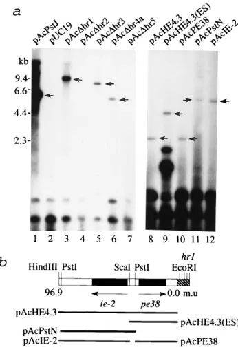
specifically essential to support DNA replication, we deleted thehrs from several plasmids. pAcDhr1 had a complete dele-tion of the hr1 region, plasmids pAcDhr2, pAcDhr3, and pAcDhr4a contained a single palindrome with a disrupted cen-tralEcoRI sequence, and pAcDhr5 contained only one half of a single palindrome. No replication of hr-deletion plasmids pAcDhr2 and pAcDhr5 was detected in standard infection-dependent transient replication assays, while the controlhr 2-containing plasmid pAcPstJ showed a strong replication signal (Fig. 1a). This result demonstrated that a single palindrome with a disrupted EcoRI core site (pAcDhr2) or half a palin-drome (pAcDhr5) disabled the ability of hrs to function as DNA replication origins, consistent with published data (20). However, deletions of the hr from pAcDhr1, pAcDhr3, and pAcDhr4a decreased but did not completely eliminate the abil-ity of these plasmids to replicate (Fig. 1a, lanes 3, 5, and 6), indicating that other non-hrsequences in these plasmids could function as DNA replication origins. To explore the nature of non-hr-stimulated replication, regions of the plasmid pAcDhr1 were subcloned and individual clones were tested in the stan-dard replication assay. Both the left and right regions flanking
[image:2.612.350.523.71.323.2]hr1 and contained withinHindIII-F fragment supported infec-tion-dependent DNA replication (data not shown). A detailed dissection of plasmid pAcHE4.3 (thehr1 left flanking region) identified two regions that correlated with non-hrreplication.
FIG. 1. Infection-dependent transient replication of plasmids containinghr
sequence deletions. (a) Sf21 cells (106) were transfected with 1mg ofhr-deletion
plasmid pAcDhr1 (lane 3), pAcDhr2 (lane 4), pAcDhr3 (lane 5), pAcDhr4a (lane 6), or pAcDhr5 (lane 7) or plasmids containing regions flankinghr1 (lanes 8 to 12). The same amounts of pAcPstJ (lane 1) and pUC19 (lane 2) were used in control transfections. Plasmid-transfected cells were infected with AcMNPV (MOI of 1) at 6 h posttransfection. Total cellular DNA was purified at 48 h postinfection and doubly digested withDpnI plusSmaI (lanes 1 to 5 and 7),KpnI (lane 6),HindIII (lanes 8 and 11), orPstI (lanes 9, 10, and 12) to linearize the replicated plasmid DNA (lanes 1 to 7, 9, 11, and 12) or to release viral inserts from vectors (lanes 8 and 10). After electrophoresis, DNA samples were trans-ferred to Qiagen nylon membranes and hybridized with32P-labeled pUC18
DNA. TheDpnI-resistant fragments are indicated by arrows. (b) Physical loca-tions of viral DNA inserts derived from the left flanking region ofhr1 and present in pAcHE4.3 (lane 8), pAcHE4.3(ES) (lane 9), pAcPE38 (lane 10), pAcPstN (lane 11), and pAcIE-2 (lane 12). m.u., map units.
on November 9, 2019 by guest
http://jvi.asm.org/
One contained the complete open reading frame of the early
pe38gene plus 94 bp of upstream sequence (Fig. 1a, lane 10), while the other contained the complete open reading frame for the earlyie-2gene plus 91 bp of upstream sequence (Fig. 1a, lane 12). During normal virus infection, both of these genes are expressed immediately after infection, but they are also regu-lated by another viral early gene, calledie-1(9, 28, 29). These experiments demonstrated that regions other than hrs can stimulate plasmid replication in virus-infected cells and sug-gested that the presence of early genes may be responsible for this property.
Initiation of DNA replication by theie-1 promoter region.
To investigate this possibility further, we analyzed the replica-tion ability of the promoter region of the ie-1 gene in the standard infection-dependent replication assay. Plasmid pIE1-P(CH) carrying the 558-bpClaI-HincII region of theie-1 pro-moter clearly replicated (Fig. 2a, lane 3), while placZ(ORF), lacking the ie-1promoter, did not (Fig. 2a, lane 2), demon-strating that DNA sequences found within theie-1promoter region could act as replication initiation sites. To identify func-tional motifs within this region, a series of plasmids containing deletions of theie-1upstream sequence was constructed (Fig. 2b) and tested in the replication assay. Replication of sub-clones individually containing only one of the five regions dem-onstrated that any one of these regions could support DNA replication. The signals from region V (Fig. 2a, lane 7) and region II (Fig. 2a, lane 14) were very weak but were visible in
longer exposures. In contrast, subclones P(CS) and pIE1-P(NH) replicated almost as efficiently as the whole promoter plasmid pIE1-P(CH), suggesting that replication efficiency in-creased with increasing insert size. Deletion of region I had little effect on initiation of DNA replication (Fig. 2a, lane 4) and therefore was not essential for replication initiation in the assays.
The ie-1 promoter region does not contain any sequence homology with previously described origins. It does contain one 24-bp imperfect palindrome within region IV, but this region could be deleted without abolishing replication (Fig. 2a, lanes 7 and 9 to 11). Three A1T-rich domains are located within regions I, II, and V (55 to 67% A1T over 70 to 119 nucleotides), but the replication of the lower-A1T-content region IV (46% A1T over 169 nucleotides), as well as the fact that one A1T-rich domain between regions I and II was dis-rupted, did not support a specific role for these A1T-rich domains in replication. Analysis of the replication negative plasmids pAcDhr2 and pAcDhr5 revealed that they also con-tain regions of about 140 nucleotides which are about 80% A1T. In addition, all of the control plasmids lacking viral inserts also contain regions with high A1T content (74% A1T over 100 nucleotides), but they were completely negative in our experiments. Therefore, high A1T content is not sufficient to impart replication ability. We conclude that retaining the viral promoter region intact is important in determining maximal replication ability. This ability did not appear to directly cor-relate with transcriptional activation but rather appeared to indicate the potential role that these regions play in binding transcription factors prior to early transcription. Theie-1 pro-moter region I, which contains the TATA box and CAGT initiator sequences, known to be essential for accurate initia-tion of transcripinitia-tion (2, 8, 29), was not essential for replicainitia-tion. In addition, although the upstream regions of the ie-1 pro-moter, including regions II to V in our study, may be nones-sential for transcription in vivo, they can enhanceie-1 expres-sion when transfected into cells (28), suggesting that other transcription factor binding sites likely exist within these re-gions. Evidence suggesting that these regions carry regulatory elements responsive to host cell factors has also been pub-lished (28). Therefore, our data are in agreement with other studies in which the presence of binding sites for a range of cellular transcription factors near the simian virus 40 origin of replication stimulated viral DNA replication but did not cor-relate with the ability to stimulate transcription (13).
Replication of plasmids containing AcMNPV early genes
and promoters.From the results presented above, we
postu-lated that viral early gene promoter regions could function as putative DNA replication origins. We therefore investigated a number of early AcMNPV genes and their promoter regions for the ability to stimulate plasmid replication. Surprisingly, almost all plasmids carrying viral DNA inserts expected to be expressed early after infection were capable of replication (Fig. 3). These plasmids carried a variety of genes (open reading frames plus their upstream regions), including theE. coli lacZ
gene (driven by the ie-1promoter), the apoptosis-repressing gene (p35), the early 39K gene, the immediate-early he65,
dnapol, p143, lef-1, lef-3, lef-4, p47, p43, and gta AcMNPV genes. The vector plasmids used in our cloning experiments without viral sequences (pBSK2, pBR322, and pUC19) were completelyDpnI sensitive under our assay conditions (Fig. 3, lanes 2 to 4). By calculating the replication efficiencies of these various plasmids, taking into account their various sizes, we determined that plasmids carrying sequences including lef-1,
[image:3.612.61.297.73.323.2]pe38,39K, andie-2showed low but detectable levels of repli-cation (less than 5% of the value for the reporter pAchr2).
FIG. 2. Theie-1promoter region functions as DNA replication origins. (a) Infection-dependent transient replication of plasmids carrying the AcMNPVie-1
gene promoter region and its subdomains. Sf21 cells were transfected with pBSK2(lane 1), placZ(ORF) (lane 2), pIE1-(CH) (lane 3), pIE1-P(CS) (lane 4), pIE1-P(CA) (lane 5), pIE1-P(CP) (lane 6), pIE1-P(CN) (lane 7), pIE1-P(NH) (lane 8), pIE1-P(PH) (lane 9), pIE1-P(AH) (lane 10), pIE1-P(SH) (lane 11), pIE1-P(NP) (lane 12), pIE1-P(PA) (lane 13), or pIE1-P(AS) (lane 14) and infected with AcMNPV as described for Fig. 1. Total cellular DNA was purified and doubly digested withDpnI plusSmaI (lanes 1 and 2) orXmnI (lanes 3 to 14). Blotting and hybridization conditions are outlined in the legend to Fig. 1. The
DpnI-resistant fragments are indicated by arrowheads. (b) Restriction map of the 558-bpClaI-HincII fragment containing theie-1promoter region. Locations of the five domains (I to V) tested in the replication assay are shown.
on November 9, 2019 by guest
http://jvi.asm.org/
Plasmids carrying the he65gene or the ie-1 promoter repli-cated at about 15 to 20% of the reporter level. Plasmids car-rying the lef-3, lef-4, p35, ordnapolgene replicated with effi-ciencies between 33 and 78% of that of the reporter, while plasmids carrying thep143gene or a region containing thep47,
p43, andgtagene promoter regions replicated as efficiently as or better than the reporter plasmid (112 to 370%). Plasmids pAcDhr2 and pAcDhr5 did not carry any early genes or pro-moters and did not replicate (Fig. 1). Given that pAcDhr5 carried the latep10promoter, these results clearly support our hypothesis that viral early genes and specifically their promoter regions can function in some cases as efficiently ashrs in sup-porting plasmid DNA replication initiation in virus-infected cells.
A correlation between transcription and DNA replication has been demonstrated in several other eukaryotic systems (for a review, see reference 12). The transcription activator NF1, which specifically prevents repression of simian virus 40 DNA replication by chromatin assembly, stimulates DNA replication 20-fold in vivo (4). The transcription factors VP16, GAL4, and p53 can bind to the large subunit of replication protein A, stimulating polyomavirus DNA replication through an influ-ence on a very early stage of the initiation process such as initiation complex assembly (11, 21). The fact that a singleie-1
promoter or its subdomains, as well as a number of other early promoter-containing regions of AcMNPV, when present in plasmids, can lead to DNA replication suggests that the pro-cesses of baculovirus early gene transcription and replication may be intimately connected. This concept is also supported by our finding that the deletion of hr2 or hr5 inactivated the plasmid’s ability to replicate; neither pAcDhr2 nor pAcDhr5 carried early promoters, although pAcDhr5 maintained an in-tact late p10 promoter. The binding of transcription factors prior to early transcription initiation may expose the DNA to the viral replication machinery, allowing for initiation of DNA replication. Alternatively, the interaction between transcrip-tion factors and the virus replicatranscrip-tion machinery may facilitate the assembly of replication factors necessary for DNA replica-tion. Finally, factors which recognize early viral promoters may inhibit or dislodge nucleosomes, allowing access to DNA do-mains, which then function as replication origins. The confor-mation of a plasmid can affect its transcriptional activity during
a transient expression assay, and supercoiling of plasmids is crucial for maximum transcription activity (26). Coincidentally, linearization of supercoiled plasmids containinghrs completely abolishes replication ability in the AcMNPV infection-depen-dent replication assay (17). Because prolonged storage of plas-mids or repeated extraction of plasmid DNA with phenol can greatly reduce the DNA’s replication efficiency (37), DNA conformation may be an important criterion for both transcrip-tional and replication activation.
Specificity of plasmid replication. One major paradox in
defining AcMNPV DNA replication origins is the nonspecific replication of plasmid DNA even in the absence of any inte-grated viral sequences upon cotransfection with AcMNPV DNA into Sf21 cells (10, 17, 37, 38). The replication efficiency for this vector DNA can be as high as that of anhr-containing plasmid and is not a result of recombination or acquisition of
hrs following cotransfection (37). In addition, when the essen-tial genes for baculovirus replication are cotransfected into insect cells, all plasmids including those not carryinghrs rep-licate (14, 18, 24), and this replication does not result from recombination between thehr-containing reporter plasmid and the plasmids expressing the replication genes (22a). These results suggest that the AcMNPV DNA replication system does not require hrs for initiation; instead, virtually any se-quences can be replicated when plasmids and viral replication-essential genes are introduced into the cells at the same time. However, if the plasmid DNA is introduced into cells prior to virus infection, the virus replication system is very sequence specific, replicating only plasmids containing certain viral se-quences.
As a working hypothesis to resolve this paradox, we propose a model (Fig. 4c) suggesting that the specificity of baculovirus replication initiation in the infection-dependent replication as-say is determined by features of chromatin structure. When a plasmid is transfected into a cell, it may assemble into a chro-matin structure with cellular proteins. Following virus infection of the cell, viral transcription factors and replication proteins are expressed. If the plasmid contains binding sites (such as viral promoter regions or hrs) recognized by the early viral proteins, binding of these factors could disrupt the chromatin structure and expose the DNA to replication proteins. If no viral factor binding sequences are present, the chromatin struc-ture could prevent replication proteins from interacting with the plasmid DNA, effectively repressing replication. Alterna-tively, the assembly of the replication proteins onto DNA may simply require binding factors to open the chromatin structure. In this case, the specificity of replication initiation would be based directly on the presence of recognizable binding se-quences. On the other hand, following cotransfection of puri-fied, naked plasmid DNA and viral DNA, the replication pro-teins may directly assemble onto the naked plasmid DNA (possibly at A1T-rich regions) prior to formation of the chro-matin structure, initiating nonspecific DNA replication.
We tested this theory by infecting cells at several time points early after transfection of vector plasmid into cells. Nonspecific replication of pUC18 DNA was detected following infection with AcMNPV at 2, 3, or 4 h after transfection. However, no replication of pUC18 DNA was detectable after 5 h posttrans-fection (Fig. 4b). Plasmid containing thehr2 region was rec-ognized as a template and replicated at all times after trans-fection (Fig. 4a). Although there is no direct evidence yet, we suspect that after 5 h, the transfected pUC18 DNA is struc-turally altered and is no longer recognizable by viral replication proteins. This hypothesis will be testable once in vitro replica-tion assays for baculovirus are available.
[image:4.612.61.295.71.184.2]We have now identified a number of regions of the genome
FIG. 3. Infection-dependent transient replication of plasmids carrying AcM
NPV genes and promoters. Sf21 cells were transfected with pAchr2 (lanes 1), pBSK2(lanes 2), pBR322 (lanes 3), pUC19 (lanes 4), pIE1-lacZ (lanes 5), pAc39K (lanes 6), pAcp35 (lanes 7), pAcHE65 (lanes 8), pAcDNApol (lanes 9), pAcp143 (lanes 10), pAclef1 (lanes 11), pAclef3 (lanes 12), pAclef4 (lanes 13), or pAcp47 (lanes 14) and infected with AcMNPV as described for Fig. 1. Total intracellular DNA was purified and digested withSmaI (lanes 1, 2, 4, 5, 6, and 9),
PstI (lanes 3, 7, 8, 10, 11, and 13), orHindIII (lanes 12 and 14) to linearize the total intracellular plasmid DNA (except that in pAcp143, which has twoPstI sites, only the 6.3-kb site hybridized). (2DpnI) or with the same restriction enzymes plusDpnI (1DpnI) to identify replicated plasmid DNA. Blotting and hybridization conditions were the same as for Fig. 1.
on November 9, 2019 by guest
http://jvi.asm.org/
in addition to hrs and ori-K which can function as putative DNA replication origins. However, we cannot exclude the pos-sibility that more DNA elements are involved in this process. The situation is reminiscent of the widely used yeast autono-mously replicating sequence (ARS) high-frequency transfor-mation assay, which successfully identified the first eukaryotic chromosomal origin of replication, ARS1, and later identified many other yeast ARS elements (25, 31, 32). However, it was subsequently discovered, by using a variety of approaches, that while all yeast chromosomal origins are ARS elements, not all
ARS elements function as origins in the yeast chromosome (7). Furthermore, not all yeast origins can initiate replication at the same time (7). We have now shown that in the infection-dependent replication assay, regions capable of binding host and viral transcription factors can function as origins of repli-cation. We suggest that these putative origins be called infec-tion-dependent replicating sequences. It remains to be deter-mined which of these sequences are used during the virus replication cycle as genuine origins of replication.
ACKNOWLEDGMENTS
We gratefully acknowledge the help of Ge Liu in establishing and fine-tuning the plasmid DNA replication assays. We thank Albert Lu and Ge Liu for the construction of plasmids pAcHE4.3, pAcHE4.5, pAchr2, pAcp143, and pAclef1; Lois Miller for pAcDNApol and pAclef3; and Paul Friesen for pIE1-lacZ.
This work was supported by grants from the Medical Research Council of Canada and the National Science and Engineering Re-search Council of Canada.
REFERENCES
1.Ayres, M. D., S. C. Howard, J. Kuzio, M. Lopez-Ferber, and R. D. Possee. 1994. The complete DNA sequence ofAutographa californicanuclear poly-hedrosis virus. Virology202:586–605.
2.Blissard, G. W., P. H. Kogan, R. Wei, and G. F. Rohrmann.1992. A synthetic early promoter from a baculovirus: roles of the TATA box and conserved start site CAGT sequence in basal levels of transcription. Virology190:783– 793.
3.Blissard, G. W., and G. F. Rohrmann.1990. Baculovirus diversity and mo-lecular biology. Annu. Rev. Entomol.35:127–155.
4.Cheng, L., and T. J. Kelly.1989. Transcriptional activator nuclear factor I stimulates the replication of SV40 minichromosomes in vivo and in vitro. Cell59:541–551.
5.Cochran, M. A., and P. Faulkner.1983. Location of homologous DNA sequences interspersed at five regions of the baculovirus AcMNPV genome. J. Virol.45:961–970.
6.Erlandson, M. A., P. Skepasts, J. Kuzio, and E. B. Carstens.1984. Genomic variants of a temperature-sensitive mutant ofAutographa californicanuclear polyhedrosis virus containing specific reiterations of viral DNA. Virus Res. 1:565–584.
7.Fangman, W. L., and B. J. Brewer.1991. Activation of replication origins within yeast chromosomes. Annu. Rev. Cell Biol.7:375–402.
8.Guarino, L. A., and M. Smith.1992. Regulation of delayed-early gene tran-scription by dual TATA boxes. J. Virol.66:3733–3739.
9.Guarino, L. A., and M. D. Summers.1987. Nucleotide sequence and tem-poral expression of a baculovirus regulatory gene. J. Virol.61:2091–2099. 10. Guarino, L. A., and M. D. Summers.1988. Functional mapping of
Autogra-pha californicanuclear polyhedrosis virus genes required for late gene ex-pression. J. Virol.62:463–471.
11. He, Z., B. T. Brinton, J. Greenblatt, J. A. Hassell, and C. J. Ingles.1993. The transactivator proteins vp16 and gal4 bind replication factor A. Cell73:1223– 1232.
12. Heintz, N. H.1992. Transcription factors and the control of DNA replica-tion. Curr. Opin. Cell Biol.4:459–467.
13. Hoang, A. T., W. Wang, and J. D. Gralla.1992. The replication activation potential of selected RNA polymerase II promoter elements at the simian virus 40 origin. Mol. Cell. Biol.12:3087–3093.
14. Kool, M., C. H. Ahrens, R. W. Goldbach, G. F. Rohrmann, and J. M. Vlak. 1994. Identification of genes involved in DNA replication of theAutographa californicabaculovirus. Proc. Natl. Acad. Sci. USA91:11212–11216. 15. Kool, M., C. H. Ahrens, J. M. Vlak, and G. F. Rohrmann.1995. Replication
of baculovirus DNA. J. Gen. Virol.76:2103–2118.
16. Kool, M., R. W. Goldbach, and J. M. Vlak.1994. A putative non-hrorigin of DNA replication in theHindIII-K fragment ofAutographa californica mul-tiple nucleocapsid nuclear polyhedrosis virus. J. Gen. Virol.75:3345–3352. 17. Kool, M., J. T. M. Voeten, R. W. Goldbach, J. Tramper, and J. M. Vlak.1993.
Identification of seven putative origins ofAutographa californicamultiple nucleocapsid nuclear polyhedrosis virus DNA replication. J. Gen. Virol. 74:2661–2668.
18. Kool, M., J. T. M. Voeten, R. W. Goldbach, and J. M. Vlak.1994. Functional mapping of regions of theAutographa californicanuclear polyhedrosis viral genome required for DNA replication. Virology198:680–689.
19. Lee, H. Y., and P. J. Krell.1992. Generation and analysis of defective genomes ofAutographa californicanuclear polyhedrosis virus. J. Virol.66: 4339–4347.
20. Leisy, D. J., C. Rasmussen, H.-T. Kim, and G. F. Rohrmann.1995. The
Autographa californicanuclear polyhedrosis virus homologous region 1a: FIG. 4. Replication potential of plasmid DNA changes with time after
trans-fection. Sf21 cells were cotransfected for 2 h with 1mg of AcMNPV DNA plus 2mg of pAchr2 (a, lane 1) or 1mg of AcMNPV DNA plus 2mg of pUC18 (b, lane 1). For infection-dependent transient replication assays, 2mg of pAchr2 (a, lanes 2 to 6) or 2mg of pUC18 (b, lanes 2 to 6) was incubated for 2 h with cells, and then monolayers were washed immediately and infected with virus (MOI of 1) at 2, 3, 4, 5, and 6 h (lanes 2 to 6, respectively) after transfection (the time of addition of DNA to cells was time zero). Total cellular DNAs were purified at 72 h after cotransfection or 48 h after virus infection and digested withDpnI plus
SmaI. Blotting and hybridization conditions were as for Fig. 1. (c) We propose that plasmid DNA, when transfected into cells, slowly forms a chromatin struc-ture which affects the way the plasmid is recognized as a template for virus-induced replication. In this model, plasmid containing a viral DNA insert rec-ognized by transcription factors would bind these factors, opening up the chromatin structure and serving as a platform for the assembly of the replisome and initiation of replication (i). Otherwise, a complete chromatin structure could preclude the assembly of the replisome (ii). If introduction of the plasmid DNA into cells is followed shortly by virus infection, there would not be enough time to form a complete chromatin structure, and so the virus replisome could as-semble on any naked region of plasmids and initiate replication (iii). This con-clusion is supported by data in panel b, lanes 2 to 4, showing that pUC18 (without any viral insert) replicated when virus infection occurred within 4 h after trans-fection. If plasmid and viral DNA were cotransfected into cells, the immediately synthesized viral replisome could assemble on any naked plasmid DNA and initiate replication (iv). These conclusions are supported by data in panels a and b, lanes 1.
on November 9, 2019 by guest
http://jvi.asm.org/
identical sequences are essential for DNA replication activity and transcrip-tional enhancer function. Virology208:742–752.
21. Li, R., and M. R. Botchan.1993. The acidic transcriptional activation do-mains of VP16 and p53 bind the cellular replication protein A and stimulate in vitro BPV-1 DNA replication. Cell73:1207–1221.
22. Li, Y., A. L. Passarelli, and L. K. Miller.1993. Identification, sequence, and transcriptional mapping oflef-3, a baculovirus gene involved in late and very late gene expression. J. Virol.67:5260–5268.
22a.Liu, G., and E. B. Carstens.Unpublished data.
23. Lu, A., and E. B. Carstens.1993. Immediate-early baculovirus genes trans-activate the p143 gene promoter ofAutographa californicanuclear polyhe-drosis virus. Virology195:710–718.
24. Lu, A., and L. K. Miller.1995. The roles of eighteen baculovirus late ex-pression factor genes in transcription and DNA replication. J. Virol.69:975– 982.
25. Newlon, C. S.1988. Yeast chromosome replication and segregation. Micro-biol. Rev.52:568–601.
26. Parvin, J. D., and P. A. Sharp.1993. DNA topology and a minimal set of basal factors for transcription by RNA polymerase II. Cell73:533–540. 27. Pearson, M., R. Bjornson, G. Pearson, and G. Rohrmann.1992. The
Auto-grapha californicabaculovirus genome: evidence for multiple replication origins. Science257:1382–1384.
28. Pullen, S. S., and P. D. Friesen.1995. Early transcription of theie-1 trans-regulator gene ofAutographa californicanuclear polyhedrosis virus is regu-lated by DNA sequences within its 59noncoding leader region. J. Virol. 69:156–165.
29. Pullen, S. S., and P. D. Friesen.1995. The CAGT motif functions as an initiator element during early transcription of the baculovirus transregulator
ie-1. J. Virol.69:3575–3583.
30. Rodems, S. M., and P. D. Friesen.1993. Thehr5 transcriptional enhancer stimulates early expression from theAutographa californicanuclear polyhe-drosis virus genome but is not required for virus replication. J. Virol.67: 5776–5785.
31. Stinchcomb, D. T., K. Struhl, and R. W. Davis.1979. Isolation and charac-terisation of a yeast chromosomal replicator. Nature (London)282:39–43. 32. Struhl, K., D. T. Stinchcomb, S. Scherer, and R. W. Davis.1979.
High-frequency transformation of yeast: autonomous replication of hybrid DNA molecules. Proc. Natl. Acad. Sci. USA76:1035–1039.
33. Summers, M. D., and D. L. Anderson.1973. Characterization of nuclear polyhedrosis virus DNAs. J. Virol.12:1336–1346.
34. Thiem, S. M., and L. K. Miller.1989. A baculovirus gene with a novel transcription pattern encodes a polypeptide with a zinc finger and a leucine zipper. J. Virol.63:4489–4497.
35. Tjia, S. T., E. B. Carstens, and W. Doerfler.1979. Infection ofSpodoptera frugiperdacells withAutographa californicanuclear polyhedrosis virus. II. The viral DNA and the kinetics of its replication. Virology99:391–409. 36. Tomalski, M. D., J. Wu, and L. K. Miller.1988. The location, sequence,
transcription, and regulation of a baculovirus DNA polymerase gene. Virol-ogy167:591–600.
37. Wu, Y., and E. B. Carstens.Unpublished data.
38. Yu, H.1990. Gene expression and replication of AcMNPV in cultured insect cells. Queen’s University, Kingston, Ontario, Canada.