A STUDY ON
MANDHARA KASAM
(DISSERTATION SUBJECT)For the partial fulfillment of the requirements
to the degree of
DOCTOR OF MEDICINE (SIDDHA)
Branch I MARUTHUVAM – POTHU
GOVERNMENT SIDDHA MEDICAL COLLEGE
PALAYAMKOTTAI -627002
The Tamil Nadu Dr.M.G.R. Medical University, Chennai-32
ACKNOWLEDGEMENT
First of all I thank to my God to finish my dissertation work successfully.
I thank the Vice Chancellor of the Tamil Nadu Dr.M.G.R.Medical University, for giving the opportunity in completing the post graduate dissertation at Government Siddha Medical College at Palayamkottai.
I thank the Commissioner of Indian Medicine and Homeopathy, Chennai and Joint Director of Indian Medicine and Homeopathy, Chennai, for giving permission to undertake the dissertation.
I thank our Principal Dr.M.Dhinakaran M.D(s), for granting me an opportunity to do my dissertation work at the Government Siddha Medical College and Hospital.
I wish honourable and greatful thanks to Dr.K.R.Revathi M.D(s) Vice Principal and Head of the Department of Post Graduate Pothu Maruthuvam Department, Government Siddha Medical College, Palayamkottai for her greatful guidance, interest and hardwork in completing my dissertation work.
I would like to thank Dr.S.Chitra M.D(s) Asst. Lecturer of Post Graduate Department of Pothu Maruthuvam, for her valuable guidance in finishing my dissertation work.
I express my sincere thanks to Prof. Dr.M.R.Vairamuthu Raja, M.D Department of Modern Medicine for his guidance in Modern Aspects.
I wish to thank Dr.J.Angelin Nirmala M.D(s) of under graduate, Maruthuvam Department, for her helpful guidance doing my dissertation work.
for their Co-operation in evaluation of the Pharmacological actions of the trial medicine.
I represent my sincere thanks to Professor N.Nagaprema M.Sc.,M.Phil., Head of the Department of Bio- Chemistry for her kind help to analyse the biochemical aspects of the trial medicine and also my thanks to the assistants in the Bio chemistry department for their co-operation in Biochemical analysis of the trial medicine.
I convey my thanks to Dr.V.S.Padma M.B.B.S., DMRD., Radiologist Government Siddha Medical College, Palayamkottai for her whole supports in radiological investigations done to the patients.
It is my duty to thank Mrs. Poonkodi M.A., Librarian of Government Siddha Medical College, Palayamkottai . For her help in referring necessary books in the library.
I must thank to Dr.R.Nepolean M.B.B.S, M.D., Consultant Microbiologist, Malar Micro Diagnostics centre, Palayamkottai to evaluate anti bacterial activity of the trial Medicine.
I should thank to my Department students for their advice , help and co-operation in finishing this work.
My cardial thanks to my parents , uncle, aunty and my family members.
My special and heartful thanks to my dearest husband Mr.N.Selvakumaran for his Overall help in completing this dissertation work.
INTRODUCTION
Nature and human beings are the wonderful creation of God. Pray and thank the god for creating nature, lands , water , air , resources rain etc for living beings for their better survival . It is the ultimate duty of the human beings to protect the nature and live in hormony with nature
Siddha system of medicine is originated from Lord siva, the supreme God and he is also considered to be chief of siddhar’s and chief of sangam poets
‘ nrhy;yplNt Njtpf;F rjhrptd;whd; nrhy;yNt NjtpAk; ee;jpf;Fr; nrhy;y ey;yplNt ee;jpjd; te;jphpf;Fr; nrhy;y
eaKld; jd;te;jphp aRtdpf;Fr; nrhy;y my;yplNt aRtdpahj; Njth; jhKk;
mfj;jpah; Fiuj;jplNt ak;KdPe;jud; Gy;yplNt Gyj;jpah;f;; FgNjrpf;f
Gyj;jpaUk; Njiuaw;Fg; Gfd;wpl;lhNu
-A+fp itj;jpa rpe;jhkzp 800
The siddha system of medicine was developed by the siddhars. Siddhars are not only physicians but also social reformers.
Siddhar’s knowledge in the field of Medicine, Natural science, and literature are extra ordinary one.
The word siddhar is derived from the term “ Siddhi” , means perfection or Achievement .
ghug;gh G+jike;J kz; ePh;thA ghpthA thfha ike;jpdhNy> Nrug;gh rlkhr;R kz;zpd; $W
nrwpkapu; Njhy; vd;gpiwr;rp euk;ige;jhFk; Neug;gh mg;Gtpd; $W jpukr;ir
ePh; %is Rf;fy Nkhile;jhFk; fhug;gh NjAf;$W gakhq;fhuq;
fLQ;Nrhk;gy; epj;jpiu ikJdq;fsQ;Nr
- rjfehb
Both the external environment and human body is composed of five basic elements called pancha bootha which includes land, air, water, fire and ether. They constitute in definite proportion according to type , land and seasons. Any aberration in the ratio of pancha bootham in nature reflects as natural calamites such as flood, famine, cyclone, and eruption of volcanoes.
“mz;lj;jpYs;sNj gpz;lk; gpz;lj;jpYs;sNj mz;lk; mz;lKk; gpz;lKk; xd;W
mwpe;Jjhd; ghh;f;Fk; NghJ”
- rl;lKdp epfz;L
The human body is composed of five base elements called land air, water, fire, and ether. Which maintain the integrity of nature humours called vadha, pitha and kapha in fixed ratio 1:1/2:1/4.
‘kpfpDk; FiwapDk; Neha; nra;Ak; E}Nyhh; tspKjyh vz;zpa %d;W”
Any deviation in this ratio affects the homeostasis of human physiology and leads to pathological condition called pini (or) Noi
The pini or Noi must be cured by “ Marunthu” Marunthu means which cure physical, mental illness which possess preventive aspects from diseases and also to postpone death
“ Neha; ehb Neha; Kjy; ehb mJ jzpf;Fk; tha; ehb tha;g;gr; nray; “
- jpUf;Fws;
According to Thiruvalluvar the disease must be identified and also then cause for the disease.
The diagnosis is based upon three dhosha theory. To diagnose the disease envagai thervu, or piniyari muraimai is carried out, which is highlighted our Valluvar as
“ kUe;njd Ntz;lhthk; ahf;iff;F mUe;jpaJ mw;wJ Nghw;wp Az;zpd;”
- jpUf;Fws;.
The treatment is based on principles of Arusuvai, mukkutram and pancha bootha principles. Further paruva kaalam, Astrology, genetic factors are also taken in to account in relation to disease.
Some facts about pathiyam and Anubanam are also considered.
Anubanam means, it is adjuvant to medicine. It acts as catalyser and enhance rate of absorption of medicine.
Anubanam is different according to type of disease, type of medicine according to season in which treatment is going on,
%d;wpnyhd;W ah;e;jij Kd;duwpe;J Ke;jpajid nahopj;jpL kUe;jpL jzpAk; Nehapd; je;jukpJNt
Ngzpf; fzpj;jpbd; gpwtpg; gpd;Fzk;
Deranged mukkutram (vadha, pitha, kaba) should be controlled first by kalichal ( purgation) vamanam (vomiting) and then only the medicine for the disease is to be prescribed.
“cw;wtd; jPh;g;ghd; kUe;Jior; nry;thndd; xg;ghdhw;; $w;Nw kUe;J”
- jpUf;Fws;
AIM AND OBJECTIVES
Aim
Millions of people all over the world are affected by Bronchial Asthma because of pollution in the environment, change in life style and diet.
Mandhara kasam is similar to bronchial asthma . According to siddha system of Medicine “ Mandhara Kasam “ is a controllable one.
Siddhar’s have enumerated lot of medicine for the disease.
The daily increasing number of asthma patients and the efficiency of siddha system of medicine, curing chronic respiratory disease prompted the author to carry out scientific clinical study on the subject.
OBJECTIVE
The prime object of this study is to do a clinical trial on Mandhara kasam affected individuals with selected siddha medicine.
1. Veliparuthi choornam 1gm tds with honey after meals – sarabendarar vaidhya muraigal kasa swasa sikitchai.
2. Thirikadathy kasayam 30 ml bd - Akasthiyar 2000. To made a detailed study of definition, aetiology, clinical feature, diagnosis investigation, treatment and dietics fo Mandhara Kasam in various siddha literatures.
3. To know the extend correlation of Aetiology, signs and symptoms and complications of Mandhara Kasam in siddha aspect is compared with Bronchial Asthma in Modern aspect.
5. To study how the disease Mandhara Kasam alters the normal condition under the headings Mukkutram, pori pulangal, udal kattugal, neerkuri, neikuri, and envagai thervugal especially in naadi nadai. 6. To make a detailed clinical evaluation of the disease by a careful
examination on aetiology, signs, and symptoms, complications treatment and prognosis during the course of disease.
7. To utilize the possible modern diagnostics to confirm the diagnosis of the disease.
8. The prime object of the present study is to explore most efficacious medicine for Mandhara Kasam.
ABSTRACT
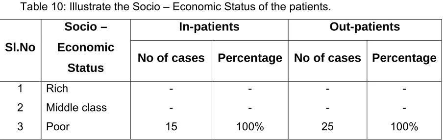
Since the number of sufferers increasing day by day, the author has chosen
the disease “Mandhara Kasam” for her dissertation work. The increasing incidence
of the disease is due to changes in life styles and environment.
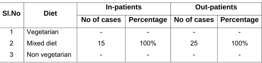
Fifteen patients of either sex were selected as In-Patients and twenty
Out-Patients were administered with the trial medicine “ Veliparuthi Choornam” 1gm
three times daily after meals and “ Thirikadathy Kasayam” 30ml twice daily after
meals during the whole study period.
The trial medicine was subjected to Biochemical and Pharmacological as well
as Microbiological analysis.
REVIEW OF LITERATURE
SIDDHA ASPECTS
The biological function of the body is governed by three distinct humours knowns as Vadha, Pitha, Kaba. In a healthy man these three humours are held in the ratio of 1:1/2:1/4 when this equilibrium is altered it leads to disease. When kaba is altered by diet, environment, factors, habits etc., the other two are also altered leading to kaba diseases.
A basic energy which is responsible for a man to be alive is known as Thathu. This one energy is divided into three factors Vadha, Pitha, and Kaba. This one life force in three ways creates, protects and fates in the body.
The human body composed of 72,000 nerves. Among this the ten are big nerves (Thasa Naadi’s)
rpwe;j,il gpq;fiyQ; RopKidap NdhL rpwg;ghd fhe;jhhp aj;jPr; rpq;Fitaha gpwe;j myk; GUlndhL FFjd;whDk;
Nguhd rq;FdpAk; tapu td;whd; jpwe;j tpit gj;Je;jhd; wr ehbahFk;.
- A+fp itj;jpa rpe;jhkzp
Yugimuni says that above ten nerves are “Thasa Naadi’s”.
‘rhUe; jrehb jd;dpy; %yk; %d;W NgUkplk; gpq;fiyAk; gpd;dYld; khWk; ciuf;ftpuw; fhw;nwhl;L zh;j;J Nkehrp tiur; Rop Nahikaj;jpy; te;J
te;j fiy %d;wpy; tha;thkghdDld; je;j gpuhzd; rkhdDk; re;jKwf;
$l;Lwthy; Nurpj;jy; $Wk; thjk; gpj;jk; ehl;Lq; fgNk ahk; ehL
According to this three naadies Edakalai, PinKalai, and Suzhumunai are basic naadies and they are called Moolathara Naadi’s.
Mukkutram Relation with Elements (Pancha Bootham):- Vadha = Vali + Ahayam
Pitham = Neruppu Kabam = Mann + Neer
Among five elements kaba has the qualities of mann and neer. This is explained as follows,
‘Nrj;Jke; jz;zPh; gpj;je; jPfhw;W thjkhNk” - mfj;jpah; ehb
Mukkutram Relation with Tastes and Elements:- Sweet = Earth + Water
Sour = Earth + Fire Salt = Water + Fire Bitter = Air + Sky Pungent = Air + Fire Astringent = Earth + Air
Vadha = Air + Sky Pitha = Fire
Kaba = Water + Earth
When vadha, pitha, kaba are in the ratio or 1:1/2:1/4 in the body it indicates that the man is physiologically normal in health according to gunavagadam.
‘ toq;fpa thjk;khj;jpiu nahd;whfpy; joq;fpa gpj;je; jd;dpyiu thrp
moFq;fge; jhdlq;fpa fhNyhby;
‘thj gpj;j ika %d;Wk; td; gyj;JlNd jj;jk; Ngj nkhd;wpy;yh tz;zk; Ngrpa jhde;jd;dpy; ePjpaha; epiyj;J epw;fpy; neLk;gpzp rpf;ftpy;iy jhJTnkhd;Nwhnlhd;W jhtpby; gpzpfs; jhNd”
- Neha; ehly; >Neha; Kjdhly; jpul;L
So the alteration of kaba thathu altered the functions of the Ezhu Udar Kattukal and other thathus indicated the disease “Mandhara Kasam”.
Mandhara kasam one of the type of kasa noi. The definition aetiology, pathology, clinical features based upon three dhosas, envagai thervugal, prognosis, treatment and preventive method are dealt here.
MANDHARA KASAM
I. VERU PEYARGAL ( Synonyms): KULIR IRUMAL
MANDHARA SWASAM
II. EYAL (Definition):
III. NOI VARUM VAZHI (Aetiology):
YUGI VAIDHYA CHINTHAMANI Says
‘Ntfpd;w tjpfkhk; GifapdhYk;
kPWfpd;w ghzj;jhy; kpFf;Fe; jhNd” -690 ‘ghzj;jhy; gukhf;fpdp kpFf;if ahYk;
ghukh kprq;fs; Grpf;if ahYk; jhzj;jhw; rQ;rhue; jtph;f;if ahYk;
rhpglh gjhh;j;jq;fs; Grpj;j yhYk; jPzj;jhw; nghrpahk ypUf;if ahYk;
Nrapioahh; Nkypd;gQ; rpijt jhYk; khzj;jhy; khJf;f kilj yhYk;
kUe;;jhYk; RthrkJ kUTq; fhNz”
‘fhzNt Njtijf;Fg; gphpj;j gz;lk; fsthb jpd;whYq; fztd; wd;id NjhzNt epe;jul;ir nrhd;d jhYk;
Rrpahd gjhh;j;jnkr;rpy; gz;z dhYk; NtzNt xUth; nra;j ed;wp jd;id
kpfkwe;J nfhLikflhd; tpsk;G Nthh;f;Fk; NgzNt rigjdpNy nrhd;d Ngr;R
gpuz;Nlhh;f;Fq; fhrk; te;J gpwf;Fe;; jhNd” - 692
Inhalation of excessive smoke.
Excessive heat.
Intake of improper diet.
Intake of different non vegetarian diet.
Too much of sorrow.
Worries, immoral habits such as talking lies.
Spoiling other’s food.
Cursing life partner.
PARA RASA SEKARAM says
‘NkT rpukjdpy; tp\ ePuhNy
tpz;zspil ke;jhu Kw;wNghJ thrKWq; Foyhu naz;nza; Nja;j;j tOTWrP jj;jhYk; gdpapdhYk; XirAW fhw;whYk; . Nth;it ahYk;
Kz;lhfp kz;il fdg;GlNd ahFk; fhrKW ehyOq; fgNk nghq;Fk;
fUJ Fz ke;jhu fhrkhKk;” --- 149
According to pararasa sekaram,
Taking oil bath during the cloudy season, excessive chillness, wind, sweating etc., leads, headache leading to the onset of the disease.
SIDDHA MARUTHUVAM (POTHU)
The disease is due to improper diet and which decreases the vitality and which increase kaba during reduced vital power of the body, husks of paddy, grass, millet, Inhalation of irritant fragrance.
SIDDHAR KAI EZHUTHTHU PIRADHI Says
‘fhy; ngUf;FzT jz;zPh; khwy ;
fUjpUky; kpfy; the;jp Fsph;e;j fhw;W khy; nra;J ehs;NjhWk; tUj;Jk; fha;r;ry;
ke;jd Kapy; epiyapy; mbfs; jhf;fy; Vy rPjNgjp tplghz;L Giffs;
,sfpa ney;yhjp kzpr; RidAl; nry;yy; Nky;topapy; rpythpDkpdhg; ghk; NehA
NkTnkd Kdpth;fs; tpsk;gpdhNu”
IV.MURKURIGAL (Preliminary signs): SIDDHA MARUTHUVAM (POTHU) Says.
Soreness of throat
Redness of throat
Pricking pain in the throat
Reduced voice
Running nose
Tightness of chest
Desire to eat hot food
THERAIYAR VAGADAM Says
‘te;jpLk; nts;Nshf;fhsk; thaJ jpj;jpg;ghFk; nehe;jpLk; gplhp kz;il ke;jK kpisg;gpNdhq;Fk; Ke;jNt jiyjh ndhe;J rhPu KfKq; Fj;Jk; Re;ju njhz;il ehrp fufud;WlNd Jk;ky;”
Belching( Regurgitation)
Feeling of sweet taste in tongue
Loss of appetite
Occipital pain
Headache, pain all over the body
Pain over the face
Soreness of throat
Irritation of nose
Sneeze
V. NOI ENN (Classification):
Mandhara kasam is described as one of the twelve types of Kasam in Yugi Vaidhya Chinthamani.
YUGI VAIDHYA CHINTHAMANI The twelve types are
3. Sudar Kasam 4. Vadha Kasam 5. Pitha Kasam 6. Swasa Kasam 7. Ratha Kasam 8. Silethma Kasam 9. Peenisa Kasam 10. Vadha Pitha Kasam 11. Pitha Setpa Kasam 12. Dondha Kasam
ROGA NIRNAYA SARAM
There are five types of Swasa Rogam. They are 1. Oorthuva Swasam
2. Arppa Swasam 3. Vicchina Swasam 4. Maha Swasam 5. Mandhara Swasam
DHANVANTRI VAIDHYAM
Mandhara kasam is classified under Dhonda Kasam. Dhonda Kasam is of five types. They are
1. Mandhara Kasam (Áó¾¡Ã ¸¡ºõ)
2. Vega Kasam (§Å¸ ¸¡ºõ)
3. Pakka Mandhara Kasam (Àì¸ Áó¾¡Ã ¸¡ºõ)
4. Sura Kasam (Íà ¸¡ºõ)
5. Vadung Kasam (Å¡Îí ¸¡ºõ)
MANDHARA KASAM: VI. KURI GUNANGAL:
‘jhdhd J}aNjhh; ehrp jd;dpy;
ryNeha; ePh; jhd; tpOe;j Jk;k Yz;lhk; khdhd khh;GneQ; rilj;J %r;R
tYthd ghk;GNghy; rPw yhFk; fhdhd fz;lNkhL KfKq; fhJk;
fhakJq; frpthfp tpah;it ahFk; Vdhd ,UkNyhL Nfhio fk;ky;
,iu g;ghF ke;jhu fhr khNk”
According to Yugi Vaidhya Chinthamani, the characteristic features of Mandhara Kasam are running nose, sneeze, tightness of chest, breath sound like hissing of snake, sweating all over the body, cough, expectoration, dyspnoea.
AGASTHIYAR -2000
‘ke;jhu fhrNk te;jhy; thq;fpLk; Rthrk; Nkyh apj;jhu nka;r;Ruk; fhZNk ,isj;jpL kpUky; nkj;j re;jhAlk;G jiyAlk;G jsutypf;F kpdsg;ghFk; ge;jhAlk;G neQ;rKfk; gj;jp typf;Fk; gz;gpNj”
The characteristic features of the disease are dyspnoea, fever, frequent cough, emaciation, pain in chest, face. This book also explains about Kaba Mandhara kasam as follows
“Ja;aNjhh; ehrp jd;dpy; Jk;kY kpf Tz;lhfp neha;A ePuha; tpOe;J NehT gl dPio thq;F ma;apd; ke;jhufhrj; jltpJ jhNd ePNfs;
nra;Akh Kdpth; nrhd;d FzkpJ njhpe;J nfhs;Ns”
Running nose, sneeze, tightness of chest, dyspnoea and cough with expectoration.
VAIDHYA CHARA SANGRAM
UYIR KAKUM SIDDHA MARUTHUVAM @ AATMA RAKSHAMIRTHAM
Áó¾¡Ã ¸¡ºò¾¢ý þÂøÒ:
KfKk; fhJk; CWk; ehrpfufuj;J Jk;ky; cz;lhFk;, ePh;tbAk; neQ;rpw;fgk; fl;b ,UKk;,isf;Fk;, neQ;R tpyhTk; typf;Fk;, ke;jhu fhyq;fspy; Neha; mjpfg;gLk;,
grpke;jk; Vw;gLk;, tapW nghUKk;> cly; mijf;Fk; fpWfpWf;Fk;.
ROGA NIRNAYA SARAM
The characteristic features of the disease are Vadha in combination with Kaba affect the nerves and causes ratting sound in throat unbearable difficulty in breathing, increased breathing and increased sputum.
VII. MUKKUTRA VERUPADUGAL (Pathology):
In Siddha system, the manifestation of all the diseases are the result of derangement of Doshas i.e., Vadha, Pitha, Kaba. The prime factor which is involved in Mandhara Kasam is Kaba, which is accompanied with vititated Vadha or Pitaha and produces the clinical symptoms of Mandhara Kasam. This is clearly indicated by Theriyar as
‘fgj;jpid ad;wp fhrk; Rthrk; fhzhJ - Njiuah;”
1. Excess of Kaba in the respiratory organs affect the Melnokkukal and Uyirkal and so the Vayu is not able to reach the terminal point of respiration leads to labored breathing.
2. Some authors say that the disease is caused by deranged Vadha. This may also be acceptable because the obstruction of vayu in the respiratory tract is abnormal.
“gpj;jNk kpFe;jh yPis apUkYk; ngyj;J epw;Fk;”
- Neha; ehly; Neha; Kjy; ehly;
So the changes in the diet and habits which increases Vadha and Kaba produce the clinical symptoms of Mandhara Kasam.
In Uyir Nilaigal, Anagatham (chest) which is the residence of Udhanan (Melnokkukal) and Pranan ( Uyirkal ) is deranged.
When Pranan, the primary Vayu is affected it leads to difficulty in breathing and involvement of Udhanan leads to cough and sneezing. Involvement of Kirugaran leads to running nose, cough, sneezing. Involvement of Devathathan leads of tiredness. Involvement of Samanan cannot control other vayus and causes loss of appetite. Involvement of Sadhagapitha leads to sluggishness. In Kaba , the derangement of Avalambagam leads to dyspnoea, cough , wheezing. In the seven udal Thathus, Saaram and Senneer are affected which leads to lethargy and depression. In severe cases Oon and Kozhuppu are also affected leads to symptoms of emaciation and body pain.
VIII. PINIYARI MURAIMAI (Diagnosis):
Diagnosis is the very important thing for physician by which, he deals the disease by finding its cause and is helpful to undertake a correct line of treatment and also prognosis. The diagnosis is based on
1. Poriyal Arithal 2. Pulalnal Arithal 3. Vinathal
4. En Vagai Thervugal
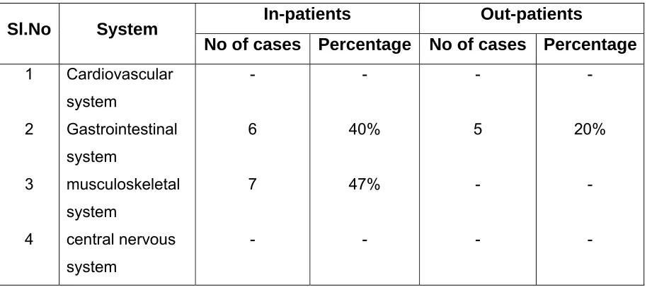
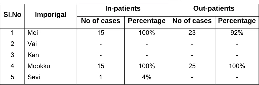
Poriyal are the five organs of perception. They are nose, tongue, eyes, skin and ears. Poriyal Arithal is examining the pori of the patient by the pori of the physician. In mandhara kasam, it is as follows
Mei(skin) : Sweating all over the body. Vai (tongue) : Dry, pale and sometimes coated. Kan (eyes) : Redness, sometimes dusky and pale.
Mookku (nose) : Visible movement of alar nasi, irritation of nose, running nose.
Sevi (ear) : Normal.
2. PULANAL ARITHAL:
Pulangal are the five objectes of senses.
Ooru (sensation) : Normal or cold due to sweating. Osai (sound) : Normal.
Ozhi (vision) : Normal.
Suvai (taste) : Diminished or normal.
Natram (smell) : Altered or absent due to running nose and inflammation of the nasal mucosa.
3. VINADHAL:
The period of human life is totally 100 years. This is divided into three stages, according to the domination of three humours as,
1. Vadha Kaalam – 1 to 33 years. 2. Pitha Kaalam – 34 to 66 years. 3. Kaba Kaalam – 67 to 100 years.
Even though in each of this stage, the other humours are also involved, but a particular humour is dominating more. According to this data, the disease Mandhara Kasam come under the type of kaba disease and so more patients are affected in the latter stage (Kaba Kaalam).
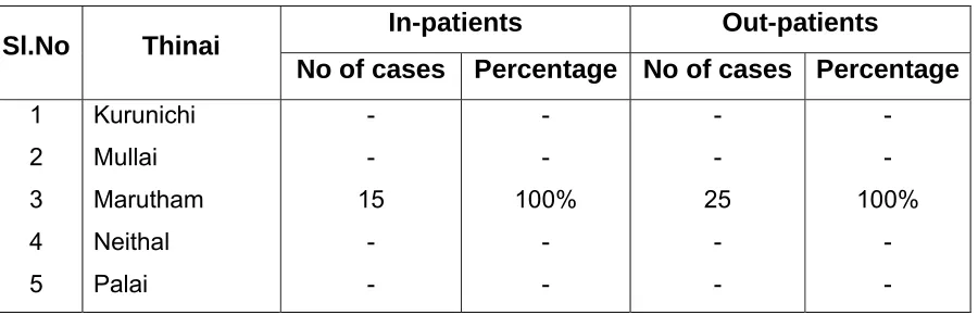
5. IVAGAI NILANGAL:
Study of Ivagai Nilangal is very important and useful because there may be possibility of the disease in some areas (e.g., Kurinchi, Mullai, Maruthauam, Neithal, Palai). Ivagai Nilangal are
• Kurinchi – Mountains and its surroundings,
• Mullai – Forests and its surroundings
• Maruthuam – Plains and its surroundings.
• Neithal – Seas and its surroundings.
• Palai – Deserts and its surroundings. A.Kurunchi
FUQ;rp tUepyj;jpw;F nfhww;Kz;b uj;jk; cwpQ;rp tU RuKz;lhk; - mwpQUiuf; ifaNk jq;Fjuj; jhikty;iy Aq;fjpf;Fk; IaNk jq;Fk; mwp
gjhh;j;j Fz rpe;jhkzp
Persons who are living in Kurunchi Nilam are usually liable for developing Kaba diseases.
‘Ky;iy epyj;jaNk hpepiu NktpDkt;
nty;iy epiyj;jgpj;j nka;FWq;fhz; - ty;iynatdpd; thjnkhop ahjj Dz;kd;W kit topNeha;g;
Ngj nkhop ahjiwag; gpd;G”
gjhh;j;j Fz rpe;jhkzp.
Though Mullai Nilam is the place of cattles, it is the place of increasing pitha, vadha also joined to that Pitha due to these Kutrams many diseases occur. It is difficult to distinguish between them.
C. Marutham:
‘kUjepy; ed;dPh; tsnkhd;iwf; nfhz;Nl nghUjdpy khjpaNeha; Nghf;Fk; - fUjepyj; jhwpujQ;R+o mUe;Jtnud; whw;gpzpnay; NywpujQ; R+o;Gtpf;F kpy;.”
gjhh;j;j Fz rpe;jhkzp.
D.Neithal:
‘nea;jdpy NkYg;ig ePq;fh J}wpDkJ nta;jdpy Nkjq;F tPlhFk; - nea;jy;
kUq; Fliy kpf;fhf;Fk; ty;YWg;ig tPf;Fk; fUq;Fliyf; fPopwf;Fq; fhz;”
gjhh;j;j Fz rpe;jhkzp.
Though Neithal Nilam has the dominatnt taste of uvarppu (salty), it is the place of Pitha Vayu. The people who dwell here are susceptible to oedema due to Kaba, Silipatha Rogam ( Filariasis), Kudalanda Viruthi ( Hernia).
‘ghiy epyk; Nghw; gliug; gpwg;gpf;f Nkiyepy kpahJ tphpj;jw;F - Ntiy epy Kg;gzpf;F kpy;yhk; KiwNa atw;wfyhk; vg;gpzpf;F kpy;yh k/njz;.”
gjhh;j;j Fz rpe;jhkzp.
Persons who live in palai are liable to develop the disease of three dhoshas (So Mandhara Kasam is found in these nilam).
6. PARUVA KAALAM (Season):
"¸¡§Ã ܾ¢÷ ÓýÀÉ¢ À¢ýÀÉ¢ º£Ã¢Ä §ÅÉ¢ø §ÅÉ¢ø ±ýÈ¡íÌ
þÕãýÚ ¾¢Èó¾Ð ¦¾Ã¢¦ÀÕõ ¦À¡Ø§¾"
- º¢ò¾ ÁÕòÐÅ¡í¸î ÍÕì¸õ
With reference to the position of the sun, year is divided into 6 seasons. They are
1. Karkaalam ( Avani and Purattasi) 2. Koothirkaalam ( iyppasi and Karthigai 3. munpanikaalam (Margazhi and Thai) 4. Pinpanikaalam (Masi and Panguni) 5. Elavenilkaalam (Chittirai and Vaigasi) 6. Mudhuvenilkaalam (Aani and Aadi)
According to literature, Mandhara kasam comes during rainy season (karkaalam). In koothirkaalam, due to kulir katru (cold wind), is also responsible for the disease.
Mandhara kasam mainly occurs due to vitiation of Kaba. Kaba thannilai sirappurum Kaalam – karthigai to masi.
jj;jk; epiyapy; jd;durpaYk; fhytiujid fpsuf; Nfz;kpd; Mbahjpaha; Ig;grp <uha; Mdpykjw;Nfh uurpay; fhyk;
kPd; Kjyhdp tPWnfhs; ke;jphp Njd; Kjd; khrp Nrdhgjpf;Nf
Njs; - fhh;j;j;pif
Neha; ehly; Neha; Kjy; ehly;
Hence the disease can occur in the later part of Koothirkalam to early part Pinpanikalam, i.e., from the last two weeks of October to the first two weeks of February.
Totally the disease’s prevalence is from August to February.
7. MUKKUTRA NILAIGAL: VADHA:
‘Kiwikahk; gpuhz Ndhlghdk; tpahdd;
%h;f;fkh %jhdNdhL rkhd dhfd; jpiwikahq; $h;kNdhL fpUf wd;wd;
Njtjj;j ndhL jdQ; raDkhFk;”
- a+fp rpe;jhkzp 800y; jj;Jttpjp 35k; nra;As;
PRANAN:
It is responsible for respiration. In Mandhara Kasam, Vayu is affected leading to difficulty of breathing.
ABANAN:
VIYANAN:
It’s main function is distribution of Saaram. In Mandhara Kasam this distribution is affected.
UDANAN
It is present in the chest umbilicus and nose . In mandhara kasam sneezing may be present due to the derangement of this vayu.
SAMANAN:
Samanan is the vayu that controls other vayus and digestion . In Mandhara kasam this vayu is affected since it cannot control the other vayus.
NAGAN:
This Vayu maintains opening and closure of eye lids and is not affected in Mandhara kasam.
KOORMAN:
This vayu is responsible for vision and yawning and is affected in some patients of Mandhara kasam.
KIRUGARAN:
This vayu is responsible for salivation, running nose, sneeze, cough and maintains appetite. In Mandhara Kasam this Vayu is deranged causing running nose, sneeze, cough and loss of appetite.
DEVATHATHAN:
It is responsible for tiredness, anger and emotional expression. In Mandhara Kasam, this vayu is deranged causing emotional stress.
DHANANJEYAN
PITHA
‘Mf;fdy; tz;znthp ahw;wyq;fp nahs;nshspj;jP Nehf;foyhk; gpj;j ike;j E}jdkh ahf;Fnkhop ghrkp uQ;rfQ; rhjfk; uhrfkh
Nyhrf nkd;wpLkh Nyh;
- kUj;Jt jdpg;ghly;
According to the Maruthuva Thani padal pitha is divided into five types .
ANAL PITHA :
This lives in the stomach and helps in digestion. In Mandhara Kasam, loss of appetite is present.
RANJAGA PITHA:
It is situated in the Stomach and increases the blood level, it is responsible for the colour of the blood.
SATHAGA PITHA:
It resides in the heart and makes correct activity with the help of mind and brain. In this disease restlessness is present
AALOSAGA PITHA
It resides in both eyes and is responsible for correct vision.
PIRASAGA PITHA :
It resides in skin and gives complexion.
KABA:
‘Mju thk; nka;f;f ftyk; gj khq;fp Nyjf khQ;Ritg; Ngj%zh;g; Nghjfkhk; jw;gfkhQ; re;jpfspw; wq;FQ; rpNylfkh kw;gkpyhr; Nrj;Jk ike;J
- kUj;Jt jdpg;ghly;
AVALAMBAGAM:
It is residing in lungs and helps other four types of Kaba to function, and also helps in the function of heart. It is deranged since the presence of tightness of chest, cough, wheezing dyspnoea.
KILETHAGAM:
It is present in the stomach and gives moisture to the food materials and also helps in digestion. In this disease, some patients have loss of appetite.
POTHAGAM:
Living in the tongue and responsible for taste sensation.
THARPAGAM
Living in the head and provides cooling to the eyes.
SANTHIGAM:
It resides in the joint and helps for free movement. In Mandhara Kasam some patients are affected by arthritis
8. EZHU UDAR KATTUKAL:
‘jd;dkhk urkpuj;jkh q;fprK Nkij jir kr;irnahL Rf;ye;jh Njohfp”
- A+fp itj;jpa rpe;jhkzp 800
They are the seven basic principles which constitute the entire body . These are otherwise called us udal Thathukkal . They are
SAARAM:
SENNEER:
It is responsible for knowledge, strength, boldness and healthy complexion. This is deranged here.
OON:
It gives structure to the body and is responsible for the movement of the body.
KOZHUPPU:
When the organs are doing their work, this Thathu gives lubrication and facilitates their work.
ENBU:
It gives the shape to the body and is responsible for protection of the vital organs.
MOOLAI :
It is present in the core of the bone which strengthens and maintains the normal condition of the bone.
SUKKILAM / SURONIHAM:
It is responsible for reproduction. When the seven Udal kattukal increase or decrease from the normal level, the normal functioning of the body is affected.
EN VAGAI THERVUGAL:
It is the basic diagnostic principle and the uniqueness of the Siddha system of medicine . The following verses reveals this as follows.
‘ehb ghprk; eh epwk; nkhop tpop kyk; ;%j;jpu kpit kUj;JtuhAjk; “
- Neha; ehly; Neha; Kjy; ehly; jpul;L Kjy; ghfk;
and presumes the vitiated Doshas in the patient
En Vagai Thervugal are
Naa
Niram
Mozhi
Vizhi
Malam
Moothiram
Sparisam
Naadi
A.NAA:
It is noted for it colour, ulcer, growth , coating, colour and consistency of the sputum that is spitted from mouth, mode of speech . In mandhara kasam patients have scanty and mucoid sputum.
B.NIRAM
Colour of the skin , in Mandhara Kasam the colour of the skin is altered.
C.MOZHI:
Mode of speech – anxiety, depressed voice. This includes the sound from lungs due to respiration due to kaba and dyspnoea. In Mandhara Kasam, mode of speech may be emotional, low pitched voice. Wheezing sound is heard.
D.VIZHI:
Type of eye – redness, ulcer, pallor, protrusion, tears, sheddding of eyelashes , excreata of eye, In mandhara kasam, the eyes are red.
E.MALAM:
abdominal pain during defecation are noted. In mandhara Kasam, the patients are having constipation
F. NEER @ MOOTHIRAM:
Colour – yellow, red, black, white, copper coloured, mixed colour, colour of fumes. Smell – Smell of fire, honey, sweet odours, fragrance of flower, fruity odour, odour of deer, flesh. Frothy or not , frequency and Quantity are noted. In mandhara kasam it is transparent and frothy.
G.SPARISAM:
Heat or coldness of the body - it may be cold due to sweating in this disease.
H. NAADI :
‘ehb vd;why; ehbay;y euk;gpy; jhNd eykhfj; Jbf;fpd;w JbjhDky;y
ehb vd;why; thj gpj;jrpNyw;gdKky;y ehb vOgj;jPuhapue;jhDky;y
ehb vd;why; mz;l nuz;lnky;yhk; ehb vOtifj; Njhw;wj;Js;sha; epd;w ehbaJ ahuha;e;J ghh;j;jhuhdhy; ehbAWk; nghUs; njhpe;J ehLthNu”
- rjf ehb
Naadi is the very important helpful observation for diagnosis and prognosis and it indicates the states of uyir thathukkal whether they are normal or abnormal .
The importance of naadi is clearly mentioned by saint Thiruvalluvar by the following verse.
‘Neha;ehb Neha; KjdhbaJ jzpf;Fk; tha;ehb thag;gr; nray;”
In Noi Nadal, noi mudhal Nadal text, naadi is defined as
‘clypy; caph; jhpj;jpUg;gjw;Ff; fhuzkhd rPtrf;jp vJNth mJNt jhJ my;yJ ehb vdg;gLk;”
GENESIS OF NAADI
The three Uyir Thathukkal are formed by the combination of three Naadis with three vayus.
Idakali + Abanan = Vatha Pinghalai + Pranan = Pitha Suzhumunai + Samanan = Kaba
‘ rhUe;jr ehbjd;dpy; %yk; %d;W
NrUkplk; gpq;fiyAk; gpd;dYld; khW ciuf;ftpuw; fhw;nwhl;Lzh;j;JNk ehrp
tiu RopNah ikaj;jpy; te;J te;j fiy %d;wpy; thAthk ghdDld;
je;j gpuhzd; rkhdDf;FQ; re;jkwf; $l;LwT Nufpj;jy; cWk; thjk; gpj;jk;
ehl;Lq;fgNk ahk; ehL.
- fz;Zrhkpak;
This can be felt one inch below the wrist on the radial artery by means of palpation by the three fingers – index, middle and ring fingers corresponding to vadha, pitha and kaba respectively
‘fhpKfdbia tho;j;jpf; ifjdpy; ehb ghh;f;fpy; ngUtpuyq;Fyj;j;py; gpbj;jb eLNt njhl;lhy; xU tpuNyhby; thjKah; eLtpuw; gpj;jk; jpUtpuy; %d;wpNyhby; Nrj;Jk ehb jhNd.”
‘toq;fpa thjk; khj;jpiu nahd;whfpy; joq;fpa gpj;je; jd;dpyiu thrp
moFq;fge; jhdlq;fpa fhNyhby;
gpwq;fpa rPth;f;Fg; gpr nfhd;W kpy;iyNa
- Fzthflk;
NAADI NADAI IN MANDHARA KASAM
When the Naadi rhythm varies from normal to aggravating kaba it causes Mandhara kasam
‘fgky;yhJ fhr Rthrk; tuhJ”
-Njiuah;
‘IaNk fjpj;j NghjwpNt nghUky; fhZk; <isA ke;jhufhrk; esph;Fsph; tpf;fy; rj;jp nra;Akh %r;rilg;ghd; jPjW fhrNuhfk;
njha;Akh kpisg;G fhrk; Njhd;W nkhd;wud; nrhd;dhNu” - gjpnzd; rpj;jh; ehb
‘cw;wpLk; Ia ehb Xq;fpNa Jbhj;J epd;why; gw;wplk; kpUkyPis gjwpNa ,isg;Gz;lhFk; nkj;jNt Nfhio thA kpFe;jpLk;”
- mfj;jpah; Fzthflk;
Kaba naadi:
‘jhdKs;s Nrj;ke; jhdpfsfpy; ntg;G rakPis kpUky; ke;jhufhrk;
<dKWQ; re;jp tplNjhlk; tpf;fy;
,Uj;Nuhfq; fug;ghd; tpuz Njhlk; khdidaPh; R+iy jpus; tpahjp tPf;fk;
tUQ;; rj;jp Rthrk; nrQ;rilg;G J}f;fk; VdKWq; fhkhiy ghz;L Nrhig
VO Ruq;fs; gygpzpAq; fhZe;jhNd”
Vatha kabha Naadi :
When the Naadi rhythm varies from normal to vatha kabam, it causes Mandhara kasam
‘ ghq;fhd thjj;jpy; Nrj;Jkehbg;
ghprpj;jhy; jpkph;NkT Kisr;ryhFk; jPq;fhd ,UkYld; re;jpNjhlk;
Nrh;e;j tplk; ntbR+iy ,Uj;Nuhfk; thq;fhj <is ke;jhufhrk;
typAlNd GwtPr;R As;tPf;fk; xq;fhd RuKlNd Rthrfhrk;
cz;lhFk; ntF Neha;f;F KWjpjhNd”
- rjf ehb
Iya ushnam :
When iyam and ushnam are combined then leads to Mandhara Kasam
‘fjpg;ghd Nrj;jkj;jp Yl;bzq; $by; fye;j Fsph; rakpUky; Rthrfhrk; kjpg;ghd Nfhio uj;jk; tpg;GUjpAlNd tsh;ehrpfh gPlkpUj;Nuhfq;
nfhjpg;ghd rpq;Fit ahf;fpuhz thA nfhl;lhtp tpf;fy; ke;jhufhrk;
Jjpg;ghd tPuyj;jpf; fha;Tuj;jk;
Njhd;Wkpf gpzp gyTe; njhe;jpg;ghNk”
- rj ehb
NEER KURI:
“te;jePh; fhpail kzk;Eiu vQ;rnyd;
iwe;jpa Xsit aiwFJ KiwNah”
- Neha; ehly; Neha; Kjy; ehly; jpul;L Kjy; ghfk;
According to this verse, the general, features of urine, Niram, edai, manam, Nurai, and enjal are analyzed.
Niram indicates the colour of the urine voided Edai indicates the specific gravity of the urine Manam indicates the smell of the urine voided Nurai indicates the frothy nature of urine voided
Enjal indicates the quantity ( Increased or Decreased)
NEI KURI:
The patient whose urine is to be tested, is asked to take regular and quality diet without any derangement in amount and quality in correct time. The urine is collected the next day in the early morning in a glass vessel. The same type of urine is collected for Neerkuri.
A drop of gingelly oil is dropped on a wide vessel containing the urine to be tested and kept it in the sun light in calm place. The derangement of the three thathus and the disease can be diagnosed by the behavior of gingelly oil on the surface of the urine.
‘mue;JkhwpujKk; mtpNuhjkjha;
m/fy; myh;jy; mfhyt+d; jtph;e;jow; Fw;ws tUe;jp cwq;fp itfiw
Mbf;fyrj; jhtpNa fhJ nga; njhU K$h;j;jf; fiyf; Fl;gL ePhpd;
epwf;Fwp nea;f;Fwp epUkpj;jy; flNd”
- rpj;j kUj;Jthq;f RUf;fk;
muntd ePz;bbd; m/Nj thjk; MopNghw; gutpd; m/Nj gpj;jk;
Kj;njhj;J epw;fpd; nkhoptnjd; fgnk
- Neha; ehly; Neha; Kjy; ehly;
♦ Oil spreading like a snake indicates vadha
♦ Oil spreading like a ring indicates pitha.
♦ Oil floating like a pearl indicates kaba
In mandhara kasam oil is floating like the pearl in the urine
LINE OF TREATMENT
The line of treatment of mandhara kasam consists of the following: 1. Kalichal maruthuvam - to bring the doshas in equillibrium. 2. Internal Medicine – Mainly anti-spasmodic, expectorant and
antihistaminic to relieve the spasm and expel the sputum 3. Diet – To give suitable diet to reduce the kaba
4. Yoga Therapy - to maintain Dhasa vayukal and to improve mental and physical health
5. Preventive methods – By practice of pranayamam
1. KALICHAL MARUTHUVAM (PURGATION ):
2. ADMINISTRATION OF INTERNAL MEDICINE:
For the treatment of the disease mandhara kasam, several remedies are suggested in ancient siddha literature. Among these remedies the author selected as follows
Veliparuthi choornam 1gm three times a day with honey after meals.
Thirikadathy kasayam 30ml two times a day after meals
DIET:
Siddhars advice the diet regimen for kaba patients and they are explained below :
Greens are to be added :
Ntis kzj;jf;fhsp nkdrPij rf;futh;j;jp gPis triy Rf;F ngz;Rzq;fd; - Ntisapiy nre;jsph; fisf;fPiu nra;gth; fgNjfh epjk; te;jspAzj;jhd; kfpo;e;J .
- gjhh;j;j Fz rpe;jhkzp
‘fhiuapU Nfhit Kd;id nrk;igg; gNlhy; Japyp tof;if neUQ;rpy;
MiuGsp ahiuKy;iy kUjnea;jy; Nkdp ey;ty; yhiu nghd;dh
thiu KRf; ifKUq;if apUgpz;zhf; NfhL gz;iz kzyp gps;isf; fPiu KRl;il Aq;fhuh kzp khlq;
fliy Gspf; fphpf; fshNt”
Greens:
rpW fPiu (Amaranthus gangeticus),
J}J tis (Solanum trilobatum),
kzj;jf;fhsp ( Solanum nigrum ) ,
KRKRf;if ( Mukia madraspatana) ,
Fg;igNkdp ( Acalypha indica),
gwl;ilf;fPiu ( Justicia madurensis).
nghd;dhq;fhzp( Alternanthera sessilis)
KUq;if fPiu (Moringa Olefera)
miuf;fPiu (Ameranthus tristis)
Vegetables to be added
mtiu (Dolicheus lab – lab )
fj;jhp (Solanum melangena),
fz;lq;fj;jphp (Solanum xanthocarpum),
mj;jp (Ficus glomavata),
<Us;sp (Allium cepa) ,
KUq;if (Moringa olefera),
thiof;fha; (Musa paradisiaca), Rz;ilf;fha; (Solanum tarvum ) ,
khtL ( mangifera indica).
Flowers and stem
thiog;G+ ( Musa paradisiaca) ,
fUizj;jz;L ( Amarphophallus poeniifolius).
Tubers:
Ks;sq;fp ( Rhaphnus sativus),
ntq;fhak; ( Allium cepa),
$iff; fpoq;F ( Maranta arundinaceae) ,
Diet Restriction :
Siddhars advised to avoid certain food items during diseased conditions. They are
Ghee except goat’s ghee,
buttermilk,
watery vegetables,
watery fruits,
cool drinks
Ice creams
Chilies
Sweets
PREVENTIVE MEASURES
The following advices are given to the asthmatic patients. Asthmatic patients are advised to,
In take of Hotwater and Hot foods,
To Avoid chill weather
To Avoid factors which causes digestive disturbances
To Avoid Allergic factors
To Avoid smoking
Taking bath strictly in Hot water
Advised to take dinner before 8 pm
Avoidance of stress
Avoid working in dust, cement, cotton mills and in husks
Advised to practice Pranayamam and yogasanam
Advised to sleep in the phoenix mat
PRANA YAMAM ( Breathing Excercise):
‘ VWjy; G+ufk; <nul;L thkj;jhy; MWjy; Fk;gfk; mWgj;J ehyjpy; CWjy; Kg;gj; jpuz;ljpy; Nurfk; khWjy; xd;wpd; fz; tQ;rfkhNk”
- jpUke;jpuk;
According to Thirumandhiram ,Pranayamam or breathing excercise mainly consists of inhalation of air by pooragam (deep inspiration) , kumbagam (holding the breath as far as possible and Resagam ( exhalation of air by expiration). The ration is 1:4:2
By this excercise, the duration of Kumbagam is increased. So that results in proper gaseous exchange, which produces increased oxygen supply to the cells particularly to alveoli.
By the regular practice of pranayamam, one can get a feeling of calmness of mind as a result of excess supply of oxygen to the brain cell . This state of mind ultimately helps in good concentration and medication. This practice also gives good appetite, strength, enthusiasm, vigour and vitality.
During breathing excercise, the lungs expand well and get proper supply of oxygen by proper expansion of chest. So pranayama practice is one of the
preventive methods for Asthma. This is expressed in the following poem,
“ehnshd;Wf;F ,Ugj;Njhuhapj;J mWE}W eykhd Rthre; jhnd Oe;jpUf;Fk; Nfh nshd;wpg; gjpdhyhapuj;J ehD}W
Ftpe;j %yhjuj;Js;nshLq;Fk; ghnshd;wp Naohapuj;JUE}W Rthrk;
ghopdpw; gha;e;jpL nkd;wwpfg; gpd;id Vnshd;wpapjidna Al;rhjpj;jhy;
vg;nghOjk; ghyuhapUf;fyhNk”
YOGA THERAPY
yoga is one of the most spiritual legacies gifted by the ancient sages of India. The practice of asanas strengthens the body and mind and the practitioner can realize it . The following asanas are helpful in Asthma . Puyankasanam, machasanam, mayurasanam, thirikonasanam savasanam, dhanurasanam, arth machasanam.
Asanas strengthen the muscles of respiration and diaphragm as well as regulate respiration . So the practice of asanas is helpful in the ashtmatic patients as supportive therapies.
Neha; fzpg;G tpthjk;
Swasa Kasam ( Ezhuppu Erumal)
tz;ikaha;f; Nfhiofl;b ,Ukp tPOk; khehfk; NghyNt thq;FQ; Rthrk; jpz;ikahr; nrUkYz;lh kbf; fbf;Fr; rPuz kpyhNy tapW %Jk;
ez;ikaha; ehrpaj jzy; NghyhFk; eype;Jlk;G tw;wptUq; FuYq; fk;Kk; cz;ikha Az;zhf; fpY}Wq; Nfzp Aoe;JNk Rthr fhrj;jp ndhg;Ng
- A+fp itj;jpa rpe;jhkzp
In Swasa Kasam, there is cough with expectoration, breathing sound like hissing of snake, hoarseness of voice, indigestion, flatulence, rhinitis, emaciation, brashing etc.
Kandakiragam:
tifahd Fwyjidg; gw;wp nehe;J khh;NghL gplhpapdpy; typAz;lhfp Efuhd rhPunky;yhk; nehe;j ohw;wp
EZf;fkha;r; RthrkJ Gwg; glhky; Kifahd ehtNy %r;R khwp
Kfj;jpNy tpah;thfp tpyhNeh Tz;lhk; gifahd td;dj;ij gUnfhl; lhJ
ghpafz;l fpufj;jpd; gz;G jhNd
In Kandagiragam , there is diffficulty in speech, pain in chest and occipital region, pain all over the body, breathlessness, sweating in face, pain in ribs, loss of appetite etc.
In mandhara kasam, there is no pain in the occipital region.
Silethuma Vadha Suronitham:
gz;ghd Tly;Fshph;e;j tapW tPq;fp
gijg;ghd tple; njhl;lhw;Nghu Nehthe; jpz;ghd rpuRnew;wp nehf; fhLz;lhk;
rpNyl;L kkha;f; NfhioNahL RthrkhFk; kz;ghd kaf;f nkhl fdT Kz;lhk;
tha; twz;l Urpapy;yh tUj;j khFk; ez;ghd ehbANk glglf;Fk;
ew;rp Nyl;k RNuhzpjkhk; ehLq; fhNy
In Silethuma Vadha Suronitham, there is chillness of body, distention of abdomen, pain while touching the abdomen, headache, expectoration, dyspnoea, fainting , dream, decreased salivation, loss of taste, abnormal pulse etc.
Iya Eraippu Noi:
‘jpwikaha; neQ;Rjdpw; Nfhio fl;Lk; rpf;nfhd;W jhdpUkp %f;filf;Fk; FWikaha;f; Fwl;nld;W Rthrq; fhZk;
FspNuhL RuKz;lha; kaf;f khFk; kwikaha; khh;Nghl neQ;r ilf;Fk;
tha; twz;l %f;fjdpy; ePNuh ghAk; ntWikaha; kpfj;jz;zPh; jhg Kz;lha;
tpL Rthr rpNyl;Lkj;jpd; tpgue; jhNd.”
- rpj;j kUj;Jtk;
In Iya Eraippu Noi, there is congestion in lungs , nasal block, dyspnoea, fever with rigor, syncope , tightness of chest, dryness of mouth, rhinitis, excessive thirst etc.
MODERN ASPECTS BRONCHIAL ASTHMA
Respiratory system
Respiration is defined as the exchange of gases between body tisue and environment.
Anatomy of The Respiratory System:
The respiratory system is divided into two parts
1. Upper respiratory tract 2. Lower respiratory tract
The dividing line being the lower border of the cricoid cartilage. It is at the level of the lower border of 6th cervical vertebra. The respiratory system is formed by the following organs.
1. Nose 2. Pharynx 3. Larynx 4. Trachea 5. Bronchi
6. Bronchial Tree 7. Lungs
8. Pleura
The upper respiratory tract
The upper respiratory tract made up of nose. Nasopharynx, Pharynx and larynx. It is lined vascular membrane covered ciliated columnar epithelium.
1.NOSE
cavity . This part is lined by olfactory mucosa. The rest of the nasal cavity is lined by respiratory mucosa.
The nose is divided into two main parts,
a) External Nose b) Nasal Cavity
a) External Nose:
The external nose has a skeletal frame work that is partly bony and partly cartilagenous. The bones are nasal bones which form the bridge of the nose and the Frontal Process of the maxillae. The cartilages are the superior and inferior nasal cartilages, the septal cartilages and some cartilages.
The prominent ridge separations the right and left halves of the nose is called the dorsum. The upper narrow end of the nose (Just below the forhead) is the root of the nose . The lower end of the dorsum is in the form a somewhat rounded tip . At the lower end of the nose we see the right and left nostrils. The nostrils are separated by a soft median partition called the columnella. This is continues with the nasal septum. Each nostril is bounded laterally by the ala.
b. Nasal Cavity:
The Nasal cavity is the entrance of the respiratory system . It is divided into right and left cavities by the nasal septum.
The root of the nasal cavity is formed by the cribriform plate of ethmoid bone.
The floor of the nasal cavity is formed by the cribriform plate of ethmoid bone.
The floor of the nasal cavity is formed by the palate.
Posterior nasal opening is called choana. It opens into the naso pharynx. The nasal sinuses communicate with nasal cavities by narrow opening and are frequently involved in nasal and nasopharyngeal infection.
Pharynx
Pharynx is a common chamber for the respiratory and digestive system. It located between the mouth and oesophagus. It is about 14 – 15 cm long.
It is situated anterior to the upper 6 cervical vertebrae It is situated posterior to the nose, oral cavity and larynx.
The pharynx has 3 parts 1. naso pharynx 2. Ora pharynx 3. laryngo pharynx
During swallowing respiration is temporarily inhibited and also the elevation of the larynx and closure of vocal cords which prevents the entry of food into larynx.
Larynx
The larynx is called voice box. it is a common chamber for respiration and sound production. It is situated in the anterior part of the neck, in front of the pharynx. It lies between the 3rd cervical vertebra to the 6th cervical vertebra.
It is formed by cartilages and muscles. The cartilages of the larynx are classified into paired and unpaired cartilages.
Paired cartilages are,
Unpaired cartilages are ,
a) Thyroid cartilage – Largest cartilage of Larynx b) Cricoid cartilage
c) epiglottis
The lower respiratory tract
The lower respiratory tract is made up of trachea, bronchi, broncheal tree, lungs and pleura.
Trachea
The trachea is otherwise called “wind pipe” It is a tubular passage extending down from the larynx. It’s length is about 10 -11cm The trachea has 16 – 20 rings formed by hyaline cartilage.
It commences at the level of the 6th cervical vertibra and terminale at the
lower border of the 4th throacic vertebra. It terminates by dividing into right and left bronchi. The trachea is lined by pseudo stratified, coloumnar ciliated epithelium, containing many glob let cells.
Bronchi
The trachea divides into
a) Right Bronchus b) Left Bronchus
a) Right Bronchus
It is about 2.5cm long
c) It is shorter, wider and vertical than left bronchus
b) Left Bronchus
It is about 5 cm long
It is longer than right bronchus It enter the hilum of the left lung It lives below the arch of aorta It divides into two lobar bronchi
BRONCHO PULMONARY SEGMENTS: The right lung has 10 segments The left lung has 8 segments
i) Right Lung Upper lobe:
1. Apical segmental bronchi 2. Anterior Segmental bronchi 3. Posterior segmental bronchi
Middle Lobe:
1. Medial Segmental bronchi 2. Lateral segmental bronchi
Lower Lobe:
1. Apical segmental bronchi
2. Anterior basal segmental bronchi 3. Posterior basal segmental bronchi 4. Medial basal segmental bronchi 5. Lateral basal segmental bronchi
Left Lung:
Upper Lobe:
Lower Lobe:
1. Apical segmental bronchi
2. Anterior basal segmental bronchi 3. Posterior basal segmental bronchi 4. Lateral basal segmental bronchi
Bronchioles
The bronchi further divide into bronchioles. The bronchioles are smaller air passages. The bronchiolar wall has no cartilage. The terminal Bronchiole open into the respiratory bronchioles.
ALVEOLI (Pulmonary Unit)
The respiratory bronchioles divide into alveolar ducts. Alveolar ducts open into air sacs called alveoli. The alveoli width is about 0.3mm
The basic unit of the lung tissue is alveoli . Alveoli are lines by flat epithelial cells. There is a network of capillaries around the alveoli. This arrangement helps in the gaseous exchange
Lungs:
The lungs are essential organs of respiration. There are right and left lungs. The lung situated within the pleural cavity found within the thorax. Before birth the lungs are solid organs and it sinks in water. After birth due to respiration it become porous and spongy and floats in water. In the young the lungs are brown or grey in colour . Gradually they become mottled black because of the deposit of inhaled carbon particles. The weight of the lung is about 650gms in adults.
The base is concave and semilunar in shape and is closely associated with the thoracic surface of the diaphram.
The costal surface is convex and in closely associated with the costal cartilages, the ribs and the intercostal muscles.
The medial surface is concave and has roughly triangular shaped area, called the hilum at the level of 5”6”7” thoracic vertebrae. Structures that enter and leave at the hilum are 1 bronchus,1 pulmonary artery 2 pulmonary veins 1, bronchial artery 1 bronchial vein., lymph vessels parasympathetic and sympathetic nerves. The area between the lungs is the mediastinum. It is occupied by heart, great vessels, trachea, right and left bronchi oesophagus, lymphnodes, lymph vessels and nerves.
Each lung is surrounded by a double layered membrane called pleura. Not all parts of the plural cavity is occupied by the lung. In costo diapharagmatic recess there is no lung.
The right lung has three lobes
a) Upper lobe b) Middle lobe c) Lower lobe
The left lung has two lobes.
a) upper lobe b) Lower lobe
The left lung is divided by the only oblique fissure which extends from the junction of the fourth or fifth rib with the vertebra column behind to the sixth costo chondral junction in front, crossing the mid axilliary line at the level of the fifth rib. The anterior border of the left lung shows the cardiac notch.
extends from the middle of the oblique fissure in the mid auxiliary line to the fourth costal cartilage.
The medial surface of each lung shows the hilum. Through the hilum structures enter and leave the lung. The structures passing through the hilum of the lung are bronchus, pulmonary artery and pulmonary vein.
7. PLEURA
The pleura is a closed serous sac . It is the second largest serous membrane of the body. It has two layers namely,
a) parietal layer (outer) b) Visceral layer (Inner)
Between the two layers the pleural cavity is situated. Thus cavity contains the firm of pleural fluid. The pleural cavity shows enlarged spaces called recesses.
PHYSIOLOGY
Respiration is the process by which oxygen is taken in and carbon di oxide is given out .
The first breath takes place only after birth. Fetal lungs are non functional. So during intra uterine life the exchange of gases between fetal blood occurs through placenta.
After the first breath the respiration is a continous process throughout life. The permanent stoppage of respiration occurs only at death.
Functions of the Lung
Supply of oxygen to the tissue and elimination of carbon-di-oxide from the tissues.
Regulates acid base balance
It helps in the maintenance of heart rate and cardiac out put
It helps in excretion of volatile, substance likes ammonia, water vapour and ketone bodies.
Mast cells present in the lung secrete substance like histamine, serotonin. 5 hydroxy tryptamine, etc., against allergy.
MECHANISM OF RESPIRATION
Respiration is the exchange of gas between the body and environment, Respiration has two phases
1. Inspiration 2. Expiration
Normal respiratory rate : 16 – 20 per minutes.
Inspiration
The following changes occur during inspiration Chest expands
The diaphragm move downwards so vertical diameter of the thorax is increased
Inercostal muscles act on the ribs so the ribs are turned out and elevated . This causes enlargement of the chest cavity
The enlargement of the chest permits the enlargement of lungs
When the lung is enlarged, pressure inside the lung is reduced. This leads to entry of more air into the lungs.
Normally the movements of the chest is equal on both sides. Inspiration is active and expiration is passive. Inspiration is shorter than expiration.
During inspiration there is downward movement of diaphragm and upward and outward movement of chest wall. In childrens the respiratory rate is greater than adult. In old age the rate is decreased.
The following changes occur during expiration
The diaphragm is relaxed and moves upwards.
Intercostals muscles are relaxed, so the ribs move inwards
As a result the capacity of thoracic cavity is reduced the intra pulmonary pressure is increased, and the air is gradually expelled.
The alternate inflation and deflations of the lungs are due to
corresponding changes in the capacity of the thoracic cage brought about by the actions of respiratory muscles.
MUSCLES OF INSPIRATION Diaphragm
External inter costal muscles
Serratus anterior and scalene muscle
Errectus muscles of the spine
MUSCLES OF EXPIRATION Abdominalis
Internal intercostal muscles
Posterior inferior serratis
THE GASEOUS EXCHANGE
Gaseous exchange takes place within various parts of the body. It occurs in two stages
1. External Respiration 2. Internal Respiration
1.External Respiration
In external respiration ,gaseous exchange takes place within the lungs. The supply of blood to the tissues depends on the oxygen tension.
Oxygen tension in the alveoli of lungs is 100mm of mercury
Oxygen tension in the blood is 40mm of mercury
Co2 tension in the blood is 46mm of mercury.
As per the diffusion principle O2 from greater pressure area diffuses into a low
pressure.
So O2 from the alveoli of the lungs enters into the blood and CO2 enters from
the blood into the alveoli of the lungs. This CO2 is expelled out during expiration
2. Internal Respiration
In internal respiration oxygen in the blood combines with heamoglobin to form oxyheamoglobin. Which is supplied to the tissues.
The O2 pressure in the tissues is less than the Co2 pressure in the tissues.
So from the tissues Co2 diffuses into the blood. As a result the blood containing
more Co2 diffuses into the blood . As a result the blood containing more Co2 is taken
to the lungs for purification.
Respiratory Volumes: 1. Tidal Volume
2. Inspiratory reserve volume 3. Expiratory reserve volume 4. The residual volume
1. Tidal Volume (TV)
Tidal volume is the volume of air passing into the lungs and expelled out of the lungs during quiet, breathing. In quiet breathing about 500ml of air leaves the lungs.
2.Inspiratory Reserve volume (IRV)
It is the additional volume of air that can be taken in by forced expiration. It about 3.3 litres
3. Expiratory Reserve volume (ERV)
It is the volume of air that can be expelled by forced expiration. It is about 1 litre.
4. The Residual Volume (RV)
The pulmonary capacities: 1. The Inspiratory capacity 2. Vital capacity
3. Functional residual capacity 4. Total lung capacity
1. The Inspiratory Capacity:
It is the maximum volume of air that can be inspired from end expiratory position. Inspiratory capacity includes tidal volume and Inspiratory reserve volume. I.C = TV + IRV 5.00+3300 = 3800ml
2.Vital Capacity
It is the maximum amount of air that can be expelled forcefully after a maximal (deep) inspiration. Vital capacity includes Inspiratory reserve volume, tidal volume and expiratory reserve volume
VC = IRC + TV + ERV
= 3300 + 500+ 1000 = 4800
3. Functional Residual Capacity
This is the volume of air remaining in the lungs after normal expiration. (after normal tidal expiration) Functional residual capacity includes expiratory reserve volume and residual volume.
FRC = ERC + RV
1000 + 1200 = 2200
4. Total Lung Capacity
Total lung capacity is the amount of air present in the lungs after a maximal (deep) inspiration. This includes all the volumes.
TLC = IRV + TV + ERV + RV
Control of Respiration or Regulation of Breathing.
The respiration is regulated in the body for the following purposes
To provide adequate O2 and to get rid of Co2 from the body.
O2 requirement and the amount of Co2 given out are proportional to the
degree of activity of the body. All the other things being equal to the pulmonary ventilatory is directly proportional to the metabolic rate.
The link between metabolism and breathing is probably the variation in Co2
tension of blood. The level of pulmonary ventilation is sufficient to eliminate Co2 in
appropriate amounts are more than adequate to meet the O2 requirements.
2. To help to regulate the H+ concentration of blood
The breathing responds in a extremely sensitive manner to the slightest changes in the H+ ion concentration of blood and it helps to restore the reaction to
its normal level.
Exchange of Respiratory gases in Lungs:
In the lungs exchange of respiratory gases takes place between the alveoli and the blood.
Respiratory unit is the structure through which the exchange of gases between blood and alveoli takes place.
Respiratory unit
The respiratory units includes
1. Respiratory bronchioles 2. Alveolar ducts
3. Antrum, Alveolar sacs 4. Alveoli
Each alveolus is like pouch with the diameter of about 0.2 to 0.5mm. It is lined by epithelial cells. The epithelial lining of the alveoli consists of two types of cells called type I alveolar cells and type II alveolar cells. Type I alveolar cells are squamous epithelial cells forming about 95% of the cell. These cells form the site of gas exchange between the alveolus and blood. Type II alveolar cells are cuboidal in nature and form about 5% of alveolar cells. Type II alveolar cells secrete the alveolar fluid and surfactant.
Respiratory Membrane
Respiratory membrane is the membraneous structure, through which the exchange of gases occurs. The blood vessels in the lung form a capillary network beyond the terminal bronchiole in the respiratory unit. The capillaries are formed by endothelial cells. The alveolar membrane and capillary membrane together form the respiratory membrane. The respiratory membrane separates air in the alveoli from the blood in capillaries.
As the capillaries are in close contact with this membrane. The alveolar gases are in close proximity to capillary blood. This facilities the gaseous exchange between air and blood.
DIFFUSING CAPACITY:
The diffusing capacity is defined as the volume of gas that diffuses, through the respiratory membrane. each membrane minute for a pressure gradient of 1mm/hg.
Diffusing capacity for oxygen is 21 ml/minutes/1mmhg. Diffusing capacity for Co2 is 400ml/minutes/1mm Hg. Thus the diffusing capacity for CO2 is 20 times more
than that of Oxygen.
Diffusion of Oxygen:
From atmosphere to the Alveoli.
The partial pressure of oxygen in the atmosphere is 159mmHg and in the alveoli It is 104 mm Hg. Because of the pressure gradient of 55 mm Hg. Oxygen easily enter the alveoli from atmospheric air.
From alveoli into the Blood:
The partial pressure of O2 in the pulmonary capillary is 40 mm Hg and in the
alveoli it is 104 mm Hg. The pressure gradient from 64 mm Hg. It facilitates the diffusion of O2from alveoli into the blood.
In the venous blood the volume of O2 is 14ml%. The content of oxygen in
arterial blood is 19ml%. Thus the diffusion of Oxygen from alveoli to blood is 5ml/100ml of the blood.
Diffusion of Co2:
From Blood into Alveoli.
The partial pressure of Co2 in alveoli is 40 mm Hg. where as in the blood it is
45 mm Hg. The pressure gradient of 5 mm Hg is responsible for the diffusion of Co2
from blood into the alveoli.
The Co2 content in the venous blood is 52ml % and in arterial blood it is
From the Alveoli into Atmosphere.
In the atmospheric air the partial pressure of Co2 is very insignificant and is
about 0.3 mm Hg. Where as in the alveoli it is 40mm Hg. So Co2 leaves alveoli
easily.
Exchange of gases of tissue:
Diffusion of O2 from blood in the Tissue:
The partial pressure of O2 in arterial blood is 95 mm Hg. It is because of
admixture of 2 venous blood resulted by 2% of shunt flow from 2% of blood reaches the heart without being oxyginated. The average O2 tension in the tissue is 40 mm
Hg. It is because of continuous metabolic activity and O2 is constantly utilized. Thus
a pressure gradient of about 55 mm Hg exists between blood and the tissue so that O2 can easily diffuse into the tissue.
The content of O2 in arterial blood is 19ml% and in the venous blood the
volume of O2 is 14 ml%. Thus the diffusion of O2 from blood to tissue is 5ml / 100ml
of blood.
Diffuse of Co2from Tissues into the Blood:
Due to the continuous metabolic activity Co2 is produced constantly in the
cells of the tissues.So the partial pressure of Co2 ishigh in the cells and is about 46
mm Hg. The partial pressure of Co2 in arterial blood is 40 mm Hg. The pressure
gradient of 6 mm Hg is responsible for the diffusion of Co2from tissues to the blood.
The Co2 content in arterial blood is 48ml%. And in the venous blood, it is
52ml%. So the diffusion of Co2from tissues to the blood is 4 ml / 100ml of blood.
The Respiratory Centre:
The respiratory neurons are sub-divide into, 1) Inspiratory centre
2) Expiratory centre 3) Pneumotoxic centre 4) Aponeurtic centre 5) Gasping centre
Regulation of Respiration:
Respiration is regulated by
1. Neural Mechanisms 2. Chemical Mechanisms 3. Reflex Mechanisms
Neural Mechanisms:
The respiratory centre is situated in the medulla oblongata of pons.
a Pneumotoxic centre – situated in the pons
b. Inspiratory centre – situated in the reticular formation of the brain stem. c. Expiratory centre – situated in the reticular formation of the brain stem.
The inspiratory centre is more powerful than expiratory centre. Respiration is automatic and has rhythmic activity.
Efferent impulses are passed from the brain to the diaphragm and intercostal muscles. Afferent impulses are carried from the lungs to the brain via the vagus.
2. Chemical Mechanisms:
In the chemical regulation of respiration if the Co2 concentration in the blood
is increased then the chemoreceptors are stimulated.
The chemoreceptors are carotid body and aortic body.
The impulses are carried from the chemoreceptors to the respiratory centres of the brain.
When the inspired air contains more than 4 cc% of carbondioxide it is dangerous. Excess Co2 interfereswith the functions of the Bundle of HIS is situated
in the heart, hence heart may fail to function.
5. Reflex Mechanisms: ( Hering Breuer reflex)
The lungs contain some stretch receptors. As a result the respiratory centre is inhibited so inspiration stops and expiration begins. During expiration the lungs contract, so inhibitor of respiratory centre stops. As a result inspiration starts again. This reflex is called Hering Breuer’s reflex.
BRONCHIAL ASTHMA
The signs and symptoms of the disease MANDHARA KASAM is roughly comparable to bronchial asthma. So the explanation about bronchial asthma is given here.
Definition:
Asthma is defined as a disorder characterized by chronic airway inflammation and increased airway hyper responsiveness resulting in symptoms of Wheeze, cough, chest tightness and dyspnoea.
In is characterised functionally by the presence of airflow obstruction which is variable over short periods of time, or is reversible with treatment.
Epidemiology:
The prevalence of Asthma increased steadily over the later part of the last century in countries with a western lifestyle and is also increasing in developing countries. Current estimates suggest that 300 million people world wide suffer from asthma. In childhood asthma is more common in boys , but following puberty females are more frequently affect.
AETIOLOGY AND TYPES OF ASTHMA
The aetiology of asthma is complex and multiple environmental and genetic determinants are implicated
May protect against asthma Living on farm
large families
Childhood infections including parasites
Predominance of Lactobacilli in gut of flora
May predispose to asthma 1. Childhood infections
(eg) respiratory syncytial virus
2. Allergen exposure (eg) house dustmite, household pets indoor pollution 3. Dietary deficiency of antioxidants exposure to pets in early life
The association between atopy – propensity to produce IgE and asthma suggests that sensitisation and exposure to allergens is an important risk factor
Warm humid centrally heated home favour multiplication of house dust mites and this may contribute to childhood asthma . Many patients with asthma appear sensitisted to pets such as cats and dogs
The rapid rise in asthma is incosistant with a genetic explanation however the development of asthma the course of the disease and the response to treatment appear to be under genetic as well as environmental control
From an aetiologic standpoint asthma is a heterogenous disease It is useful for epidemiologic and clinical purposes to classify asthma by the principal stimulas that are associated with acute episodes. However it is important to emphasize that this distinction may often be artificial and the response of a given sub classification usually can be initiated by more than one type of stimulas, with there reservations in mind, one can describe two broad types of asthma
a) Allergic Asthma b) Idiosyncratic Asthma
a)Allergic Asthma