A STUDY ON MANAGEMENT OF CARCINOMA
PENIS
Dissertation submitted in partial fulfillment of regulation for the award of
M.S. Degree in General Surgery
(Branch I)
THE TAMILNADU
Chennai March 2010
A STUDY ON MANAGEMENT OF CARCINOMA
PENIS
Dissertation submitted in partial fulfillment of regulation for the award of
M.S. Degree in General Surgery
THE TAMILNADU
DR. M.G.R. MEDICAL UNIVERSITY
ChennaiMarch 2010
COIMBATORE MEDICAL COLLEGE
Coimbatore - 641 014
CERTIFICATE
Certified that this is the bonafide dissertation done by
Dr.A.JOSEPH STALIN A.MUTHU and submitted in partial fulfillment of the requirements for the Degree of M.S., General Surgery, Branch I of
The Tamilnadu Dr. M.G.R. Medical University, Chennai.
Date : Unit Chief
Date : Professor & Head
Date : Dean
Coimbatore Medical College Coimbatore - 641 014
DECLARATION
I solemnly declare that the dissertation titled “A STUDY ON MANAGEMENT OF CARCINOMA PENIS” was done by me from 2007 onwards under the guidance and supervision of
ProfessorDr. A. Ramamoorthy M.S.
This dissertation is submitted to the Tamilnadu Dr. MGR Medical University towards the partial fulfillment of the requirement for the award of MS Degree in General Surgery (Branch I).
Date :
ACKNOWLEDGEMENT
I express my gratitude to Dr. V. Kumaran, the Dean Coimbatore Medical College Hospital for providing facilities to carry out this project work successfully. I sincerely thank Dr. P.Govindaraj, Professor and HOD, Department of General Surgery for his constant guidance and encouragement through out the period of this study.
I would like to express my gratitude to my Guide Prof. A. Ramamoorthy for his valuable guidance and support without which this project work would not have been possible.
Prof. G. Mohan, Prof. P.M. Nanjundappan,
Prof. Vasanthakumar,Prof .Kattabomman
for their constant encouragement and support to carry out this study.
I would like to thank the Assistant Professors of the Department of Surgery, CMC Hospital, for their voluntary and useful guidance and support.
CONTENTS
HISTORY 03
ANATOMY 04
EMBRYOLOGY
GROSS ANATOMY
BLOOD SUPPLY
NERVE SUPPLY
ETIOLOGY 08
MODES OF PRESENTATION 10
DIAGNOSIS 12
STAGING 13
DIFFERENTIAL DIAGNOSIS 20
TREATMENT 21
SURGERY
RADIOTHERAPY CHEMOTHERAPY
LASER THERAPY
CASE ANALYSIS 41
DISCUSSION 52
SUMMARY 54
CONCLUSION 55
BIBLIOGRAPHY 56
ANNEXURES
PROFORMA PHOTOS
HISTORY
• Sushrutha around 600 B.C. identified and described incurable
penile growths namely Raktja Arubas and Granthis and suggested
amputation.
• Mac Cormac in 1886 described 5 cases treated by radical
penectomy and bilateral block dissection of regional 1ymph nodes.
• 1912, Gibson described in detail how to perform Total penectomy
• Young in 1935 described partial penectomy with Inguinal
lymphadenectomy and it remains the standard of cure.
• Classical lymphadenectomy was described by Dasler in 1948.
• Modified Inguinal lymphadenectomy was introduced by Catalona.
• LASER Theraphy without functional impairment was used by
Hofstetter and Frank in 1976.
• Organ sparing surgery was made possible using Mohs
ANATOMY
EMBRYOLOGY
Penis has two separate origins for its three erectile bodies.
When the paired genital tubercles meet in midline, they are responsible
for the formation of the paired corpora cavernosa and their crura which
are attached to bony pelvis. The development of the caudal end of the
urogenital sinus and the paired urethral folds are responsible for the
development of the bulbous and penile urethra with the surrounding
spongy tissue which expands distally to form the glans penis.
GROSS ANATOMY
Penis is divided into 3 parts. The root, the body and the
glans. The root lies in the superficial perineal pouch and provides
fixation and stability. The body which constitutes the major part is
composed of erectile tissues completely covered by skin. The glans is the
distal expansion of the corpus spongiosum; it is conical and normally
The two cavernous bodies lie on the dorsum of the penis,
and are surrounded by a double layer of dense fibrous connective tissue
named Buck’s fascia. They are incompletely separated by a layer of
same tissue, the septum penis, through the major part of the penile body;
approaching the perineum, the corpora diverge from each other to form
the crura. Each crus penis diverges from its fellow, undergoes some
enlargement, and gains attachment to the public arch all the way down to
the tuberosity of the Ischia. Each crus is firmly adherent to the ramus of
the ischium and pubis and is surrounded by the fibres of the
ischiocavernous muscles.
The corpus spongiousum of the penis is an erectile mass
similar to that of the corpus cavernosum, but of finar construction. It
surrounds the urethra, is central in anterior position and lies in the ventral
aspect of the penis. The anterior end of corpus spongiosum is expanded
and forms the glans penis which fits closely over the blunt rounded ends
of the corpora cavernosa.
The urethra opens by a vertical slits at the end of the glans.
The elongated masses of erectile tissue constituting the body of penis are
capable of considerable enlargement when they are engorged with blood
during erection.
The skin covering the penis is remarkable for its thinness
and looseness of connection with the fascial sheath of the organ. The skin
of the penis is folded upon itself to form the prepuce, which overlaps the
The internal layer of the prepuce is confluent with the skin
that covers and adheres firmly to the glans and is continuous with the
mucous membrane of the urethra at the external meatus. The prepuce is
separated from the glans by potential space- the preputial sac. The penis
is supported and suspended by two ligaments. T he fundiform ligament is
continuous with the lower end of linea alba; then it splits into lamina that
surrounds the body of the penis ant units underneath it and fuses with the
septa of the scrotum.
The suspensory ligament which is deep to fundiform
ligament is triangular and attached above to the front of the pubic
symphisis and below it blends with the fascia of the penis on each side of
the organ.
BLOOD SUPPLY - ARTERIES
It is a highly vascular organ. Most of its blood supply is from
the internal pudendal artery, a branch of the internal iliac artery. This
provides 3 main branches, the deep artery of the penis, the bulbal artery
and the urethral artery. The deep artery of the penis runs through the
entire corpus cavernosum.
The urethral artery supplies the bulbous urethra as well as
the bulbous spongiosus. There is also deep dorsal artery of the penis
which runs below the transverse pubic ligament, to proceed forward to
dorsum of penis between the layers of the suspensory ligament. It lies
between the two dorsal veins and the dorsal nerves of the penis, below the
VENOUS DRAINAGE
It is mainly through 3 main channels. The cavernous veins,
the deep and superficial dorsal veins. The cavernous vein is responcible
for venous drainage from the corpus carvernosum. The circumflex veins
join the deep dorsal vein of the penis, which the superficial dorsal veins
lying outside Back’s fascia drain prepuce and the skin of the penis and
empty into the prostatic plexus.
NERVE SUPPLY
It is primarily from pelvic plexus; parasympathetic
component from S2-S4, sympathetic from hypogastric plexus. A portion
of the pelvic plexus is known as the prostatic. From these prostatic plexus
cavernous nerves arise which supply the corpus cavernosum and are
ETIOLOGY
The incidences of Carcinoma of penis varies markedly with
the hygienic standards and the cultural and religious practices of different
countries. Circumcision has been well established as a prophylactic
measure that virtually eliminates the occurrence of penile carcinoma. The
development of the tumour in uncircumcised men has been attributed to
the chronic irrigative effects of smegma, a by – product of bacterial
action on desquamated epithelial cells that are within the preputial sac.
Such exposure is accentuated by phimosis, which is found in 25% to 75%
of patients reported in large series.
Although definitive evidence that smegma is a carcinogen
has not been established (Reddy and Baruah, 1963) its relationship to the
development of the penile carcinoma has been widely observed.
Carcinoma of penis is so rare amoung the Jewish where
neonatal circumcision is a universal practice. In India, carcinoma of penis
is extremely rare amoung the neonatal circumcision Jewish population
but somewhat more comman amoung Muslims who practice prepubertal
circumcision. It is quite common among the uncircumcised Christian and
Hindu population (Paymaster and Gangadharan, 1967).
The tumour is rare among the neonatally circumcised
individuals but more frequent when it is delayed until puberty. Adult
Development of the disease. This suggests that some period of exposure
to smegma may account for the decrease in effectiveness of prepubertal
circumcision and negligible protective effect of adult circumcision.
Although a history of trauma may predate the development
of carcinoma of penis, it is thought this finding is coincidental rather that
causal.
No consistent etiologic relationship of penile cancer to
venereal disease-syphilis, granuloma inguinale, and chanchroid has been
found, and association of the disease with penile cancer is probably
coincidental. Penile cancer has also been associated with sexually
transmitted HPV
PREMALIGNANT PENILE LESIONS:
Following are considered as premalignant lesions.
1. Condyloma accuminata.
2. Pseudo-epitheliomatous hyperplasia.
3. Erythroplasia of Queyrat.
4. Leucoplakia.
5. Penile horns.
6. Balanitis xerotica obliterans.
7. Paget’s disease.
8. Buschke-Lowenstein disease, Giant Condylomata.
MODES OF PRESENTATION
SIGNS
The presentation ranges from relatively subtle indurations or small
excrescence to a small papule, pustule, warty growth, or more luxuriant
exophytic lesion. It may appear as a shallow erosion or deep excavated
ulcer with elevated or rolled edges. Phimosis may obscure a lesion and
allow for a tumor to progress silently.
Eventually, erosion through the prepuce, foul preputial odour, and
discharge with or without bleeding call attention to the disease. Penile
tumours may present anywhere on the penis but occur most commonly on
the glans (48%) and prepuce (21%). Other tumors involve glans and
prepuce (9%), Coronal sulcus (6%) and shaft (<2%).
Rarely a mass, ulceration, suppuration, or hemorrhage may present
in the inguinal area because of the presence of nodal metastases from a
lesion concealed within phimotic foreskin. Urinary retention or urethral
SYMPTOMS
Pain does not develop in proportion to the extent of the local
destructive process and usually is not a presenting complaint. Weakness,
weight loss, fatigue and systemic malaise can occur secondary to chronic
suppuration. Occasionally, significant blood loss from the penile lesion,
DIAGNOSIS
Patients with cancer of the penis, more than patients with other
types of cancer, seem of delay seeking medical attention.15% of patients
have been noted to delay medical care for more than a year. Delayed
diagnosis will decrease the survival. Earlier diagnosis and time bound
treatment will improve the outcome.
At presentation the majority of lesions are confined to the penis.
The penile lesion is assessed with regard to size, location, fixation and
involvement of the corporeal bodies. Inspection of the base of the penis
and scrotum is necessary to rule out extension into these areas. Rectal and
bimanual examination provides information about perineal body
involvement and the presences of a pelvic mass. Careful bilateral
STAGING
Current methods of clinical staging are as under:
1. HANSON’S STAGE
Stage I: A superficial growth confined to glans or
Prepuce.
Stage II: Extension to shaft; palpable but mobile
Regional 1ymph nodes.
Stage III: Tumor confined to penis; lymph nodes fixed.
Stage IV: Tumor encroaching on to perineum or Scrotum;
nodes fixed or distant metastasis.
2. MODIFIED JACKSON
Stage I: Confined to glans or Prepuce.
Stage II: Disease extension to shaft (involving Corpora
cavernosa). No palpable regional lymph nodes.
Stage III: Inguinal nodes palpable and mobile.
Stage IV: Primary tumor encroaching on to perineum or
Scrotum, etc or /and nodes fixed or distant
3. UICC, TNM CLASSIFICATION
A. Primary Tumor(T)
Tx Primary tumor cannot be assessed.
T0 No evidence of Primary tumor.
Tis Carcinoma in situ.
Ta Noninvasive verrucous carcinoma.
T1 Tumor invades subepithelial connective tissue.
T2 Tumor invades corpus spongiosum or cavernosum.
T3 Tumor invades urethra or prostate.
T4 Tumor invades other adjacent structures.
B. Regional Lymph Nodes (N)
Nx regional lymph nodes cannot be assessed.
N0 No regional lymph nodes metastasis.
N1 Metastasis in a single, superficial lymph nodes.
N2 Metastasis in multiple or bilateral superficial inguinal
lymph nodes.
N3 Metastasis in deep inguinal or pelvic 1ymphnodes(s),
C. Distance Metastasis (M)
Mx Presence of distant metastasis cannot be assessed.
M0 No distant metastasis.
BIOPSY
Confirmation of the diagnosis of carcinoma of the penis and
assessment of the depth of invasion of the lesion by microscopic
examination of a biopsy specimen are mandatory before the initiation of
therapy.No harmful effects related to tumour dissemination from biopsy
of the penis have been reported. An alternative approach is biopsy with
frozen section confirmation followed by partial or total penectomy. Full
informed consent must be obtained before the procedure.
Propreative procedure in growth of the penis as
circumcision, FNAC, and wedge biopsy and for inguinal nodes as FNAC
and excision will be done to confirm the diagnosis. Post operatve excision
biopsy of the growth and node will be done to rule out positive margin.
HISTOLOGY
The majority of tumour of the penis squamous cell
carcinomas demonstrating keratinisation, epithelial pearl formation, and
various degrees of mitotic activity. The normal rate pegs are disrupted;
invasive lesions penetrate the basement membrane and surrounding
GRADING
Most malignancies of the penis are of low grade, reduced
survival among patients with anaplastic tumour. Loss of cell surface
blood group antigens has been associated with invasion and metastasis.
DNA ploidy determination using archival pathology material has been
related to prognosis in a number of urogenital and non-urologic
tumours.Aneuploidy may predict disease of greater biologic potential for
growth and metastasis. The stroungest prognostic indicator for survival
continues to be absence of nodal metastasis.
LABORATORY STUDIES
Laboratory studies in patients with penile cancer are usually
normal. Anemia leukocytosis and hypo-albuminemia may be present in
patients with chronic illness, malnutrition, and extensive suppuration at
the area of the primary and inguinal metastatic sites. Azotemia may
develop secondary to urethral or urethral obstruction.
Hypercalcemia without detectable osseous matastasis has
been associated with penile cancer. It is often associated with inguinal
metastasis and may resolve following excision of involved inguinal
nodes. Para hormonal substances may be produced by both tumour and
metastasis. Medical treatment of hypercalcemia includes saline hydration
RADIOLOGICAL STUDIES
Intravenous urography is generally not indicated unless
massive retroperitoneal nodes are present. Although lymphangiography
can opacify the three major nodes groups-external iliac,common iliac and
obturator nodes the hypogastric and presacral nodes are generally not
seen. The technical difficulty of the procedure, combined with increased
availability of computed tomography scanning and magnetic resonance
imaging has made lymphangiography now largely obsolete in this
disease.Fine needle biopsy used in lymph node prescribed before
prophylactic radiotherapy. Sentinel node biopsy in inguinal nodal
involvement is done where there is no clinical involvement found.
Staging laparotomy which is suggested but not carried due to
increased morbidity and mortality.
Miscellaneous; Bone scan, X-Ray chest is done where
MINIMAL DIAGNOSTIC CRITERIA FOR CARCINOMA
OF PENIS:
Primary tumour
Clinical examinationIncisional – excisional biopsy of lesion and histological examination for
grade and depth of invasion
Regional and juxtaregional lymph nodes (N)
Clinical examinationCT scan
Superficial inguinal node dissection for high grade or invasive histology
Lymphangiography and aspiration cytology (optional)
Distant Metastasis
Clinical examinationChest radiograph, CT scan
MRI, Bone scan (optional)
DIFFERENTIALDIAGNOSIS
A number of penile lesions must be considered in the
differential diagnosis of penile carcinoma. They include condyloma
accuminata, Bushke-Lowenstein tumour as well as number of
inflammatory lesions – chancre, chancroid, herpes, 1ymphogranuloma
venereum, granuloma inguinale and tuberculosis. These diseases can be
identified by appropriate skin tests, tissue studies, serological
TREATMENT OF PRIMARY NEOPLASM
The gold standard of therapy for cancer penis is partial or
total penectomy. The low incidence of distant metastasis, the significant
morbidity that can result from untreated local disease, and the success of
long term palliation and survival even with advanced disease support
aggressive local therapy whenever possible.
CONVENTIONAL SURGICAL TREATMENT
For lesions involving the glans and distal shaft, even when
apparently superficial, partial amputation with a 2 cm margin proximal to
the turmour is necessary to minimize local recurrence. Frozen section of
the proximal margin is recommended for confirmation of a tumour-free
margin of resection. No local recurrences following total penectomy and
a 6% local recurrence rate after partial penectomy. Local wedge
resection has been associated with recurrences of up to 50%. Adequate
partial penectomy in the absence of inguinal nodes can provide 5 years
LOCAL SURGERY FOR TUMOUR
1. MOHS
MICROGRAPHIC
SURGERY:
MMS is a method of removing skin cancer by excising tissue
in thin layers. First introduced in 1941 by Mohs was described as
microscopically controlled chemosurgery. The capability of MMS to
trace out silent tumours “extensions” with a cure rate equivalent to the
more radical surgical techniques, while allowing the maximum
preservation of normal, uninvolved tissue, makes it an attractive modality
for the treatment of some carcinoma of the penis.
After MMS, the areas of excision are allowed to heal well by
secondary intention, and meatal stenosis can be a complication. Meatal
stenosis is treated by standard techniques with island flaps to reconstruct
the meatus or a Y-V advancement technique to relieve the stenosis. The
appearance of the distal shaft of the penis can be improved by recreating
a coronal margin.
For small lesions and in selected patients, however, MMS
penectomy, while leaving the patient with less long term functional and
cosmetic disability.
2. LOCAL EXCISION
As the recurrence rate is high, it is better to restrict local
excision to tiny, warty lesions and where the diagnosis is in doubt or
where the primary lesion is inconspicuous.
3. CIRCUMCISION
Circumcision is possible only when a lesion is confined to
the prepuce, and encouraging results have been reported. After retraction
of the prepuce, with a slit if necessary, the lesion is found to be
superficial and is at least 2 to 2.5 cm from the corona glandis. The are to
be excised is carefully marked out before making incisions.
4. RADIOTHERAPY
Primary radiation therapy allows for preservation of penile
structure and function in carefully selected patients.
Radiation therapy may be considered in a select group of patients:
1. Young individuals presenting with small (2-3 cm),
superficial, exophytic, non-invasive lesions on the glans or
coronal sulcus.
3. Patients with inoperable tumour or distant metastasis who
require local therepy to the primary tumour but who express
a desire to retain the penis.
Radiation therapy usually administered over a period of 3 to 6 weeks.
Disadvantages to radiation therapy will be development of
urethral fistula, stricuture stenosis with or without penile necrosis, pain
and edema. The Squamous cell carcinoma is characteristically radio
resistant. Infection is frequently associated with newly diagnosed penile
cancer, and this markedly reduces the therapeutic effects of radiation.
The success of radiation therapy depends on treatment
schedules. Total radiation dosages, and modality-external beam, electron
been radium mold and interstitial therapy. Small, superficial tumors
respond well to radiotherapy and with careful planning, complications
can be minimized.
5. LASER SURGERY
Laser therapy has been employed to treat many benign and
premalignant penile lesions as well as stage Tis, Ta, T1, and some T2
penile cancer. The aim is destruction of the lesion with preservation of
normal structure and function.
Currently, four different types of laser are used in the
treatment of penile lesions; CO2, Nd: YAG, argon, and potassium titany1
The CO2 laser has a wavelength of 10,600 nm. The beam
energy is absorbed by intracellular water, which is then heated to a high
temperature. This results in vaporization of the tissue being treated. The
thus produces a ‘scalpel’ effect limited to the surface of the tissue under
treatment. This laser can coagulate only small blood vessels (0.5 mm), so
it is not effective in producing hemostasis.
The Nd: YAG laser has a wavelength of 1060 nm end
produces tissue penetration, depending on the power used, of 3 to 6 mm,
making it ideal for treating superficial skin lesions. It can coagulate
vessels up to 5 mm in diameter effectively. There is a 20% to 30%
‘forward scatter’ of energy, which may inadvertently produce tissue
changes beyond the immediate field of treatment.
Both the argon and XTP lasers have similar wavelengths
(488-515 nm for argon and 532 nm for XTP), which are maximally
absorbed in tissue pigments such as hemoglobin and melanin. Both
beams have less tissue-penetrating ability than the CO2 laser.
6. OTHER LOCAL THERAPY
5-FU cream, electro fulguration is useful in premalignant
PARTIAL AMPUTATION
A flag method is commonly employed. A fine catheter or a
piece of soft rubber tubing or a non crushing intestinal clamp is applied
around the base of penis as tourniquet. The combined lengths of two
flaps, the cut edges of which must be at least 2.5 cm clear of the growth,
should be a little more than the diameter of the penis. The inferior flap
should be longer than the superior one so that the suture line is at a higher
level than the urethra which is brought out through a stab incision
subcutaneous tissue. Down to Buck’s fascia which covers the corpora
cavernosa and the corpus spongiosum and reflecting them back to their
bases. The corpora cavernosa are divided at this level but the corpus
spongiosum and the urethra are divided some 1.5 to 2 cm more distally.
The tourniquet is now removed and all bleeding vessels secured. The
stump of the urethra is brought through a suitably placed stab wound in
the inferior flap. The end of the emerging urethra is split into two halves
by short laterl incisions and each half is sutured beyond the margin of the
TOTAL AMPUTATION OF PENIS
The patient is placed in the lithotomy – Trendelenberg position
and the penis is enclosed in a polythene or rubber glove to allow cleaning
of the whole area. A racquet incision is made encircling the base of the
penis and is carried backwards in the midline of the scrotum and
perineum to a point some 2-3 cm in front of the anus. Alternatively, an
inverted U scrotal flap is used extending the incision forwards to encircle
the base of the penis.
By dissection exactly in the midline towards a bougie which has
been placed in the urethra, the scrotum is split into two halves which are
retracted laterally. The penis is then mobilized anteriorly by dividing the
suspensory ligament and ligating the dorsal vessels. The penis is further
separated from the public arch, and the deep vessels (especially the deep
dorsal vein of the penis) ligated. The perineal part of the incision is
deepened to expose the bulb of the penis. (Covered by the
bulbospongiosus muscle) and the crura (each covered by ischiocavernous
muscle). The bulb is separated from the anterior part of the perineal
membrane, and the crura are detached from the margins of public arch
and divided, leaving a thin rim of tissue which is secured with sutures to
control bleeding. The bougie is now removed; the urethra is dissected out
of the muscular fibers surrounding the bulb and is divided about 5 cm
The anterior part of the wound is repaired by suturing the
skin flaps together in the midline; the posterior part is sutured around the
stump of the urethra so that this now pursues a direct course from the
bladder neck to the perineum and protrudes some 5 cm beyond the skin.
The protruding part of the urethral stump is split into two halves by
making short anterior and posterior incisions; these are then separated and
loosely sutured to overlap the surrounding skin edges. The catheter is left
in the bladder for some days. Ther perineal urethral meatus functions
well and seldom becomes stenosed. In the operation described, the
scrotum and testis are retained. Many patients find that the perineal
urethrostomy is difficult to manage so long as the scrotum remains,
because it tends to get in the way of the urinary stream and becomes wet
and excoriated. It may be preferable, therefore, to remove the scrotum
RECONSTRUCTIVE SURGERY OF PENIS
PHALLUS LENGIHENING
Fair amount of additional length can be obtained by simple web
plasty at the penoscrotal junction. Further length is obtained by dividing
the infundibular ligament and the suspensory ligament. If the length is
still insufficient, the corpora cavernosa is freed from the public
attachment and reefed together using cotton thread. Additional skin is
provided by lateral scrotal flaps. The urethra is left 1 cm longer, quilled
and flaps stitched in a manner as to achieve an appearance resembling
the normal phallus.
TOTAL PENILE RECONSTRUCTION
The aim is to provide urinary function and the feeling of having a
phallus initially, all procedures for phallic construction involved delayed
formation and transfer of tubed abdominal flaps. These tubes were
produced from random flaps of skin and because of their size were based
on a tenuous blood supply. To allow new vascular patterns to become
delay between the stages. In the tube-within in-a-tube design, the inner
tube allowed for placement of a baculum during intercourse and the
inner tube provided skin coverage. Patients voided through a proximal
urethrostomy. Orticochea described total reconstruction of the penis
using the gracilis musculocutaneous flap; in 1978, Puckett and montie
reported a series in which they constructed the penis using a tubed groin
flap. Today, forearm flaps are the most commonly employed method for
total phallic construction and penile reconstruction. The forearm flap is a
fasciocutaneous flap vascularized by the radial artery. The forearm flap
can be elevated and transferred on the superficial aspect. The lateral and
medial ante brachial, basilica and medial antebrachinal veins are also
included in the flap and constitute a portion of the venous drainage.
In the forearm flap as described by Chang and Hwang the
shaft coverage is accomplished using the radial aspect of the skin paddle.
A de-epitheliazed strip is created and a second skin island, on the ulnar
aspect of the skin paddle is tubed to form the urethra. The urethral tube
is them rolled within tube of skin to form a tube within a tube design. In
the white population, this flap has demonstrated a tendency to lead to
ischemic stenosis of the lateral paddle, where the urethra is constructed in
the cricket bat modification the urethral tube extends distally, closed
overlying either the radial or the ulnar artery.
The urethral portion is tubed and transported by inverting it into the
center of the shaft portion of the skin paddle. The advantage of this
artery in contrast to the Chinese design, in which the ulnar aspect is far
distal from the radial artery with the potential for ischemic stenosis or
loss of the portion. Biemer’s modification also centers the urethral
portion of the flap over the artery.
Modifications of the Biemer design also include the glans
construction technique that was originally described by Puckett and
Montie in the original Biemer design, a central strip becomes the urethra,
and lateral to that strip two epithelialized portions and two lateral islands
(Lateral aspects of that skin paddle) are fused dorsally and ventrally to
cover the shaft. With the Puckett modification (Pockett et al, 1982) a
large island is left distal and flared back over penis. The Biemer design
especially when it is combined with Puckett’s design offers superior
cosmetic results. The disadvantages to the use of forearm flap for phallic
construction is an unsightly scar and cold intolerance.
MODIFIED PERINEAL URETHROSTOMY
Preserving the perineal part of the urethra and bringing it out
through a button hole in perineum. They found that this facilitate
micturition.
ESMASCULATION
Complete removal of scrotum and testis
(EMASCULATION) was done along with total amputation to avoid
scrotal skin excoriation and to improve the patient convenience while
passing urine.But it doesnot have any survival advantage and not
SURGERY FOR REGIONAL LYMPH NODES
The strategy for dealing with regional nodes may
possibly be evolved on the following basis:
1. Only about 50% of the initially palpable nodes are
histologically positive
2. About 20% of non-palpable nodes are likely to harbour
malignancy.
3. Unless grossly enlarged pelvic nodes are usually difficult to
palpate particularly in the obese.
4. In the case of unilateral involvement, chances of positive nodes
on the opposite side are only about 25%. Estrom and Elsymer
however reported 18 out of 30 cases positive in their series.
5. Even if inguinal nodes are positive, the chances of involvement
of external iliac nodes are very low. Even when external iliac
are involved, the disease seldom goes beyond the common iliac.
6. Palpable lymph nodes on initial examination often regress
completely.
7. Lymph node biopsy or lymphangiography has limited
8. Wait and watch policy is widely accepted as safe, provided
patient can be trust to report for a regular follow up. Catulano
cases coming in advanced stage if synchronous block was not
carried out.
9. In countries like India, patients first report with advanced stage
and if lymph nodes are not dealt with at the time of first time
admission, they cannot be trusted to come for a regular follow
up due to financial and other constraints.
10. Large fixed nodes need not be neoplastic. Part of the
mass or sometimes the entire mass can be
inflammatory.
11. Hemipelvectomy patients had 5 years survival rate more than
others.
TECHNIQUE
Incisions: Vertical, Lazy vertical, Root and horizontal parallel, Lazy S
and combination are used.
For standard inguinal bloc dissection an incision is made 2
cm above and parallel to the whole length of the inguinal ligament. The
upper skin flap is reflected and the incision is deepened through the fat to
expose the external oblique aponeurosis some 5 cm above the ligament.
All fascial and glandular tissue is then stripped cleanly off the
arteries, the superficial circumflex iliac, the superficial epigastric and the
superficial external pudendal together with their accompanying veins.
The lower margin of the wound is strongly retracted, the long suphenous
vein is divided between ligatures at least 10 cm below the ligarment and
its stump is turned upwards together with all surrounding fat and lymph
glands. The partially separated tissue is now stripped off the inguinal
ligament from lateral to medial side. As this dissection proceeds, the
small arteries already secured and the long saphenous vein are divided
again at their junctions with the femoral vessels, which are now left
clearly away from the medial side of the femoral vein and from the
femoral canal.
PLAN OF TREATMENT OF INGUINAL NODES
Stage I and II (Tis, T1 – T4, N0, and M0) patients with
carcinoma of penis with no palpable adenopathy require periodic
examination of the inguinal area after treatment of the primary tumour.
Most inguinal metastasis occurs within 2 to 3 months interval. Such a
program requires also co-operation of the patient.
Stage III (Tis, T1 – T4, N1-N2, M0) patients with penile
carcinoma and initial palpable adenopathy require control of the primary
tumour followed by reevaluation of the inguinal nodes 2-6 weeks after
Simultaneous bilateral redical lymphadenectomy is varranted if
Iymphadenopathy persists.
Stage IV (Tis, T1-T4, N3 & M1). This category includes
patients with distant metastases or inguinal adenopathy that is inoperable
due to invasion and fixation or to recurrence following surgery.
Treatment is limited to palliative chemotherapy or radiotherapy.
COMPLICATIONS OF INGUINAL LYMPHADENECTOMY
Early complications are phlebitis, pulmonary embolism,
wound infection and flap necrosis. Late complication is lymph edema.
RADIATION THERAPY
Assessment of the treatment of the inguinal area by primary
radiation therapy is hampered by the uncertainty.
arising from the inaccuracy of the clinical staging and the frequent lack
of histologic confirmation of nodal metastasis.
Objection to the treatement of the inguinal node metastasis are
Maceration and ulceration. Radiation therapy may be considered in
patients presenting with inoperable fixed and ulcerative inguinal lymph
nodes. Radiation therapy to inguinal area is not as effective
therapeutically as lymph node dissection, but it may be used for palliation
in the situation of the inoperable nodes.
CHEMOTHERAPY
This modality has the least application in the management of
carcinoma of the penis, inclusive of carcinoma of urethral origin,
Methotrexate, Blemycin, or Cisplatinum as a single drug therapy.
DOSAGE
Methotrexate 250 mg/m2 IV+leucovorin. To avoid adverse
effects, 24 hours later 150 mg/m2 of leucovorin IV given. This was
followed by 8 additional injections through a course lasting 2-4 weeks.
Bleomycin 3-20 mg/m2 as an infusion drip (10-30 mg/day or
weekly bolus) till mucositis develops.
Cis platinum 70-120 mg/m2 IV every 3-5 weeks after
administration was desirable. Pre hydration and mannitol diuresis is
The course was repeated after 4 weeks till 4-7 courses.
Day 1. Vincristine 1.5 mg/m2
Day 2. Bleomycin 7.5 mg/m2, Methotrexate 12.5 mg/m2
Day 3. Bleomycin 7.5 mg/m2, Methotrexate 121.5 mg/m2
COMBINED THERAPY
Combined modality approaches have been employed with some
success in patients presenting with unresectable disease to convert the
NON SQUAMOUS MALIGNANCEY
BASAL CELL CARCINOMA
Although basal cell carcinoma is frequently encountered on other
cutaneous surfaces, it is rare on the penis. Treatment is by local excision,
which is virtually always curative.
MELANOMA
Melanoma presents as a blue-black or reddish brown pigmented
papule, plaque, or ulceration on the glans penis. It occurs on the prepuce
less frequent. Diagnosis is made by histologic examination of biopsy
speciments, which demonstrate atypical junction cell activity with
displacement of the pigment cells into the dermis. Distant metastatic
spread has been found in 60% of the patients.
Haematogenous metastasis occurs by means of the vascular
structures of the corporeal bodies; 1ymphatice spread to the regional
ilioinguinal nodes occurs by 1ymphatic permeation. Surgery is the
primary mode of treatment, with radiotherapy and chemotherapy being
of only adjunctive or palliative benefit. For Stage I melanoma (localized
one regional area), adequate excision of the primary tumour by partial or
total penile amputation together with enbloc bilateral ilioinguinal block
SARCOMAS
Primary mesenchymal tumors of the penis are rare. The patients
ranged in age from newborn to the seventies. The presenting signs and
symptoms of subcutaneous mass, penile pain and enlargement, priapism,
and urinary obstruction where the same for both benign and malignant
lesions. Malignant lesions were found more frequently on the proximal
shaft. The most common malignant lesions were those of vascular origin
(hemangioepithelioma), followed in frequency by those of neural,
myogenic, and fibrous origin.
Wide local surface excision and partial penile amputation for the
superficial tumours have been suggested. Total penile amputation has
been reserved for tumours of deep corporeal origin. Local recurrences
however are characteristic of sarcomas. (Dehner and Smith, 1970). To
avoid local recurrences, a total amputation, even for superficial
malignancies of any cell type, should be considered.
PAGET’S DISEASE
Paget’s disease of the penis is extremely rare. It appear grossly as
an erythematous, eczematoid, well demarcated area that cannot be
clinically distinguished from erythroplasia of Queyrat, Bowen’s disease,
Discomfort, pruritus, and occasionally a serosanguinous discharge. On
microscopic examination, identification is clearly made by the presence
of large, round or oval, clear staining hydropic cells with hypo chromatic
nuclei (i.e. Paget cells)
Paget’s disease may often herald a deeply seated carcinoma with
Paget cells moving through ducts or 1ymphatics to the epidermal surface.
In the penis, a sweat gland carcinoma (Mitsudo et all, 1981) or
periurethral gland adenocarcinorma (Jenkins, 1989) may be the primary
neoplasm. Complete local excision of the skin and the subcutaneous
tissue is the recommended form of therapy. If inguinal adenopathy is
present, radical node dissection is advised (Hagan et al, 1975). Careful
observation for recurrence at the margins is necessary.
LYMPHORETICULAR MALIGNANCY
Primary 1ymphoreticular malignancy rarely occurs on the penis.
Leukemia may infiltrate the corpora, resulting in pain and priapism.
When 1ymphomatous infiltration of the penis diagnosed, a thorough
search for systemic disease is necessary. If the penile lesion is indeed a
primary tumour, treatment with systemic chemotherapy may be used. It
is the most effective therapy for local disease, for potential occult
deposits that may exist elsewhere, and for preservation of form and
function (Marks et al 1988). Local low-dose irradiation has also been
manifestation of a generalized 1ymphoreticular disorder, may produce
genital lesions and is now most frequently associated with AIDS.
METASTASIS
Metastatic lesions to the penis are unusual. Renal and respiratory
neoplasms have also metastasized to the penis. The most frequent sign of
penile metastasis is priapism. Penile swelling, nodularity, and ulceration
have also been reported. Penile metastasis represents an advanced form
of virulent disease and usually appears rather rapidly after recognition
and treatment of the primary lesion. Because of the association of a
penile metastatic lesion with advanced disease, survival after its
presentation is limited, and the majority of patients die within one year.
PROGNOSIS OF CARCINOMA PENIS
In the early cases an 80-100% 5 year survival can be expected in
Jackson’s Stage I disease, 67% in Stage II, 29% in Stage III, and 0% in
Stage IV. If 1ymph nodes are histologically negative the survival is
80-90% but once the nodes are positive, survival drops to 70% if one noe is
involved and 25-40% if more than one is involved. Age and amount of
CASE ANALYSIS
30 patients with a proven diagnosis of carcinoma of penis,
admitted between May 2007 and September 2009 in this institution was
taken up for the study.
AIM OF STUDY
The aim of the study is to analyse the predisposing factors,
stage at presentation, regional 1ymph nodes in the stages, the type of
surgery done and the prognosis in the patients with carcinoma of penis.
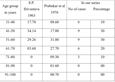
AGE:
Most of the patients, who attended the hospital with carcinoma
penis, were between 40 and 70 years of age. The youngest was found to
TABLE 1
Age group in our series as compared with other Authors
Age group
in years
S.P.
Srivastava
1963
Prabakar et al
1976
In our series
No of cases Percentage
31-40 17.70 08.60 6 10
41-50 34.14 17.80 9 30
51-60 29.26 31.00 9 30
61-70 03.60 27.70 6 20
71-80 0 09.30 3 10
81-90 0 01.60 0 00
91-100 0 00.70 0 00
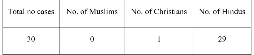
RELIGION
Among the 30 cases 29 were Hindus. Only one Christian. There
were no Muslims detected in the study, probably because of the practice
TABLE 2
Religion incidence in our series
Total no cases No. of Muslims No. of Christians No. of Hindus
30 0 1 29
INCIDENCE
Most reports are based on hospital statistics which consists
of a highly filtered material and may reflect the current incidence. In the
absence of National or Regional registry therefore the incidence figures
are low for countries of religious of religious groups which practice ritual
circumcision but they are also low in countries like Denmark or Japan
which do not. Hanash found an incidence of 2.64% of male
malignancies in U.S.A. Narayanan et al from the same country reported
an incidence of 2% of all male genital malignancies. They are also
observed a drop in incidence with years. Norman et al from Canada
found an incidence of 4.92/1, 00,000 males reported by Reddy et al from
Gieu tribe in the same country; this tribe practices the ritual
circumcision. The incidence reported in Koreans is 4% while it was
10.3% among the Chinese. In U.S.A. again the Blacks are 3.5 times more
prone to it than Whites. Dodge reviewed the tumor registry at Kampala
in East Africa and found that constituted 1% of all cancers and 1.9% male
carcinoma were that of penis. Paymaster found an incidence of 2.8% of
all tumours in Mumbai. Reddy et al reported an incident of 6.9% of all
tumors in Visakapatnam and 13.79% of all male malignancies. Panda
and Nayal from Orissa reported an incidence of 4.5-5% of all male
carcinoma. The overall incidence based on hospital records at
Pondicherry was 2.64% of all cancers and 4.9% of cancer in the male.
The figures for Agra were 30.2% and 63.79% respectively. The disease
constituted 87.1% of male genitor urinary cancer. A figure remarkably
high from Western standards, where the commonest tumour would be
prostatic followed by the urinary bladder, carcinoma penis being the least
common.
Carcinoma of the penis was detected in 2% of all the male patients
admitted with malignancies. The incidence has reduced recently. In the
late 90’s roughly more than 50 patients with carcinoma penis, were
treated in this institution. Most of the patients who are admitted with
carcinoma penis were from poor socio economical status, uneducated,
coolies, who do not have the habit of cleaning the prepuce and the glans
penis during their daily bath. For the past 2 years only 23 cases of
Most of the patients developed growth or ulcer in
uncircumcised penis. The onset was spontaneous without any history of
trauma.
The history of STD was found in only one case in which VDRL
was reactive. The other patients were non reactive.
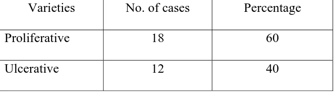
Out of the 30 patients 18 patients of Ca. Penis presented with
[image:60.612.106.444.484.577.2]proliferative growth and 12 patients with ulcer.
TABLE 3
Showing different varieties of Ca. of penis in our series
Varieties No. of cases Percentage
Proliferative 18 60
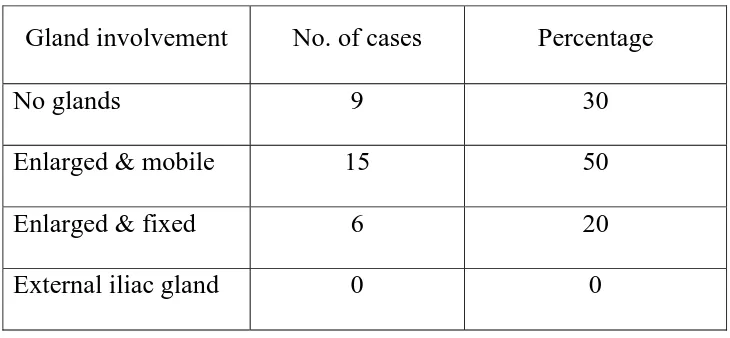
Out of the 30 patients in this study, 6 cases that had ulcerative
lesions and 9 cases that had proliferative growth had inguinal deposits. 6
patients of the 30 patients had hard, fixed, inguinal 1ymph nodes. The
involvement of the superficial inguinal nodes was more. The deep
[image:61.612.104.469.435.604.2]inguinal nodal involvement was found in only one case.
TABLE 4
STATUS OF REGIONAL LYMPH NODES
Gland involvement No. of cases Percentage
No glands 9 30
Enlarged & mobile 15 50
Enlarged & fixed 6 20
External iliac gland 0 0
15 patients had secondary deposits in inguinal nodes discovered by
FNAC. The remaining was found to be of inflammatory origin. This
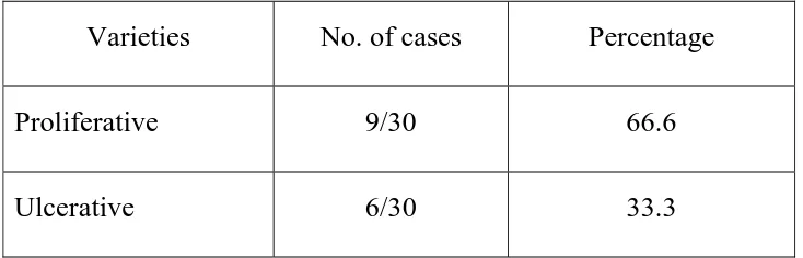
TABLE 5
Showing different varieties of Ca. penis with 1ymph node involvement
Varieties No. of cases Percentage
Proliferative 9/30 66.6
Ulcerative 6/30 33.3
Most of the patients with Ca. Penis presented at stages IIor
TABLE 6
Stage No. of cases Percentage
Stage I 3 10
Stage II 12 40
Stage III 9 30
Stage IV 6 20
Most of the patients with Ca. Penis presented at stages II or III. 3
patients presented with stage I and 6 were in stage IV
The main complaints were growth in the penis, pain and foul
smelling discharge
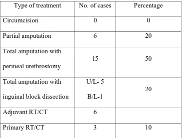
Out of the 30 Patients with Ca. Penis, 6 patients underwent partial
amputation of penis. 12 patients underwent total amputation with
perineal urethrostomy. Among 15 patients with inguinal nodal
involvement 4 underwent unilateral and 1 underwent bilateral inguinal
[image:63.612.105.469.69.290.2]After confirming tissue biopsy for primary tumour and FNAC for
1ymph nodes the treatment was planned.
Biopsy reports were positive for squamous cell carcinoma in all the
cases. Of 22 cases with nodes for whom FNAC was sent, the reports
were positive in 15 cases for secondary deposits. Due to the non
availability of frozen section biopsy this was not used preoperatively.
Out of 6 cases with fixed inguinal nodes 2 cases treated with
[image:64.612.105.469.440.717.2]palliative radiotherapy. 4 combined(RT+CT)
TABLE 7
DIFFERENT MODALITIES OF TREATMENT GIVEN TO OUR
PATIENTS
Type of treatment No. of cases Percentage
Circumcision 0 0
Partial amputation 6 20
Total amputation with
perineal urethrostomy
15 50
Total amputation with
inguinal block dissection
U/L- 5
B/L-1
20
Adjuvant RT/CT 6
COMPLICATIONS & FOLLOW UP
Partial Amputation
:
Out of 6 patients who underwent partial amputation 1
developed meatal stenosis for which urethral dilatation was
done. Only 2 were passing urine in standing posture & 3 patients
were able to resume their sexual activity.
Total Amputation
:
Of the 15 patients , 5 had wound gaping , managed
conservatively.
Inguinal block Dissection :
Out of 6 patients , 1 died on 1st POD due to uncontrolled
haemmorage from femoral vessel. 2 patients had seroma, 1 had
skin necrosis.
DISCUSSION
Most of the patients with carcinoma penis belonged to the forty to
sixty age group. Muslims were free of the disease due to early
circumcision.
Of late the incidence of CA penis has been reducing due to the
awareness among public population.
Most of the patients with CA penis belong to poor socio economic
groups were uneducated and are not aware of personal hygiene and this
disease.
Majority of the patients with CA penis presented with proliferated
growth and few inguinal node involvement. Minority of patients
presented with ulcerative growth but had more involvement of inguinal
nodes.
The glans penis and prepuce were the most involved in CA penis.
Biopsy reports of all the patients were reported as squamous cell
carcinoma.
Most of the patients underwent partial or total amputation with
perineal urethrostomy.
For operable inguinal node involvement, a ‘Wait and Watch”
policy was adopted in majority and after six weeks inguinal block
dissection was planned, but only a few turned up . In others Ilioinguinal
block dissection along with partial/Total amputation was carried out.
Most of patients are able to cope with morbidity following surgery .
For inoperable cases RT / CT given.
Few patients who turned up for followup presented with inoperable
SUMMARY
This dissertation is submitted with collection of 30 cases of
carcinoma of penis.
1. Most patients are between 41-60 years.
2. Majority of the patients are from rural areas and from
socially low economic group.
3. Phimosis and poor personal hygiene of cases form major
predisposing factors.
4. Total amputation is the treatment of choice for shaft
involvement.
5. Block dissection should be done for all FNAC proven
mobile metastatic nodes.
6. Wait &Watch policy regarding inguinal lymphadenopathy
should be weighed with caution.
7. RT/CT is the treatment of choice for advanced / inoperable
cases.
8. Much of morbidity/mortality can be avoided through proper
personal hygiene and seeking proper medical advice at early
CONCLUSION
The carcinoma of penis occurs more common in the age group
between 40-60 yrs.
The neonatal circumcision is the only way to prevent the carcinoma
of penis.
The early diagnosis and treatment decrease the mortality and
improve the 5 year survival rate.
The surgery is the mainstay of treating the carcinoma of penis.
The other modalities of treatment will be adjuvant not the main
treatment.
Creating awareness in public is the only way to reduce the
BIBLIOGRAPHY
1. Panda, K. Nayak, C.R. Clinico-pathological studies of
cancer penis. A review of 120 cases, J. Med. Aesoc
75:25:1980.
2. Rangabashyam, N. Gnanaprakasam, D. Meyappan, R, et al.
Carcinoma penis. J. Royal college of Surgeons. Edinburgh,
26.104, 1981
3. FI Demiry. M.I.M., Oliver, R.T.D, Hopestone, H.F et al.
Reprisal of the role of radiotherapy and surgery in the
management of carcinoma of penis. But J. Urol. 56:724,
1984
4. Fraley, B.E. Zhang, G.Sazama. R et al, cancer penis
prognosis and treatment plans 55:1618, 1985.
5. Roseman. D.S. Ansell, Sexually transmitted disease. Uro.
Clin.N. Amer, 11.27, 1984.
6. Narayanan, A.S., Olney, I carcinoma of penis 49:2185,
1982. Chapman, W H Sexually transmitted diseases and
carcinogenesis. Olney, L.E. Loening, S.A-nis-Analysis of
219 cases et al. Cancer. Norman R.W., Milliard O.H., Mark
F.G. et al. Carcinoma of penis a year review. Canad.J.Surg,
7. Peison, B. Benisch, B. Nicora’s multicentric hasal cell
carcinoma of penis. Urology 5:322, 1985.
8. Bosch. P, Forbe, K.A., Kollin, J et al. Secondary carcinoma
of penis J. Uroi 132, 990, 1982.
9. Mugarbil. Z.H., Child S.C., Tannenbaum, M et al.
Carcinoma of prostate metastasing to penis.
10.Urology 25:314. 1985. II. Robe Y.E.L.I Schell Hamer, PF,
Four cases of metastasis to the penis With a review of
literature J. Urol. 132:992, 1984.
11.Zungri, E. Algaba, F. Sasatarial. J.M. Epithelial sarcoma of
penis. Fur. Urol 9:53, 1983, Isa. S.S., Azmaraz. R.
Magoven, J. Leiomyosarcoma of penis: A case report of
review of literature
12.Cancer 54:39, 1984, Conger, X. Spore, A Kaposi sarcoma.
Limited to glans penis. Urology 26:173, 1985. Willscher
M.K. Daly K.J Convoy, J.F. et al. Penile horns: A case
13.Metcalf, J.S., Lee. Maize, J.C. Epidermoid urothelial
carcinoma involving the glans penis. Arch Dermatol.
121:552, 1985.
14.Lowe, D., Mckee, P.H. Verrucous carcinoma of the penis.
A study. Britt J. Urol 35:427, 1983.
15.Horl. D.B., Redman, J.R. Janset. C.T Papulosquamous
lesions of the glans penis: Urology 23:1, 1984.
16.International Union against Cancer. UICC-TNM
classification of malignant tumours, 3 rd edition, Geneva,
De Burren, 1978, PP 126-128.
17.Catalona W.L. Sentinel node biopsy – Role of
1ymphadenectomy in carcinoma penis. Urol. Clin. N. Amer
73:785, 1980
18.Luciani L. Pisciolli, F. Scappini. P. Value and role of
percutaneous regional aspiration in the management of
19.Fowler, J.E, Sentinel 1ymph node biopsy for staging penile
cancer, Urology, 23:352, 1982.
20.Uehiling D.T. Staging 1aparotomy for carcinoma of penis.
J. Urol. 110:213, 1973.
21.Skinner, D.G. Leadbetter, W.F. Kelly, S.B. The surgical
management of squamous cell carcinoma of penis. J. Urol.
107:272, 1972.
22.Finkensteil L.H, CO2 laser surgery in urology, Surg. Clin.
N. America. 64:913, 1984.
23.Young H.H. A radical operation for the cure of cancer of the
penis. J. Urol. 26:285, 1981.
24.Block, N.1., Rosen, P. Whitmore, W.F. Jr,
Hemipelviectomy for advanced penile cancer. J. Urol,
110:703, 1973.
25.Voder Malik, J.B.M., Harrison, D.H, Surgical approaches to
block dissection of the inguinal nodes. Brit. J.Plastic Surg.
381:321, 1985.
26.Sagerman, R.H.Yu, W.S. Chung, C.T. et al. External beam
irradiation of carcinoma of penis. Radiology 152:1831,
27.Ahmed, T. Sklaroff, R. Yagoda, A sequential of
methotrexate, Cis Platinum and Bleomycin for penile
cancer. J. Urol. 132:465, 1984.
28.Maicke, M.G., Adjuvant using bleomycin in squamous cell
carcinoma of the penis – A study of 19 cases. Brit. J. Urol
55:542, 1984.
29.Roshan Lal Guptha, Recent advances in Surgery, 1987,
119-155.
30.Campbell, Text book of urology.
31.Oxford Text book of Surgery.
32.Short Practice of Surgery, Bailey and Love.
33.Principles of surgery, Swartz.
PROFORMA
Name :
Age :
IP No. :
Address :
Occupation :
Religion :
D.O.A :
D.O.S :
D.O.D. :
Complaints 1. Proliferative growth –
2. Pain & Dysuria –
3. Bleeding on touch –
4. Ulcer
5. Foul smelling discharge –
6. Itching –
7. Fistulous tract from the growth
8. Retention of urine –
9. Inguinal swelling
History of Present illness:
Ulcer : 1. Site –
2. Size –
3. Duration –
Past history : History of
1. Phimosis
2. Balanoposthitis
3. Warty lesions
4. Previous surgery
5. Trauma
6. Exposure to STD
H/o Diabetes Mellitus / Hypertension
Personal History: 1. Diet 2. Smoking 3. Alcoholic General Examination: Built
Nutritional status
Anemia Weight Pulse BP
Generalized 1ymphadenopathy
Local Examination:
Primary tumor :
Inspection :
Growth/Ulcer : Site Size
Edge Margin Floor Palapation: Warmth Tenderness
Inspectory findings
Base Induration
Regional Lymph Modes:
Size Number
Mobility Mobile
Fixed
Consistency Firm
Hard Unilateral/Bilateral
P/A : Liver –
Spleen –
Other masses –
P/R : Perineal body involvement
Other masses
Management
Investigations:
a) General
1. Urine - Albumin
- Sugar - Deposits
2. CHG
Hb% TC DC ESR
3. Blood urea
4. Blood sugar
5. Serum creatinine
6. LFT
7. USG abdomen
8. Chest X-ray PA view
9. ECG
b) For Primary:
Biopsy
c) For Regional Nodes:
FNAC/Biopsy
CT Scan
d) For distant metastasis:
1. CT Chest 2. MRI 3. Bone scan 4. Serum calcium
Treatment:
For primary tumour
1. Surgery:
a) Partial amputation
b) Total amputation with perineal urethrostomy
2. Radiotherapy
For regional 1ymph node metastasis
1. Operable
a. Unilateral – I1io – inguinal block dissection
b. Bilateral I1io inguinal block dissection
2. Inoperable:
a. Radiotherapy
b. Chemotherapy
c. Combined
Follow up for 2 years
Disease free
Inguinal metastasis
Stump recurrence (Partial amputation)
PARTIAL AMPUTATION INCISION
PERINEAL URETHROSTOMY
VERRUCOUS.CA
MASTER CHART
S.No IP No Age Stage Amputation Lymphadenectomy RT CT Partial Total U/L B/L
1. 20081 48 II T
2. 25269 52 III T +
3. 28528 68 IV T + +
4. 29526 38 II P
5. 31238 47 III T +
6. 33508 53 II T
7. 35562 72 III T + +
8. 36825 58 IV T + +
9. 39583 56 II T
10. 39862 32 II P
11. 41326 45 I P
12. 42791 73 IV + +
13. 43998 43 II T
14. 45526 39 I P
15. 46282 61 III T +
16. 47921 49 II T
17. 48128 57 III T + + +
18. 48562 36 II P
19. 49327 68 III T
20. 51262 63 III T + +
21. 51899 58 II T
22. 52908 78 IV + +
23. 53007 52 III T +
24. 53286 38 II T
25. 54921 65 IV + +
26. 55282 54 II T
27. 56821 37 II P
28. 57988 61 III T +
29. 58061 56 IV T + +
A STUDY ON MANAGEMENT OF
CARCINOMA PENIS
Dissertation submitted in partial fulfillment of regulation for the award of M.S. Degree in General Surgery
(Branch I)
THE TAMILNADU
DR. M.G.R. MEDICAL UNIVERSITY
A STUDY ON MANAGEMENT OF
CARCINOMA PENIS
Dissertation submitted in partial fulfillment of regulation for the award of M.S. Degree in General Surgery
(Branch I)
THE TAMILNADU
DR. M.G.R. MEDICAL UNIVERSITY
Chennai March 2010
COIMBATORE MEDICAL COLLEGE
CERTIFICATE
Certified that this is the bonafide dissertation done by
Dr.A.JOSEPH STALIN A.MUTHU and submitted in partial fulfillment of the requirements for the Degree of M.S., General Surgery, Branch I of
The Tamilnadu Dr. M.G.R. Medical University, Chennai.
Date : Unit Chief
Date : Professor & Head
Department of Surgery
Date : Dean
Coimbatore Medical College
DECLARATION
I solemnly declare that the dissertation titled “A STUDY ON
MANAGEMENT OF CARCINOMA PENIS” was done by me from
2007 onwards under the guidance and supervision of
ProfessorDr. A. Ramamoorthy M.S.
This dissertation is submitted to the Tamilnadu Dr. MGR Medical
University towards the partial fulfillment of the requirement for the award
of MS Degree in General Surgery (Branch I).
Place : Dr. A.JOSEPH STALIN