CLINICOPATHOLOGICAL ANALYSIS OF OVARIAN
TUMORS AND THE ROLE OF p53 AND Ki- 67 IN
SURFACE EPITHELIAL TUMORS OF OVARY
DISSERTATION
SUBMITTED FOR M.D(PATHOLOGY)
BRANCH III
APRIL – 2013
CERTIFICATE
This
is
to
certify
that
this
dissertation
titled
“CLINICOPATHOLOGICAL ANANLYSIS OF OVARIAN
TUMORS AND THE ROLE OF p53 AND Ki-67 IN SURFACE
EPITHELIAL TUMORS OF OVARY”
is the original and bonafide
work
done
by
Dr.G.Gayathiri
under
the
guidance
of
Dr.N.ARUMUGAM, M.D
., Professor&Head, Department of pathology
at the Thanjavur medical college and Hospital, Thanjavur, during the
tenure of her course in M.D. Pathology from May-10 to April 13 held
under the regulation of the Tamilnadu Dr.M.G.R. Medical
University,Guindy, Chennai- 600032.
PROF. N. ARUMUGAM, M.D., PROF.C.GUNASEKARAN , M.D, D.ch; Professor and Head Dean in-charge
Department of pathology Thanjavur Medical College Thanjavur Medical college Thanjavur- 613 004 Thanjavur- 613004.
CERTIFICATE BY THE GUIDE
This is to certify that this dissertation titled
“
CLINICOPATHOLOGICAL
ANALYSIS
OF
OVARIAN
TUMORS AND THE ROLE OF p53 AND Ki-67 IN SURFACE
EPITHELIAL TUMORS OF OVARY”
is the original and bonafide
work done by
Dr.G.Gayathiri
under my guidance and supervision at
the Thanjavur Medical college & Hospital, Thanjavur- 613 004,
during the tenure of her course in M.D. Pathology from May-2010 to
April 2012 held under the regulation of the Tamilnadu Dr.M.G.R.
Medical University, Guindy, Chennai-600032.
PROF.N.ARUMUGAM, M.D.,
Professor
Department of pathology
Thanjavur Medical college
Thanjavur- 613 004.
Place: Thanjavur
ACKNOWLEDGEMENT
First of all I offer my humble obeisance to Almighty for blessing me with courage, strength and mental tenacity to accomplish this endeavour.
I wish to express my sincere and profound gratitude to
DR.N.ARUMUGAM.,MD; PROFESSOR and Head of the Department of Pathology, Thanjavur Medical College, for his valuable guidance, constant encouragement, words of advice and judicious help during the course of this project.
I take immense pleasure in thanking Dr.M.SARASWATHY,MD;DGO, & Dr. AL. SHANTHI, M.D,DGO., PROFESSORS; Dr. A.VASAHAR M.D, and Dr. M. SENTHIL KUMAR , M.D,DCP., ASSOCIATE PROFESSORS, for their valuable suggestions, encouragement and guidance throughout my study.
I would also like to express my sincere thanks to my Assistant Professors
Dr. K.G.Padmanaban, M.D., Dr.S.Jenita Christiana Ranjana, M.D.,and
Dr. V.Sindhu, M.D., for their help and cooperation throughout my study.
I am highly indebted to all the Lab technichians , all staffs of Pathology Department and Librarians for their timely help throughout my study.
CONTENTS
SL.NO TOPICS PAGE NO.
1. INTRODUCTION 1
2. AIM OF STUDY 4
3. MATERIALS AND METHODS 5
4. REVIEW OF LITERATURE 7
5. OBSERVATION AND RESULTS 47
6. DISCUSSION 68
7. CONCLUSION 88
INTRODUCTION
Ovarian tumors accounts for 6% of all cancer in women49 .Ovarian tumors are the 5th leading cause of cancer death in women in India49. Annual incidence rate in India is 9.0 per 100,000 population.Ovarian tumors accounts for 30% among all tumors of female genital tract 58.
Tumors of ovary generally are more prevalent in the upper socio-economic groups due to their low fertility rate and there is aracial predisposition of ovarian cancers with increased risk of Caucasians and lower risk for black women 70.
About two-thirds of ovarian tumors occurs in reproductiveage group. Many risk factors are associated with increasedprevalence of ovarian tumors most importantly ,age ,positive family history,genetic factors,hormonal and reproductive factors42.
Most cases are sporadic, only around 5-10% of ovariancancers are Hereditary. Women having inherited mutations in BRCA-1 & BRCA2 tumor suppressor gene are at increased risk for developing the tumor 14,42.
Abdominal USG & Serum CA-125 measurement were usedas screening methods for diagnosis of early ovarian carcinoma. Fine needle aspiration cytology is used for primary diagnosis in a patient with advanced disease and also to monitor recurrences after treatment with an overall accuracy of differentiating benign from malignant ovarian tumor ranging from 90-95% 51.
Despite the development of new diagnostic andtherapeutic strategies to improve the 5- yr survival rate ,ovariancancer still remains the deadliest due to the fact that most of them are diagnosed only in advanced stages of disease where 5-yr survival rate falls less than 20% and partly it is due to paucity of knowledge about exact etiological factors48.
Any persistent ovarian enlargement is an immediateindication for surgical assessment and actual diagnosis rests with the histopathological examination of specimen. WHO Histological Classification is used for the diagnosis of ovarian tumors. They are categorised into 3 major categories 1.Surface epithelial -stromal.2. Sex cord –stromal 3. Germ cell tumors 44,49.
Histological subtyping of surface epithelial stromal tumorsinto Benign, Borderline & Malignant has therapeutic and prognostic significance. Histological grade is an important independentprognostic factor in patients with surface epithelial stromal tumor 49.
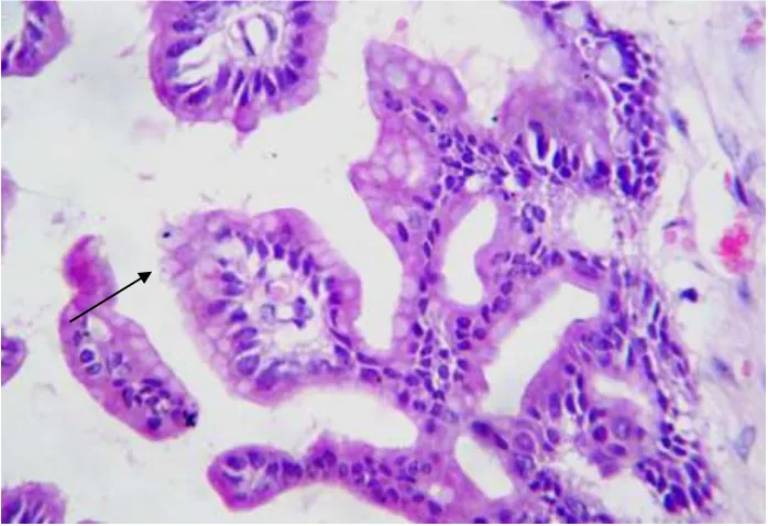
Ovarian serous carcinoma is classified according to 2-tier grading system into low and high grade and it is based on the biological evidence that these tumors develop from different pathways of gene alterations3. p53 immunohistochemical staining is done to provide an update on pathogenesis of low & high grade serous carcinoma and also helps in understanding the pathogenesis of Type I & Type II ovarian carcinomas3.
Ki-67 is a cell proliferation marker. MIB1 is a murinemonoclonal antibody against ki-67 antigen8. Ki-67 labelling index helps in the differential diagnosis of surface epithelial stromal tumors of ovary34,35.
AIM OF THE STUDY
1.To study and compare the incidence of ovarian tumors in our institution along with clinical correlation.
2. To study and compare the incidence of malignant ovarian tumors in our institution in relation to malignancies occurring in female genital tract .
3. To justify the use of Two-tier grading system in ovarian serous carcinomas in routine practice.
4. To determine whether p53 mutations seperates Type I and Type II epithelial ovarian carcinomas and to provide support for new theory of ovarian carcinogenesis.
MATERIALS AND METHODS
A total of 150 cases of ovarian neoplasms referred from Raja Mirasudhar Govt.Hospital (RMH) , Thanjavur Medical college during 2010 to 2012 were included in this study. We received unilateral, bilateral salphingo-oophorectomy along with total abdominal hysterectomy and ovariotomy specimens. Specimens were fixed in toto in 10% neutral buffered formalin and processed routinely.
In cystic ovarian neoplasms, 4-5 bits were taken from the wall alongwith papillary excrescencess if present. In solid tumors,3-4 bits were taken if the tumors were less than 5 cm. If more than 5 cm, one block per 1 cm of the tumor were taken across its greatest dimension, particularly if the appearance is variegated. 3-4 micrometre sections were cut and stained with haematoxylin and eosin (Appendix I) .
H&E stained sections were reviewed in all cases. The following clinicaland histological parametres were evaluated in particular patients age ,tumorsize,stage of disease(FIGO staging),Histological Type & Subtypes were done according to WHO Classification criteria. For serous carcinomas ,histological grade was done according to recent two-tier grading system.
Ki-67 Immunostaining:
The conventional 3-4 micro metre sections were cut from paraffinblocks and the immunohistochemical staining procedure was performed using the heat induced antigen retrieval method with specific murine monoclonalantibody-MIB1 and labelling index was done. Labelling index was measured as percentage of MIB-1 positive cells in a 1000 randomly selected tumor cells.Only nuclear staining was regarded as positive; weak nuclear or cytoplasmic staining was regarded as negative.
P53 Immunostaining:
REVIEW OF LITERATURE
GROSS ANATOMY
Ovaries are paired, almond shaped pelvic organs weighing 5-8 gms. Each ovary measures 3x2x1 cm in an adult, lies on either side of uterus close to the lateral pelvic wall.37,74
Each ovary is attached to posterior aspect of broad ligament by the mesoovarium and is attached at its medial pole to ipsilateral uterine cornu by ovarian ligament. The lateral pole of ovary attaches to pelvic wall by infundibulopelvic ligament which contains the principal vascular supply andlymphatic drainage of the ovary74.
Cut surface
Shows narrow white outer cortex and a greyish pink,medulla thatforms the bulk of the organ. In adults,ovary shows thin walled fluid filled cystic follicles and bright yellow corpora lutea is readily visible .
EMBRYOLOGY:
HISTOLOGY:
The ovary is covered by a single layer of cells originates from the coelomic epithelium. This is a highly specialised mesothelial layer called as surface epithelium becomes continues with the mesothelium of peritonealcavity. The cells can be flattened, cuboidal, columnar or focally pseudo-stratified.
Beneath the surface epithelium lies the cortex,which is roughly divisible into outer fibrous acellular collagenous zone termed as ‘Tunica albuginea’ and inner more cellular active cortex. Inner cellular cortex contains the primordial follicle, ripening follicle and mature follicles. The stroma.consists of uniformed spindle cells in bundles often with storiform pattern(75)
The central portion of the ovary is the medulla and it also has activefollicles and cellular stroma. The blood vessels enter at the hilum areaccompanied by a small amount of connective tissue.
OVARIAN TUMORS:
Tumors of the ovary represents about 30% of all cancers of femalegenital tract system. Age adjusted incidence rates are highest in economically advanced countries(5).
Environmental,genetic and life style factors all influence ovarian cancer risk.Genetic susceptibility is evident from numerous epidemiological investigations and there was increased susceptibility in patients with positive family history. A number of specific genes most important of which is BRCA1and BRCA 2 . Ovarian cancer is a minor feature of the hereditary nonpolyposis colon cancer syndrome(53)
WHO HISTOLOGIC CLASSIFICATION OF TUMORS OF
THE OVARY
Surface epithelial – stromal Serous tumors
Malignant
Adenocarcinoma Borderline tumor
Benign - Cystadenoma, adenofibroma, cystadenofibroma
Mucinous tumors
Malignant
Adenocarcinoma Borderline tumor
Benign - Cystadenoma,adenofibroma,cystadenofibroma Mucinous cystic tumor with pseudomyxoma peritonei
Endometrioid tumors
Malignant
Adenocarcinoma
Malignant mixed mullerian tumor Endometrial stromal sarcoma Borderline tumor
Benign - Cystadenoma,adenofibroma,cystadenofibroma
Clear cell tumors
Malignant
Benign - Cystadenoma , adenofibroma, cystadenofibroma
Transitional tumors
Malignant
Transitional cell carcinoma(non –Brenner type) Malignant Brenner tumor
Borderline
Benign - Brenner tumor
Squamous cell carcinoma Mixed epithelial tumors
Malignant Borderline Benign
Undifferentiated and unclassified tumors
Undifferentiated carcinoma
Adenocarcinoma, not otherwise specified Sex cord – stromal tumors
Granulosa – stromal cell tumor Granulosa cell tumor
Adult granulosa cell tumor Juvenile granulosa cell tumor
Thecoma-fibroma group
Thecoma,not otherwise specified Typical
Cellular fibroma Fibrosarcoma
Stromal tumor with minor sex cord elements Sclerosing stromal tumor
Signet ring –stromal tumor
Sertoli – stromal cell tumors
Sertoli- Leydig cell tumor group Well differentiated
Of intermediate differentiation
Variant with heterologous elements Poorly differentiated
Variant with heterologous elements Retiform
Variant with heterologous elements Sertoli cell tumor
Stromal –Leydig cell tumor
Sex cord –stromal tumors of mixed or unclassified cell type Sex cord tumor with annular tubules
Gynandroblastoma
Sex cord-stromal tumors,unclassified
Steroid cell tumors
Stromal luteoma Leydig cell tumor group
Hilus cell tumor
Leydig cell tumor .non hilar type
Well differentiated Malignant
Germ cell tumors
Primitive germ cell tumors Dysgerminoma
Yolk sac tumor
Embryonal carcinoma Polyembryoma
Non gestational chorio carcinoma Mixed germ cell tumor
Biphasic or triphasic teratoma Immature teratoma
Mature teratoma -Solid
-Cystic
-Fetiform teratoma
Monodermal teratoma and somatic type tumors associated with dermoid Cyst
Thyroid tumor group
Struma ovarii Benign
Malignant
Carcinoid tumor
Neuro ectodermal tumor group Carcinoma group
Melanocytic nevus Sarcoma group
Sebaceous tumor group Pituitary type tumor group Retinal anlage tumor group
Germ cell sex cord – stromal tumor
Gonadoblastoma - Variant with malignant germ cell tumor
Mixed germ cell – sex cord – stromal tumor - Variant with malignant germ cell tumor
Tumors of the rete ovarii
Adenocarcinoma Adenoma
Cystadenoma Cystadeno fibroma
Miscellaneous tumors
Small cell carcinoma, hypercalcemic type Small cell carcinoma, pulmonary type Large cell neuro endocrine carcinoma Hepatoid carcinoma
Primary ovarian mesothelioma Wilm’s tumor
Gestational choriocarcinoma Hydatidiform mole
Adenoid cystic carcinoma Basal cell tumor
Paraganglioma Myxoma
Soft tissue tumors not specific to the ovary
Tumor-like conditions
Luteoma of pregnancy Stromal hyperthecosis Stromal hyperplasia Fibromatosis
Massive ovarian edema
Lymphoid and haematopeitic tumors Secondary tumors
SURFACE EPITHELIAL STROMAL TUMOR:
Each of the tumor type is subdivided into Benign, Borderline, Malignant category. Benign tumors of serous, mucinous, endometroid, clear cell are further subtyped into cystadenoma, cystadenofibroma, adenofibromas(94).
SEROUS TUMORS:
The most common type of surface epithelial neoplasms areserous tumors . Approximately 60% Benign, 10% Borderline, 30% malignant(43).
BENIGN SEROUS TUMORS:
Occurs most commonly in 5th decade.
They are bilateral in 10-20% cases.They are usually unilocular , entirely cystic or can be partly cystic. Cysts filled with watery thin serous fluid ,innersurface of the cyst wall can be smooth or papillary excrescences can beseen(43).
HISTOLOGY:
Cysts and polypoid excrescencess has single layer of ciliatedepithelium similar to that of fallopian tube. Nuclear atypia is seen. Psammoma bodies are infrequent with rare mitotic figures. Papillae when found are almost entirely made up of fibrous stroma(44).
SEROUS BORDERLINE TUMOR:
They are common in 4th& 5th decade.
GROSS
They are bilateral in 25% of cases. They are usually cystic, containing watery or thick mucinous fluid and have surface or intracystic numerous papillary projections(28).
HISTOLOGY:
Three most important diagnostic features (29)are:
1. Arborising papillae (hierarchial branching) form increasingly smallerbranches ending in clusters of epithelial cells, detached from stroma .
2. Varying degree of mild –moderate nuclear atypia.
3. Absence of frank stromal invasion/solid sheets of tumor with cribriform pattern (43).
1. Typical(90%)
2. Micropapillary(10%) .
Typical serous borderline tumorsmakes up the majority and has aclassic branching papillary architecture and epithelilal tufts overlying the papillae.
Micropapillary pattern shows focal or diffuse proliferation of tumor cells in elongated,thin micropapillae with little or no stromal support emergingdirectly from the cyst lining. The micropapillae are atleast five times as long asthey are wide,arising directly from the papillae with thick fibrous stalk.
Having diagnosed a serous borderline tumor following features(81) should be examined for:
1. Surface involvement:
Cross examination of ovarian surface is done carefully and it is oriented such that surface is evident on slide. The presence of surface involvement are associated with increased frequency of high stage tumor. Surface involvement enables peritoneal spread.
2. Stromal microinvasion:
Generous and judicious sampling is required to exclude microinvasion. Atleast one section per centimetre of tumor diameteris required. Invasive foci measuring less than 10mm2 and less than 3mm2 in greatest dimension is called as microinvasion.
borderline ovarian tumors,but will co-seggregate with more aggressive tumors; in which case micropapillary pattern is more ominous.
3. Lymph node metastases:
Pelvic and paraaortic lymphnode metastases are found in as many as 27% of cases .These metastases are present in sinusoidal spaces rather than substance of node. Its presence does not alter the prognosis of serous borderline tumors, however they have lower disease free survival.
4. Implants on peritoneal surface:
Implants can be invasive and non invasive. Presence of peritoneal implants and whether or not they are invasive are the most important indicators of outcome in serous borderline tumors. on invasive implants are of 2 types. Epithelial & Desmoplastic (12) .
Epithelial implants: implants composed of papillae resembling those within ovarian serous borderline tumor. they are found either in mesothelial lined invaginations of peritoneal surface or between the lobules of fat.
Desmoplastic implants : Papillae, glands, cell cluster or single cell within inflamed ;dense fibroblastic or granulation t
issue that appears
to be plastered onto serosal surface.Invasive implant:
1. Implants with haphazardly arranged glands involving the normal tissues such as omentum.
2. Loose or dense fibrous reaction without significant inflammation 3. Dominant epithelial proliferation
5. Irregular borders and
6. Aneuploidy
SEROUS BORDERLINE ADENOFIBROMA AND
CYSTADENOFIBROMA:
In this variant epithelial lining of glands and or cysts of the adenofibroma or cystadenofibroma has the features of serous borderlinetumor instead of benign epithelium.(81)
MALIGNANT SEROUS CYSTADENOCARCINOMA: GROSS:
They are bilateral in approximately 70% of cases. They range from microscopic size to more than 20cm dm. Mostly they are cystic and solid with papillae within cysts or on surface. Poorly differentiatedtumors are predominantly solid, multinodular masses with necrosis and haemorrhage(43)
MICROSCOPY:
GRADING:Various grading systems are in use .The most commonly used grading system was Silverbergs grading system which is as follows (94)
TABLE 1: Silverberg grading system:
Score Architecture Cytologic atypia Mitotic figures/10hpf
1 Glandular Mild 0-9
2 Papillary Moderate 10-24
3 Solid Severe >25
Score 3-5: Grade 1 Score 6-7: Grade 2 Score 8-9: Grade 3
2 –TIER GRADING SYSTEM FOR SEROUS CARCINOMA3:
According to Anais Malpaica et al ,Serous carcinoma is graded according to degree of nuclear atypia and mitotic rate is used as secondary feature.
Low grade:
1. Mild to moderate nuclear atypia and as secondary feature 2.< 12 mitoses/10hpf.
High grade
Significance of 2 tier grading system:
Serous carcinomas are graded into low and high grade which reflect a difference in the pathogenesis of these tumors(3). Since the system is based on defined criteria that are easy to follow and because it involves only two diagnostic categories ,it provides better reproducibility in the grading of serous carcinoma(80).
Accordingly low grade serous carcinomas will not show p53expression as these tumors have intact p53 gene 80.In contrast more than 95%of high grade serous carcinomas shows strong positivity with p53 immunostaining indicating that this gene is involved in their tumorigenesis.
MUCINOUS TUMORS OF OVARY
The second most common type are mucinous tumors .About 80% ofmucinous tumors are benign,10% are borderline,10% malignant(6).
BENIGN MUCINOUS TUMORS: GROSS:
They are large,unilateral,multilocular cystic masses containing viscous mucoid material. Cystadenofibromas are partially to almost completely solid.
MICROSCOPY:
Mucinous columnar epitheliumcontaining intracytoplasmic mucin lines the glands and cysts, which resembles endocervical or gastrointestinal type epithelium.Cystadenofibromas shows mucinous glands and cysts uniformly distributed in a fibromatous stroma(93).
INTESTINAL TYPE: GROSS:
About 85% of mucinous borderline ovarian tumors are of intestinal type. Bilateral in only 5% cases. They are large,cystic,multi/unilocular with viscous fluid. Borderline foci can be solid but typically reside within fleshy polypoid areas in cyst wall.(6,21)
MICROSCOPY:
In addition to conventional benign mucinous
cystadenoma,borderlineareas shows stratification ,no more than 3 layers and may form filliformintracystic papillae with minimal support. Nuclei are enlarged hyperchromaticand have increased mitotic activity. Goblet cells are seen distributed haphazardly. Glands and cysts lumen contain mucin(6).
ENDOCERVICAL TYPE: GROSS:
About 10-15% of borderline tumors are endocervical type. They arebilateral in 40% cases and sometimes arise within endometriotic cysts(94).
MICROSCOPY:
MUCINOUS CYSTADENOCARCINOMAS GROSS:
Only about 5 % of these tumors are bilateral. They are large,unilateral, smooth surfaced; multilocular cystic masses containing viscous fluid. Haemorrhagic, necrotic, solid, papillary areas are relatively frequent. For all grossly suspicious areas, one histological section per 1-2 cm of tumor diameter is recommended.
MICROSCOPY:
There are 2 forms of stromal invasion. Expansile and infiltrative. Inexpansile invasion, glands and cysts are lined by malignant cells forms complexpapillary areas or back- back arrangement of glands with little/no discernableintervening stroma. To qualify as frankly invasive, such areas should be atleast10mm2.(77)
In infiltrative type of invasion; glands, tubules, cords, cell nestshaphazardly infiltrate the stroma.The invasive cells has nondescript eosinophilic cytoplasm(77).
MUCINOUS CYSTIC TUMOR WITH PSEUDOMYXOMA PERITONEI:
Pseudomyxoma peritonei is a clinical terminology that describe the presence of abundant gelatinous material within pelvis and abdominal cavity surrounded by fibrous tissue. First step is to exclude the presence of appendiceal neoplasm or other gastrointestinal primary mucinous tumor metastasis to peritoneum.
ENDOMETROID TUMORS:
GROSS:
These tumors average 8-10cm in dm. They are solid, firm,tan tumors with cysts of varying sizes(84).
MICROSCOPY:
They are well differentiated,benign appearing glands or cysts lined byendometrial type cells with or without squamous differentiation(84).
BORDERLINE ENDOMETROID TUMORS: GROSS:
These tumors are predominantly unilateral ,ranging in size from 2-40cm; cut surface gry-white to tan,can be solid and cystic or predominantly solid. Larger tumors shows haemorrhage and necrosis(92)
MICROSCOPY:
Islands of crowded endometroid glands or cysts lined by cellsdisplaying grade 1 to grade 3 cytologic atypia proliferate in an adenofibromatous stroma. Absence of stromal invasion and low mitotic activity is seen. About 15-50 % of patients have endometriosis in same ovary as well as at extraovarian sites(92).
ENDOMETROID ADENOCARCINOMAS: GROSS:
MICROSCOPY:
Well differentiated tumors shows round,oval or tubular glands lined bystratified nonmucin containing epithelium. Squamous differentiation occurs in30-50% cases often in the form of morules(74).
Table2:FIGOGrading scheme for Endometrioid adenocarcinoma:
MALIGNANT MIXED MULLERIAN TUMOR: GROSS:
They are bilateral in 90% cases. They are large tumors partly solid andpartly cystic, bosselated masses with hemorrhage and necrosis(44).
Grading Histological feature
Grade 1 or well differentiated Well formed glands resembling villoglandular carcinoma of uterine corpus, < 5% solid tumor growth. Grade 2 or moderately
differentiated
More complex glandular architecture,increased nuclear stratification,6-50% solid tumor growth.
MICROSCOPY:
Biphasic neoplasm that has malignant epithelial and mesenchymal component. Epithelial component can be any type of surface epithelial carcinoma.mesenchymal component most commonly seen are fibrosarcomaor endometroid stromal sarcoma or leiomyosarcoma(44).
ADENOSARCOMA: GROSS:
Almost always unilateral and is predominantly solid with small cysts.
MICROSCOPY:
Biphasic tumor in which mesenchymal component is sarcomatous butepithelium is benign. Cellular stroma forms periglandular cuffing. Adenosarcoma of ovary has worse prognosis when compared to its uterinecounterpart.(44)
ENDOMETRIAL STROMAL SARCOMA:
GROSS:More than 70% tumors are unilateral. most of them are solid and firm but some have cysts filled with mucoid or haemorrhagic fluid.
MICROSCOPY:
CLEAR CELL TUMORS: BENIGN TUMORS: GROSS:
External surface is smooth lobulated . Cut surfaceshows honey- combing appearance with tiny cysts embedded in rubbery stroma.(45)
MICROSCOPY:
Tubular glands lined by 1 or 2 layers of epithelium that containsPolygonal cells whose cytoplasm is clear with minimalnuclear atypia.(45)
BORDERLINE CLEAR CELL TUMORS:
GROSS:Smooth lobulated external surface with cut section appearing moresoftier and fleshier.
MICROSCOPY:Tumor composed of tubules and small cysts lined by one or several layers of cuboidal epithelial cells with clear cytoplasm or hobnail nuclei. Epithelial cells shows mild to moderate nuclear atypia and occasional mitotis(44)
CLEAR CELL ADENOCARCINOMA: GROSS:
These tumors have an average size of about 15cm. Cut section shows unilocular cyst with yellow nodules with some cysts containing watery/mucinous fluid. They are seen associated withendometriosis ovary(44).
These tumors shows tubulocystic, papillary and solid patterns.Individual cells are polyhedral cells with abundant clear cytoplasm separated by delicate fibrovascular stroma in solid pattern(44).
TRANSITIONAL TUMORS:
BENIGN BRENNER TUMOR: GROSS:
More than 50% tumors are less than 2 cm dm. They are well circumscribed firm,white gritty on sectioning with focal areas of calcification. Brenner tumors are associated with mucinous cystadenoma in about 25% Cases(76)
MICROSCOPY:
Nests of transitional type of epithelial cells withcentrally grooved coffee bean nuclei with abundant eosinophilic cytoplasm and distinct cell membranes. These nests lie in a predominantly fibromatous stroma.(76)
BOREDRLINE BRENNER TUMOR: GROSS:
They are typically large with mean diameter of 16-20cm, usually has solid and cystic component.
MICROSCOPY:
MALIGNANT BRENNER TUMOR: GROSS:
They are large tumors, has solid component as well as cystscontaining papillary or polypoid masses.
MICROSCOPY:
These tumors exhibits stromal invasion associated with a benignor borderline Brenner component. Invasive element is usually high gradetransitional cell carcinoma(93).
TRANSITIONAL CELL CARCINOMA: GROSS:
They are bilateral in about 15% cases , often partly solid & cystic.
MICROSCOPY:
They have papillary architecture lined by multilayered transitional epithelium, cells shows features of malignancy and these tumorsshould not have a benign or borderline Brenner component.(93)
MIXED EPITHELIAL TUMOR: MICROSCOPY:
These tumors consists of admixture of 2 or more of 5major cell types: serous, endometroid, clear cell, Mucinous and transitional.(76)
MICROSCOPY:
It is a primary ovarian tumor with no differentiation and marked cytological atypia.(76)
B. SEX CORD – STROMAL TUMOR : GRANULOSA CELL TUMOR:
These tumors constitute for about 1.5% of all tumors of ovary(76).
ADULT TYPE: GROSS:
They are unilateral in 95% of cases. On an average size of 12 cm and encapsulated. Cut section shows solid and cystic,areas of necrosis and haemorrhage giving a variegated appearance.(1)
MICROSCOPY:
There is proliferation of granulosa cells often with a stromal component of fibroblasts, theca or lutenised cells. Individual cells have scanty cytoplasm with longitudinal nuclear grooves. There is minimal to no cytological atypia and a low mitotic activity (1,76).
JUVENILE TYPE:
Occurs predominantly before 30 yrs age group . In prepubertalgirls 90% are associated with isosexual pseudo precocity.(76)
GROSS:
These tumors are indistinguishable from adult type granulosa cell tumor .
MICROSCOPY:
These tumors shows nodular or diffuse cellular growth punctuatedby macrofollicles of varying sizes and shapes. Their lumen contain eosinophilic or basophilic fluid. Typically rounded neoplastic granulosa cells has abundant eosinophilic and or vacuolated cytoplasm and almost all nuclei lack grooves. Abundant mitotic figures seen.(1,76)
THECOMA –FIBROMA: THECOMA
GROSS:
These tumors occur in postmenopausal women .They have anaverage size of about 5-10 cm. Cut section shows solid and yellow and almost always unilateral.(93)
MICROSCOPY:
thecomas contain lutein cells ,individually or in nests, in a background more often fibromatous than thecomatous. Oedema and microcyst formation is striking(93).
FIBROMA:GROSS:
They account for 4% of all ovarian tumors.they are hard whitetumors. Majority of them are unilateral.(40). About 10-15% of tumors are large and often associated with ascites and Meigs syndrome(4).
MICROSCOPIC:
These tumors composed of spindle shaped cells with uniform bland nuclei and scant cytoplasm, arranged in fascicles or in a storiform pattern. Mitoses are absent. About 10% are uniformly and densely cellular and are referred as cellular fibromas.(4,40)
FIBROSARCOMA: GROSS:
Large, solid tumors, haemorrhagic and necrotic and usually unilateral.
MICROSCOPIC:
These tumors are densely cellular with moderate to severecytological atypia, high mitotic rate and atypical mitotic figures with haemorrhage and necrosis.(94)
SCLEROSING STROMAL TUMOR:
MICROSCOPY:
These tumors shows pseudolobulation of cellular areasseparated by hypocellular areas of densely collagenous stroma. Cellular areas shows prominent thin walled vessels, some of them having HPC-like pattern.
SIGNET-RING STROMAL TUMOR : GROSS:
These tumors may be both solid and cystic or uniformly solid.
MICROSCOPY:
H& E section shows a diffuse proliferation of spindle and round cells, latter shows eccentric nuclei with a single large intracytoplasmic vacuole and resemble signet ring cells(93).
SERTOLI-STROMAL CELL TUMOR:
SERTOLI-LEYDIG CELL TUMOR GROUP:
They are rare and comprises of less than 0.5% of all ovarian tumors.
GROSS:Majority of them are unilateral.they are solid or solid and cystic.Solid areas are fleshy,pale yellow with areas of haemorrhage and necrosis.(94)
MICROSCOPY:
2. Intermediate,( Meyer’s type II)- Composed of cords, sheets and nests of sertoli like cells separated by spindle stromal cells
3. Poorly differentiated( sarcomatoid ,Meyers type III)-they are arranged in sarcomatoid pattern with masses of spindle shapedcells.(94)
SERTOLI CELL TUMOR:
They are commonly seen in women of reproductive age group.
GROSS:
Pure forms of sertoli cell tumors are rare. They are mostly unilateraland average size of 5-7 cm diametre in size.(76)
MICROSCOPY:
These tumors composed of sertoli cells that line tubules or trabeculaeor grow in nests or solid sheets. Cells are columnar or polygonal and have small round to oval nuclei and granular or eosinophilic cytoplasm, with minimal nuclear atypia. (76).
GYNANDROBLASTOMA:
Gynandroblastoma is a rare tumor ,containing both sertoli cells orsertoli-leydig cell and granulosa cell differentiation. They are usually unilateral with size ranging from 1-18cm. Microscopically tubules and trabeculae similarto those in well differentiated sertoli or sertoli-leydig cell tumors mixed with nests and sheets of granulosa cells.(76)
GROSS:
They are large and usually well circumscribed often lobulated appearance. Sectioned surface ranges from yellow to brown or black.
MICROSCOPY:
These tumors shows solid aggregates of cells withoccasional nests or trabeculae. Tumor cells are polygonal with granular and eosinophilic cytoplasm,nuclei are usually bland. Areasof haemorrhage and necrosis can be seen(94).
C. GERM CELL TUMORS: INTRODUCTION :
These tumors account for 30% of primary neoplasms of ovary, 95% of which are dermoid cysts(mature cystic teratomas). Median age at presentation is 18 years. Malignant GCT are common cancers among children and adolescent females. (94)
DYSGERMINOMAS: GROSS:
Well encapsulated tumor ,unilateral in 90% of cases. Average tumor size is 15cm dm. Section shows solid,uniform or lobular and creamy white. Foci of coagulative necrosis and cystic change or macroscopic calcification can be seen.(15)
MICROSCOPY:
with lymphocytes. syncytiotrophoblastic cells are found in around 5% of cases.(15)
YOLK SAC TUMOR: GROSS:
They are well encapsulated tumor with an average size of 15cm dm.Cut section shows grey yellow areas with frequent areas of necrosis, haemorrhage and liquefaction. Cysts are also seen.(39)
MICROSCOPY:
Tumor shows the characteristic reticular pattern formed by aloose, myxoid stroma harbouring a network of microcysts. These cysts are lined by clear or flattened epithelial cells. PAS +ve hyaline globules are seen tumor. Only around 10-20% of tumors shows schiller-duval bodies.(39)
EMBRYONAL CARCINOMA AND POLYEMBRYOMA:
They are rare tumors mostly reported as a component of mixedgerm cell tumors.(46)
MICROSCOPY:
Tumor shows dysorganised sheets of large primitive appearing cells forming papillae or crevices coexist with syncytiotrophoblastic cells as well as early teratoid differentiation.When abundant embryoid bodies are seenthis tumor is called as Polyembryoma .(46)
MIXED GERM CELL TUMOR:
MICROSCOPY:
Most common combination found is yolk sac tumor anddysgerminoma. Some of the additional elements are immature/mature teratoma, polyembryoma, embryonal carcinoma, may be present.(46)
BIPHASIC TERATOMA:
These are tumors composed of derivatives of 2 or 3 germ layers.
IMMATURE TERATOMA: GROSS:
Typically unilateral ,large,variegated predominantly solid.
MICROSCOPY:
Tumor shows immature embryonal type tissues, in the form of neuroepithelial rosettes and tubules admixed with mature tissue.
MATURE TERATOMA:
Most mature cystic teratomas occur in the reproductive agegroup. Mature solid teratomas occur mainly in first two decades of life.(76)
GROSS:
Dermoid cysts presents as cystic mass with an average size of 15cm dm, they are bilateral in 8-15% of cases. Cysts are often filled with hair and sebaceous material. Rokitansky protruberance is seen protruding into the cyst(76).
MICROSCOPY:
smooth muscle, teeth, bone, respiratory epithelium, gastrointestinal epithelium are also seen.(76)
MONODERMAL TERATOMAS: STRUMA OVARII:
Most common type of monodermal teratoma. Accounts for 2-7% of all ovarian teratomas.(92)
GROSS:
They are unilateral and size varies from 0.5-10 cm dm. Cut section brown solid and gelatinous nodules within a dermoid cyst(92).
MICROSCOPY:
Tumor is composed of normal or hyperplastic thyroid tissue withmicro or macrofollicular pattern trabecular and solid pattern. Most common malignancy in struma ovarii is papillary carcinoma with similar characteristichistological features as in tumors arising from thyroid gland.(92)
CARCINOID TUMOR:
Ovarain carcinoids account for 0.5-1.7% of all carcinoids(63).
MICROSCOPY: Insular carcinoid consists of islands and nests of round cells withabundant eosinophilic cytoplasm and round uniform nuclei. Trabecularcarcinoids exhibits wavy and anastomosing ribbons composed of columnarcells with long axes ofcell parallel to one another(63).
NEUROECTODERMAL TUMOR:
GROSS:They are unilateral and an average size of 14 cm dm. Sectioned surfaceis solid with friable, gray pink ,tissue and cysts with papillary excrescencess. (85)
MICROSCOPY: Well differentiated tumors forms like ependymomas and poorlydifferentiated tumors like medulloepithelioma and primitive neuro ectodermaltumor are seen. Anaplastic forms such as glioblastoma multiforme is alsoseen(85).
CARCINOMA: Carcinoma secondarily develops from dermoid cysts. They usually occur in postmenopausal women(46).
GROSS:Cauliflower like exophytic growth or infiltrative grey white plaqueswith necrosis and haemorrhage may be seen.
MICROSCOPY:Most common type of carcinoma arises is squamous cell carcinoma accounting for 80% of cases. Second most common malignancy isadenocarcinoma.(46)
SARCOMAS:
MELANOCYTIC TUMORS:They are rare tumors , dermoid cyst with melanoma in stage I are alive even after 2 yrs of diagnosis .(92).
SEBACEOUS TUMORS:
MICROSCOPY:Tumors like basal cell carcinoma with sebaceous differentiation, sebaceous carcinoma and sebaceous adenoma are common. Hallmark ofthese lesions is presence of large number of mature sebaceous cells with oil red O positive staining.(76)
PITUITARY TYPE TUMORS:Corticotroph cell adenoma and prolactinomas are common(76).
RETINAL ANLAGE TUMOR: Pigmented progonoma and malignant tumors derivedfrom the retinal anlage are seen.(95)
GERM CELL-SEX CORD STROMAL TUMOR(93):
GONADOBLASTOMA:
Most of these patients have gonadal dysgenesis and more than 90% cases have a Y chromosome.Typically seen in children or young adults.gonadoblastoma is a benign tumor unless a malignant germ cell component is seen.(93)
GROSS:They are small with a yellow to tan grey cut surface and areas ofcalcification.(93)
MIXED GERM CELL-SEX CORD-STROMAL TUMOR:
Occurs in infants or children under the age of 10. One fourth of cases have isosexual pseudoprecocity.(94)
GROSS:They are large tumors predominantly solid or partly solid &cystic.
MICROSCOPY:Tumor is composed of germ cells and sexcord derivatives similar in apperaence to immature sertoli/ granulosa cells intimately admixed with each other.(94)
D: TUMORS OF RETE OVARII:Most of these lesions are incidental finding in a postmenopausal women.To diagnose, these lesions tumor must be located in ovarian hilus.(16)
MICROSCOPY:Tumor is composed of cuboidal cells or columnar nonciliated cells arranged in retiform spaces.dilated areas and cyst are most frequently seen.(16)
METASTATIC TUMORS:These tumors comprise about 5-10% of all ovariantumors. Primary tumors from gastrointestinaltract (especially large intestine, stomach, appendix), are most common followed by Breast, Uterine corpus and uterine cervix. In young girls ovarian metastases are common with neuroblastoma, rhabdomyosarcoma, Ewing sarcoma. (82,98).
GENERAL FEATURES OF OVARIAN METASTASES:
1. Bilaterality.
2. Small superficial; Multinodular tumor 3. Vascular invasion
4. Desmoplastic reaction
GROSS:Ovarian metastases are bilateral in 70% of cases. Size of ovarian metastase varies from microscopic size to more than 10cm(16).
MICROSCOPY:Metastatic tumors grow as superficial or parenchymatous solid nodules. Histologic nature depends on the primary tumor(16).
KRUKENBERG TUMOR:They are defined characterised by thepresence of mucin –filled,signet ring tumor cells within cellular stroma of the ovary. Most krukenberg tumors represent ovarian metastases from the gastrointestinal tract, especially stomach(59).
p53:
p53 is a tumor suppressor gene situated on chromosome 1745P53 gene mutation results in uncontrolled cell proliferation. Approximately 50% of malignant tumors in humans have mutations in p53 gene and it is the Most common tumor suppressor gene involved with human malignancies.(66)
Studies have shown that p53 gene is mutated in about 50 - 80% ofovarian carcinomas31. It has been identified that immunohistochemical detection of p53 protein overexpression, is an adverse prognostic factor for survival in human ovarian cancer67.
The number of cases with mutant Tp53 among ovarian serous and endometroid carcinomas is 57% and 25% respectively with maximum value in the poorly differentiated tumors in patients with stage III or stage IV disease.One of the reasons for deranged p53 gene functions can be related to BRCA1 & BRCA2 mutations which are frequent event in hereditary ovarian Carcinoma66.
arise as denovo and majority of them has p53 mutations and lack KRAS, BRAF gene mutations(Type II pathway)17.Epithelial ovarian malignancies showing p53 aberrations aresignificantly less sensitive to chemotheraphy and more aggressive than those with functional p53 gene.7
Ki-67:
Ki-67 is a cell proliferation marker. Ki-67 antigen immunostaining isused to estimate the proliferation index of a tumor34. This antigen ispreferentially expressed in late G1,S,G2& M phase except in G0 phase8
Determining the proliferative activity of ovarian tumors has reportedto be of diagnostic and prognostic value22,34. In paraffin section, MIB1 antibody is equivalent to ki-67. MIB1 is a murine monoclonal antibody reacts withthe Ki-67 protein expressed by the proliferating tumor cells23.
MIB1 antibody staining provide a reliable means of identifyingproliferating normal and neoplastic human cells in histological sections89,99.
MASTER CHART
18. 1096/10 45 MASS ABDOMEN R-CYSTIC,L-CYST WITH PAPILLARY EXCRESENCES R-BORDERLINE MUCINOUS CYSTADENOMA L-SEROUS PAPILLARY CYSTADENOMA I
19. 1107/10 40 PAIN ABDOMEN UNILATERAL- CONGESTED I
Sl.No HPE.No AGE-YRs CLINICAL
PRESENTATION
GROSS FEATURE HPE DIAGNOSIS FIGOSTAG
ING
1. 1/10 47 MASS ABDOMEN
UNILATERAL-CYSTIC
BENIGN serous CYSTADENOMA
I
2. 66/10 45 MASS ABDOMEN
UNILATERAL-CYSTIC
BENIGN SEROUS CYSTADENOMA
I
3. 96/10 25 PREGNANCY
ASSOCIATED UNILATERAL-CYSTIC MATURE CYSTIC TERATOMA I
4. 232/10 24 MASS ABDOMEN
UNILATERAL-CYSTIC
BENIGN SEROUS CYSTADENOMA
I
5. 258/10 28 MASS ABDOMEN UNILATERAL –
CYSTIC
MATURE CYSTIC TERATOMA
I
6. 314/10 50 ASYMPTOMATIC
UNILATERAL-CYSTIC
BENIGN SEROUS CYSTADENOMA
I
7. 337/10 21 MASS ABDOMEN UNILATERAL-
CYSTIC
BENIGN SEROUS CYSADENOMA
I
8. 425/10 42 MASS ABDOMEN
UNILATERAL-CYSTIC
MATURE CYSTIC TERATOMA
I
9. 442/10 50 MASS ABDOMEN
WITH ASCITES BILATERAL, SOLID&CYSTIC BILATERAL PAPILLARY SEROUS CYSTADENOCARCIN OMA III
10. 528/10 27 MASS ABDOMEN UNILATERAL –
CYSTIC
BENIGN serous CYSTADENOMA
I
11. 589/10 40 MASS ABDOMEN
UNILATERAL-CYSTIC
BENIGN SEROUS CYSTADENOMA
I
12. 613/10 33 MASS ABDOMEN
UNILATERAL-CYSTIC
MATURE CYSTIC TERATOMA
I
13. 824/10 46 MASS ABDOMEN
UNILATERAL-CYSTIC
MATURE CYSTIC TERATOMA
I
14. 1001/10 42 MASS ABDOMEN R-SOLID, L-CYSTIC R-BENIGN
BRENNER,L-BENIGN MUCINOUS CYSTADENOMA
I
15. 1023/10 42 ASYMPTOMATIC
UNILATERAL-CYSTIC
BENIGN serous CYSTADENOMA
I
16. 1025/10 23 MASS ABDOMEN
UNILATERAL-CYSTIC&SOLID
GRANULOSA CELL TUMOR
I
17. 1031/10 32 MASS&PAIN
CYSTIC,HAEMO RRHAGIC FLUID
SEROUS
CYSTADENOMA 20. 1173/10 39 MASSABDOMEN
,ASCITES
BILATERAL,SOLI D
KRUKENBERG TUMOR 21. 1181/10 25 MASS ABDOMEN
UNILATERAL-CYSTIC
BENIGN SEROUS CYSTADENOMA
I 22. 1314/10 34 ASYMPTOMATIC
UNILATERAL-CYSTIC
BORDERLINE MUCINOUS CYSTADENOMA
I
23. 1362/10 12 MASS ABDOMEN UNILATERAL-SOLID,CYSTIC,V ARIEGATED
MIXED GERM CELL TUMOR
I
24. 1364/10 60 MASS ABDOMEN BILATERAL,CYST WITH PAPILLARY EXCRESCENCE BILATERAL BENIGN SEROUS CYSTADENOFIBR OMA I
25. 1459/10 40 MASS ABDOMEN UNILATERAL CYSTIC WITH PAPILLARY EXCRESCENCE BENIGN SEROUS CYSTADENOMA I
26. 1553/10 70 MASS ABDOMEN UNILATERAL CYSTIC
BENIGN SEROUS CYSTADENOMA
I 27. 1606/10 22 MASS ABDOMEN UNILATERAL
CYSTIC
BENIGN SEROUS CYSTADENFIBRO MA
I
28. 1643/10 47 MASS ABDOMEN UNILATERAL-CYSTIC,SOLID,V ARIEGATED
GRANULOSA CELL TUMOR
I
29. 1654/10 58 MASS ABDOMEN UNILATERAL-CYSTIC
BENIGN SEROUS CYSTADENOMA
I 30. 1656/10 38 MASS ABDOMEN UNILATERAL
SOLID,CYSTIC,V ARIEGATED
GRANULOSA CELL TUMOR
II
31. 1698/10 29 MASS ABDOMEN
UNILATERAL-CYSTIC BENIGN CYSTADENOMA serous I 32. 1763/10 50 MASS ABDOMEN UNILATERAL,SO
LID,CYSTIC,VARI EGATED
GRANULOSA CELL TUMOR
I
33. 1809/10 70 MASS ABDOMEN, ASCITES BILATERAL,SOLI D,CYSTIC,VARIE GATED BILATERAL PAPILLARY SEROUS ADENO CARCINOMA II
34. 1853/10 27 PAIN ABDOMEN UNILATERAL CYSTIC
MATURE CYSTIC TERATOMA
I 35. 1892/10 24 MASS ABDOMEN UNILATERAL
CYSTIC
BENIGN SEROUS CYSTADENOMA
I 36. 1894/10 28 MASS ABDOMEN UNILATERAL
CYSTIC
BENGN SEROUS CYSTADENOMA
I 37. 1934/10 50 MASS ABDOMEN UNILATERAL,CY
STIC
MATURE CYSTIC TERATOMA
38. 1985/10 52 MASS ABDOMEN UNILATERAL -CYSTIC
MATURE CYSTIC TERATOMA
I 39. 2024/10 26 MASS ABDOMEN UNILATERAL
-CYSTIC
MATURE CYSTIC TERATOMA
I 40. 2050/10 50 MASS ABDOMEN UNILATERAL
CYSTIC
MATURE CYSTIC TERATOMA
I 41. 2107/10 23 MASS ABDOMEN UNILATERAL
CYSTIC
MATURE CYSTIC TERATOMA
I 42. 2119/10 29 MASS ABDOMEN UNILATERAL
SOLID
FIBROMA I 43. 2124/10 40 MASS ABDOMEN
UNILATERAL-CYSTIC
MATURE CYSTIC TERATOMA
I 44. 2151/10 45 MASS ABDOMEN
UNILATERAL-CYSTIC
BORDERLINE MUCINOUS CYSTADENOMA
I
45. 2165/10 24 PAIN&MASS ABDOMEN UNILATERAL-CYSTIC,HAEMO RRHAGIC FLUID BENIGN SEROUS CYSTADENOMA I
46. 2358/10 65 MASS ABDOMEN UNILATERAL CYSTIC
BENIGN MUCINOUS CYSTADENOMA
I
47. 2438/10 48 MASS ABDOMEN, ASCITES BILATERAL, SOLID,CYSTIC, HAEMORHAGIC BILATERAL SEROUS ADENO CARCINOMA II
48. 2499/10 50 MASS ABDOMEN UNILATERAL, CYSTIC
BORDERLINE MUCINOUS CYSTADENOMA
I
49. 2605/10 35 MASS ABDOMEN UNILATERAL, SOLID,CYSTIC
MUCINOUS CYSTADENO CARCINOMA
I
50. 2611/10 54 MASS ABDOMEN UNILATERAL CYSTIC
BENIGN SEROUS CYSTADENOMA
I 51. 2634/10 36 PAIN ABDOMEN UNILATERAL
CYSTIC
BENIGN SEROUS CYSTADENOMA
I 52. 2666/10 35 MASS ABDOMEN UNILATERAL,
CYSTIC
BENIGN SEROUS CYSTADENOMA
I 53. 2722/10 40 MASS ABDOMEN UNILATERAL,
CYSTIC
BENIGN
SEROUSCYSTADE NOMA
I
54. 2723/10 32 PAIN&MASS ABDOMEN UNILATERAL, CYSTIC, HAEMORHAGIC FLUID BENIGN SEROUS CYSTADENO FIBROMA. I
55. 2799/10 60 MASS ABDOMEN UNILATERAL CYSTIC
BORDERLINE MUCINOUS CYSTADENOMA
I
56. 2906/10 63 MASS ABDOMEN UNILATERAL SOLID
FIBROMA I 57. 2911/10 21 PAIN&MASS
FLUID 58. 3067/10 32 MASS ABDOMEN
UNILATERAL-CYSTIC
BENIGN
SEROUSCYSTADE NOMA
I
59. 3136/10 38 MASS ABDOMEN UNILATERAL SOLID& CYSTIC, VARIEGATED SEROUS CYST ADENO CARCINOMA III
60. 3198/10 60 MASS ABDOMEN BILATERAL, R& L-CYSTIC R- SEROUS CYSTADENOMA L-MATURE CYSTIC TERATOMA I
61. 3223/10 26 MASS ABDOMEN UNILATERAL-CYSTIC
MATURE CYSTIC TERATOMA
I 62. 3257/10 60 MASS ABDOMEN,
ASCITES UNILATERAL, SOLID,CYSTIC, PAPILLARY EXCRESCENCES SEROUS CYSTADENO CARCINOMA III
63. 3288/10 45 PAIN ABDOMEN UNILATERAL, CYSTIC,SOLID, VARIEGATED
GRANULOSA CELL TUMOR
I
64. 3417/10 40 PAIN ABDOMEN UNILATERAL CYSTIC,HAEMO RRHAGIC FLUID CONGESTED SEROUS CYSTADENOMA I
65. 3454/10 21 PAIN ABDOMEN WITH ECTOPIC PREGNANCY UNILATERAL , CYSTIC MATURE CYSTIC TERATOMA I
66. 3528/10 38 MASS ABDOMEN UNILATERAL-SOLID,CYSTIC, VARIEGTAED
GRANULOSA CELL TUMOR
I
67. 3529/10 48 MASS ABDOMEN, ASCITES
UNILATERAL, SOLID , CYSTIC , PAPILLARY EXCRESCENCESS ,NECROSIS PAPILLARY SEROUS CYSTADENO CARCINOMA III
68. 3544/10 26 MASS ABDOMEN UNILATERAL, CYSTIC
SEROUS
CYSTADENOMA I 69. 3601/10 50 MASS ABDOMEN UNILATERAL,
SOLID,CYSTIC, VARIEGATED
GRANULOSA CELL TUMOR
I
70. 3619/10 55 MASS ABDOMEN UNILATERAL, SOLID,CYSTIC
GRANULOSA CELL TUMOR
I 71. 3669/10 42 MASS ABDOMEN UNILATERAL,
CYSTIC
BENIGN MUCINOUS CYSTADENOMA
I
72. 3708/10 35 MASS ABDOMEN UNILATERAL CYSTIC
BENIGN MUCINOUS CYSTADENOMA
I
73. 3760/10 35 MASS ABDOMEN UNILATERAL,CY STIC
BENIGN MUCINOUS CYSTADENOMA
I
STIC MUCINOUS CYSTADENOMA 75. 3837/10 58 ASYMPTOMATIC UNILATERAL,CY
STIC WITH PAPILLARY EXCRESCENCESS BENIGN SEROUS ADENOFIBROMA I
76. 3860/10 40 MASS ABDOMEN UNILATERAL CYSTIC
BENIGN MUCINOUS CYSTADENOMA
I
77. 3922/10 40 MASS ABDOMEN UNILATERAL CYSTIC PAPILLARY SEROUS CYSTADENO FIBROMA I
78. 3927/10 26 MASS ABDOMEN UNILATERAL CYSTIC
BENIGN MUCINOUS CYSTADENOMA
I
79. 3938/10 50 MASS ABDOMEN UNILATERAL CYSTIC WITH PAPILLARY EXCRESCENCESS PAPILLARY SEROUUS CYSTADENO FIBROMA I
80. 3945/10 28 MASS ABDOMEN UNILATERAL CYSTIC PAPILLARY EXCRESCENCESS PAPILLARY SEROUS CYSTADENO FIBROMA I
81. 4005/10 28 MASS ABDOMEN UNILATERAL CYSTIC
MATURE CYSTIC TERATOMA
I
82. 4183/10 30 MASS ABDOMEN UNILATERAL, CYSTIC PAPILLARY SEROUS CYSTADENO FIBROMA I
83. 4247/10 37 MASS ABDOMEN UNILATERAL CYSTIC
MUCINOUS CYSTADENOMA
I 84. 4275/10 27 MASS ABDOMEN UNILATERAL
SOLID AND CYSTIC MUCINOUS CYSTADENO CARCINOMA I
85. 4352/10 28 MASS ABDOMEN UNILATERAL, CYSTIC
MUCINOUS CYSTADENOMA
I 86. 4473/10 23 MASS ABDOMEN UNILATERAL,
CYSTIC
MATURE CYSTIC TERATOMA
I 87. 133/11 22 MASS ABDOMEN UNILATERAL
CYSTIC PAPILLARY SEROUS CYSTADENO FIBROMA I
88. 181/11 50 MASS ABDOMEN UNILATERAL,SO LID, CYSTIC
SEROUS
CYSTADENOCARC INOMA
II
89. 294/11 45 MASS ABDOMEN UNILATERAL, CYSTIC
BORDERLINE MUCINOUS CYSTADENOMA
I
CYSTIC,HAEMO RRHAGIC FLUID
MUCINOUS CYSTADENOMA 91. 493/11 21 PAIN ABDOMEN UNILATERAL,
CYSTIC,HAEMO RRHAGIC FLUID
BENIGN SEROUS CYST
I
92. 604/11 27 PREGNANCY ASSOCIATED UNILATERAL CYSTIC MATURE CYSTIC TERATOMA I 93. 642/11 45 MASS ABDOMEN UNILATERAL ,
CYSTIC
MUCINOUS CYSTADENOMA
I 94. 644/11 30 MASS ABDOMEN UNILATERAL
CYSTIC
MUCINOUS CYSTADENOMA
I 95. 700/11 32 MASS ABDOMEN UNILATERAL,
CYSTIC
MUCINOUS CYSTADENOMA
I 96. 758/11 35 MASS ABDOMEN UNILATERAL,
CYSTIC WITH PAPILLARY EXCRESCENCESS PAPILLARY SEROUS CYSTADENO FIBROMA I
97. 760/11 50 MASSABDOMEN ,ASCITES UNILATERAL SOLID AND CYSTIC MUCINOUS CYSTADENO CARCINOMA I
98. 1023/11 26 ASYMPTOMATIC UNILATERAL CYSTIC
BENIGN SEROUS CYSTADENOMA
I 99. 1035/11 31 MASS ABDOMEN UNILATERAL,
CYSTIC
BENIGN MUCINOUS CYSTADENOMA
I
100. 1217/11 47 MASS ABDOMEN UNILATERAL, CYSTIC
BENIGN MUCINOUS CYSTADENOMA
I
101. 1381/11 50 MASS ABDOMEN BILATERAL SOLID
BILATERAL KRUKENBERG TUMOR 102. 1481/11 50 MASS ABDOMEN UNILATERAL,
SOLID,CYSTIC PAPILLARY SEROUS CYSTADENO CARCINOMA III
103. 1495/11 19 PREGNANCY ASSOCIATED UNILATERAL CYSTIC BENIGN MUCINOUS CYSTADENOMA I
104. 1667/11 16 MASS ABDOMEN UNILATERAL CYSTIC WITH PAPILLARY EXCRESCENCESS BORDERLINE PAPILLARY SEROUS CYSTADENO FIBRMA I
105. 1682/11 38 MASS ABDOMEN UNILATERAL CYSTIC ,PAPILLARY EXCRESCENCESS PAPILLARY SEROUS CYSTADENOFIBR OMA I
106 1872/11 26 MASS ABDOMEN UNILATERAL CYSTIC
BENIGN MUCINOUS CYSTADENOMA
I
STIC CYSTADENOMA 108. 2112/11 50 MASS ABDOMEN UNILATERAL
CYSTIC
BORDERLINE MUCIONUS CYSTADENOMA
I
109. 2134/11 22 PREGNANCY ASSOCIATED UNILATERAL, CYSTIC BENIGN MUCINOUS CYSTADENOMA I
110. 2364/11 60 ASYMPTOMATIC UNILATERAL, CYSTIC
BENIGNSEROUS CYSTADENOMA
I 111. 2439/11 47 MASS ABDOMEN UNILATERAL,
SOLID , CYSTIC, VARIEGATED
GRANULOSA CELL TUMOR
I
112. 2593/11 25 MASS ABDOMEN UNILATERAL, CYSTIC
MUCINOUS CYSTADENOMA
I 113. 2810/11 25 MASS ABDOMEN UNILATERAL,
CYSTIC, PAPILLARY EXCRESCENCESS SEROUS CYSTADNO FIBROM I
114. 2897/11 38 MASS ABDOMEN UNILATERAL, CYSTIC
MUCINOUS CYSTADENOMA
I 115. 2985/11 52 MASS ABDOMEN UNILATERAL,
SOLID MALIGNANT SEROUS CYSTADENO CARCINOMA III
116. 3080/11 35 MASS ABDOMEN UNILATERAL CYSTIC
MUCINOUS CYSTADENOMA
I 117. 3391/11 44 MASS ABDOMEN UNILATERAL
CYSTIC
MUCINOUS CYSTADENOMA
I 118. 3427/11 25 MASS ABDOMEN UNILATERAL
CYSTIC
MATURE CYSTIC TERAOMA
I 119. 3480/11 45 MASS ABDOMEN UNILATERAL
SOLID
FIBROMA I 120. 3622/11 21 MASS ABDOMEN UNILATERAL
CYSTIC
MUCINOUS CYSTADENOMA
I 121. 3624/11 35 MASS ABDOMEN UNILATERAL,
SOLID,CYSTIC
MUCINOUS CYSTADENO CARCINOMA
I
122. 3627/11 45 MASS ABDOMEN UNLAERAL, CYSTIC
BENIGN MUCINOUS CYSTADENOMA
I
123. 3684/11 25 MASS ABDOMEN UNILATERAL, CYSTIC
BENIGN MUCINOUS CYSTADENOMA
I
124. 3685/11 60 MASS ABDOMEN BILATERAL CYSTIC, PAPILLARY EXCRESCENCESS B/L BORDERLINE SEROUS CYSTADENO FIBROMA I
125. 3763/11 25 MASS ABDOMEN UNILATERAL, CYSTIC
BORDERLINE SEROUSTUMOR
I 126. 3971/11 42 MASS ABDOMEN UNILATERAL,
CYSTIC
BENIGN MUCINOUS
CYSTADENOMA 127. 4048/11 22 ASYMPTOMATIC UNILATERAL,
CYSTIC PAPILLARY SEROUS CYSTADENO FIBROMA I
128. 4203/11 55 ASYMPTOMATIC, UNILATERAL, SOILD &CYSTIC METASTATIC ADENO CARCINOMATOU S DEPOSITS -
129. 4239/11 27 MASS ABDOMEN UNILATERAL CYSTIC
BENIGN MUCINOUS CYSTADENOMA
I
130. 4345/11 40 MASS ABDOMEN UNILATERAL CYSTIC
MATURE CYSTIC TERATOMA
I 131. 4488/11 45 MASS ABDOMEN UNILATERAL
CYSTIC
BENIGN SEROUS CYSTADENOMA
I 132. 4489/11 27 MASS ABDOMEN UNILATERAL
,CYSTIC
MATURE CYSTIC TERATOMA
I 133. 4508/11 34 MASS ABDOMEN BILATERAL,
CYSTIC, PAPILLARY EXCRESCENCESS BILATERAL PAPILLARY CYSTADENO FIBROMA I
134. 4544/11 1
25 MASS ABDOMEN UNILATERAL CYSTIC
MATURE CYSTIC TERATOMA
I 135. 4564/11 50 MASS ABDOMEN UNILATERAL,
CYSTIC
BENIGN MUCINOUS CYSTADENOMA
I
136. 4689/11 45 MASS ABDOMEN UNILATERAL, SOLID
FIBROTHECOMA I 137. 4758/11 43 MASS ABDOMEN BILATERAL
CYSTIC
BILATERAL MATURE CYSTIC TERATOMA
I
138. 35/12 45 MASS ABDOMEN UNILATERAL CYSTIC
BENIGN SEROUS CYSTADENOMA
I 139. 48/12 35 MASS ABDOMEN UNILATERAL
CYSTIC PAPILLARY SEROUS CYTSADENO FIBROMA I
140. 97/12 49 MASS ABDOMEN UNILATERAL CYSTIC
BENIGN SEROUS CYSTADENO FIBROMA
I
141. 343/12 50 MASS
ABDOMEN,ASCIT ES
BILATERAL SOLID,CYSTIC
B/L SEROUS CYSTADENO CARCINOMA
III
142. 800/12 25 MAS S ABDOMEN UNILATERAL, CYSTIC
BENIGN MUCINOUS CYSTADENOMA
I
143. 921/12 45 MASS ABDOMEN BILATERAL, SOLID
BILATERAL KRUKENBERG 144. 1131/12 55 MASS ABDOMEN UNILATERAL,
SOLID AND
BENIGN
CYSTIC BENIGN BRENNER 145. 1371/12 45 MASS ABDOMEN UNILATERAL,
CYSTIC
MUCINOUS CYSTADENOMA
I 146. 1376/12 55 ASYMPTOMATIC UNILATERAL,
SOLID
BENIGN BRENNER TUMOR
I 147. 1693/12 32 MASS ABDOMEN UNILATERAL,
CYSTIC ,FOCAL SOLID
BORDERLINE MUCINOUS CYSTADENOMA
I
148. 1869/12 16 MASS ABDOMEN UNILATERAL CYSTIC
BENIGN MUCINOUS CYSTADENOMA
I
149. 1870/12 58 MASS ABDOMEN UNILATERAL CYSTIC
BORDERLINE MUCINOUS CYSTADENOMA
I
150. 2121/12 20 PAIN & MASS ABDOMEN
UNILATERAL, SOLID
OBSERVATION AND RESULTS
Thisprospective study covered a total number of 150 ovarian neoplasms referred from Raja Mirasudhar Government hospital(RMH),Thanjavur Medical college during the study period from 2010 to May 2012.
We received bilateral,unilateral salphingo-oophorectomy along with total abdominal hysterectomy and ovariatomy specimens. Specimens were fixed in to in neutral buffered formalin and processed routinely.
I: INCIDENCE:
The following table (table 3) shows the total number of ovarian neoplasms among female neoplasms.
TABLE 3: TOTAL NUMBER OF OVARIAN NEOPLASMS IN RELATION TO TOTAL FEMALE NEOPLASMS.
SL.NO PERIOD TOTAL NO
OF FEMALE NEOPLASMS
TOTAL NO OF
OVARIAN NEOPLASMS
%
1. Jan10-May10 396 32 8.08%
2. Jun10-Dec10 424 36 8.4%
3. Jan11-May11 389 26 6.6%
4. May11-Dec11 422 33 7.8%
5. Jan12-May12 289 23 7.9%
Total 1920 150 7.8%
II. AGE INCIDENCE :
In this study ovarian neoplasms were in the age group ranging from 10 to 79 years. The patients were divided into 7 groups according to their age(i.e, 10-19 yrs,20-29 yrs,30-39 yrs,40-49 yrs, 50-59 yrs,60-69 yrs and 70-79 yrs), Age incidence of ovarian neoplasms is shown in the following Table 4.
TABLE 4: AGE INCIDENCE OF OVARIAN NEOPLASMS
SL.NO AGE IN YEARS TOTAL NO.OF CASES %
1 10-19 5 3.3 %
2 20-29 44 29.3 %
3 30-39 29 19.3 %
4 40-49 37 24.6 %
5 50-59 25 16.6 %
6 60-69 8 5.3 %
7 70-79 2 1.3 %
TOTAL 150
From the above table it is evident that the highest incidence of ovarian neoplasms is seen in the age group between 20-29 yrs and the lowest
CHART 1: COMPARISON OF OVARIAN NEOPLASMS IN RELATION TO TOTAL OVARIAN NEOPLASMS
CHART 2: AGE INCIDENCE OF OVARIAN NEOPLASMS 1920
150
Total No.of female neoplasms Total No.of ovarian neoplasms
0 500 1000 1500 2000 2500 To tal N o . o f n e o p lasm s 0 5 10 15 20 25 30
20-29 30-39 40-49
29 19 24 p e rc e n tage
Age in yrs
20-29
30-39
III. AGE INCIDENCE OF CATEGORIES OF OVARIAN NEOPLASMS :
The ovarian neoplasms are divided into Benign, Borderline, Malignant categories as given in the following table.5
TABLE 5:
SL.NO AGE – YEARS BENIGN BORDERLINE MALIGNANT
1 10-19 3 1 1
2 20-29 40 1 3
3 30-39 19 2 8
4 40-49 28 3 6
5 50-59 11 3 11
6 60-69 5 2 1
7 70-79 1 - 1
TOTAL 109
(72.6%)
12 (8%)
29 (19.3%)
CHART 3: AGE INCIDENCE OF CATEGORIES OF OVARIAN NEOPLASMS
0 10 20 30 40 50 60 70 80
Benign Borderline Malignant
72
8
19
p
e
rc
e
n
tagee Benign
Borderline
TABLE 5A: FREQUENCY DISTRIBUTION OF INDIVIDUAL BENIGN TUMORS IN DIFFERENT AGE GROUPS.
Diagnosis Age
10-19 yrs Age 20-29 yrs Age 30-39 yrs Age 40-49 yrs Age 50-59 yrs >60 Yrs Total(%) Serous cystadenoma
- 11 5 8 2 2 28
(25.6%) Serous
cystadenofibroma
- 4 5 7 2 2 20
(18.3%) Mucinous
cystadenoma
1 9 10 10 1 1 32
(29.3%)
Benign Brenner - - - 1 2 - 3
(2.75%)
Fibroma - 1 - 1 - 1 3
(2.75%) Mature cystic
teratoma
- 10 4 5 3 - 22
(20.1%)
Fibrothecoma - - - 1 - - 1
(0.9%)
Total 1 35 24 33 10 6 109
TABLE 5B: FREQUENCY DISTRIBUTION OF INDIVIDUAL MALIGNANT TUMORS IN DIFFERENT AGE GROUPS
Diagnosis Age
10-19 yrs Age 20-29 yrs Age 30-39 yrs Age 40-49 yrs Age 50-59 yrs Age >60 yrs. Total%
Papillary Serous cystadenocarcinoma
- - 1 2 5 2 10(34.4%)
Mucinous
cystadenocarcinoma
- 1 2 - 1 - 4(13.6%)
Granulosa cell tumor
- 1 2 3 3 - 9(31.2%)
Dysgerminoma - 1 - - - - 1(3.4%)
Mixed germ cell tumor
1 - - - 1(3.4%)
Metastatic
adeno/krukenberg
- - 1 1 2 - 4(13.6%)
Total 1 3 6 6 11 2 29(100%)
Of all patients with serous carcinomas(figure2) 50 %(5/10 cases)
IV. CLINICAL EVALUATION:
All the cases were evaluated clinically at the time of admission as in the following table 6.
TABLE 6: CLINICAL FEATURES OF VARIOUS OVARIAN TUMORS
SL.NO CLINICAL FEATURES NO.OF CASES %
1. Mass abdomen 119 79.3 %
2. Pain abdomen 11 7.3 %
3. Pregnancy associated 4 2.6 %
4. Ascites 7 4.6 %
5. Asymptomatic 9 6 %
CHART 4: PIE CHART DEPICTING THE PERCENTAGE OF SIGNS ANDSYMPTOMS AMONG OVARIAN
TUMOR PATIENTS.
79% 7%
5% 4% 2% Mass abdomen
Pain abdomen
Asymptomatic
Ascites
V. LATERALITY:
Likewise tumors are also categorised as with unilateral/bilateral ovarian involvement as in the given table.7 .
TABLE 7: DISTRIBUTION OF OVARIAN TUMORS IN RELATION TO LATERALITY OF INVOLVEMENT.
L.NO TUMORS UNILATERAL % BILATERAL %
1. SEROUS
Benign Borderline Malignant 44 2 6 4 1 4
TOTAL 52 38.2% 9 65%
2 MUCINOUS
Benign Borderline Malignant 31 9 4 1 - -
TOTAL 44 32.3% 1 7%
3. TRANSITIONAL 3 -
4.
SEXCORD-STROMAL
13 -
5. GERM CELL
TUMOR
23 23% 1 7%
6. METASTATIC 1 -
7. KRUKENBERG 3 21%
Grand Total 136 14
CHART 5: DISTRIBUTION OF OVARIAN TUMORS IN RELATION TO LATERALITY OF INVOLVEMENT
90
10
0 10 20 30 40 50 60 70 80 90 100
unilateral bilateral
Pe
rc
e
n
tage
Laterality
unilateral
% ) were unilateral at the time of presentation and 9/61,( 14.7%) cases were b