COMP
RE
ENDO
RES
THE TA
P
PARATIV
ESISTANC
ODONTIC
STORED W
PO
AMILNAD
In par
MAS
PROSTHO
VE EVALU
CE AND F
CALLY T
WITH TW
STS –AN
Dissertati
DU DR. M
rtial fulfill
STER OF D
BR
ODONTICS
AP
UATION
FRACTUR
REATED
WO DIFFE
IN VITRO
ion Submi
M.G.R. ME
lment for t
DENTAL
RANCH I
S AND CR
PRIL 2011
OF THE
RE PATTE
D ANTERI
ERENT E
O STUDY
itted to
EDICAL U
the Degree
SURGER
ACKNOWLEDGEMENT
This thesis is the result of work with immense support from many people
and it is a pleasure now that I have the opportunity to express my gratitude to all
of them.
I would be failing in my duty if I do not adequately convey my heartfelt
gratitude and my sincere thanks to my Head of the Department, Dr. N.S.
Azhagarasan, M.D.S., Professor, Department of Prosthodontics and Crown &
Bridge, Ragas Dental College & Hospital, Chennai, for his exceptional guidance,
tremendous encouragement, well-timed suggestions and heartfelt support
throughout my postgraduate programme which has never failed to drive the best
out of me. I would like to profoundly thank him for giving an ultimate sculpt to
this study. I will remember his help for ages.
I wish to express my gratitude to Dr. S. Ramachandran, M.D.S.,
Principal, Ragas Dental College, Chennai, for his encouragement throughout my
postgraduate course. I also thank him for permitting me to make use of the
amenities in the institution.
I would like to express my real sense of respect, gratitude and thanks to
Dr. K. Chitra Shankar M.D.S., Professor, for her guidance, constant support,
back up and valuable criticism extended to me during the period of my study. The
timely help and encouragement rendered by her had been enormously helpful
I would like to solemnly thank Dr. K. Madhusudan, M.D.S., Professor, for
the valuable guidance and encouragement rendered by him. This dissertation has
been the fertile outcome of his massive endurance, support, proficient guidance
and counsel.
I would also like to thank Dr.S.Jayakrishnakumar M.D.S., Dr.Manoj
Rajan M.D.S., Dr. Saket Miglani, M.D.S., Dr. Manikandan M.D.S., Dr. Saravana Kumar M.D.S., Dr.R. Hariharan M.D.S., Dr. Vallabh
Mahadevan M.D.S., for their valuable suggestions and help given throughout my
study.
I thank Mr. Bhupati for helping me with the statistical analysis for the
study.
I would also like to thank Mr. Amanullah, Manager, CIPET, Chennai for
extending his support and expertise in the measurement phase of my study.
It would not be justifiable on my part if I do not acknowledge the help of
my fellow colleagues, my seniors, my juniors and friends for their
encouragement and continuous support throughout my postgraduate course.
Last but not the least, even though words wouldn’t do much justice, I would like to specially thank my parents for their blessings and love.
1
INTRODUCTION
The success of current endodontic procedures has resulted in the
preservation of many teeth with minimum or without remaining coronal tooth
structure. Endodontically treated teeth have been found to exhibit higher risk
of fracture than vital teeth because of desiccation or premature loss of
moisture supplied by the vital pulp, coronal destruction from dental caries,
trauma, previous restorations and excessive removal of radicular dentin during
endodontic treatment.51 The restoration of endodontically treated teeth is
important to ensure successful treatment outcome. Restoration provides
protection and reinforcement of the tooth, and also prevents the passage of
microorganisms and organic liquids into root canals. Endodontically treated
teeth with extensive loss of coronal tooth structure are commonly restored
with a post and core and a crown. 20
The available post and core designs can be divided in to custom
fabricated metal post and core and prefabricated post to which core is
adapted.26 The advantage of custom post and cores are that they can be used in
teeth with very little remaining coronal tooth structure that have less fracture
resistance. They have high rigidity and improve the fracture resistance of the
endodontically treated teeth.18,26 The disadvantages of cast post and core
includes root fractures, corrosion, discoloration of gingiva and greyish
appearance of all-ceramic crowns due to light reflection from the post and
2
The use of prefabricated posts with core offer a number of advantages
like biocompatibility, resistance to corrosion and fatigue, ease of removal and
mechanical properties similar to the teeth.26 The use of prefabricated post
systems are preferred more as they are more practical, less expensive,
eliminates casting procedure and in some situations less invasive than
customized post and core system.35
Prefabricated posts can be classified as metallic and non-metallic posts.
The metallic posts can be made of different materials like titanium and its
alloys, stainless steel, platinum-gold-palladium, chromium containing alloys
and brass.26 The non-metallic posts are of various types like fibre posts
including carbon fibre, glass fibre, quartz fibre, woven polyethylene fibre,
glass fibre plus zirconia posts, ceramic and zirconia posts. But these
prefabricated metallic and carbon fibre posts do not satisfy the esthetic
requirements for anterior teeth. Increase in demand for esthetic restorations in
dentistry have led to the implementation of tooth colored, metal free,
translucent post and core systems.17,50
There is a wide range of availability of non-metallic esthetic posts such
as fibre reinforced posts, ceramic posts and zirconia posts.42 These posts have
number of advantages over the metallic and carbon fibre posts. They have
superior natural appearance than the metallic posts, good strength, resiliency
that reduces fracture potential commonly seen with traditional metal posts,
3
They do not discolour teeth or gingival tissue. They are insoluble and
impermeable to oral fluids and are corrosion resistant.12,42
Laboratory studies have investigated a number of physical properties
such as rigidity, flexural strength, fracture resistance of the post and post/root
relationship, retention testing of posts in the canal, core retention on the post,
scanning electron microscopy of the post/root interface, microleakage,
corrosion of metals with fibre posts, thermal stress, spectrophotometric
analysis, cytotoxic properties and radiopacity of various post-core systems.8
Fracture resistance is of greater importance than retention because the
post can be recemented if dislodged from tooth. However, if root fractures the
tooth is invariably lost. 18 Different methods like photoelastic analysis, finite
element method and mechanical studies with models (in vitro) have been
suggested to determine the fracture resistance of post and core systems.8 The
direct application of photoelastic and finite element methods to the clinical
situation is limited where as the laboratory model studies have been made to
be clinically parallel since the structures modelled (i.e. bone, tooth, postcore
and crown) are much more dynamic.18
In the literature, various factors have been evaluated with respect to
fracture resistance of endodontically treated teeth that includes post length,
post diameter, post material, post design, post adaptability, amount of
4
design, biocompatibility of the post material, position of tooth, use of treated
tooth and the load experienced by the restored teeth.18
Factors such as the amount of remaining tooth structure, ferrule effect
of the crown, and magnitude and direction of functional loads probably have
greater influence on survival rates of the post and core system.36 Studies have
concluded that fracture resistance of endodontically treated teeth were higher
for teeth that had a ferrule of 2mm or more when compared to those without
ferrule. Incorporating a ferrule into the design of the crown, embracing the
circumference of the root, protects the root where maximum force occurs.
Ferrule effect plays a key role in increasing the failure threshold of post
treated teeth.2, 18,19,30,38,45,48,51
The correlation between post material and fracture of the
endodontically treated teeth has been reported in literature.8,18 The post
material should have the same modulus of elasticity (rigidity) as root dentin to
distribute the applied forces evenly along the length of post and root. The
elastic modulus of fibre post is closer to that of dentin compared to that of
metal posts. It was hypothesized that the dentin like rigidity would allow for
reduction of stress concentrations between the dentin post interface and forces
could be more evenly transferred to the root. Consequently the incidence of
root fracture might decrease. On the other hand rigid metal post resisted lateral
forces without distortion and resulting in stress transfer to the less rigid dentin
5
as a result distribute stresses between post and the dentin thereby decreasing
the incidence of root fracture.18,26
Currently available fibre posts are carbon fibre post, glass fiber post,
quartz fibre post and glass fibre plus zirconia post. The carbon fibre post is
black in color and do not lend themselves to esthetic restorations with all
ceramic restoration. This led to introduction of glass fibre and quartz fiber post
which are transparent and more tooth colored. These new tooth colored posts
can potentially improve esthetics of anterior teeth.42
A prefabricated zirconia post system has been introduced in 1989 to
satisfy esthetic need presented by endodontically treated anterior teeth. This
post is biocompatible, radiopaque and possesses high flexural strength,
fracture toughness and provides optical properties similar to all ceramic
crowns.1
The glass fibre posts and zirconia posts have gained popularity as
esthetic posts because of their purported favourable biomechanical properties.
Studies on fracture resistance of maxillary anterior teeth restored with esthetic
posts are less in literature. Since there is a greater need for esthetic posts in the
maxillary anterior region, the role of glass fibre and zirconia posts on fracture
resistance needs to be evaluated.
Thus, the aim of the present in vitro study was to comparatively evaluate
6
teeth restored with two different esthetic posts namely the glass fibre and
zirconia posts.
The objectives of the study included the following:
1. To evaluate the fracture resistance of endodontically treated
anterior teeth restored with glass fibre posts.(Group A)
2. To evaluate the fracture resistance of endodontically treated
anterior teeth restored with zirconia posts. (Group B)
3. To compare the fracture resistance of endodontically treated
anterior teeth restored with glass fibre and zirconia posts.( Group A
and Group B)
4. To evaluate the fracture patterns of endodontically treated anterior
teeth restored with glass fibre posts. (Group A)
5. To evaluate the fracture patterns of the endodontically treated
anterior teeth restored with zirconia posts. (Group B)
6. To compare the fracture patterns of endodontically treated anterior
teeth restored with glass fibre and zirconia posts. (Group A and
1
INTRODUCTION
The success of current endodontic procedures has resulted in the
preservation of many teeth with minimum or without remaining coronal tooth
structure. Endodontically treated teeth have been found to exhibit higher risk
of fracture than vital teeth because of desiccation or premature loss of
moisture supplied by the vital pulp, coronal destruction from dental caries,
trauma, previous restorations and excessive removal of radicular dentin during
endodontic treatment.51 The restoration of endodontically treated teeth is
important to ensure successful treatment outcome. Restoration provides
protection and reinforcement of the tooth, and also prevents the passage of
microorganisms and organic liquids into root canals. Endodontically treated
teeth with extensive loss of coronal tooth structure are commonly restored
with a post and core and a crown. 20
The available post and core designs can be divided in to custom
fabricated metal post and core and prefabricated post to which core is
adapted.26 The advantage of custom post and cores are that they can be used in
teeth with very little remaining coronal tooth structure that have less fracture
resistance. They have high rigidity and improve the fracture resistance of the
endodontically treated teeth.18,26 The disadvantages of cast post and core
includes root fractures, corrosion, discoloration of gingiva and greyish
appearance of all-ceramic crowns due to light reflection from the post and
2
The use of prefabricated posts with core offer a number of advantages
like biocompatibility, resistance to corrosion and fatigue, ease of removal and
mechanical properties similar to the teeth.26 The use of prefabricated post
systems are preferred more as they are more practical, less expensive,
eliminates casting procedure and in some situations less invasive than
customized post and core system.35
Prefabricated posts can be classified as metallic and non-metallic posts.
The metallic posts can be made of different materials like titanium and its
alloys, stainless steel, platinum-gold-palladium, chromium containing alloys
and brass.26 The non-metallic posts are of various types like fibre posts
including carbon fibre, glass fibre, quartz fibre, woven polyethylene fibre,
glass fibre plus zirconia posts, ceramic and zirconia posts. But these
prefabricated metallic and carbon fibre posts do not satisfy the esthetic
requirements for anterior teeth. Increase in demand for esthetic restorations in
dentistry have led to the implementation of tooth colored, metal free,
translucent post and core systems.17,50
There is a wide range of availability of non-metallic esthetic posts such
as fibre reinforced posts, ceramic posts and zirconia posts.42 These posts have
number of advantages over the metallic and carbon fibre posts. They have
superior natural appearance than the metallic posts, good strength, resiliency
that reduces fracture potential commonly seen with traditional metal posts,
3
They do not discolour teeth or gingival tissue. They are insoluble and
impermeable to oral fluids and are corrosion resistant.12,42
Laboratory studies have investigated a number of physical properties
such as rigidity, flexural strength, fracture resistance of the post and post/root
relationship, retention testing of posts in the canal, core retention on the post,
scanning electron microscopy of the post/root interface, microleakage,
corrosion of metals with fibre posts, thermal stress, spectrophotometric
analysis, cytotoxic properties and radiopacity of various post-core systems.8
Fracture resistance is of greater importance than retention because the
post can be recemented if dislodged from tooth. However, if root fractures the
tooth is invariably lost. 18 Different methods like photoelastic analysis, finite
element method and mechanical studies with models (in vitro) have been
suggested to determine the fracture resistance of post and core systems.8 The
direct application of photoelastic and finite element methods to the clinical
situation is limited where as the laboratory model studies have been made to
be clinically parallel since the structures modelled (i.e. bone, tooth, postcore
and crown) are much more dynamic.18
In the literature, various factors have been evaluated with respect to
fracture resistance of endodontically treated teeth that includes post length,
post diameter, post material, post design, post adaptability, amount of
4
design, biocompatibility of the post material, position of tooth, use of treated
tooth and the load experienced by the restored teeth.18
Factors such as the amount of remaining tooth structure, ferrule effect
of the crown, and magnitude and direction of functional loads probably have
greater influence on survival rates of the post and core system.36Studies have
concluded that fracture resistance of endodontically treated teeth were higher
for teeth that had a ferrule of 2mm or more when compared to those without
ferrule. Incorporating a ferrule into the design of the crown, embracing the
circumference of the root, protects the root where maximum force occurs.
Ferrule effect plays a key role in increasing the failure threshold of post
treated teeth.2, 18,19,30,38,45,48,51
The correlation between post material and fracture of the
endodontically treated teeth has been reported in literature.8,18 The post
material should have the same modulus of elasticity (rigidity) as root dentin to
distribute the applied forces evenly along the length of post and root. The
elastic modulus of fibre post is closer to that of dentin compared to that of
metal posts. It was hypothesized that the dentin like rigidity would allow for
reduction of stress concentrations between the dentin post interface and forces
could be more evenly transferred to the root. Consequently the incidence of
root fracture might decrease. On the other hand rigid metal post resisted lateral
forces without distortion and resulting in stress transfer to the less rigid dentin
5
as a result distribute stresses between post and the dentin thereby decreasing
the incidence of root fracture.18,26
Currently available fibre posts are carbon fibre post, glass fiber post,
quartz fibre post and glass fibre plus zirconia post. The carbon fibre post is
black in color and do not lend themselves to esthetic restorations with all
ceramic restoration. This led to introduction of glass fibre and quartz fiber post
which are transparent and more tooth colored. These new tooth colored posts
can potentially improve esthetics of anterior teeth.42
A prefabricated zirconia post system has been introduced in 1989 to
satisfy esthetic need presented by endodontically treated anterior teeth. This
post is biocompatible, radiopaque and possesses high flexural strength,
fracture toughness and provides optical properties similar to all ceramic
crowns.1
The glass fibre posts and zirconia posts have gained popularity as
esthetic posts because of their purported favourable biomechanical properties.
Studies on fracture resistance of maxillary anterior teeth restored with esthetic
posts are less in literature. Since there is a greater need for esthetic posts in the
maxillary anterior region, the role of glass fibre and zirconia posts on fracture
resistance needs to be evaluated.
Thus, the aim of the present in vitro study was to comparatively evaluate
6
teeth restored with two different esthetic posts namely the glass fibre and
zirconia posts.
The objectives of the study included the following:
1. To evaluate the fracture resistance of endodontically treated
anterior teeth restored with glass fibre posts.(Group A)
2. To evaluate the fracture resistance of endodontically treated
anterior teeth restored with zirconia posts. (Group B)
3. To compare the fracture resistance of endodontically treated
anterior teeth restored with glass fibre and zirconia posts.( Group A
and Group B)
4. To evaluate the fracture patterns of endodontically treated anterior
teeth restored with glass fibre posts. (Group A)
5. To evaluate the fracture patterns of the endodontically treated
anterior teeth restored with zirconia posts. (Group B)
6. To compare the fracture patterns of endodontically treated anterior
teeth restored with glass fibre and zirconia posts. (Group A and
25
MATERIALS AND METHODS
The present in vitro study was conducted to comparatively evaluate the fracture
resistance and fracture patterns of endodontically treated anterior teeth restored
with two different esthetic posts.
The following materials were used in the study:
1. 20 recently extracted maxillary central incisors (Fig.1 )
2. Saline (Nirlife, India) (Fig. 2)
3. Sodium hypochlorite 2.5% (Comet, Comodent Corporation, Mumbai,
India) (Fig.3 )
4. Gutta-percha points (Dentsply, Germany) (Fig.4)
5. Rootcanal sealer (Ah plus, Dentsply, Germany) (Fig.5 )
6. Glassfibre posts (Exacto, Angelus, Brasil) (Fig. 6)
7. Zirconia posts (Icelight, Danville, California, U.S.A.) (Fig.7 )
8. Silane (Angelus, Brasil) (Fig.8 )
9. 37% phosphoric acid etching gel (Etch, D- tech, India) (Fig.9)
10.Dual cure Resin luting cement (Rely X U 100, 3M ESPE, Germany)
(Fig.10 )
11.Bonding agent (Adper single bond 2, 3M ESPE, Germany) (Fig.11 )
12.Restorative light cure Composite (Z100 restorative,3M ESPE, Germany)
(Fig.12 )
13.Polyvinyl siloxane putty and light body impression material (Aquasil,
Dentsply, Germany) (Fig.13)
26
15.Die hardener (Han Dae Chemicals, Germany) (Fig15 a )
16.Die spacer (Yeti Dental, Germany) (Fig15 b )
17.Die lubricant (Yeti Dental, Germany) (Fig 15 c)
18.Inlay wax (G C Corporation, Tokyo, Japan) (Fig.16 )
19.Sprue wax (Bego, Germany) (Fig.17a)
20.Surfactant spray (Uni Coat, Delta, India) (Fig.17b)
21.Investment ring and crucible former (Sili Ring, Delta, India) (Fig.17c )
22.Phosphate bonded investment material (Bellasun, Bego, Germany)
(Fig.17d )
23.Investment liquid (Begosol, Bego, Germany) (Fig.17e)
24.Separating discs 0.7 mm thickness (Dentorium, New York, USA) (Fig.17f)
25.Base metal alloy (Bellabond plus, Bego, Germany) (Fig.17g)
26.Aluminum oxide powder 110 microns (Aluminox 110, Delta , India)
(Fig.18 )
27.Metal trimming burs (Edenta, Switzerland) (Fig.19a)
28.Metal polishers (Edenta, Switzerland) (Fig.19b)
29.Universal polishing paste (Ivoclar Vivadent AG, Liechtenstein, Germany)
(Fig.19c)
30.Type I Glass ionomer cement (G C Corporation, Tokyo, Japan) (Fig. 20)
31.Dipping wax (Duodip, Yeti Dental, Germany) (Fig.21)
32.Auto polymerizing clear acrylic resin (Cold cure, DPI- RR, India) (Fig.22)
33.Custom made metal mold (Fig. 23)
27
The following instruments and equipments used in this study:
1. Ultrasonic scaler (Pizeon, Switzerland) (Fig.25)
2. Airotor hand piece (Pana air, NSK, Japan) (Fig.26a )
3. Contra angle handpiece (NSK, Japan) (Fig.26b)
4. Barbed broach (Mani, India) (Fig.27)
5. Endodontic K Files (Mani, India) (Fig.28)
6. Flat end tapered diamond abrasive (Dia Burs, Mani, India) (Fig.29 )
7. Torpedo diamond abrasive (Sunshine diamond, Germany) (Fig.30)
8. Peaso Reamer (Angelus, Brasil) (Fig.31)
9. Light cure unit (Confident, India) (Fig.32)
10.P.K. Thomas wax up instruments (Dispodent, India) (Fig.33)
11.Wax calliper (API, Germany) (Fig.34)
12.Vaccum mixer (Whipmix, U.S.A) (Fig.35)
13.Burnout furnace (Technico, Technico Laboratory Products Pvt. Ltd.,
Chennai, India) (Fig.36a)
14.Induction casting machine (Fornax GEU, Bego, Germany) (Fig.36b)
15.Sandblaster (Delta, India) (Fig.37)
16.Alloy Grinder (Demco, California, USA) (Fig.38)
17.Wax pot (Schuler Dental, Germany) (Fig.39)
18.Dental surveyor (Bego, Germany) (Fig.40)
28 Description of universal testing machine:
In the present study, the fracture resistance of endodontically treated anterior
teeth restored with two different esthetic posts were tested with the universal
mechanical testing machine (Lloyd instruments, Farnham, U.K.). This machine
rests on a table top. It consists of a lower chamber, upper chamber, a display
board to display the amount of force needed and a computer. The upper member
houses the hydraulic pressure machine. It also has the fixture to hold the vertical
straight rod. The lower portion has a bench vice test specimen fixture to hold the
test specimens. The whole unit is attached to a computer for recording and
29
METHODOLOGY
The following methodology was adopted for preparation and for testing the
samples.
1. Selection of teeth
2. Preparation of teeth for root canal treatment
3. Tooth preparation
4. Post space preparation
5. Cementation of posts
6. Core buildup
7. Preparation of Ni-Cr copings
8. Cementation of Ni-Cr copings
9. Embedding teeth with post core and copings in acrylic blocks
10.Simulation of periodontal ligament
11.Testing of samples for fracture resistance
1. Selection of teeth:
Twenty freshly extracted maxillary central incisors free of cracks, caries,
fractures, and restorations were selected for the study (Fig 1). The external
debris was removed with an ultrasonic scaler (Pizeon, Switzerland), and the
teeth were stored in saline solution (Nirlife, India) until testing. The root lengths
were measured from the cemento enamel junction of the proximal side to the
30
for all the specimens.They were randomly divided into two groups A and B of
10 samples each.
2. Preparation of teeth for root canal treatment:
The anatomic crowns of all teeth were removed perpendicular to the long
axis of the tooth, from the most incisal point to 2mm above the proximal
cementoenamel junction (CEJ), with the use of a water cooled flat end tapered
diamond abrasive (Diabur, Mani, India) to simulate grossly destroyed tooth
(Fig.42). All twenty teeth were prepared in a similar manner. Access opening
was done and the pulp extirpated with a barbed broach. All the 20 samples were
endodontically treated using K-files (Fig.28) by a step-back procedure. The root
space was sequentially prepared from size 15 to 50 size file. After intermittent
rinsing with 2.5% sodium hypochlorite, (Fig.3) the endodontic treatment was
completed (Fig.43) with lateral condensation of gutta-percha points (Gutta
Percha Points; DENTSPLY) (Fig.4) and eugenol-free sealer (Ah plus; Dentsply,
Germany) (Fig.5). The root canal fillings were allowed to set for 24 hrs.
3. Tooth preparation:
All the teeth were prepared with a torpedo diamond abrasive to establish
a chamfer finish line at the level of cement enamel junction. The finish line was
established circularly for standardization purpose and to get a crown ferrule of
2mm (Fig.44). Core ferrule of 0.5mm was obtained by preparing a contrabevel
of 0.5mm at the coronal end of the tooth with a thin tapering diamond abrasive
31 4. Postspace preparation:
The standardization followed for post space preparation in teeth are the
post length of 13mm length to be prepared in the root.
Group A:
Tapered Glass fibre (Exacto, Angelus, Brasil) (Fig.6) were used for
Group A samples. The length and diameter of the Glass fibre posts measured
17mm and 1.4mm at the broadest end respectively. Guttapercha was removed
from the root canals with the peaso reamer (Fig.45) provided by manufacturer to
create the space of 13mm from the coronal edge of tooth, leaving a minimum of
3mm of guttapercha apically. Post space preparation was checked by placing the
post in the space created in the root. All the ten samples of group A were
prepared for post space in a similar manner.
Group B:
Tapered Zirconia posts (Icelight, Danville, California, U.S.A) were used
for Group B samples. The length and diameter of the zirconia dowel measure
17mm and 1.4mm at the broadest end respectively. Guttapercha was removed
from the root canals with the peaso reamer provided by manufacturer to create
the space of 13mm from the coronal edge of tooth, leaving a minimum of 3mm
of guttapercha apically. Preparation of the post space was repeatedly checked by
placing the post in the space created in the root. All the ten samples of Group B
were prepared for post space in a similar manner (Fig.45).
32
Group A: Glass fibre posts were luted to the post space of the 10 samples of
Group A
Pretreatment of glass fibre post was done by application of silane
(Angelus, Brasil) and air drying it (Fig.46). Self adhesive dual cure resin cement
(Rely X U 100 , 3M ESPE, Germany) was applied in to the canal and thin layer
of resin cement on the posts with help of periodontal probe. The posts were
inserted in to the canal and the excess cement was removed (Fig.47). The
coronal end of each dowel was positioned directly in contact with the tip of the
light unit and was light polymerized for 40 seconds with a light cure (Confident,
India) unit as per the manufacturer recommendation.
Group B: Zirconia posts were luted to the post space of the 10 samples of
Group B
Pretreatment of zirconia posts was not done as it was recommended. Self
adhesive dual cure resin cement was coated on the walls of the post space. A
thin layer of resin cement is applied on the posts with help of periodontal probe.
The posts were then inserted into the canal and the excess cement was removed
(Fig.48). The coronal end of each dowel was positioned directly in contact with
the tip of the light unit and was light polymerized for 40 seconds with a light
33 6. Core buildup:
Dentin was prepared by etching the tooth surface with 37% phosphoric
acid (D-tech, India) (Fig.8) for 15 seconds. Etchant was rinsed off with water for
10 seconds. Once etching was done the tooth surface was dried with compressed
air. 2-3 coats of bonding agent (Adper single bond 2, 3M ESPE, Germany)
(Fig.11) was applied on the etched tooth surface. It was then thinned out by air
blowing gently and light cured for 10 seconds as per the manufacturers’
recommendations. A light-polymerizing composite core (Z 100, Restorative, 3M
ESPE, Germany) was built on the sample in an incremental manner. The height
of the core was maintained to 4.0 mm from the coronal edge of the tooth and
finished with an ultrafine diamond abrasive (Fig.49). Core was built for all the
samples of Group A and B in a similar manner.
7. Preparation of Ni-Cr copings:
a) Pattern fabrication:
i) Making impression of samples:
The impression of the teeth restored with post and core were made using one
stage putty wash impression technique. The light body consistency Poly vinyl
siloxane (Aquasil, Dentsply, Germany) (Fig.13) was syringed to the sample
surface when the putty consistency Polyvinyl siloxane (Aquasil, Dentsply,
Germany) (Fig.13) was mixed by another operator and loaded on to the tray.
The loaded tray was impressed on the tooth and the tooth and the impression
was made (Fig.50). A total of twenty impressions were made, for the samples
34 ii) Fabrication of die:
Type-IV dental stone (Ultrarock, Kalabhai, Mumbai, India) (Fig.14) was
mixed with water as per manufacturer’s recommended ratio of 3:1 and poured
into the sample impression. After the die was allowed to set for 1 hour and
then removed from the impression. The set die (Fig.48) was then trimmed
apically using a pear shaped bur to demarcate the finish line. All the dies
were prepared in the same manner.
iii) Wax pattern fabrication:
A single coat of die hardener (Handae Chemicals, Germany) (Fig.15a) was
applied over the finish line area. This was followed by the application of a
single layer of die spacer (Yeti Dental Products, Germany) (Fig.15b) to
provide relief over the entire die, stopping short of the finish line area by 2
mm(Fig.52). Once this dried satisfactorily the die was coated thoroughly with
die lubricant (Yeti Dental Products, Germany) (Fig.15c) and left to allow the
lubricant to soak in. Excess lubricant was then removed using a gentle stream
of compressed air. The initial wax coping was then formed by dipping the die
into molten wax and the excess wax was removed using a P.K.T. no.4. This
was followed by the addition of cervical wax using P.K.T. no. 1 to complete
the coping. A notch was then carved on the lingual surface 3 mm below the
incisal edge (Fig.53) using P.K.T. no. 4. Care was taken to maintain a
thickness of 1 mm all around, which was verified using a wax calliper (API,
35 b) Spruing the patterns:
The wax pattern was sprued with preformed wax sprue (Bego, Germany) of
2.5 mm diameter. The wax sprue was attached to the incisal edge of the
pattern and a reservoir was placed on the sprue 1.5 mm away from the
pattern. The pattern was directly sprued to the crucible former (Fig.55a) of
the ring less casting system (Sili Ring, Delta, India) (Fig.17c). All the
patterns were sprued in an identical manner.
c) Investing the patterns:
All patterns were invested individually using graphite free, phosphate bonded
investment material (Bellasun, Bego, Germany) (Fig.17d). A 6 mm distance
was provided between the patterns and top of the ring. All patterns were
sprayed with surfactant spray (Aurofilm, Bego, Germany) (Fig.17b), to aid in
better wetting of the investment material. As per the manufacturer’s
recommendation, 160 gm of phosphate bonded investment was mixed with
38ml of investment liquid which was prepared by mixing 30 ml of colloidal
silica and 8 ml of distilled water in the ratio of 75:25 respectively. The
investment powder and liquid were first hand mixed with a spatula until the
entire material was wetted thoroughly followed by a vacuum mixing for 30
seconds. Once the investment was mixed the entire pattern was painted with a
thin layer of investment using a small paint brush. The sili ring was
positioned on the crucible former and the remainder of investment was
vibrated slowly in to the ring (Fig.55b). The invested patterns were allowed
36 d) Pattern elimination:
All the invested patterns were placed in a burn-out furnace after setting of the
investment (Technico, Technico laboratory products Pvt Ltd Chennai, India)
(Fig.36a) for pattern elimination. Investments with the patterns were left in
the burnout furnace for a period of three hours. During the first hour, the
temperature was raised from room temperature to 380°C; in the second hour,
the temperature was raised to 900°C and during the last hour the temperature
was sustained at 900°C to accomplish complete burnout of the pattern
without any residue. The investment mold was initially placed in the furnace
such that the crucible end was in contact with the floor of the furnace for the
escape of molten material. The investment mold was reversed later near the
end of the burnout cycle with the sprue hole facing upward to enable escape
of the entrapped gases and also to allow oxygen contact to ensure complete
burnout of the pattern.
e) Casting:
Casting was accomplished with Ni-Cr alloy (Bellabond plus, Bego, Germany)
(Fig17g) melted in an induction casting machine (Fornax GEU, Bego,
Germany) (Fig.36b). The casting procedure was performed quickly to prevent
heat loss resulting in the thermal contraction of the mold. The Ni-Cr alloy
was heated sufficiently till the alloy ingot turned to molten state and the
crucible was released. The centrifugal force ensured the complete flow of the
37 f) Divesting and finishing the metal substructure:
Following casting the hot casting was cooled to room temperature. A knife
was used to trim the investment at the button end of the ring. It was then
broken apart and the remaining investment was slowly removed (Fig.56a).
Adherent investment was removed from the casting by air abrading with
110µm alumina (Delta, INDIA) (Fig.18) at 80 psi pressure in a sand blasting
machine (Delta, India) (Fig.56b). Sprue was cut using 0.7mm thin separating
discs (Dentorium, New York, USA) (Fig.17f). The casting was inspected
under magnification for casting defects. Casting with irregularities in the
internal margin, distorted surfaces were discarded. External surfaces were
relieved of all nodules with a round carbide bur and steam cleaned. Thickness
of the metal substructure was measured using an Iwanson’s gauge to ensure
that the required thickness of 1mm of metal substructure was achieved
(Fig.57). This procedure was repeated for all twenty specimens. All the metal
copings were finished and polished using metal trimming burs (Edenta,
Switzerland) (Fig.19a), metal polishers (Edenta, Switzerland), silicon carbide
rubber points, white and grey (Dentsply, India) (Fig.19b) and finally by
Universal polishing paste (Ivoclar Vivadent AG, Liechtenstein, Germany)
(Fig.19c).
8. Cementation of Ni-Cr copings:
Type I glass ionomer powder and liquid (G C Corporation, Japan) (Fig.20)
was taken in the mixing pad in a ratio based on manufacturers’
38
and used to lute the copings on the tooth specimens (Fig.58). Excess material
was removed and copings were allowed to set.
9. Embedding teeth with post core and copings in acrylic blocks:
All the teeth with post core and coping (Fig.59) were embedded in acrylic
block for testing purpose, the root surface of the teeth were dipped in the
dipping wax till the level of cement enamel junction to get a even thicknesses
of 0.3mm. This was done to simulate space for the periodontal ligament
(Fig.60). A custom made silicone mold was fabricated. The silicone mold was
fabricated with polyvinyl siloxane impression material with an internal mold
block size is 25mm height and 15mm width. Sample was placed perpendicular
to the floor with the help of surveyor in to the mold space (Fig.61).
Autopolymerizing clear acrylic resin was poured in to the mold and the tooth
was positioned till the level of wax near cementoenamel junction. The resin
was allowed to polymerize. This procedure was done for all the samples to
obtain individual resin blocks for all the test samples.
10. Simulation of periodontal ligament:
The teeth in the resin block were labelled. After the polymerization of acrylic
resin, the teeth were removed from the resin block and the wax eliminated.
After removal of wax light body was syringed into the resin block and the
tooth specimen placed into it (Fig.62). This was done to simulate the
periodontal ligament around the tooth. Once the polymerization was
complete, the excess was removed. This was done for all the test samples.
39
simulation of periodontal ligament, embedded in resin block were ready for
testing (Fig.63, 64).
11. Testing of samples for fracture resistance:
The samples were tested for fracture resistance under static load by using
Universal testing machine. To simulate the load in the anterior teeth, the force
was applied at 450 to the long axis of the tooth. A metal jig was fabricated to
position the resin block so that it would create an angle of 450 to the floor.
The metal jig with the resin block was mounted in the lower member and the
upper member had the vertical straight rod. A shear force was applied to the
metal coping at a cross head speed of 1mm / min until fracture occurred
(Fig.65). The maximum fracture loads were recorded in Newton. The fracture
47
DISCUSSION
The restoration of endodontically treated teeth, although practiced for
many years, remains a major concern in dentistry.18 The main factors that
make endodontically treated teeth more prone to failures are thin walled,
weakened roots predisposing them to root fractures and reduced retentive
surfaces resulting in high stress levels in the cements.48
Posts play a significant role in reducing the fracture of endodontically
treated teeth which are weaker due to desiccation or premature loss of
moisture supplied by a vital pulp.51 They strengthen the weakened
endodontically treated teeth against intraoral forces by distributing torquing
forces within the radicular dentin to supporting tissues along their roots.
Various post designs like cast post and core, prefabricated metallic
posts and prefabricated non-metallic posts are being used in dentistry in an
attempt to retain the core and to improve the fracture resistance of the
endodontically treated teeth.
Custom cast post and cores have superior physical properties.
However, they produce a greyish discolouration of gingival and translucent all
ceramic crowns.17 Also they are very rigid with high flexural strength
predisposing the tooth to root fracture.
Prefabricated posts like stainless steel, brass, titanium, fibre, ceramic
and zirconia posts are being increasingly used due to their advantages like
biocompatibility, resistance to corrosion and fatigue, ease of removal and
48
systems are preferred more as they are more practical, less expensive,
eliminates casting procedure and in some situations less invasive than
customized post and core system.35 A disadvantage of metallic prefabricated
posts are they do not satisfy the esthetic requirements for anterior teeth.
Glass fibre posts were introduced in 1992, they are composed of
unidirectional glass fibre embedded in a resin matrix that strengthen the
dowels without compromising the modulus of elasticity.
Zirconia has been used for root canal post in 1989 and they have good
optical & biological properties. These posts are made from fine grained
tetragonal zirconium polycrystals and have high fracture toughness, high
flexural strength and excellent resistance to corrosion.1
These non metallic posts have number of advantages over the metallic
posts. They have superior natural appearance than the metallic posts, good
strength resiliency that reduces fracture potential commonly seen with
traditional metal posts, modulus of elasticity close to dentin and provide
excellent biocompatibility. Use of these prefabricated posts also eliminates
the extra procedure of pattern fabrication for casting as in a custom post. They
do not discolour teeth or gingival tissue. They are insoluble and impermeable
to oral fluids and are corrosion resistant.12,42 Reviews show that they have been
unanimously suggested for preventing coronal microleakage.52
Various factors related to fracture resistance of endodontically treated
teeth have been evaluated with respect to post length, post diameter, post
49
used and the method of cementation, core material, core design,
biocompatibility of the post material, position of tooth, use of endodontically
treated teeth and the load experienced by the restored teeth.18
Considering the need for esthetic posts for anterior teeth, glass fibre
posts and zirconia posts were tested in the study.
In order to stimulate clinical conditions, a mechanical model study was
done wherein extracted natural maxillary incisors were endodontically treated
and restored with glass fibre and zirconia posts. Composite core was built and
base metal alloy copings were cemented onto the prepared teeth. Periodontal
ligament was simulated using light body and the samples were then treated for
fracture resistance.
This type of model studies have shown to be advantageous over other
methods like photoelastic analysis and finite element method. Different
methods like photoelastic analysis, finite element method and mechanical
studies with models (in-vitro) have been suggested to determine the fracture
resistance of post and core systems.8 The direct application of photoelastic and
finite element methods to the clinical situation is limited where as the
laboratory model studies have been made to be clinically parallel since the
structures modelled (i.e. bone, tooth, post core and crown) are much more
dynamic.18
Retention of posts can be achieved by using luting agents like zinc
phosphate, zinc polycarboxylate, glass ionomer and resin cements. It has been
50
uniform stress distribution between the post and the canal.7,18 Mendoza et al
showed that resin cements give additional resistance to fracture compared to
brittle, non bonding zinc phosphate cement. They bond well to the glass fibre
posts and zirconia posts. All the posts in the current study were luted with dual
cure resin cements after appropriate preparation of the post space and the post
to ensure proper bonding with the root. It has been demonstrated that
resin-based cements have greater retention than do conventional cements, such as
zinc phosphate. 46
The choice of core material also plays a role in fracture resistance. The
ability of a post to distribute stress can be affected by the core material. The
use of composite, glass-ionomer or amalgam core have been described in
literature.18The modulus of elasticity of the core material affects the
distribution of the stress. Composite resin core has been reported to help in
distribution of stresses to the surface underneath the core, thus creating less
cervical stress. Also bonding composite cores to the fibre posts has improved
the retention and resistance of the cores. Composite cores have been shown to
have higher fracture strength than ceramic cores.52 Considering esthetics
offered by the composite core and its good physical properties, composite core
was used in this study. Teixeira ECN et al 46 has done a study he reported the
load to fracture was 45.1N for glass fibre post with no core and crown.
Tooth preparation was done to establish a 2 mm crown ferrule and 0.5
mm core ferrule. Studies have concluded that fracture resistance of
51
more when compared to those without ferrule. Incorporating a ferrule into the
design of the crown, embracing the circumference of the root, protects the root
where maximum force occurs. Ferrule effect plays a key role factor in failure
threshold for post treated teeth.2,18,19,30,38,45,48,51
Ni-Cr copings were fabricated and cemented with Type I glass
ionomer cement to simulate clinical situations. The coping was designed to
have a notch in the palatal aspect to provide a positive stop for the vertical rod
of the universal testing machine. It was positioned 3mm cervical to the incisal
edge of the coping to simulate normal occlusal contacts between the maxillary
and mandibular anterior teeth.
Teeth mounted in resin have limited resiliency. The use of
autopolymerizing resin liberates heat which can affect the dentin. It can lead to
deceased moisture content, crazing and weakening of the sample, which will
indirectly affect the fracture resistance value. In order to simulate periodontal
ligament for incorporating the resilience factor, polyvinyl siloxane was used in
this study.18
A custom made jig was fabricated to position the acrylic blocks in a
45˚ angulation to the floor. This was to establish a 135˚ angle with the vertical
rod of the testing machine. This was done to simulate normal relationship
between the anterior teeth, since clinically, the anterior teeth are placed at an
angle to the occlusal plane; the forces are therefore not directed along their
52
The results obtained in the current study evaluating the fracture
resistance of teeth restored with glass fibre and zirconia posts were tabulated
and subjected to statistical analysis. The mean maximum load for fracture
resistance of samples with glass fibre post was 798.2N. Study done by
Akkayan B et al 2 has reported a highest fracture resistance of 998.4N for
maxillary canine with glass fibre posts. Kianoosh et al 47 has reported mean
failure loads of 1015.2N for mandibular premolars with glass fibre posts.
Giovani AR et al20 has reported mean value of strength for compressive load
of 31.7N in maxillary canines, the absence of periodontal simulation,
incomplete coverage of roots with acrylic resin and shorter posts could have
resulted in lower values. Comparison with this study emphasizes the need for
periodontal simulation for distributing forces. McLaren JD et al34 evaluated
the mean ultimate failure load of different post systems. He found out that
premolar with glass fibre post and composite core exhibited a mean ultimate
failure load of 166.7N when tested without a crown.
The mean maximum load value obtained for zirconia posts obtained in
the study was 840.6N. Study done by Akkayan B et al 2 has reported a highest
fracture resistance of 954.2N for maxillary canine with zirconia posts. Fracture
loads reported by Oblak C et al37 for zirconia posts have ranged from 385 to
993N, based on the diameter of the post. Kern et al have published
preliminary, encouraging results on the use of zirconia posts for the esthetic
53
521N for maxillary central incisors with zirconia posts restored with
composite posts.
In the literature, the maximum incisal forces of anterior teeth varied,
but the amount was almost always below 200N, which is much lower than the
the failure loads of glass fibre posts (798.2N) and zirconia posts (840.6N) used
in this study.40 Therefore, it may be suggested that anterior teeth with a 2mm
ferrule, restored with glass fibre and zirconia posts, would resist normal
occlusal forces. However, this study did not consider the influence of
parafunctional habits such as bruxism.
The sites of fracture for samples with glass fibre post and zirconia
posts samples were also recorded. All the samples exhibited a predominant
tendency to fracture at the cervical third of crown or the middle third of root.
Glass fibre posts exhibited an increased tendency to fracture at the cervical
third of crown, whereas zirconia posts exhibited an increased tendency to
fracture at the middle third of root. The glass fibre posts samples also
exhibited fracture at the middle third of crown and apical third of root whereas
zirconia post samples had no fracture in these sites.
Giovani AR et al20 has reported high percentage of fractures at the
cervical third of root with glass fibre posts while McLaren JD et al34 has
reported no root fractures when tested only with cores. The presence of base
metal alloy copings and ferrule in this study might be additional factors that
can be accounted for high fracture resistance and fracture of the tooth at the
54
Fractures in the cervical third of crown has been reported to be
repairable by Heydecke G et al22 while fractures below that have been termed
as catastrophic by Heydecke G et al.22 The high elastic modulus of zirconia
posts could be the cause of greater percentage of root fractures.52 Considering
the high percentage of root fractures in zirconia post samples in comparison
with glass fibre posts there is a greater need for further evaluation of other
factors like flexural strength and modulus of elasticity of the post to reduce the
incidence of root fractures for maxillary central incisor. Further studies need to
be conducted with esthetic all ceramic crowns instead of metal crowns to
simulate the esthetic restorations as in a clinical situation. The role of post
length, remaining dentin and the root configuration in reducing root fractures
needs to be assessed for these esthetic posts.
The present in-vitro study has several limitations testing the samples in
static loading; it does not directly replicate forces in the oral cavity with regard
to size of load and nature of load. Most pulpless teeth in vivo probably fail as
a result of fatigue failure, so resistance to static loads is not the only issue of
interest. The specimens were not thermal cycled and ageing was not done,
which has the effect of degradation of the luting agent and may possibly
influence the outcome. Only maxillary central incisors were used, these results
can only be applied to that group of teeth. Furthermore, cement pressure was
not standardized, as only finger pressure was used. It is also important that
55
well as their mechanical properties. Future research is necessary to clarify the
40
RESULTS
The present in- vitro study was conducted to comparatively evaluate
the fracture resistance and fracture patterns of endodontically treated anterior
teeth restored with two different esthetic posts, namely glass fibre and zirconia
posts.
A total of twenty recently extracted maxillary central incisors were
endodontically treated. Tooth preparations were done maintaining 2mm
ferrule length from proximal cementoenamel junction. Post space preparation
was done for all 20 samples. Ten samples received glass fibre posts and were
considered as Group A. The remaining ten samples received zirconia posts and
were considered as Group B. The luting of the posts in the both the Group A
and B was done with self adhesive resin cement. Core build was done for all
the test samples with light curable composite. Nickel chromium cast copings
were fabricated to the test samples and luted with Type I glass ionomer
cement. The test samples were embedded in acrylic block wherein periodontal
ligament simulation was done. These samples were placed in position at a 450
angulation to the loading cell of the testing apparatus. The test samples were
than subjected to static loading in universal testing machine until they
fractured, and maximum load in Newton were recorded. The results obtained
from the study were tabulated and subjected to statistical analysis. The fracture
patterns with respect to various locations of fracture of endodontically treated
anterior teeth were tabulated and compared for Group A and Group B
41
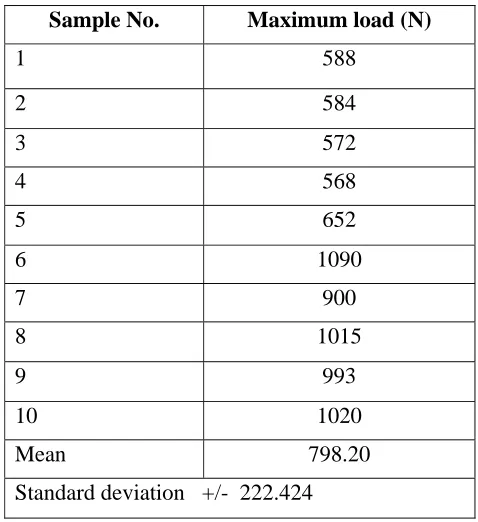
• Table I shows basic values, mean and standard deviation of maximum
load for fracture resistance of Group A samples.
• Table II shows basic values, mean and standard deviation of maximum
load for fracture resistance of Group B samples.
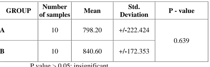
• Table III shows the Comparison between mean values of maximum
load for fracture resistance of Group A and B samples using
Independent student’s T-test.
• Table IV shows the fracture patterns with respect to locations of
fractures of Group A samples.
• Table V shows the fracture patterns with respect to locations of
fractures of Group B samples.
• Table VI shows the comparison of fracture patterns with respect to
locations of fractures of Group A and Group B samples.
• Graph I shows the basic values of maximum load for fracture
resistance of Group A samples.
• Graph II shows the basic values of maximum load for fracture
resistance of Group B samples.
• Graph III shows the comparison of mean values of maximum load for
fracture resistance of Group A and B samples
• Graph IV shows the comparison of fracture patterns with respect to
42
Table I: Basic values, mean and standard deviation of maximum load for
fracture resistance of Group A samples
Sample No. Maximum load (N)
1 588 2 584 3 572 4 568 5 652 6 1090 7 900 8 1015 9 993 10 1020 Mean 798.20 Standard deviation +/- 222.424
Table II: Basic values, mean and standard deviation of maximum load
for fracture resistance of Group B samples
Sample No. Maximum load (N)
[image:43.612.186.430.486.666.2]43
Table III: Comparison between mean values of maximum load for
fracture resistance of Group A and B using Independent
student’s T-Test
GROUP Number
of samples Mean
Std.
Deviation P - value
A 10 798.20 +/-222.424
0.639 B 10 840.60 +/-172.353
P value > 0.05; insignificant
Inference – On statistical comparison of the mean fracture resistance
values of Groups A and B, P value > 0.05, denoting no statistical significance
[image:44.612.141.453.450.684.2]between the two Groups.
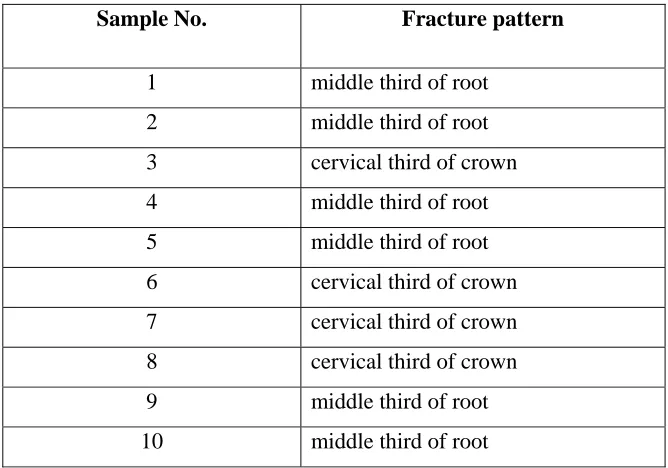
Table IV: Fracture patterns with respect to locations of fractures of
Group A samples.
Sample No. Fracture pattern
44
Table V: Fracture patterns with respect to locations of fractures of
Group B samples.
Table VI: Comparison of fracture patterns with respect to locations of
fractures of Group A and Group B samples
Groups No. of samples
(n)
Middle third of crown
Cervical third of
crown
Middle third of root
Apical third of
root
A 10 1 5 3 1
B 10 0 4 6 0
Sample No. Fracture pattern
[image:45.612.118.493.528.621.2]45
Graph I: Basic values of maximum load for fracture resistance of
Group A samples
Graph II: Basic values of maximum load for fracture resistance of Group
B samples
588 584 572 568 652
1090
900
1015 993 1020
0 200 400 600 800 1000 1200
1 2 3 4 5 6 7 8 9 10
Sample number
678 690 755 686 702 820 1035 850 1100 1090 0 200 400 600 800 1000 1200
1 2 3 4 5 6 7 8 9 10
46
Graph III : Comparison of mean values of maximum load for fracture
resistance of Group A and B samples.
Graph IV: Comparison of fracture patterns with respect to location of
fracture of Group A and B samples.
798.2 840.6
0 100 200 300 400 500 600 700 800 900 1000
New
ton
56
CONCLUSION
The following conclusions were drawn from this present in vitro study,
which was conducted to comparatively evaluate the fracture resistance and
fracture patterns of endodontically treated anterior teeth restored with two
different esthetic posts:
1. The mean maximum load for the fracture resistance of endodontically
treated anterior teeth with glass fibre posts was found to be 798.20
Newtons.
2. The mean maximum load for the fracture resistance of endodontically
treated anterior teeth with zirconia posts was found to be 840.60
Newtons.
3. On comparison, the mean maximum load value for fracture resistance
for test samples with zirconia posts was higher than that for the test
samples with glass fibre posts. However, the difference in mean values
was found to be statistically insignificant. (p value >0.05)
4. The fracture patterns with respect to locations of fractures for Group A
samples were as follows: Middle third of crown – 1 tooth (10%),
cervical third of crown -5 teeth (50%), middle third of root -3 teeth
(30%) and apical third of root -1 tooth (10%)
5. The fracture patterns with respect to locations of fractures for Group B
samples were as follows: cervical third of crown -4 teeth (40%) and
57
6. On comparison, Group A samples exhibited 10% of fracture in middle
third of crown while Group B samples exhibited no fracture in middle
third of crown. Group A samples exhibited 50% of fracture in cervical
third of crown while Group B samples exhibited 40% of fracture in
cervical third of crown. Group A samples exhibited 30% of fracture in
middle third of root while Group B samples exhibited (60%) of
fracture in middle third of root. Group A samples exhibited 10% of
fracture in apical third of root while Group B samples exhibited no
fracture in apical third of root. Overall, samples with glass fibre posts
exhibited greater crown fracture (60%) while samples with zirconia
58
SUMMARY
The present in vitro study was conducted to comparatively evaluate the
fracture resistance and fracture patterns of endodontically treated anterior
teeth restored with two different esthetic posts, namely glass fibre posts and
zirconia posts.
A total of twenty freshly extracted, maxillary central incisors were
used in this study. They were randomly divided into two test Groups A and B
of ten samples each. All the samples were endodontically treated. Tooth
preparation was done to obtain a 2mm ferrule for the crown and post space
preparation was done uniformly for all the samples and the teeth. Group A test
samples were restored with glass fibre posts and Group B with zirconia posts.
All the samples were then subjected to core build up with light cure composite
material. Nickel chromium cast copings were fabricated and cemented to the
prepared teeth with post-core. All the samples were embedded in acrylic resin
block with the periodontal ligament simulation and subjected to static loading
in universal testing machine. The maximum load at which fractures occurred
for all the test samples were recorded and tabulated.
Statistical analysis was made with independent student’s T test and P
value less than 0.05 was considered significant. Samples with zirconia posts
exhibited greater fracture resistance than those with glass fibre posts.
59
statistically significant. This study indicates that both the esthetic posts,
namely glass fibre and zirconia posts exhibited similar fracture resistance
statistically.
The fracture patterns with respect to locations of fractures were also
tabulated for Group A and Group B samples. Visual inspection of the fracture
patterns of the endodontically treated anterior teeth restored with glass fibre
and zirconia posts have revealed that use of zirconia posts exhibited 60% of
the root fracture while glass fibre posts exhibited 60% of crown fractures.
The choice of an appropriate restoration of endodontically treated teeth
is guided by strength and esthetics. Cast metal post and core foundation have a
long history of successful use due to the superior physical properties.
However, esthetics properties of these materials are limited when used to
support the all-ceramic restorations, in the prosthetic rehabilitation of
maxillary anterior region. The Prefabricated posts made of tooth coloured
material such as glass fibre or zirconia have become popular because they
increase the transmission of light within the roots and overlying tissues.
In this in vitro study, the post and core foundations with glass fibre and
zirconia posts exhibited higher values of fracture resistance (798.20 N, 840.60
N) than the maximum physiological forces of 200N acting on the teeth in the
oral cavity.40 Therefore, it may be suggested that endodontically treated
60
Hence, these posts can be used as an alternative to cast post core or
prefabricated metallic posts in the maxillary anterior region.
Further studies to examine fracture mechanics, as well as long-term
clinical investigations are needed to evaluate the performance of glass fibre
and zirconia posts.
61
BIBLIOGRAPHY
1. Akgungor G, Sen D, Aydin M. Influence of different surface treatments
on the short term bond strength and durability between a zirconia post
and a composite resin core material. J Prosthet Dent 2008; 99(5):
388-399.
2. Akkayan B. An in vitro study evaluating the effect of ferrule length on
fracture resistance of endodontically treated teeth restored with
fiber-reinforced and zirconia dowel systems J Prosthet Dent 2004; 92(2):
155-62.
3. Al-Hazaimeh N, Gutteridge DL. An in vitrostudy into the effect of the
ferrule preparation on the fracture resistance of crowned teeth
incorporating prefabricated post and composite core restorations. Int
Endodon J 2001; 34: 40-45.
4. Aquilino SA, Caplan DJ. Relationship between crown placement and
the survival of endodontically, treated teeth. J Prosthet Dent 2002; 87(3):
256-63.
5. Asmussen E, Peutzfeldt A, Sahafi A. Finite element analysis of stresses
in endodontically treated, dowel-restored teeth. J Prosthet Dent 2005;
94(4): 321-9.
6. Assif D, Oren E, Marshak BL, Aviv I. Photoelastic analysis of stress
transfer by endodontically treated teeth to the supporting structure using
7. Balbosh A, Kern M. Effect of surface treatment on retention of
glass-fiber endodontic posts. J Prosthet Dent 2006; 95(3): 218-23.
8. Bateman G, Ricketts DNJ, Saunders WP. Fibre-based post systems: a
review. British Dental J 2003; 195(1): 43-8.
9. Bex RT, Parker MW, Judkins JT, Pelleu GB. Effect of dentinal
bonded resin post-core preparations on resistance to vertical root
fracture. J Prosthet Dent 1992;67(6):768-72.
10.Burgess J.O, Summitt J.B, Robbins J.W. The resistance to tensile,
compression, and torsional forces provided by four post system. J
Prosthet Dent 1992; 68(6): 892-903.
11.Burns AD, Krause WR, Douglas HB, Burns DR. Stress distribution
surrounding endodontic posts. J Prosthet Dent 1990; 64(4): 412-8.
12.Cohen BI, Musikant L, Deutsch AS. Comparison of retentive
properties of four post systems. J Prosthet Dent 1992; 68(6): 264-268.
13.Cooney JP, Caputo AA, Trabet KC. Retention and stress distribution
of tapered-end endodontic posts. J Prosthet Dent 1986; 55(5): 540-6.
14.Creugers NHJ, Mentink AGM, Fokkinga WA, Kreulen CM. 5-Year
Follow-up of a Prospective Clinical Study on Various Types of Core
Restorations. Int J Prosthodont 2005; 18(1): 34-39.
15.Deutsch AS, Cavallari J, Musikant BL, Silverstein L, Lepley J,
Petroni G. Root fracture and the design of prefabricated posts J Prosthet
16.Deutsch AS, Musikant BL, Cavallari J, Bernardi S. Retentive
properties of a new post and core system. J Prosthet Dent 1985; 53(1);
12-14.
17.Dilmener FT, Sipahi C, Dalkiz M. Resistance of three new esthetic
post-and-core systems to compressive loading. J Prosthet Dent 2006;
95(2): 130-6.
18.Fernandes AS, Dessai GS. Factors affecting the fracture resistance of
post- core reconstructed teeth: A review. Int J Prosthodont 2001; 14(4):
355-363.
19.Gegauff AG: Effect of crown lengthening and ferrule placement on
static load failure of cemented cast post-cores and crowns. J Prosthet
Dent 2000; 84(2): 169-79.
20.Giovani AR, Vansan LP, Neto MDS, Paulino SM. In vitro fracture
resistance of glass-fiber and cast metal posts with different lengths. J
Prosthet Dent 2009; 101(3): 183-188.
21.Hatzikyriakos AH, Reisis GI, Tsingos N. A