TESTICULAR FINE NEEDLE ASPIRATION
CYTOLOGY AND HISTOPATHOLOGY
CORRELATION IN MALE INFERTILITY
Dissertation submitted in partial fulfillment of the requirements for the degree of
M.D. (Pathology) – Branch III
THE TAMILNADU DR.M.G.R.MEDICAL UNIVERSITY CHENNAI
CERTIFICATE
This is to certify that this dissertation entitled “TESTICULAR FINE NEEDLE ASPIRATION CYTOLOGY AND HISTOPATHOLOGY CORRELATION IN
MALE INFERTILITY” is a bonafide work done by Dr. M. SRIDEVI in partial fulfillment of the requirements of The TAMIL NADU DR.M.G.R. MEDICAL UNIVERSITY, Chennai for the award of M.D. Pathology Degree.
DIRECTOR GUIDE
Prof. Dr. A.SUNDARAM. M.D., Director and Head,
Institute of Pathology, Madras Medical College, Chennai – 600 003.
Prof. Dr. P. KARKUZHALI. M.D.,
Professor of Pathology, Institute of Pathology , Madras Medical College, Chennai – 600008.
DEAN
Prof. Dr. J. MOHANA SUNDARAM. M.D., Ph.D., D.N.B.
DECLARATION
I declare that this dissertation entitled “TESTICULAR FINE NEEDLE ASPIRATION CYTOLOGY AND HISTOPATHOLOGY CORRELATION IN
MALE INFERTILITY” done by me under the guidance and supervision of Prof. Dr. P. Karkuzhali M.D. It is submitted in partial fulfillment of the requirements for the award of the M.D., Pathology degree by The Tamilnadu Dr. M.G.R. Medical University, Chennai. This has not been submitted by me for the award of any degree or diploma from any other University.
ACKNOWLEDGEMENT
I express my sincere thanks to Prof. Dr. J. MOHANA SUNDARAM, M.D., Ph.D., D.N.B. Dean, Madras Medical College and Government General Hospital, for permitting me to utilize the facilities of the Institution.
I express my unfeigned thanks to Prof. Dr. A. SUNDARAM M.D., Director and Head, Institute of Pathology, Madras Medical College and Government General Hospital, for his constant encouragement.
I am extremely grateful to Prof. Dr. P. KARKUZHALI M.D., Professor of Pathology, Institute of Pathology, Madras Medical College and Government General Hospital, for her valuable guidance and inspiration throughout this study.
I am thankful to Prof. Dr. GEETHA DEVADAS M.D., D.C.P., Professor of Pathology, Institute of Pathology, Madras Medical College and Government General Hospital, for her helpful suggestions in carrying out this work.
I also wish to thank Prof. Dr. PAPPATHI M.D., D.C.H., Associate Professor of Pathology, Institute of Pathology, Madras Medical College and Government General Hospital, for her support.
I also wish to thank Prof. Dr. SUDHA VENKATESH M.D., Professor of Pathology, Institute of Pathology, Madras Medical College and Government General Hospital, for her encouragement.
I wish to thank Prof. Dr. T.B. UMADEVI M.D., Professor of Pathology, Institute of Child Health, Madras Medical College and Government General Hospital, for her support.
I am thankful to the Prof. Dr. SHANTHA RAVISHANKAR M.D., D.C.P., Professor of Neuro-Pathology, Institute of Pathology, Madras Medical College and Government General Hospital, for her helpful suggestions in carrying out this work.
I wish to thank Prof. Dr. K. RAMA M.D., Professor of Pathology, Institute of Govt. Kasturba Gandhi Hospital, Madras Medical College and Government General Hospital, for her support.
Institute of Obstetrics & Gynecology, Madras Medical College and Government General Hospital, for her encouragement.
I wish to thank Prof. Dr. T. CHITRA M.D., Professor of Pathology, Institute of Ophthalmology, Madras Medical College and Government General Hospital, for her support.
I wish to thank all the Assistant Professors of the Institute of Pathology, Madras Medical College and Government General Hospital, for their continuous support.
I would not like to miss thanking all my colleagues and friends, to whom I am indebted for their valuable time, generous support and motivation without which this study project would not have seen the light. I would also like to thank Dr. S. Y. Jagannathan for helping me in the statistical analysis.
I am also thankful to all the technicians who supported me in histotechnics and special stains.
I also express my gratitude to all the patients who were subjects of this study for their cooperation.
Words are not enough to thank my family for their understanding, moral support and encouragement.
CONTENTS
S.NO. PAGE NO.
1. INTRODUCTION 01
2. AIMS OF THE STUDY 04
3. REVIEW OF LITERATURE 05
4.
5.
MATERIALS AND METHODS
RESULTS
28
40
6. DISCUSSION 54
7. SUMMARY AND CONCLUSION 64
8. BIBLIOGRAPHY
9.
10.
MASTER CHART
INTRODUCTION
Male infertility is a common problem that can be devastating to a couple trying to
conceive. The statistics of infertility shows that 15% of all marriages face in future the
problems of infertility. The WHO has reported a core global prevalence of 5%
infertility in the mid 70s. In approximately 30% of cases, significant abnormalities are
found in the man alone, in another 20% of cases abnormalities are found in both the
man and the woman. Thus in roughly 50% of infertile couples, the male factors is at
least partially responsible for the failure to conceive. The diagnostic approach to a case
of male infertility includes a detailed clinical history and a thorough physical
examination supported by various laboratory investigations like semen analysis,
hormonal evaluation, detection of anti-sperm antibodies, sperm function assays,
ultrasound, vasography and invasive tests like testicular biopsy (or) FNAC (Fine
Needle Aspiration Cytology). FNAC has become one of the important methods of
investigations in cases of azoospermia and oligospermia. The technique of testicular
FNAC is more than 30 to 50 years old, but has become popular only in recent years.
Unlike FNA done in other sites, testicular FNA requires local anesthesia which could
be the reason for the non-popularity of the technique. Therefore only few studies have
been done so for.
Testicular biopsy can help us to differentiate a post-testicular, obstructive etiology
of male infertility from an intrinsic testicular cause. Though biopsy provides some
seminiferous tubules, it has its own complications like hematoma, fibrosis (very rarely),
scarring and sampling only a small volume of tissue. FNAC on the other hand is
reliable, quick, easy, less invasive and associated with no or minimal complications.
FNAC could give better morphological details of different stages of
spermatogenesis.
Studies have shown a good correlation between FNA and biopsy findings and
abnormal findings in FNA can be followed up and evaluated further with a formal
testicular biopsy. The concordance rate of FNA and histological diagnosis reached
>85% in many studies with high specificity and sensitivity approaching >95%.
In the past, findings of ↑FSH (Follicular stimulating hormone) concentration often
predetermined that infertility couples were recommended to use donor without further
investigations. However, focal areas of spermatogenesis may exist in men previously
considered to be devoid of spermatogenesis, such as in Sertoli Cell Only Syndrome
(SCOS) & maturation arrest. In such cases it is now possible for testicular FNA
‘mapping' for sperm recovery from different regions of the male reproductive system
for Intracytoplasmic sperm Injection (ICSI) enabling fertilization. By performing FNA
map prior to In Vitro Fertilization (IVF) cycle we can maximize testis parenchymal
conservation in non-azoospermic men with varied pathology. When post-testicular
obstructive cause is demonstrated, surgical correction may be indicated.
because of the heterogenecity of the pathologic process. The organ may contain more
mature germ cell lineage in small foci far from the site of biopsy. The wider sampling
area in the testis by FNA may result in a more accurate representation of pathology in
AIMS AND OBJECTIVES OF THE STUDY
1. To evaluate cytological features of testicular FNAC for diagnosis of Male Infertility
and to determine the diagnostic values and reliability of testicular FNA as a
cytological sampling technique.
2. Considering histopathology as the ‘gold standard’ to study the correlation between
cytological and histological diagnosis.
3. To correlate Johnson’s Score of histopathology with that of histological diagnosis.
4. To evaluate the possibility of replacing biopsy of azoospermic testes by FNA for
REVIEW OF LITERATURE
HISTORY:
Technique of testicular FNAC is more than 30-50 years old. The statistics of
infertility shows that 15% of all marriages face in future the problems of infertility.
Testicular FNAC was first attempted by Posner in 1905 and Huhner in 19281. Later most
of the work was done by Orbant in 19652 and Perssons in 19713 who characterized
different cell types in cytological smears, and demonstrated good correlation of
cytological diagnosis with histological categories. Mallidis and Baker in19944, AI-Jitawi
et al in 19975 and Abdulla et al in 20006 had described on FNA biopsy technique for
histological assessment in male infertility.
The role of testicular FNAC as a diagnostic parameter in the evaluation of
azoospermic patients were studied by Foresta et al in 19927 and Shoshana et al in 19938.
Dajani et al in 19989 had studied the use of testicular FNAC by grading the cytological
smears in 1000 infertile men. Later Rewat et al in 200710, AI-Rayes et al in 200011,
Kurien et al in 200312, Bettella et al in 200513 and Srivastava et al in 200414 had also
studied the role of testicular FNAC in male infertility. Bettella et al in 200513 studied the
role of FNAC in Testicular sperm extraction (TESE) and Gupta et al in 200615 had
described the role of testicular FNAC in patients with clinically obstructive
azoospermia. Angelocarpiel et al in 2002 16 had studied the use of FNAC in Cryptorchid
Craft et al in 199317, Schoysman et al in 199318, Tournaye et al in 199819 and
Bourne et al in 199520 had described about the recovery of mature sperms from
obstructive azoospermic patients for Intracytoplasmic Sperm Injection (ICSI). Later
studies reported the successful recovery of mature sperm in patients with
non-obstructive azoospermia (Devroy et al in 199521 and Friedler et al in 199722).
Craft and Tsirigotis in 199523 had described that multiple needle biopsies could
retrieve more spermatozoa than a single open biopsy. To determine more accurately
which men with non-obstructive azoospermia (NOA) are candidates for ICSI, diagnostic
and therapeutic testicular sperm extraction (TESE) with cryopreservation (Mulhall et al,
1997)24 and real-time, multi-biopsy approaches were performed with either optical
magnification (Schlegal et al, 1996)25 or classic testis biopsy (Tournaye et al, 1996)26.
Turek et al in 199927 had proved that by Testicular Sperm Extraction (TESE), In
Vitro-Fertilization (IVF) Cycle cancellation can be reduced and can maximize testis
parenchymal conservation in Non-Obstructive Azoospermia (NOA) men with atrophic
testes. Turek et al in 200028 had studied the diagnostic findings from testes FNA
mapping in obstructive azoospermia.
Infertility:
Infertility is defined as the inability to achieve pregnancy after one year of
CAUSES OF MALE INFERTILITY
1. Pre-testicular Causes :
• Disorders of the hypothalamic or pituitary endocrine diseases (Thyroid or Adrenal
disorders or Diabetes Mellitus)
• Metabolic disorders (Renal and Liver disease)
• Chronic Infection
• Drugs
2. Testicular Causes:
• Idiopathic Hypospermatogenesis or aspermatogenesis
• Developmental and genetic disorders (agonadism, cryptorchidism, SCOS and
Klinefelter’s Syndrome).
• Circulatory - varicocele or torsion.
• Inflammatory lesions – infections or immune causes
• Iatrogenic – chemical, radiation or surgical
• Environmental.
• Congenital Block: anomalies of excretory ducts or accessory glands.
• Acquired Block:
Inflammatory lesions of the excretory ducts and accessory glands.
Iatrogenic or post-traumatic lesions of the excretory ducts, accessory glands or
ejaculation nerve plexus29.
EVALUATION OF MALE INFERTILITY
1. Clinical History and Examination
2. Semen Analysis
3. Transrectal Ultrasonography
4. Vasogram
5. Testicular FNA
6. Testicular Biopsy
7. Others – Tests for antisperm antibodies, ova penetration, cervical mucus interaction,
hormonal assays, karyotype analysis.30
CLINICAL HISTORY AND EXAMINATION
smoking/alcoholism/intake of drugs, diseases like DM/TB/HT/STD/Thyroid problems
and also surgical history for maldescended testes/hernia repairs. They should be
examined for external genital abnormalities, testicular size, testicular consistency and
presence of varicocele. Then they should undergo routine examinations like HB%, blood
sugar, urea, creatinine and urine routine examination followed by other investigations
listed below:
INVESTIGATIONS
1. SEMEN ANALYSIS: Semen analysis should be the first step in the investigation. If
any gross abnormalities detected, then the couple should be councelled for the need
Collection of semen for analysis:
Collection is best done by masturbation failing which by coitus interruptus. The
semen is collected in a clean wide mouthed dry glass jar. Sample so collected should be
sent to the lab as early as possible so that the examination can be performed within 2
hours. Coitus should be avoided for at least 2 to 3 days prior to the test.
Normal Semen values (WHO 1999)32
In the selected cases, acid phosphatase, zinc (prostatic fluid content) and fructose
(seminal vesicle fluid content) are to be estimated. If the values are found lower than
mentioned, it is not wise to interpret the analysis as abnormal. However, one should
repeat the test at least twice at about two and half month’s interval, before signing out Volume – 2 ml or more
PH – 7.2 to 7.8
Sperm concentration – 20 million/ml or more Total Sperm Count ≥ 40 million per ejaculate
Motility – 50% or more progressive forward motility Morphology – 30% or more normal forms
Vitality – 75% or more living Leucocytes – <1 million/ml
Total Fructose – >13 mmol/ejaculate
Micro Agglutination Reaction (MAR) test – <10% sperms with adherent particles
the report as abnormal. In the semen analysis, patients are said to have normal study
when the above criteria’s are fulfilled. If the sperm count is less than 20 million it means
Oligospermic and Azoospermic if no sperms are found in the semen. Aspermia is a
condition where there won’t be any seminal ejaculate.
3. TRUS (Trans rectal Uretheroscopy): To visualize the seminal vesicles, prostate
and ejaculatory ducts obstruction.
4. VASOGRAM: It is a radiographic study done to evaluate ejaculatory duct
obstruction. It has been mostly replaced by TRUS.
5. TESTICULAR FNAC:
Diagnosis involves two steps:
1. Identification of the cell types present
2. The proportions of the cell population represented by each.
Two cell populations are evident in cytology.33
• Sertoli cells
• Cells of various stages of spermatogenesis
The spermatogenetic cells are divided into spermatogonia, primary spermatocytes,
Thus accurate recognition of the normal cell types in FNA of testis is the key to
diagnosis. The MGG Staining method is used throughout. The following descriptions
apply to MGG stained FNA smears.34, 35
Cytological features of these cells are described below:
Sertoli cells: They have round or oval nucleus with a rather smooth chromatin
pattern. Large pale or blue nucleoli are usually present. The cytoplasm is abundant, pale
slate blue and is usually foamy with poorly-defined borders. Although occurring singly,
these cells usually form sheets or a loose matrix. When spermatogenesis is present the
spermatogenic cells are interposed within the matrix. Bare nuclei presumably extruded
from the matrix are common. Sertoli cells are invariably present even in the total
absence of spermatogenesis. Their presence therefore gives the cytologist the confidence
that the testis has been sampled correctly.
Spermatogenetic cells (in order of maturation):
Spermatogonia: They contain 16-20µm round or oval, slightly eccentrically
placed nucleus with smooth finely condensed nuclear chromatin which is either pale
staining (light) or dark staining. Nucleoli are not usually seen. The cytoplasm is
homogenous and has well defined border. Spermatogonia are outnumbered by
spermatocytes in normal spermatogenesis and may only occur in relatively small
Primary Spermatocytes: It’s the largest germ cell. It contains a round nucleus,
14-20 µm, the size depending upon the state of maturation, with a heavy coarse
chromatin pattern. The nuclear chromatin shows a ‘chunky’ appearance with a clear
dark/light effect. The cytoplasm stains deeply hyper-basophilic and is moderate in
amount. Nucleoli are not seen.
Secondary Spermatocytes: It contains a round nucleus, variable in size
depending on maturation; from 8-16 µm. Binucleate forms are common. The chromatin
pattern is coarse but to a lesser extent than that seen in the primary spermatocytes and
exhibits a similar pattern. The cytoplasm is moderate in amount, basophilic but not
hyperbasopholic as seen in the primary spermatocyte. As the cell matures towards
spermatid the cytoplasm is often reasonably abundant. Nucleoli are not seen. These cells
are rarely identified because of their shorter life span and immediate transformation to
spermatids
Spermatids: A small cell, although the size is variable. The nucleus is less than
8 µm depending, as in the other spermatogenic cells, upon maturation stage. In the
‘close to mature’ stage, the nucleus of course resembles a sperm head. The nuclear
chromatin is darkly staining and smooth. The cytoplasm is grey blue and often shows a
ragged, uneven border. Sperm tails are commonly seen either in or protruding from the
cytoplasm.
is found on opposite side of acrosome. This end point is proof that, spermatogenesis, the
transformation of spermatid to spermatozoa, is functional. A maturation arrest at the
spermatid level is not uncommon which would of course produce azoospermia and
infertility and therefore a cytological conclusion that the testis is functioning normally
must include observation of all stages of maturation.
Leydig (interstitial) cells: Leydig cells are relatively uncommon in testicular
cytology when compared with the other cellular components. They are however usually
present in small numbers if careful scrutiny is applied. The nucleus is 10-12 µm, round,
darkly staining with a relatively smooth chromatin. The cytoplasm is abundant and
stains basophilic. The cell borders are usually clearly defined in contrast to the poorly
defined sertoli cell borders. The cytoplasm is also cleaner and smoother when compared
with sertoli cells. Scattered green/blue granules are seen lying within the cytoplasm.
These cells can occur singly but are usually seen in small sheets, similar in pattern to
those cells seen in tissue sections lying between the tubules of the testis in clusters.
Mesothelial cells:
Mesothelial cells are an expected finding, picked up on the way in from the
scrotal lining and occur in large monolayer sheets with moderately high N/C ratio. They
have moderate amount of bluish cytoplasm & well demarcated cell borders, large nuclei
& prominent nucleoli. Cytoplasmic vacuoles may be seen. It is important to identify the
sample is indeed from the testis and not a failure.
Large numbers of degenerate autolysing cell forms are a feature of testicular
cytology. This effect is presumed to be due to the rapid ‘turnover’ of the spermatogenic
cells within the organ and consequent loss of many, not an artifact produced by the
heavy handedness of the sampler.
Specimen adequacy for FNA:
If at least 200 cells could be counted on minimum in one well spread slide, the
specimen is considered adequate. Approximately 97% testicular FNA yield adequate
specimen for evaluation of spermatogenesis.
Based on various proportions of the different cell types, the smear is categorized
into six groups15:
1. Normal spermatogenesis – This pattern is reported when the smears show spermatogonia, spermatocytes, spermatids, many spermatozoa and a proportional
numbers of sertoli cells forming roughly one third of the total spermatogenetic cells.
2. Hypospermatogenesis – This pattern is described when all types up to spermatozoa are present and the proportion of sertoli cells to spermatogenic cells is increased.
spermatozoa.
4. Late Maturation Arrest – In these the smears were characterized by the total absence of spermatozoa and significant relative increase in proportions of round and
elongated spermatids along with spermatocytes and sertoli cells.
5. Testicular Atrophy – The smears have scanty cellularity and few sertoli cells.
CYTOLOGICAL GRADING
Cytological smears were also graded according to Dajani et al9 by counting 100
consecutive spermatogenic and sertoli cells in a well spread area with good cellularity in
a random manner using light microscopy as follows:
Grade A: spermatozoa were detectable
A1: Adequate production of fully developed spermatozoa
A2: Low, scanty or rare fully developed spermatozoa.
Grade B: Germ cells seen but no spermatozoa present. Includes maturation arrest at the
level of spermatid, spermatocyte or spermatogonia stages.
Grade C: Sertoli cell only pattern; no germ cells detected.
CELL INDICES:
Various indices were calculated according to Agarwal et al37 as follows:
Spermatic index (S.I):
SI is the number of spermatozoa per 100 spermatogenic cells. Its normal value is
51.6 ± 12.4.
Sertoli cell index (SEI):
SEI is the number of sertoli cells per 100 spermatogenic cells. Its normal value is
42.4 ± 11.4.
Sperm- sertoli index (SPSEI): SPSEI is an expression of number of
spermatozoa per 100 sertoli cells.
IN NORMAL SPERMATOGENESIS: SI and SPSEI will be higher than SEI.
IN HYPOSPERMATOGENESIS: SI and SPSEI will be lower than in normal
spermatogenesis and SEI will be higher than in normal spermatogenesis and maturation
arrest.
IN MATURATION ARREST: SI and SPSEI will be zero and SEI will be higher
than in normal spermatogenesis.
spermatogenic cells.
TESTICULAR BIOPSY:
The specimens were studied histopathologically for evaluation of the following
criteria39, 40:
- General architecture (150-250 µm diameters of tubules)
- Number of seminiferous tubules in specimen (usually 20-30 cross sections of
tubules)
- Seminiferous tubules pattern
- Germ cell/ Sertoli cell ratio
- The Basement membrane
- Interstitial tissue
- Leydig cells
- Tubular Hyalinization
- Tunica albuginea
Methods to estimate the degree of spermatogenesis:
1. Johnson Scoring41
Score 10 -Complete Spermatogenesis and perfect tubules
Score 9– Many spermatozoa present but disorganized spermatogenesis
Score 8 – Only a few spermatozoa present
Score 7 - No spermatozoa but many spermatids present
Score 6 - No spermatozoa, only a few spermatids present
Score 5 - No spermatozoa or spermatids present, but many spermatocytes present
Score 4 –Only a few spermatocytes present
Score 3 -Only Spermatogonia present
Score 2 - No germ cells, only sertoli cells present
Score 1 - No germ cells or sertoli cells
The mean Johnson Score of normal testis is 9.39±0.24 and 60% or more of the
tubules should score at10.
In pituitary hypogonadism → 3.95±1.4
In Klinefelter’s Syndrome → 1.3±0.3
2. Silber-Rodiguer-Rigarr Method :
Counting round mature spermatids (small round, dark nuclei) per tubule cross
section. At least 20 round spermatids/tubules – Ideally counting in 20 tubules. If it is 45,
40, 20 and 6-10 then the sperm count is approximately 85, 45, 10 and 3 million/ml. If
sperm count is lower than expected it indicates partial obstruction42.
3. Estimating Germ Cells: Sertoli Cell Ratio (Sertoli Index): Counting at least
30 tubule cross sections .This ratio is constant at about 13: 1 in young healthy men.
Average of 12 sertoli cell per tubular cross sections is considered normal.
In the histopathology, five patterns are observed40. They are:
1. Normal spermatogenesis
2. Hypospermatogenesis
3. Maturation Arrest
4. Germ Cell Aplasia
5. Tubular hyalinization
with portions of septa. Germ cells in all stages of spermatogenesis are seen within the
seminiferous tubules in cases of normal spermatogenesis. Seminiferous tubules are so
convoluted that they are mostly seen in cross sections. Maturation proceeds in an
orderly overlapping helical pattern along the length of a tubule. So not all stages are
seen within the tubules. Normal spermatogenesis is found in azoospermic patterns
with excurrent duct obstruction.
2. Hypospermatogenesis: A reduction in the number of all germinal elements,
including the late spermatids/spermatozoa is present in cases of
hypospermatogenesis. Thus, histologically germinal epithelium may be hypoplastic
and therefore thinner than normal. The organization of the germinal epithelium may
be disrupted and immature germ cell may be found in the lumen in some instances.
The interstitial (Leydig) cells are normal. It is associated with oligospermia in the
range of 2 to 20 million sperms per ml and severe cases may show azoospermia.
Associated changes may include tunica propria thickening, focal interstitial fibrosis
and focal tubular sclerosis. In some cases the spermatogenic cells occupy only a
small portion of the entire circumference of the tubules, whereas other tubules
contain only sertoli cells. Many genetic, toxic, infectious and vascular disorders may
produce the same morphologic changes.
3. Maturation Arrest: Histological examination reveals spermatogenesis proceeding
is identified. The arrest may occur at the primary spermatocyte, secondary
spermatocyte or early spermatid stage but most often occurs at the level of the
primary spermatocyte. The tubules in these settings usually show identical
morphologic changes in that all appear arrested at the same stage of spermatogenesis.
Patients with complete maturation arrest at any stage shows no late
spermatids/spermatozoa, whereas those with incomplete maturation arrest show
arrest at different stages of spermatogenesis. In such cases, some tubules show
spermatogenetic cells arrested at one stage (for e.g., spermatids) and adjacent tubules
at another stage (for e.g., primary spermatocytes). Thus patients with complete arrest
exhibit azoospermia, whereas patients with incomplete arrest may have varying
degrees of oligospermia.
4. Germinal Cell Aplasia: It is also known as sertoli cell only syndrome
(SCOS). It is the severe form of these disorders and is invariably accompanied by
azoospermia. This finding is noted in 10 to 20% of all testicular biopsy specimens
obtained from infertile men. Testicular histology reveals seminiferous tubules lined
by sertoli cells with a complete absence of all germ cells. The oval nuclei of the
sertoli cells are typically located in the mid-portion of their cytoplasm with their
longitudinal diameter perpendicular to the basement membrane of the tubules. The
diameter of the seminiferous tubule is reduced and the interstitium is usually
minimally altered. Patients with this syndrome have small to normal sized testis
predominantly sertoli cell only patterns, but careful evaluation reveals a few scattered
germ cells, mostly spermatogonia. This represents germ cell hypoplasia of variable
degrees. Rarely, testis biopsy may show a combination of areas with sertoli cell only
patterns in combination with maturation arrest.
Testis biopsies in Klinefelter syndrome usually show absent spermatogenesis,
tubular sclerosis and prominent Leydig cells often in the form of nodules. However
some biopsies may also show some degree of spermatogenesis. Spermatozoa have been
observed in the wet preparations of testis biopsies and a successful ICSI have been done.
5. Tubular hyalinization: It is also known as tubular sclerosis or “end-stage” testis
disease as it is the end-product of many forms of tubular injury. All disturbances of
spermatogenesis may ultimately evolve into this end-stage disease. Hyalinization of
the tubules is associated with a loss of germinal epithelium, obliteration of the
lumens and fibrosis of the interstitium. Sertoli cells may or may not be present.
Leydig cells may be absent or decreased in number within the sclerotic interstitium.
Foci of spermatogenesis may be seen in some of the preserved tubules, but even
these tubules are probably obstructed by fibrous tissue and not connected to
excretory ducts. Clinically, these testes are bilaterally atrophic and firm. This
appearance may ultimately be found in undescended testes, remote chronic orchitis or
ischemia, acquired gonodotropin deficiency, testes of karyotypic abnormalities, or
Other patterns: In cases of hypogonodotropic hypogonadism, congenital or
acquired lesions of the hypothalamus and pituitary the development and function of
testes are affected by these disorders. It can be caused by tumours, inflammation or
genetic and developmental syndromes. Various drugs, trauma, or irradiation of the
hypothalamus may have the same consequences. All these conditions are characterized
by low levels of serum gonadotropins. The testes are small and lined with immature
sertoli cells that have round nuclei and inconspicuous nucleoli. Leydig cells are absent.
In cases of isolated LH deficiency, normal spermatogenesis may be reduced. Treatment
with GnRH given in a pulsatile manner may initiate and maintain spermatogenesis and
cure infertility.
Excurrent Duct obstruction:-
Normal spermatogenesis is seen associated with dilated tubular lumens,
disorganized germ cells, germ cells sloughing and thickened tunica propria.
The testicular biopsy is rarely pathognomonic of a single etiology. In addition,
several patterns may be present in an individual biopsy specimen. Thus in most cases, a
testicular biopsy does not result in the identification of a specific etiologic factor of a
patient’s infertility.
Fine Needle Aspiration versus biopsy:
of infertility as compared with biopsy. Many have claimed that FNA is superior to
biopsy. FNA cytology sometimes provides information not evident on histology mainly
because of the heterogeneity of the testes, the organ may contain more mature germ cell
lineage in small foci far from the site of biopsy. The wider sampling area in the testis by
FNA may result in a more accurate representation of true histological geography and
variation that exists within the organ. Despite a histological finding of no germ cells
within the seminiferous tubules, clinical experience in men undergoing testicular sperm
extraction and intracytoplasmic sperm injection has demonstrated that testis with this
histology can harbor fertile sperm. This fact underlines 2 important practical points:
(1) The importance of careful examination of a testicular biopsy to find mixed
histological patterns of infertility
(2) The benefit of wider geographic sampling, which can be achieved more practically
by FNA than with multiple biopsies.
Many studies have revealed the concordance rate of FNA and histological
diagnosis as >85% with high specificity approaching >95%.
MATERIAL AND METHODS
Urology OT (Operation theatre), Madras Medical College, performed during the period
of June 2007 – August 2009. All patients signed informed consent prior to aspiration.
Indications43:
ABSOLUTE
1. Azoospermia
2. Oligozoospermia
3. Teratozoospermia (Abnormal form)
4. Atypical cells in ejaculate
RELATIVE
1. Varicocele
2. Cryptorchidism
3. Chronic infection
Contraindication
1. Severe anaemia
2. Unco-operative patients
Patient’s selection: The study was done in patients in whom three consecutive
semen analyses showed oligospermia or azoospermia. In case of primary hypogonadism,
biopsy was not done in these cases. All semen analyses were done only after a period of
abstinence of at least 4 days. A clinical examination was then conducted and relevant
personal and clinical data were noted. Patients were then subjected to a FNA of the
testes for cytological evaluation and open testicular biopsy was done for
histopathological correlation.
Patient preparation:
Procedures and risks involved in the procedures were explained and informed
consent was obtained. Routine investigations like Hb%, blood sugar, urea, creatinine
and urine routine examination were done.
Semen Analysis:
All semen analyses were done only after a period of abstinence of at least 4 days.
The semen was examined for the following: colour, volume, ph, liquefaction time,
sperm count, motility, morphology and leucocytes. Then the results are given as either
normal (If sperm count > 20 million) or oligospermic (If sperm count <20 million) or
azoospermic (If no sperms are seen in the semen).
FNA technique:
They were done in the Urology OT (Operation theatre); Madras Medical College
findings. Then spermatic cord block was done by injecting 5 ml of 1% xylocaine. Five
minutes after injection of local anaesthesia, larger of the two testes was held with (L)
hand and was positioned with epididymis in the posterior position. Testis was gently
held between the thumb and the index finger. Using (R) hand, the testis was punctured
with a 23 gauge needle attached to a 5 ml of disposable syringe. Needle was passed in an
axis perpendicular to longitudinal axis of testes in the middle of the anterior surface of
testis opposite to epididymis. With a to and fro motion of the needle, aspiration was
done twice or thrice under negative pressure. Negative suction was released and tissue
fragments were then expelled onto a clear glass slide, gently smeared. If too much
pressure is applied it may lead to marked distortion and crushing artifacts. Open
testicular biopsy was performed immediately following the procedure of FNAC of the
testis9. Smears were air-dried and stained with May Grunwald - Giemsa (MGG) as well
as fixed in 95% alcohol and stained with H&E.
Procedures:
1. May Grunwald - Giemsa (MGG) stain:
1. Smears are air dried, fixed in acetone free methanol for 30 minutes.
2. Stain in May Grunwald for 5 minutes
3. Stain in Giemsa for 15 minutes
5. Air dry the slides
6. Clear in xylene (optional) for 5 minutes
7. Mount the slides with DPX(Dibutyl Pthalate Xylene).
2. Hematoxylin and Eosin stain:
1. Smears are fixed in 85% Isopropyl alcohol for 20 minutes
2. Stain in Harris Hematoxylin for 5 minutes
3. Wash well in tap water
4. Differentiate in 0.5% acid alcohol for 3 to 5 seconds
5. Wash well in tab water for blueing for 10-20 minutes
6. Dip in 1% aqueous eosin for once
7. Wash in running tap water for 1 to 5 minutes
8. Slides are dried and mounted in DPX (Dibutyl Pthalate Xylene).
Testicular biopsy technique:
A 1 to 1.5 cm incision was made on the convexity of the centre of anterior
portion of testes, through the portal of entry of needle, through the skin and tunica
albuginea with size 11 scalpel blade. Gentle pressure on testis extruded a small amount
(2 to 3 mm) of testicular parenchyma from the incision, which was then excised with a
Bouin’s fluid, specimen processed and stained with hematoxylin and eosin for HPE
examination. After securing homeostasis, Tunica albuginea, Tunica Vaginalis and skin
with subcutis were closed in layers with 3/0 vicryl sutures. A dry scrotal dressing was
then applied40.
Staining Procedure for HPE:
Hematoxylin and Eosin stain:
1. Dewax sections, hydrate through graded alcohols to water.
2. Stain in Harris Hematoxylin for 5 minutes
3. Wash well in tap water
4. Differentiate in 1% acid alcohol
5. Wash well in tab water for blueing for 10-15 minutes
6. Stain in 1% eosin for 1 to 2 minutes
7. Wash in running tap water for 1 to 5 minutes
8. Dehydrate in alcohols, clear in Xylol and mount in DPX.
Aftercare (follow -up):
The patients stayed in the recovery room for 30-90 minutes and were sent home
on recovery from anaesthesia. No antibiotics or pressure scrotal dressings were applied.
swelling in the suture site. They were seen 2 weeks after the procedure, and enquiries
were made about pain, swelling, infection and intercourse. Testicular volume was
estimated with a Prader Orchidometer and the biopsy sites were examined.
The study populations of 35 infertile cases were studied on the basis of the following parameters:-
Age group (ranging from 20-40 years).
Duration of infertility (ranging from 2-14 years).
Sperm count and sperm count correlation with HPE and Cytological diagnosis.
Based on sperm count, the cases were grouped under azoospermia if no sperms are seen
in semen and Oligospermia if sperm count was <20 millions. In addition semen was also
examined for colour, volume, ph, liquefaction time, sperm count, motility, morphology
and leucocytes .
Cytological grading: Cytological smears were also graded according to Dajani et
al9 by counting 100 consecutive spermatogenic and sertoli cells in a well spread area
with good cellularity in a random manner using light microscopy as follows:
Grade A: spermatozoa were detectable
A1: Adequate production of fully developed spermatozoa
Grade B: Germ cells seen but no spermatozoa present. It includes maturation
arrest at spermatid, spermatocyte or spermatogonia stages.
Grade C: Sertoli cell only pattern; no germ cells detected.
Grade D: Sclerosis or lack of both germinal and sertoli cells.
Cytological diagnosis:
Based on various proportions of the different cell types, the smear is categorized
into six groups15:
1. Normal spermatogenesis – This pattern is reported when the smears show
spermatogonia, spermatocytes, spermatids, many spermatozoa and a proportional
numbers of sertoli cells forming roughly one third of the total spermatogenetic cells.
2. Hypospermatogenesis – This pattern is described when all types up to spermatozoa
are present and the proportion of sertoli cells to spermatogenic cells is increased.
3. Early Maturation Arrest – In this category smears will show a high percentage of
spermatogonia and primary spermatocytes along with absence of spermatids and
spermatozoa.
4. Late Maturation Arrest – In these the smears were characterized by the total
absence of spermatozoa and significant relative increase in proportions of round and
5. Testicular Atrophy – The smears have scanty cellularity and few sertoli cells.
CELL INDICES:
Various indices were calculated according to Agarwal et al37.100 consecutive
spermatogenic and sertoli cells were counted in a well spread portion of the smear and
the percentage of spermatozoa per 100 spermatogenic cells (Spermatic index) and the
number of sertoli cells per 100 spermatogenic cells (Sertoli cell index) were calculated
based on Agarwal et al37. The mean value of these indices in each category of the
cytological diagnosis is then taken and compared with different categories of cytological
diagnosis.
Spermatic index (S.I):
SI is the number of spermatozoa per 100 spermatogenic cells.
Sertoli cell index (SEI):
SEI is the number of sertoli cells per 100 spermatogenic cells.
Sperm- sertoli index (SPSEI):
SPSEI is an expression of number of spermatozoa per 100 sertoli cells
In normal spermatogenesis: SI and SPSEI will be higher than SEI.
In hypospermatogenesis: SI and SPSEI will be lower than in normal
arrest.
In maturation arrest: SI and SPSEI will be zero and SEI will be higher than in
normal spermatogenesis.
In SCOS: SI will be zero and SEI & SPSEI cannot be calculated as there are no
sertoli cells.
HISTOLOGICAL DIAGNOSIS:
In the histopathology, the predominant pattern was taken into account and
classified into the following five patterns 40. They are:
i. Normal spermatogenesis: Germ cells in all stages of spermatogenesis are seen
within the seminiferous tubules in cases of normal spermatogenesis
ii. Hypospermatogenesis: A reduction in the number of all germinal elements,
including the late spermatids/spermatozoa is present in cases of
hypospermatogenesis.
iii. Maturation Arrest: Histological examination reveals spermatogenesis
proceeding normally through a specific stage at which point no further
maturation of germ cells is identified. The arrest may occur at the primary
iv. Germ Cell Aplasia: Testicular histology reveals seminiferous tubules lined by
sertoli cells with a complete absence of all germ cells.
v. Tubular hyalinization: Hyalinization of the tubules is associated with a loss of
germinal epithelium, obliteration of the lumens and fibrosis of the interstitium.
Sertoli cells may or may not be present. Leydig cells may be absent or decreased
in number within the sclerotic interstitium.
JOHNSON SCORING41:
100 consecutive seminiferous tubules were counted and the mean was taken and
compared with histological report.
Score 10 - Complete Spermatogenesis and perfect tubules
Score 9 - Many spermatozoa present but disorganized spermatogenesis
Score 8 - Only a few spermatozoa present
Score 7 – No spermatozoa but many spermatids present
Score 6 - No spermatozoa, only a few spermatids present
Score 5 - No spermatozoa or spermatids present, but many spermatocytes present
Score 4 - Only a few spermatocytes present
Score 3 - Spermatogonia only
Score 1 - No germ cells or sertoli cells
The mean Johnson Score of normal testis is 9.39±0.24
Statistical analysis:
The patients were statistically analyzed on the basis of the following formulas:
Sensitivity a / ( a + b) x100
Specificity d / (c + d) x 100
Positive Predictive value a / (a + c) x 100
Negative Predictive value d / (b +d) x 100
a True positive
b False negative
c False positive
d True negative
a + b Diseased
c + d Not diseased
RESULTS
This study was done on 36 cases, out of which 1 case has been excluded as the
sample did not contain testicular tissue. The results of 35 cases taken for study were
tabulated. The age group ranged from 20 to 40 years (Table 1) & (Fig 1) with a mean
[image:42.612.71.463.228.454.2]age of 30.4 yrs.
Age No. of cases(35) % of total
20-30 16 45.7%
31-40 18 51.4%
41-50 1 2.9%
MEAN AGE 30.4 yrs
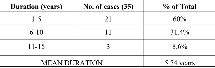
The duration of infertility ranged from 2 years to 14 years (Table 2) & (Fig 2)
[image:43.612.61.455.54.192.2]with mean duration of 5.7 years.
Table 2: study population- distribution of infertility duration
Duration (years) No. of cases (35) % of Total
1-5 21 60%
6-10 11 31.4%
11-15 3 8.6%
MEAN DURATION 5.74 years
[image:43.612.66.477.347.477.2]Fig 2:
Almost all patients had Primary infertility except for one patient who had
secondary infertility. In semen analysis, the sperm count ranged from azoospermia to
[image:44.612.113.502.359.611.2]cytology diagnosis.
Azoospermia is correlated with maturation arrest, germ cell aplasia and tubular
hyalinization. Oligospermia is correlated with hypospermatogenesis and normal
[image:45.612.52.491.230.350.2]spermatogenesis (table 3) & (fig.4):
Table 3: Sperm count correlation with HPE and Cytological diagnosis
Sperm count No. of cases
% of Total
HP correlation Cytology correlation No. of
cases
% No .of cases
%
Azoospermia 27 77.14% 18 67% 17 63%
Oligospermia 8 22.86% 4 50% 6 75%
Total 35 100% 22 58.5% 23 69%
Out of 35 cases, 27 cases (77.14%) were azoospermic. Out of this 18 cases (67%)
correlated with HP diagnosis and 17 cases (63%) correlated with cytological diagnosis.
Discordance of 9 cases in HPE and 10 cases in cytology was found to be due to
obstructive causes except in two cases. Out of 35 cases, 8 cases (22.86%) were
oligospermic, out of which 4 cases (50%) correlated with HP diagnosis and 6 cases
Fig:4
Fig:4
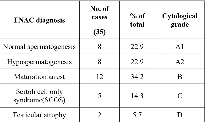
et al35 and Foresta et al7. Cytological smears were graded according to Dajani et al9 and
[image:47.612.67.460.181.414.2]correlated with the cytological diagnosis (Table 4) & (Fig 5).
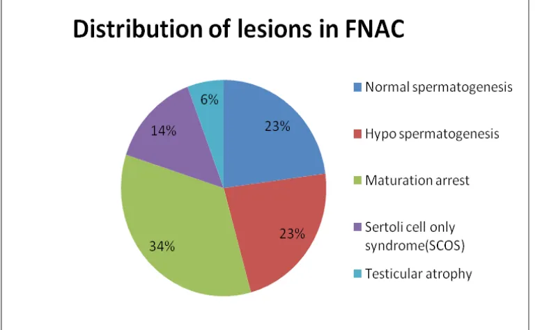
Table 4: Distribution of Cytological diagnosis and cytological grade
FNAC diagnosis
No. of cases
(35)
% of total
Cytological grade
Normal spermatogenesis 8 22.9 A1
Hypospermatogenesis 8 22.9 A2
Maturation arrest 12 34.2 B
Sertoli cell only
syndrome(SCOS) 5 14.3 C
Testicular atrophy 2 5.7 D
The most frequent diagnosis on cytological report was Maturation arrest (Fig. no.
9, 11, 13) with 8 cases. Next common diagnoses were Normal spermatogenesis (Fig.
no.1-4) and Hypospermatogenesis (Fig. no.7) with 8 cases each. SCOS (Fig. no.16, 17)
was seen in 5 cases and testicular atrophy (Fig. no. 20, 21) seen in 2 cases. Consequently
the common cytological grades in the descending order of frequencies were B, A2 &
A1, C & D.
Fig 5:
100 consecutive spermatogenic and sertoli cells were counted in a well spread
portion of the smear and the percentage of spermatozoa per 100 spermatic cells
[image:48.612.116.500.58.295.2]cell index) were calculated based on Agarwal et al37. The mean value of these indices in
[image:49.612.61.470.173.382.2]each category of the cytological diagnosis is given in Table 5.
Table 5: various cell indices in different categories of cytological diagnosis
Cytological diagnosis No. of cases
Indices Spermatic
Index (S.I)
Sertoli Cell Index (SEI)
Normal spermatogenesis 8 54.25 43.75
Hypo spermatogenesis 8 6.2 123
Maturation arrest 12 0 99.3
Sertoli cell only syndrome
5 0
-Testicular atrophy 2 0
Progressive decreasing values of SI were seen in normal spermatogenesis,
hypospermatogenesis and maturation arrest. SEI was found to be the most important
index in distinguishing hypospermatogenesis from normal spermatogenesis since SEI
was found to be elevated in hypospermatogenesis. Maturation arrest was distinguished
from hypospermatogenesis by the SI which was severely decreased with absent
spermatozoa. In SCOS, SI and SPSEI were zero.
In histological classification, the predominant pattern was taken into account and
presented in Table 5 & Fig 6
Histological classification No. of cases
(35) % of Total
Normal spermatogenesis 8 23%
Hypospermatogenesis 5 14.2%
Maturation arrest 13 37.1%
Germ cell Aplasia 7 20%
Tubular hyalinization 2 5.7%
Most frequent diagnosis was Maturation arrest (Fig. no.10, 12, 14, 15) with 13 cases
(37.1%). Next common diagnosis was normal spermatogenesis (Fig. no.5, 6) with 8
cases (23%). Tubular hyalinization (Fig. no.22) was seen in 2 cases (5.7%). Germ cell
aplasia (Fig. no.18,19) was seen in 7 cases and hypospermatogenesis (Fig. no.8) in 5
cases.
In all the cases, Johnson’s scoring was done. To do Johnson’s scoring 100
consecutive seminiferous tubules were counted and the mean was taken and compared
[image:51.612.62.477.198.443.2]with histological report (Table 6 & Fig 7).
Table 6: Correlation between Johnson’s scoring and histology diagnosis
Johnson’s scoring No. of cases % of total Histological diagnosis No. of cases % of correlation
9 &10 7 20% Normal
spermatogenesis
7 100%
8 6 17.1% Hypo
spermatogenesis
5 83.3%
7-3 13 37.2% Maturation arrest 12 92.3%
2 7 20% Germ cell
Aplasia
7 100%
1 2 5.7% Tubular
Hyalinization
2 100%
Total 35 100% Total 33 94.3%
Most of the cases scored between 3 to 7 which means maturation arrest. Scores 9
& 10 correlated well with Normal spermatogenesis and score 2 and 1 with Germ cell
Aplasia and Tubular Hyalinization. There were discordance in 2 cases, where score 7
was reported as Hypospermatogenesis and score 8 as Normal spermatogenesis.
Considering histopathology as the gold standard for definitive diagnosis of any
lesion, of the 35 cases of our study, 30 cases correlated well with FNAC. The overall
percentage of correlation with respect to HPE was 86%. 8 cases diagnosed as Normal
spermatogenesis by HPE were also diagnosed the same in FNAC with 100% correlation.
Similarly 5 cases diagnosed as hypospermatogenesis and 2 cases diagnosed as tubular
hyalinization were diagnosed the same in FNAC with 100% correlation. Of 13 cases
diagnosed as Maturation arrest in HPE, 10 cases were diagnosed the same in FNAC.
Remaining 3 cases were diagnosed as Hypospermatogenesis since the smears contained
few mature spermatozoa. Of the 7 cases diagnosed as SCOS in HPE, 5 cases were
diagnosed the same in FNAC. Remaining 2 of the cases were diagnosed as early
maturation arrest, since some spermatogonia were seen in the FNAC. The results of
[image:52.612.57.482.631.720.2]HPE and FNAC correlation are given in Table 7 & Fig 8.
Table 7: FNA-Histopathology Correlation
Patterns HPE FNAC % correlation
Normal spermatogenesis 8 8 100%
Maturation arrest 13 10 76.9%
Germ cell Aplasia (SCOS) 7 5 71.4%
Tubular Hyalinization 2 2 100%
Total 35 30 86%
Fig 8:
[image:53.612.60.480.55.181.2] [image:53.612.56.474.57.465.2]Statistical Analysis:
FNA finding of normal spermatogenesis and tubular hyalinization showed
[image:54.612.106.508.55.320.2]sensitivity,specificity, positive predictive value, negative predictive value of 100%.
Table 8: Statistical Analysis
Patterns Sensitivity Specificity
+ ve
Predictive value (%)
-ve
Predictive value (%)
Normal
Hypo
spermatogenesis 100% 90% 62.5% 100%
Maturation
arrest 77% 91% 83.3% 87%
Germ cell
Aplasia 71.4% 100% 100% 93.3%
Tubular
Hyalinization 100% 100% 100% 100%
Total 89.7% 96.2% 89.2% 96.1%
FNA finding of hypospermatogenesis showed a sensitivity of 100%, specificity of
90%, positive predictive value of 62.5%, and negative predictive value of 100%. FNA
finding of maturation arrest showed a sensitivity of 77%, specificity of 91%, positive
predictive value of 83.3% and negative predictive value of 87%.FNA finding of SCOS
showed a sensitivity of 71.4%, specificity of 100%, positive predictive value of 100%
DISCUSSION
Open testicular biopsy has remained the cornerstone in the diagnosis of male
infertility for decades. Testicular FNAC has picked up in recent years following Obrant
and person2, Papic et al35, Schenck and Schill34 and Foresta et al7 who characterized
different cell types in cytological smears and demonstrated good correction of
cytological diagnosis with histological categories. FNAC is a minimally invasive
technique which is one of the important investigations in diagnosis and management of
infertility. In current era, microassisted fertilization techniques are of great help for
infertile couples as nowadays the only requirement in these techniques is a viable sperm
and ovum. Neither quality nor degree of motility is essential6. Therefore in cytological
smears a report of presence or absence of sperm is also adequate. In cases of
hypospermatogenesis and maturation arrest, these patients may be helped to some extent
by hormonal therapy. FNAC serves the purpose with minimum side effects where as
biopsy may result in fibrosis which hamper the process of sperm extraction for ICSI
(Intra Cytoplasmic Sperm Injection).
This study has been done in 35 infertile males in Urology OT, MMC with mean
age of 30.4 years and mean duration of infertility of 5.74 years to evaluate the
cytological features of testicular FNAC for the diagnosis of male infertility, and to study
the correlation between cytological and histological diagnosis. Out of these 35 cases, 27
27 azoospermic cases, 9 cases in HPE and 10 cases in FNAC were found to have mature
sperms. This discordance was found to be due to obstructive causes except in two cases.
In the present study, maturation arrest with a cytological grade of B and Johnson’s
score of 3 to 7 was found to be the most frequent cause of male infertility. Correlation of
various cell indices with cytological diagnosis showed an increase in Spermatic index
(SI) and decrease in Sertoli index (SEI) in case of normal spermatogenesis, whereas
progressive decrease in SI and increase in SEI were observed in hypospermatogenesis
and maturation arrest. SEI was found to be more increased in hypospermatogenesis than
in maturation arrest. In SCOS, SI and SEI was found to be zero. In considering all the
results, the overall accuracy of FNAC in this was found to be 86%. FNAC was found to
be 100% accurate in diagnosing normal spermatogenesis, hypospermatogenesis and
tubular hyalinization. Overall sensitivity of this study was found to be 89.7% with
specificity of 96.2%, positive predictive value of 89.2% and negative predictive value of
96.1%.
The results of this study of comparison of testicular FNAC and HPE in male
infertility is in concordant with various studies conducted in the past and is compared
with some of these studies here. The mean age of patients in various studies is given
below (Table 1):
Studies Mean Age(years)
Foresta et al ( 1992)7 27 yr
Mallidis & Baker et al (1994)4 34 yr
Dajani et al (1998)9 36 yr
Gupta et al (2006)15 27 yr
Current study 30.4 yr
The duration of infertility was found to be 2-10 yrs according to Mona et al and a
mean duration of 2 yrs according to Foresta et al. In our study the mean duration is 5.7
yrs.
Bettella et al 200513 studied the role of FNAC in TESE (Testicular Sperm
Extraction) and found mature spermatozoa by FNAC in 33 of 125 men (26.4%), in 24 of
42 patients with severe hypospermatogenesis (57.1%), 9 of 14 subjects with maturation
arrest (64.3%) and none with SCOS. In our study mature spermatozoa were identified in
5 out of 5 patients with hypospermatogenesis, 3 out of 13 patients with maturation arrest
and none with SCOS.
Agarwal et al 200437 had correlated the sperm count with HP report. Comparison
our study with Agarwal et al is given below
[image:58.612.58.473.56.220.2](Table 2).
Sperm Count
Current Study Agarwal et al ,200437
No. of cases (35)
% correlation
No. of cases (50)
% correlation
Azoospermia 27 67% 27 81.5%
Oligozoospermia 8 50% 23 65.2%
Testicular cytomorphological patterns given in various studies are compared with
(TABLE 3): COMPARISON OF INCIDENCE OF CYTOLOGICAL TYPES WITH OTHER STUDIES
A ut h or/ year N or m al sp er m at ogen es is H yp o s p er m at ogen es is Mat u rat io n arre st S ert ol i cel l on ly sy nd ro m e T es ti cu lar A trop hy
Al-Jitawi et al, 19955
10.2% 31.4% - 30.2% 28.5%
Meng et al, 200145
13.8% 17.2% 33.3% 35.6%
-Qublan et al, 200246
20.6% 26.5% 23.5% 29.4%
-Singh et al, 199940
15.63% 65.63% 3.3% 3.13% 12.5%
Plas et al,
199947 14% 3% 36% 22% 7%
Seo et al, 200148 - 41.6% 13.5% 44.9%
-Rayes et al, 200011
31% 13% 11% 39% 5%
Verma et al 199249
30.3% 42.3% 6.7% 2.6%
-Current study 22.9% 22.9% 34.2% 14.3% 5.7%
There is a wide variation among several studies and so the incidence varies in
different studies. Our study showed a predominance of maturation arrest whereas some
Two studies have shown germ cell aplasia as the predominant cause for infertility.
Dajani et al9 had studied the use of testicular FNAC by grading the cytological
smears in 1000 infertile men and found the common grade to be grade C which indicates
SCOS. Our study showed that grade B is the commonest grade. Table 4 gives the
[image:61.612.51.486.293.448.2]comparison of our study with that of Dajani et al.
TABLE 4: COMPARISON OF CYTOLOGICAL GRADES
Cytological
Grade A1 A2 B C D
Dajani et al9 (1998)
25.3% 17.1% 20.6% 34.6% 0.24%
Current study
22.9% 22.9% 34.2% 14.3% 5.7%
Mona et al39 2008 had studied the patterns of testicular histopathology in male in
infertility and ranked the testicular biopsies according to the Johnsons scoring system
.Table 5 gives the comparison of our study with Mona et al 2008.
TABLE 5: COMPARISON OF JOHNSONS SCORE AND HP REPORT
Johnsons Score
Mona et al 200839 Current study
Number of cases (50)
Percentage of total (%)
Number of cases (35)
[image:61.612.95.522.637.718.2]9 & 10 12 24% 7 20%
8 4 8% 6 17.1%
7-3 14 28% 13 37.2%
2 17 34% 7 20%
1 3 6% 2 5.7%
In Mona et al the predominant Johnson score was 2 indicating SCOS. In our study
the predominant score ranged between 3 and 7 indicating maturation arrest at
spermatogonia, spermatocyte and spermatid.
Several references are available on correlation between FNAC and biopsy as
[image:62.612.97.516.54.204.2]given below in table 6.
TABLE 6: LITERATURE ON CORRELATION BETWEEN FNA AND BIOPSY
STUDIES ( Year) NUMBER OF PATIENTS
CYTOLOGIC & HISTOLOGIC AGREEMENT (%)
Gottschalk- Sabbag et al (1993)50
47 87%
Mallidis et al (1994)4 46 94%
Mahajan et al (1999)51 60 97%
Rammou- Kinia et al (1999)52
30 87%
Odabas et al (1997)54 24 90%
Meng et al(2001)44 87 94%
Qublan et al (2002)46 34 96%
Srivastava et al (2004)14 46 95.6%
Current study 35 86%
Most of the available references show an accuracy rate of >85% and our study
shows accuracy (% agreement) of 86%. Correlation between histology and cytology in
evaluating spermatogenesis exceeds 90% in most studies. Meng et al44 found discordant
diagnosis between cytology and histology in 6% of cases. In half of these, the
discordance was due to additional information provided by FNAC. It is likely that the
thin sections performed during the histological preparation “cut” tails of some
spermatozoa, but these are well preserved when the whole FNA specimen is smeared on
the glass.
In our study discordance was observed in 5 of 35 cases (14.3%). Out of this 5
cases, 3 cases reported as maturation arrest in biopsy were found to have mature
spermatozoa (hypospermatogenesis) in cytological smears and the two other cases
reported as Sertoli cell Only Syndrome in biopsy contained spermatogonia (maturation
arrest).
[image:63.612.55.476.56.162.2]Inour study the overall sensitivity is 90% and specificity is 96.2%. The following
table 7 gives the comparison of sensitivity and specificity with that of various studies:
Study Sensitivity Specificity
Hussein et al 200555 98% 100%
Betella et al ,200513 44.6% 100%
Current study 90% 96.2%
In our study the sensitivity was 90% and specificity was 96.2%.
Thus FNAC is less invasive and gives informative data on spermatogenesis of the
entire testes. Report can be issued quickly as compared to biopsy. Complications related
to procedure are rare. It is simple, quick and inexpensive because surgical instruments
are not required. Local scarring doesn’t occur. It is well tolerated by patient. Infertile
patients feel more secure with aspiration than with biopsy. The material shows excellent
preservation and various cell types can be identified. FNAC guided TESE is useful
alternative to blind biopsy44. There are also some limitations as FNAC is unable to
provide architectural information of testes. It doesn’t give information about thickness of
tubular basement membrane, tubular diameter or status of interstitial tissue. Testicular
disorders leading to azoospermia such as atrophy, fibrosis and Leydig cell hyperplasia
can be diagnosed better on basis of histology but are difficult to assess by FNA. Some
patients complain of prolonged pain but this can be relieved by scrotal support and
analgesics. Fairly experienced pathologist is needed to interpret the smears. Neurogenic
shock has been reported in patients who failed to rest after the procedure. Hematoma