0022-538X/97/$04.0010
Copyrightq1997, American Society for Microbiology
Regulated Processing of Hepatitis C Virus Core Protein
Is Linked to Subcellular Localization
QINGYAN LIU,1CHARLES TACKNEY,1RAMESH A. BHAT,2
ALFRED M. PRINCE,1ANDPEI ZHANG1*
Laboratory of Virology and Parasitology, The Lindsley F. Kimball Research Institute of the New York Blood Center, New York, New York 10021,1and Department of Molecular Biology and Bone Metabolism,
Wyeth-Ayerst Research, Radnor, Pennsylvania 190872
Received 6 June 1996/Accepted 16 September 1996
Posttranslational processing and subcellular localization of the HCV core protein are critical steps involved in the assembly of hepatitis C virus (HCV). In this study, both of these events were investigated by in vitro translation and transient COS-1 cell transfection of core protein expression constructs. Mutations at amino acid residues 173 to 174 and 191 to 192 disrupted processing events at the two putative cleavage sites in the C-terminal hydrophobic region of the core protein, indicating that these residues are implicated in the pathway of core protein maturation. As a result, two forms of core protein, C173 and C191, were detected by immu-noblotting. Indirect immunofluorescence experiments showed that core proteins C173 and C191, when pro-duced from HCV full-length protein or various polyprotein precursors, displayed a cytoplasmic localization. The C173 species, however, was translocated to the nucleus when expressed in the absence of C191. These findings indicate that preferential cleavage may occur during core protein maturation and that the association of the C191 with the C173 species may contribute to the distinct subcellular distribution of core protein. This may provide a possible mechanism for the control of the diverse biological functions of core protein during HCV replication and assembly.
Hepatitis C virus (HCV), a major etiologic agent of trans-fusion associated hepatitis (21), is a positive, single-stranded RNA virus (2, 12, 13, 32). Based on structural resemblance, it has been proposed that HCV is related to the flaviviruses and
the pestiviruses (9, 20, 33). The 59untranslated region of the
HCV genome contains an internal ribosome entry site that is sufficient for the initiation of translation (35, 36). A polypro-tein precursor of about 3,000 amino acids is encoded by a single large open reading frame and is further processed into various precursors and mature viral proteins (3, 11). The
pu-tative structural proteins are encoded in the order NH2
-Core-E1-E2-P7, which are processed into core (C), E1, E2, and P7 by host membrane-associated signal peptidase(s) (8, 9, 15, 22, 27, 30), while the contiguous nonstructural polyprotein com-ponent NS2 to NS5 is processed by a virus encoded serine protease located at the N terminus of NS3 (4–6, 17, 34). The boundary between NS2 and NS3 is cleaved by a viral metallo-protease (7, 10).
The amino acid sequence of core protein is well conserved among different HCV isolates. Its N-terminal region is highly basic, while its C terminus is hydrophobic (1, 5, 8, 27). Previous studies have shown that HCV core protein binds to cellular membranes, RNA molecules, and the 60S ribosomal subunit (26). The RNA and ribosome-binding domains of core protein have been mapped to its N terminus (26). Recent data have indicated that core protein forms dimeric and multimeric com-plexes (18). In addition, core protein can be phosphorylated by protein kinase A and protein kinase C and may engage in gene transcriptional regulation (24, 28, 29). These observations sug-gest that the HCV core protein is a multifunctional viral prod-uct.
Several research groups have shown that core protein accu-mulates in either the cytoplasm or nucleus, depending on the gene construct and the cell line (16, 19, 23, 26, 31). However, the localization of core protein and its relationship to proteo-lytic processing have not been systematically studied.
Here we report that during maturation, core protein under-goes two consecutive membrane-dependent cleavage events at amino acid residues 173 to 174 and 191 to 192. The cleavage at residues 173 to 174 follows that at 191 to 192. As a result, two forms of core protein, C173 (amino acids [aa] 1 to 173) and C191 (aa 1 to 191) are generated. Core protein products rep-resenting both C173 and C191, produced from either an entire HCV polyprotein or various polyprotein precursors, display a cytoplasmic localization, while C173 expressed in the absence of C191 is able to translocate into the nucleus. These obser-vations suggest that an indirect or direct interplay between these two forms of core protein, generated by preferential cleavages, may determine their distinct subcellular localization.
MATERIALS AND METHODS
Construction of recombinant plasmids and site-directed mutagenesis.The constructs used in this study were cloned by standard techniques (25). HCV cDNA sequences were derived from pRC/B2, a plasmid containing the entire open reading frame of the HCV BK strain (genotype 1b) inserted into the vector pRC/CMV (Invitrogen). The HCV cDNA clones were originally provided by H. Okayama (32). All other partial HCV protein coding regions were cloned into the expression vector pRC/ATG. This vector was generated by inserting a syn-thetic double-stranded oligonucleotide containing the Kozak ATG consensus sequence (14), produced by annealing the oligonucleotides 59-AGCTTGCCAC CATGGC-39and 59-GGCCGCCATGGTGGCA-39, into the HindIII and NotI sites of the polylinker of pRC/CMV. This construct places the T7 promoter sequences immediately upstream of the ATG initiator codon.
cDNA encoding core protein (C191; aa 2 to 191, nucleotides [nt] 336 to 905), its C-terminal truncation (C173; aa 2 to 173, nt 336 to 851), and polyprotein precursors, core-E1 (CE1; aa 2 to 383, nt 336 to 1481) and core-E2 (CE2; aa 2 to 809, nt 336 to 2759), were generated by PCR methods with appropriate primers. To generate a cDNA fragment encoding C191, primers C1 (59-AAGC GGCCGCAAGCACGAATCCTAAACCTCAAAG-39; nt 336 to 358) and C191-2 (59-TTTCTAGATAGCGGAAGCTGGGGTGG-39; nt 889 to 905) were used. Similarly, primers C1 and E1-2 (59-AATCTAGATGCCGTCAACGCCA
* Corresponding author. Mailing address: Laboratory of Virology and Parasitology, The Lindsley F. Kimball Research Institute of the New York Blood Center, 310 E. 67th St., New York, NY 10021. Phone: (212) 570-3096. Fax: (212) 570-3180.
657
on November 9, 2019 by guest
http://jvi.asm.org/
GCAAA-39; nt 1464 to 1481) or primers C1 and E2-2 (59-AATCTAGATGGC GTAAGCTCGCGGTGG-39; nt 2742 to 2759) were used to produce CE1 and CE2, respectively. Primers C1 and C173-2 (59-TTTCTAGATAGAGCAACCG GGCAGATTC-39; nt 833 to 851) were used to obtain the truncated core protein C173. All resulting PCR products were isolated and cloned into pRC/ATG at the
NotI and XbaI sites (Fig. 1A). The coordinates of the HCV sequence described
here were derived from the GenBank genetic sequence data bank. All constructs were verified by restriction enzyme digestion, and the junction and mutation sites were confirmed by automated DNA sequencing.
Mutations introduced into the C terminus of core protein were generated by PCR-mediated site-directed mutagenesis (Fig. 1B). Amino acid residues Ser173
and Phe174were replaced by Met and Leu, respectively, and the product was
termed M1. Ala191and Tyr192were substituted with Val and Asp, respectively,
and the product was termed M2. To construct pRC/CE1M1, a 545-bp cDNA fragment was initially generated by PCR with primers C1 and M1 (59-GAAGA TAGAAAGCATGCAACCGGG-39; nt 840 to 863). M1 oligonucleotide was designed to be an internal primer containing a SphI site and introducing a mutation of Ser-Phe to Met-Leu. This product was then used as a primer, along with primer E1-2, to generate CE1M1 with a mutation at positions 173 to 174. CE1M1 was cloned into pRC/ATG. Similarly, using primers E1-2 and M2 (59 -CCAGCTTCCGTCGACGAAGTGCAC-39; nt 894 to 917), a 599-bp cDNA fragment was generated by PCR. The M2 oligonucleotide contained a SalI site to mutate residues at positions 191 to 192. This PCR product was then used with primer C1 in a second PCR. The resultant CE1M2 product was cloned into the
NotI-XbaI sites of pRC/ATG. To generate constructs pRC/CE2M1 and pRC/
CE2M2, pRC/CE1M1 and pRC/CE1M2 were digested with BstEII-XbaI, and replaced by the fragment from pRC/CE2. Using CE1M1 as a template and C1 and C191-2 as primers, pRC/C191M1 was constructed by cloning PCR fragment C191M1 into the NotI and XbaI sites of pRC/ATG. In this way, the various HCV polyprotein intermediates were mobilized into a mammalian expression vector system.
Anti-HCV core antibody.Monoclonal anti-HCV core antibody C7-50 (kindly provided by Jack R. Wands) was used to identify the expression of HCV core protein.
Cell culture and transfections.COS-1 cells were grown at 378C in Dulbecco’s modified Eagle’s medium containing 4.5 mg of glucose per ml and 10% fetal bovine serum under 5% CO2–air. Cells were seeded into six-well plates with 23
105cells per well prior to transfection and cultured overnight. Transfection was
performed the following day with Lipofectin reagent (GIBCO-BRL) as in-structed by the manufacturer. The DNA transfection mixture comprised 2mg of the various constructs, respectively, or the control expression vector, pRC/ATG. At 48 h after transfection, the cells were analyzed by immunoblotting or immu-nofluorescence.
In vitro translation assay.In vitro translation assays were performed by using the TNT-coupled transcription/translation system (Promega). Plasmids with a T7
promoter were prepared and linearized with XbaI. The reaction mixtures con-tained 0.25mg of DNA template in 12.5ml of rabbit reticulocyte lysate reaction mixture in the presence of [35
S]methionine. The reaction mixtures were incu-bated at 308C for 90 min, after which the reactions were stopped by the addition of an equal volume of 23sodium dodecyl sulfate (SDS) loading buffer contain-ing 100 mM Tris hydrochloride (pH 6.8), 200 mM dithiothreitol, 4% (wt/vol) SDS, 20% glycerol, and 0.2% bromphenol blue. The mixture was then heated at 1008C for 3 to 5 min and analyzed by SDS-polyacrylamide gel electrophoresis (PAGE) (12% polyacrylamide) and immunoblotting.
Immunoblotting.Transfected COS-1 cells were lysed with 13SDS loading buffer, boiled for 10 min, sheared by sonication, and centrifuged for 5 min at room temperature. Similarly, in vitro-translated35
S-labeled proteins (2 to 5ml) were directly mixed with an equal volume of 23SDS loading buffer and heated. Samples were separated by SDS-PAGE (12 or 15% polyacrylamide) and elec-trotransferred to nitrocellulose membrane with a blotting apparatus (Bio-Rad Laboratories). The membranes were blocked in 5% nonfat dry milk in phos-phate-buffered saline at room temperature for 1 h. Monoclonal core anti-body (1:1,000 dilution) and horseradish peroxidase-labeled goat anti-mouse im-munoglobulin G (heavy plus light chains) IgG (H1L) (1:5,000 dilution; Kirkegaard & Perry Laboratories) were used to detect the expression of HCV core protein and developed by enhanced chemiluminescence (Amersham). The intensity of the resultant bands was quantified by measuring the optical density with the Eagle Eye II still video system (Stratagene).
Indirect immunofluorescence analysis.Transfected COS-1 cells were washed with phosphate-buffered saline and fixed with acetone at2208C for 2 min. Fixed cells were incubated at 378C for 1 h with mouse monoclonal anti-core antibody (1:200 dilution). Fluorescein isothiocyanate-conjugated (FITC) goat anti-mouse antibody (Kirkegaard & Perry Laboratories) diluted 1:50 was applied as the secondary antibody. Cells were examined under a fluorescence microscope (Ni-kon Optiphot). FITC emission was excited with 495-nm irradiation, and emission signals were filtered with a 520- to 560-nm filter.
RESULTS
Identification of the cleavage site at the C terminus of HCV
core protein in vitro.A previous study (26) has suggested that
the HCV core polypeptide, residues 1 to 191 (9), may require further processing for its complete maturation. These results indicated that two putative cleavage sites are located between
amino acid residues (P1/P19) Ser173-Phe174and Ala191-Tyr192.
Cleavage at these positions resulted in the removal of a frag-ment (H1) spanning residues 173 to 191 (26). As shown in Fig. 1, we constructed clones terminating the core protein at resi-dues 173 (pRC/C173) and 191 (pRC/C191), as well as clones bearing mutations at the putative cleavage sites 173 to 174 (M1) and 191 to 192 (M2). To further examine the details of core protein processing, C173, C191, and C191M1 were ana-lyzed by in vitro translation in the presence and absence of canine pancreatic microsomal membranes with constructs pRC/C191, pRC/C173, and pRC/C191M1, respectively. The translated protein products were subjected to immunoblotting analysis with monoclonal anti-core antibody. As shown in Fig. 2, in the presence of canine pancreatic microsomal membrane, two bands of approximately 21 and 19 kDa, corresponding to core proteins C191 and C173, respectively, were detected from the products translated by pRC/C191. In the absence of canine pancreatic microsomal membranes, however, only the 21-kDa core protein C191 form could be observed. When construct pRC/C173, which encodes the C-terminal truncated core pro-tein, was used as the template for translation, only the 19-kDa band, corresponding to C173, was observed regardless of the presence or absence of membrane. No further processed prod-ucts could be detected. These results indicate that the C ter-minus of core protein may contain a membrane-associated cleavage site around residue 174.
When mutations were introduced to replace residues Ser173
-Phe174to Met-Leu, as shown in pRC/C191M1 (Fig. 1B), core
protein C173 was no longer detected in either the presence or absence of membrane, although the unprocessed C191 form was noted (Fig. 2). This may be interpreted as indicating that there is a membrane-associated cleavage site, besides that ob-served at residues 191 to 192 (9), located between residues 173
FIG. 1. HCV genome structure and expression constructs. (A) Diagram of the HCV BK strain genome RNA with the 59and 39untranslated regions (UTR) indicated by lines and the polyprotein precursor open reading frame denoted as a stippled box. The putative HCV gene products are also indicated. The con-structs encoding a full-length HCV polyprotein and its various C-terminal dele-tions are indicated as open boxes with the posidele-tions of amino acid residues indicated. (B) The sequence of the HCV core protein C-terminal hydrophobic region and the mutated residues are shown. The constructs for the expression of HCV polyproteins with mutations are presented as open boxes, and the mutated positions are indicated by arrows.
on November 9, 2019 by guest
http://jvi.asm.org/
and 174 and that mutations of these two amino acids block cleavage at this site in vitro.
Core protein cleavage events are preferentially regulated in
vivo.A transient-transfection system was used to study aspects
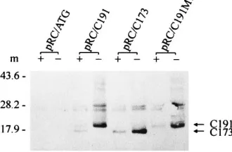
of HCV core protein processing in vivo. COS-1 cells were transfected with various constructs encoding core proteins and polyprotein precursors, as well as the cleavage site mutants. At 48 h after transfection, lysates from transfected cells were analyzed by immunoblotting with monoclonal core anti-body. As shown in Fig. 3, we found that core protein produced from a full-length HCV polyprotein (pRC/B2) and various precursors (CE2, CE1, and C191) resulted in two core protein products, C191 (21 kDa) and C173 (19 kDa). Although C173 was the major product, C191 was always detected. The ratio between C173 and C191, as determined by the intensity of the bands, was approximately 5:1 to 10:1. These data suggest that core protein C191 is sufficient to generate two forms of core protein, C173 and C191. When the C terminus of core protein was truncated up to residue 173, C173 was the only core pro-tein detected (Fig. 3).
To further examine core protein processing, cleavage sites 173 to 174 and 191 to 192 were mutated to assess their sus-ceptibility to peptidase(s). As shown in Fig. 3, in lysates of cells transfected by pRC/C191M1 and pRC/CE1M1, which carry mutations at residues 173 to 174 (M1), both the C173 and C191 products could be observed. However, the C173/C191 ratio was significantly reduced (0.5:1) compared with that in the wild type (5:1 to 10:1). This finding indicated that cleavage at the 173 to 174 site is substantially blocked by mutation. Similar results were obtained with construct pRC/CE2M1 (data not shown). When an analogous mutation was introduced into residues 191 to 192 (M2), no C191 species was detected. In-stead, a protein of approximately 52 kDa, corresponding to the unprocessed precursor CE1, accumulated. This result indi-cated that the amino acid substitutions at cleavage site 191 to 192 inhibited the cleavage events occurring at this site. Addi-tionally, the mutation introduced at site 191 to 192 appeared to interfere with the cleavage occurring at site 173 to 174, result-ing in only trace amounts of C173. This may suggest that the cleavage at site 173 to 174 is preceded by and perhaps depen-dent on that at site 191 to 192. Similar results were obtained with construct pRC/CE2M2 (data not shown). These data are consistent with the results obtained from in vitro translation experiments, indicating that there may be two membrane-de-pendent cleavage sites associated with core protein maturation and that these cleavage events may be regulated in a prefer-ential or sequprefer-ential manner.
Subcellular localization of the HCV core proteins.The
C-terminal hydrophobic domain of core protein has been impli-cated in the nuclear and cytoplasmic distribution of core pro-tein. Although our immunoblotting data demonstrate that there are two observable forms of core protein, the relation-ship between their processing and subcellular localization has not been well established. To address this issue, various con-structs expressing HCV full-length polyprotein and C-terminal truncations thereof were transfected into COS-1 cells. At 48 h later, the transfected cells were examined by indirect immuno-fluorescence with monoclonal anti-core antibody. Core protein released from the full-length HCV polyprotein (pRC/B2) showed a reticular or punctate cytoplasmic immunofluorescent staining pattern (Fig. 4A). When the nonstructural region, including NS2 to NS5, was deleted from the full-length HCV polyprotein (pRC/CE2), similar immunofluorescent staining patterns were noted (Fig. 4B). The subcellular localization of core protein remained the same when E2 or E1 was sequen-tially truncated (pRC/CE1 or pRC/C191) (Fig. 4C and D). Upon deletion of the C-terminal hydrophobic region between residues 174 and 191 (pRC/C173), nuclear localization of core protein was noted (Fig. 4E). As shown in Fig. 3, both C173 and C191 forms could be observed during the processing of core protein from its polyprotein precursors. As expected, only the C173 form was detected when core protein was truncated up to residue 173. Therefore, the cytoplasmic staining observed here (Fig. 4A to D) must result from both C173 and C191 forms while the nuclear staining (Fig. 4E) is the result of the accu-mulation of C173 alone. This suggests that C191 may interact with C173 in vivo, and the interplay between C173 and C191 may prevent the nuclear translocation of the C173 form.
[image:3.612.94.259.74.182.2]When the cleavage site at residues 173 to 174 was mutated, as shown in COS-1 cells transfected with either the construct pRC/C191M1 or the construct pRC/CE1M1, immunofluores-cent staining was observed only in the cytoplasm (Fig. 4F and G). As discussed above, data obtained from immunoblotting analysis demonstrated that these two constructs were capable of generating C173 and C191M1 forms in the ratio 0.5:1. Such cytoplasmic immunostaining (Fig. 4F and G) may represent
FIG. 2. HCV core protein proteolytic processing in vitro. Constructs pRC/ C191, pRC/C173, and pRC/C191M1, as well as vector (pRC/ATG) alone, were transcribed and translated in vitro in the presence (1) or absence (2) of canine pancreatic microsomal membrane (m), using the TNT-coupled reticulocyte ly-sate system (Promega). Translated products labeled with [35S]methionine were
separated by SDS-PAGE (12% polyacrylamide) and analyzed by immunoblot-ting. The positions of core proteins (arrows) and of the molecular mass standards (in kilodaltons) are indicated at the sides of the gel.
FIG. 3. HCV core protein processing in COS-1 cells. Constructs expressing various HCV proteins and their mutants were transfected into COS-1 cells with Lipofectin reagent (GIBCO). Cell lysates were harvested 48 h later, separated by SDS-PAGE (15% polyacrylamide), and analyzed by immunoblotting. The con-structs used are indicated above the gel. The positions of core proteins, polypro-tein precursors, and their mutants are indicated by arrows on the right, and the molecular mass standards (in kilodaltons) are indicated on the left.
on November 9, 2019 by guest
http://jvi.asm.org/
[image:3.612.72.287.508.666.2]coexistence of C173 and C191M1 products in the cytoplasm. When the cleavage mutation at residues 191 to 192 (M2) was introduced into CE1 and expressed in transfected cells, immu-nofluorescence of core protein was seen in the nucleus, al-though cytoplasmic staining was still noted (Fig. 4H). Since, in this case, no C191 product was generated, the nuclear fluores-cence is probably due to translocation of the successfully cleaved C173 product to the nuclear compartment. The cyto-plasmic fluorescence is the result of uncleaved core protein precursor that extends into the E1 domain. These data further suggest that C173 accumulates in the nucleus and that coex-pression of C191 leads to the sequestering of C173 in the cytoplasm.
DISCUSSION
One of the important questions in the study of HCV con-cerns the relationship of HCV core protein maturation to the many interactive events that occur during virus replication. The results presented here are directed to this issue. Regard-ing HCV core protein processRegard-ing, our data, by demonstratRegard-ing that mutations at amino acid positions 173 to 174 and 191 to 192 disrupt or prevent processing at the two putative cleavage sites at the C terminus of core protein (9, 26), imply that these
residues are actual cleavage sites (P1/P19) or closely associated
with them. Through two membrane-associated proteolytic cleavages, core protein is released by the host signal
pepti-FIG. 4. Immunofluorescence analysis of the subcellular localization of HCV core proteins. COS-1 cells transfected with the various expression constructs were fixed with2208C acetone for 2 min. The mouse monoclonal anti-core antibody diluted 1:200 in 1% bovine serum albumin was used as the primary antibody. The FITC-conjugated goat anti-mouse antibody was used as secondary antibody. The immunofluorescent staining patterns are shown, respectively, as cells transfected by pRC/B2 (A), pRC/CE2 (B), pRC/CE1 (C), pRC/C191 (D), pRC/C173 (E), pRC/C191M1 (F), pRC/CE1M1 (G), and pRC/CE1M2 (H).
on November 9, 2019 by guest
http://jvi.asm.org/
dase(s) from the HCV polyprotein, and two mature forms of core protein, C191 and C173, are observed. The cleavage event occurring at 173 to 174 is preceded by that at 191 to 192. These results are consistent with and further extend previous reports (8, 9, 26) and allow us to conclude that these cleavage events may be regulated in a preferential or sequential manner, al-though the mechanisms of this regulation are unknown. One possible explanation is that the cleavage event at 191 to 192 is required to expose the cleavage site at 173 to 174, allowing for recognition by host signal peptidase. That is, cleavage at 173 to 174 is not readily accomplished without prior release of the C191 moiety from the polyprotein precursor. If this is the case in vivo, the formation of C191 is likely to be a limiting step for the generation of C173, so as to balance the molar ratio of the resulting core proteins. In addition, since previous studies have suggested that the hydrophobic region between residues 174 and 191 may function as a signal sequence involved in the trafficking of E1 glycoprotein (9, 26), it is possible that the preferentially or sequentially regulated cleavage at the C ter-minus of the core protein influences E1 maturation as well. In this way, the levels of both viral structural proteins are ex-pected to be coordinately balanced.
Previous studies have indicated that various C-terminal trun-cations of core protein, up to residue 164, result in the nuclear localization (16, 19, 23, 31). The relationship between core protein proteolytic processing and its subsequent subcellular localization is still unclear. In this study, we have provided data to demonstrate that the localization of core protein is linked to cleavage events at the C terminus. We found that when core protein C173 was expressed alone, it was able to translocate to the nucleus. However, core protein, when released from the longer precursors, CE1, CE2, or full-length HCV polyprotein, was capable of generating C173 as well as C191. In this case, both species of core protein were restrained in the cytoplasm. Although the mechanism by which both forms of core protein are retained in the cytoplasm is not apparent, these results suggest the presence of a subtle interaction between these two core protein products. The interaction between C191 and C173 in the cytoplasm may prevent C173 nuclear translocation, thereby resulting in the cytoplasmic localization of both forms. In support of this hypothesis, recent study has indicated that HCV core protein can form homotypic multimers in vitro and in vivo and that specific core-core interactions are mediated by amino acid residues 1 to 115 (18), indicating that C173-C191 heteromultimers may also exist. In the study presented here, we demonstrate that in transiently transfected COS-1 cells, the amount of C173 was 5 to 10 times greater than that of C191 (Fig. 3), suggesting that if an interaction between C173 and C191 does occur, it is likely to result in heteromultimers with a defined molar ratio. Therefore, it is reasonable to believe that the association between C173 and C191 in the cytoplasm is the driving force keeping core proteins sequestered within the cytoplasm. As noted for other flaviviruses, viral RNA pack-aging and assembly are accomplished in the cytoplasm. The requirement of cytoplasmic localization of HCV core proteins may be regarded as a necessary biological event in the viral life cycle. In addition, the HCV core protein C-terminal hydropho-bic region has been demonstrated to be associated with the endoplasmic reticulum membrane (26). Thus, the hydrophobic region between residues 174 and 191 may function as an an-chor to facilitate cytoplasmic localization. Further studies on the formation of core protein heteromultimers may elucidate our understanding of the virion assembly pathway. As an al-ternative mechanism, it is equally possible that amino acid residues 174 to 191 mediate multimerization itself and/or act to block nuclear localization of C173 in trans.
The biological significance of the translocation of core pro-tein product to the nucleus is of great interest. Previous studies have shown that core protein may be involved in the regulation of transcription of host and viral genes (24, 28, 29). The N terminus of core protein contains clusters of basic amino acids which may play a role in core protein nuclear localization (19, 31). However, it is unknown whether the C173 species will be accumulated in the nucleus during natural viral infection. In fact, the precise maturation of HCV core protein in vivo is uncharacterized. If the proposed processing occurs, C173 may utilize a similar signal to translocate itself into the nucleus, thereby realizing its possible regulatory function on host gene expression. Whether the nuclear translocation of C173 ob-served here and the interplay between C173 and C191 are associated with modulating these regulatory effects needs to be determined. The expression of core polypeptide may affect pleiotropic responses in the infected host cell, beyond its cur-rently presumed structural role.
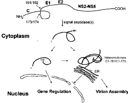
Based on these results, we propose a model as shown in Fig. 5. According to this model, during core protein maturation, the polyprotein precursor undergoes two consecutive cleavage events, resulting in the production of two forms of core pro-tein, C173 and C191. If these two species are present in a defined molar ratio, they can form a complex, which associates with the endoplasmic reticulum membrane, thereby sequester-ing C173 within the cytoplasm. In the absence of this regula-tory ratio, C173 translocates into the nucleus. Cytoplasmically localized core protein subunits may interact with each other and function as a scaffold for viral assembly and genomic RNA packaging, while the species in the nucleus may serve to reg-ulate gene expression in some obligate manner. Clearly, HCV core protein engages in a multifaceted relationship with its host cell as well as with other viral proteins. A better under-standing of these associations will help to clarify the life cycle of this refractory agent.
ACKNOWLEDGMENTS
We thank Alan McLachlan and Eckard Wimmer for critical com-ments. We are indebted to Jack Wands for the gift of anti-core mono-clonal antibody, to Oscar Pogo and David Russo for assistance with fluorescence microscopy, and to Tellervo Huima-Byron for artwork.
[image:5.612.327.539.72.243.2]Q. Y. Liu was supported by a postdoctoral fellowship from Texaco Foundation. This work was supported in part by Pasteur Merieux.
FIG. 5. Hypothetical model for HCV core protein maturation.
on November 9, 2019 by guest
http://jvi.asm.org/
REFERENCES
1. Bukh, J., R. H. Purcell, and R. H. Miller. 1994. Sequence analysis of the core gene of 14 hepatitis C virus genotypes. Proc. Natl. Acad. Sci. USA 91:8239– 8243.
2. Choo, Q.-L., G. Kuo, A. J. Weiner, L. R. Overby, D. W. Bradley, and M. Houghton.1989. Isolation of a cDNA clone derived from a blood-borne non-A, non-B viral hepatitis genome. Science 244:359–362.
3. Choo, Q.-L., K. H. Richman, J. H. Han, K. Berger, C. Lee, C. Dong, C. Gallegos, D. Coit, A. Medina-Selby, P. J. Barr, A. J. Weiner, D. W. Bradley, G. Kuo, and M. Houghton.1991. Genetic organization and diversity of the hepatitis C virus. Proc. Natl. Acad. Sci. USA 88:2451–2455.
4. Eckart, M. R., M. Selby, F. Masiarz, C. Lee, K. Berger, K. Crawford, C. Kuo, G. Kuo, M. Houghton, and Q.-L. Choo.1993. The hepatitis C virus encodes a serine protease involved in processing of the putative nonstructural pro-teins from the viral polyprotein precursor. Biochem. Biophys. Res. Commun. 192:399–406.
5. Grakoui, A., C. Wychowski, C. Lin, S. M. Feinstone, and C. M. Rice. 1993. Expression and identification of hepatitis C virus polyprotein cleavage prod-ucts. J. Virol. 67:1385–1395.
6. Grakoui, A., D. W. McCourt, C. Wychowski, S. M. Feinstone, and C. M. Rice. 1993. Characterization of hepatitis C virus-encoded serine proteinase: de-termination of proteinase-dependent polyprotein cleavage sites. J. Virol. 67:2832–2843.
7. Grakoui, A., D. W. McCourt, C. Wychowski, S. M. Feinstone, and C. M. Rice. 1993. A second hepatitis C virus-encoded proteinase. Proc. Natl. Acad. Sci. USA 90:10583–10587.
8. Harada, S., Y. Watanabe, K. Takeuchi, T. Suzuki, T. Katayama, Y. Takebe, I. Saito, and T. Miyamura.1991. Expression of processed core protein of hepatitis C virus in mammalian cells. J. Virol. 65:3015–3021.
9. Hijikata, M., N. Kato, Y. Ootsuyama, M. Nakagawa, and K. Shimotohno. 1991. Gene mapping of the putative structural region of the hepatitis C virus genome by in vitro processing analysis. Proc. Natl. Acad. Sci. USA 88:5547– 5551.
10. Hijikata, M., H. Mizushima, T. Akagi, S. Mori, N. Kakiuchi, N. Kato, T. Tanaka, K. Kimura, and K. Shimotohno.1993. Two distinct proteinase activities required for the processing of a putative nonstructural precursor protein of hepatitis C virus. J. Virol. 67:4665–4675.
11. Houghton, M., A. Weiner, J. Han, G. Kuo, and Q.-L. Choo. 1991. Molecular biology of hepatitis viruses: implications for diagnosis, development and control of viral disease. Hepatology 14:381–388.
12. Inchauspe, G., S. Zebedee, D.-H. Lee, M. Sugitani, M. Nasoff, and A. M. Prince.1991. Genomic structure of the human prototype strain H of hepa-titis C virus: comparison with American and Japanese isolates. Proc. Natl. Acad. Sci. USA 88:10292–10296.
13. Kato, N., M. Hijikata, Y. Ootsuyama, M. Nakagawa, S. Ohkoshi, T. Sug-imura, and K. Shimotohno.1990. Molecular cloning of the human hepatitis C virus genome from Japanese patients with non-A, non-B hepatitis. Proc. Natl. Acad. Sci. USA 87:9524–9528.
14. Kozak, M. 1991. Structural features in eukaryotic mRNAs that modulate the initiation of translation. J. Biol. Chem. 266:19867–19870.
15. Lanford, R. E., L. Notvall, D. Chavez, R. White, G. Frenzel, C. Simonsen, and J. Kim.1993. Analysis of hepatitis C virus capsid, E1, and E2/NS1 proteins expressed in insect cells. Virology 197:225–235.
16. Lo, S.-Y., F. Masiarz, S. B. Hwang, M. M. C. Lai, and J.-H. Ou. 1995. Differential subcellular localization of hepatitis C virus core gene products. Virology 213:455–461.
17. Manabe, S., I. Fuke, O. Tanishita, C. Kaji, Y. Gomi, S. Yoshida, C. Mori, A. Takamizawa, I. Yosida, and H. Okayama.1994. Production of nonstructural proteins of hepatitis C virus requires a putative viral protease encoded by NS3. Virology 198:636–644.
18. Matsumoto, M., S. B. Hwang, K.-S. Jeng, N. Zhu, and M. M. C. Lai. 1996. Homotypic interaction and multimerization of hepatitis C virus core protein. Virology 218:43–51.
19. Matsuura, Y., T. Harada, M. Makimura, M. Sato, H. Aizaki, T. Suzuki, and T. Miyamura.1994. Characterization of HCV structural proteins expressed in various animal cells. Intervirology 37:114–118.
20. Miller, R. H., and R. H. Purcell. 1990. Hepatitis C virus shares amino acid sequence similarity with pestiviruses and flaviviruses as well as members of two plant virus supergroups. Proc. Natl. Acad. Sci. USA 87:2057–2061. 21. Prince, A. M., B. Brotman, G. F. Grady, W. J. Kuhns, C. Hazzi, R. W.
Levine, and S. J. Millian.1974. Long-incubation post-transfusion hepa-titis without serological evidence of exposure to hepahepa-titis B virus. Lancet ii:241–246.
22. Ralston, R., K. Thudium, K. Berger, C. Kuo, B. Gervase, J. Hall, M. Selby, G. Kuo, M. Houghton, and Q.-L. Choo.1993. Characterization of hepatitis C virus envelope glycoprotein complexes expressed by recombinant vaccinia viruses. J. Virol. 67:6753–6761.
23. Ravaggi, A., G. Natoli, D. Primi, A. Albertini, M. Levrero, and E. Cariani. 1994. Intracellular localization of full-length and truncated hepatitis C virus core protein expressed in mammalian cells. J. Hepatol. 20:833–836. 24. Ray, R. B., L. M. Lagging, K. Meyer, R. Steele, and R. Ray. 1995.
Transcrip-tional regulation of cellular and viral promoters by the hepatitis C virus core protein. Virus Res. 37:209–220.
25. Sambrook, J., E. F. Fritsch, and T. Maniatis. 1989. Molecular cloning: a laboratory manual, 2nd ed. Cold Spring Harbor Laboratory Press, Cold Spring Harbor, N.Y.
26. Santolini, E., G. Migliaccio, and N. La Monica. 1994. Biosynthesis and biochemical properties of the hepatitis C virus core protein. J. Virol. 68: 3631–3641.
27. Selby, M. J., Q.-L. Choo, K. Berger, G. Kuo, E. Glazer, M. Eckart, C. Lee, D. Chien, C. Kuo, and M. Houghton.1993. Expression, identification and sub-cellular localization of the proteins encoded by the hepatitis C viral genome. J. Gen. Virol. 74:1103–1113.
28. Shih, C.-M., S. J. Lo, T. Miyamura, S.-Y. Chen, and Y.-H. Lee. 1993. Sup-pression of hepatitis B virus exSup-pression and replication by hepatitis C virus core protein in Huh-7 cells. J. Virol. 67:5823–5832.
29. Shih, C.-M., C.-M. Chen, S.-Y. Chen, and Y.-H. Lee. 1995. Modulation of the
trans-suppression activity of hepatitis C virus core protein by
phosphoryla-tion. J. Virol. 69:1160–1171.
30. Spaete, R. R., D. Alexander, M. E. Rugroden, Q.-L. Choo, K. Berger, K. Crawford, C. Kuo, S. Leng, C. Lee, R. Ralston, K. Thudium, J. W. Tung, G. Kuo, and M. Houghton.1992. Characterization of the hepatitis C virus E2/NS1 gene product expressed in mammalian cells. Virology 188:819–830. 31. Suzuki, R., Y. Matsuura, T. Suzuki, A. Ando, J. Chiba, S. Harada, I. Saito, and T. Miyamura.1995. Nuclear localization of the truncated hepatitis C virus core protein with its hydrophobic C terminus deleted. J. Gen. Virol. 76:53–61.
32. Takamizawa, A., C. Mori, I. Fuke, S. Manabe, S. Murakami, J. Fujita, E. Onishi, T. Andon, I. Yoshida, and H. Okayama.1991. Structure and orga-nization of the hepatitis C virus genome isolated from human carriers. J. Virol. 65:1105–1113.
33. Takeuchi, K., Y. Kubo, S. Boonmar, Y. Watanabe, T. Katayama, Q.-L. Choo, G. Kuo, M. Houghton, I. Saito, and T. Miyamura.1990. The putative nu-cleocapsid and envelope protein genes of hepatitis C virus determined by comparison of the nucleotide sequences of two isolates derived from an experimentally infected chimpanzee and healthy human carriers. J. Gen. Virol. 71:3027–3033.
34. Tomei, L., C. Failla, E. Santolini, R. De Francesco, and N. La Monica. 1993. NS3 is a serine protease required for processing of hepatitis C virus polypro-tein. J. Virol. 67:4017–4026.
35. Tsukiyama-Kohara, K., N. Iizuka, M. Kohara, and A. Nomoto. 1992. Internal ribosome entry site within hepatitis C virus RNA. J. Virol. 66:1476–1483.
36. Wang, C., P. Sarnow, and A. Siddiqui. 1993. Translation of human hepatitis C virus RNA in cultured cells is mediated by an internal ribosome-binding mechanism. J. Virol. 67:3338–3344.