Copyright © 2000, American Society for Microbiology. All Rights Reserved.
The Human Immunodeficiency Virus Type 1 Tat Protein Up-Regulates
the Promoter Activity of the Beta-Chemokine Monocyte
Chemoattractant Protein 1 in the Human Astrocytoma
Cell Line U-87 MG: Role of SP-1, AP-1,
and NF-
B Consensus Sites
SIEW PHENG LIM1ANDALFREDO GARZINO-DEMO2*
Institute of Molecular and Cell Biology, Singapore 117609, Singapore,1and Institute of Human Virology,
University of Maryland Biotechnology Institute, Baltimore, Maryland 212012
Received 12 March 1999/Accepted 12 November 1999
It has been shown that the human immunodeficiency virus type 1 (HIV-1) Tat protein can specifically enhance expression and release of monocyte chemoattractant protein 1 (MCP-1) from human astrocytes. In this study, we show evidence that Tat-induced MCP-1 expression is mediated at the transcriptional level. Transient transfection of an expression construct encoding the full-length Tat into the human glioblastoma-astrocytoma cell line U-87 MG enhances reporter gene activity from cotransfected deletion constructs of the MCP-1 promoter. HIV-1 Tat exerts its effect through a minimal construct containing 213 nucleotides upstream of the translational start site. Site-directed mutagenesis studies indicate that an SP1 site (located between nucleotidesⴚ123 andⴚ115) is critical for both constitutive and Tat-enhanced expression of the human MCP-1 promoter, as mutation of this SP1 site significantly diminished reporter gene expression in both instances. Gel retardation experiments further demonstrate that Tat strongly enhances the binding of SP1 protein to its DNA element on the MCP-1 promoter. Moreover, we also observe an increase in the binding activities of transcrip-tional factors AP1 and NF-B to the MCP-1 promoter following Tat treatment. Mutagenesis studies show that an upstream AP1 site and an adjacent NF-B site (located atⴚ128 toⴚ122 andⴚ150 toⴚ137, respectively) play a role in Tat-mediated transactivation. In contrast, a further upstream AP1 site (ⴚ156 toⴚ150) does not appear to be crucial for promoter activity. We postulate that a Tat-mediated increase in SP1 binding activities augments the binding of AP1 and NF-B, leading to synergistic activation of the MCP-1 promoter.
The Tat protein of human immunodeficiency virus type 1 (HIV-1) is an 86-amino-acid protein encoded by two exons (2, 17, 46). The first exon, which encodes amino acids 1 to 72, is sufficient for directing viral gene expression through transacti-vation of the HIV-1 long terminal repeat (LTR) and is essen-tial for viral replication (2, 17, 46). Tat protein is released from HIV-1-infected and Tat-transfected cells and can be taken up by nearby uninfected or nontransfected cells through endocy-tosis (19, 31). Several reports have shown that Tat has pleio-tropic effects on cell survival, growth, and function of unin-fected cells (13, 14, 19, 31, 51). It inhibits antigen-induced proliferation of isolated T cells (51) and promotes the growth of Kaposi’s sarcoma-derived cells (13). In addition, Tat has been shown to play a role in apoptosis (41) and to induce the production of several cytokines, including interleukin-2 (IL-2) (52), IL-6 (41), tumor necrosis factor alpha (TNF-␣) (5), and monocyte chemoattractant protein 1 (MCP-1) (10).
The-chemokine MCP-1 is a monocyte-specific chemotactic factor thought to play a significant role in monocytic infiltra-tion to sites of injury or inflammainfiltra-tion (27, 43). HIV-1 infecinfiltra-tion in the central nervous system (CNS) causes a dementing ill-ness, the pathological correlate of which is monocytic infiltra-tion of the CNS (21, 37). The mechanisms by which monocytic infiltration of the CNS may lead to dementia include the ability
of activated monocytes to release TNF-␣, nitric oxide, platelet-activating factor, and quinolinate, which in turn may cause neural cell death (4, 15, 30, 49). Patients with HIV-1-associated dementia express elevated levels of MCP-1 in their brain tissue and in cerebrospinal fluid (10). Exogenous Tat is capable of enhancing the expression of MCP-1 transcripts and secreted protein in human primary fetal astrocytes (10). In a related study, it has been shown that treatment of human primary fetal astrocytes with Tat can increase NF-B binding to its cognate binding motif (9). It is conceivable that NF-B and/or some other Tat-induced factor(s) thus play a role in Tat-mediated induction of MCP-1 expression. In this study, we sought to de-termine the mechanism for Tat-induced expression of MCP-1 in human astrocytes by studying its effect on the MCP-1 pro-moter.
MATERIALS AND METHODS
Cells and cell culture.The preparation of astrocyte cultures from human fetal tissue has been described previously (12). The cells are grown in Eagle’s minimal essential medium (EMEM) supplemented with 10% fetal calf serum, 2 mM L-glutamine, and 5g of gentamicin per ml. The human glioblastoma-astrocy-toma cell line U-87 MG and the epithelioid cervical carcinoma cell line HeLa were purchased from the American Type Culture Collection (Manassas, Va.). HeLa-tat-III was obtained from the AIDS Research and Reference Reagent Program (National Institutes of Health, Bethesda, Md.) (38) and was kindly provided by Marvin S. Reitz, Jr. (Institute of Human Virology, University of Maryland Biotechnology Institute, Baltimore). The cells were cultured in EMEM containing 2 mML-glutamine, 1.5 g of sodium bicarbonate per liter, 0.1 mM nonessential amino acids, 1 mM sodium pyruvate, and 10% fetal bovine serum and maintained at 37°C in 5% CO2. The growth media of HeLa-tat-III cells was further supplemented with 200g of gentamicin per ml.
* Corresponding author. Mailing address: Institute of Human Vi-rology, University of Maryland Biotechnology Institute, 725 W. Lom-bard St., Baltimore, MD 21201-1192. Phone: (410) 706-4676. Fax: (410) 706-4694. E-mail: ihvinfo@umbi.umd.edu.
1632
on November 9, 2019 by guest
http://jvi.asm.org/
Plasmids and constructs.Plasmid pGL2-Basic (Promega, Madison, Wis.) was used to generate deletion constructs of the human MCP-1 (hMCP-1) promoter. To obtain phMCP486, phMCP213, and phMC128, which covered positions⫺486 to⫹6,⫺213 to⫹6, and⫺128 to⫹6, respectively, DNA fragments were ampli-fied by PCR with chemically synthesized oligonucleotides that corresponded to nucleotides⫺486 to⫺456 of the sense strand (5⬘GGGCTAGCAAGCTTGA GAGCTCCTTCCTGG3⬘),⫺213 to⫺183 of the sense strand (5⬘GGGCTA GCCTTCCTGGAAATCCACAGGATG3⬘),⫺128 to⫺75 of the sense strand (5⬘GGGCTAGCGACTCCGCCCTCTCTCCCTCTG3⬘), and⫺17 to⫹6 of the antisense strand (5⬘GGAGATCTTTCATGCTGGAGGCGAGAGTGC3⬘). Hu-man genomic DNA derived from a healthy adult was used as the template. The PCR products were digested withNheI andBglII and cloned into theNheI-BglII site of pGL2-Basic. The mammalian expression construct pCMHV1-tat, encod-ing a Tat cDNA, was kindly provided by S. K. Arya, National Cancer Institute, National Institutes of Health (20).
PCR mutagenesis.Site-directed mutation was introduced with a QuikChange site-directed mutagenesis kit (Stratagene, La Jolla, Calif.). Briefly, 5 ng of phMCP213 plasmid DNA and 125 ng of each of two complementary oligonu-cleotides containing the desired mutation (Table 1) were incubated with 2.5 U of
PfuDNA polymerase and cycled 18 times with the following conditions: dena-turation at 95°C for 30 s, annealing at 55°C for 1 min, and extension at 68°C for 10 min. The reaction mix was cooled on ice for 2 min and digested with 10 U of
DpnI at 37°C for 1 h, and 1l of the reaction mix was transformed into Epicurian Coli XL1-Blue supercompetent cells. Plasmids were isolated from colonies grown overnight at 37°C on ampicillin-resistant agar plates, and sequencing was carried out to identify those that contained the mutated sequences of interest.
DNA sequencing.DNA sequencing on all constructs created was carried out in the Biopolymer Sequencing Facility, University of Maryland, Baltimore. Two hundred nanograms of the double-stranded templates and 10 ng of the primer were used for the dideoxy method with aTaq Dye Deoxy terminator cycle sequencing kit and an automated DNA sequencer 377 (PE Applied Biosystems, Foster City, Calif.).
Cell transfections.Transfection experiments were performed with Superfect transfection reagent (Qiagen, Valencia, Calif.). The day before transfection, 1.5⫻105U-87 MG or HeLa cells were seeded into six-well plates in 2.5 ml of complete medium. The cells were 40 to 60% confluent on the day of transfection. For transfection, 150l of serum-free EMEM containing 5g of total plasmid DNA was mixed well with 75g of Superfect transfection reagent and allowed to stand at room temperature for 10 min to allow complex formation. Growth medium was aspirated from the cells, and the cells were washed once with 4 ml of phosphate-buffered saline (PBS). The DNA-Superfect mixture was diluted with 1 ml of complete growth medium and added to the cells. Cells were then reincubated at 37°C and 5% CO2for 3 h, after which the medium was aspirated and the cells were washed again with 4 ml of PBS; 2.5 ml of complete medium was added to the cells, and incubation at 37°C and 5% CO2was continued for another 48 to 72 h, after which the cells were harvested and luciferase activity was measured. Either 4g of test plasmid and 1g of pSV--galactosidase plasmid DNA or 2g of test plasmid, 1 to 2g of pCMtat-HIV-1 (20), and 1g of pSV--galactosidase plasmid DNA were used in the transfections. For phorbol myristate acetate (PMA) induction, a final concentration of 10⫺7M PMA was added to the cells for the indicated periods of time.
Luciferase assays.Luciferase activity was measured with a luciferase assay kit from Promega (Madison, Wis.). Following a 48- to 72-h incubation period, cells were washed twice with PBS and lysed with 150 l of reporter lysis buffer (Promega). The lysate was allowed to stand at room temperature for 10 to 15 min
and collected into 1.5-ml Eppendorf tubes. These were quick-spun for 1 min in a microcentrifuge, and 10l of each lysate was mixed with 100l of buffer and measured for luciferase activity in a Turner luminometer (Turner Designs, Sunnyvale, Calif.) over an integration period of 15 s. Values obtained were normalized to the levels of-galactosidase in the cell lysates.-Galactosidase activities were determined with an assay kit (Promega) and exhibited ⬍20% variation between samples.
Treatment of cells with recombinant HIV-1 Tat.Recombinant Tat1-86, a kind gift from B. C. Nair, Advanced Bioscience Laboratories, Kensington, Md., was prepared as described previously (6). Briefly, Tat was expressed inEscherichia coliand purified by heparin affinity chromatography. Further purification was done by reversed-phase high-performance liquid chromatography (HPLC) (data not shown). The protein was found to be homogeneous and⬎95% pure follow-ing sodium dodecyl sulfate-polyacrylamide gel electophoresis with Coomassie blue staining. Its biological activity was assessed by the HIV rescue assay with a cell line (HLM-1) containing an integrated nonreversible Tat-defective provirus (data not shown) (6). U-87 MG cells (107) were treated with 40 or 100 nM Tat for 2 h in serum-free EMEM in a 37°C CO2incubator, after which they were harvested for the preparation of nuclear extracts. For heat inactivation, Tat was incubated at 60°C for 30 min (30).
Nuclear extracts and EMSA.Nuclear extracts were prepared from 107cells by the method of Andrews and Faller (1). Protein concentration was determined with a bicinchoninic acid protein assay from Pierce (Rockford, Ill.) and deter-mined to be between 1 and 5g/ml. The DNA fragment from⫺213 to⫹6 of the hMCP-1 promoter was excised from plasmid phMCP213 withNheI andBglII and filled in with [␣-32P]dCTP (New England Biolabs, Inc., Beverly, Mass.) and Klenow fragment. Oligonucleotides containing consensus SP1, AP1, NF-B, or NF1 binding elements (Table 1) were annealed and end labeled with [␥-32P]ATP and T4 kinase. Electrophoresis mobility shift assay (EMSA) mixtures contained 0.25 ng of 32P-labeled DNA fragment or double-stranded oligonucleotides (15,000 cpm), 10 mM Tris-HCl (pH 7.5), 1 mM dithiothreitol, 1 mM EDTA, 0.5 mM MgCl2, 5% glycerol, 0.5g of poly(dI-dC), 0.1g of sonicated salmon sperm DNA, and 4g of nuclear extract. The binding reaction mixtures were incubated on ice for 20 min and electrophoresed through 5% native polyacryl-amide gels in 25 mM Tris borate–0.5 mM EDTA at 270 V for 3 h. The gels were dried down and exposed to X-ray films for 12 to 16 h at⫺70°C.
Competition assays were performed with 100-fold molar excess of cold DNA fragments from positions⫺213 to⫹6 or double-stranded oligonucleotides con-taining SP1, AP1, NF-B, and NF1 binding elements (Table 1). Supershift assays were performed with SP1, AP1, NF-B p50 and p65, and C/EBP polyclonal antibodies (Santa Cruz Biotechnology, Santa Cruz, Calif.) by preincubating the nuclear extracts with 2.5l of the polyclonal antibody in the reaction buffer for 10 min and continuing the gel retardation assay as described above.
RESULTS
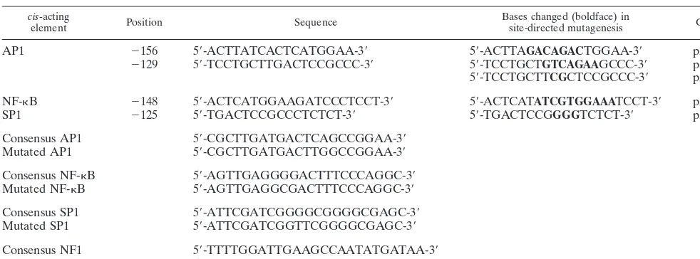
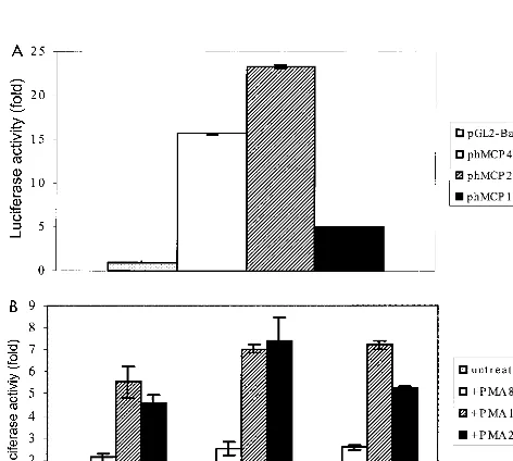
[image:2.612.54.545.83.265.2]Analysis of deletion constructs of the hMCP-1 promoter.To confirm transcriptional regulatory protein binding regions, lu-ciferase reporter constructs bearing progressive 5⬘deletions of the hMCP-1 promoter were generated and transfected into the human glioblastoma cell line U-87 MG (Fig. 1). All three constructs, phMCP486, phMCP213, and phMCP128, exhibited significantly higher luciferase activity than the control plasmid TABLE 1. ciselements on the hMCP-1 promoter region analyzed in this study
cis-acting
element Position Sequence Bases changed (boldface) insite-directed mutagenesis Construct
AP1 ⫺156 5⬘-ACTTATCACTCATGGAA-3⬘ 5⬘-ACTTAGACAGACTGGAA-3⬘ p⌬AP1A
⫺129 5⬘-TCCTGCTTGACTCCGCCC-3⬘ 5⬘-TCCTGCTGTCAGAAGCCC-3⬘ p⌬AP1B1 5⬘-TCCTGCTTCGCTCCGCCC-3⬘ p⌬AP1B2 NF-B ⫺148 5⬘-ACTCATGGAAGATCCCTCCT-3⬘ 5⬘-ACTCATATCGTGGAAATCCT-3⬘ p⌬NF-B
SP1 ⫺125 5⬘-TGACTCCGCCCTCTCT-3⬘ 5⬘-TGACTCCGGGGTCTCT-3⬘ p⌬SP1
Consensus AP1 5⬘-CGCTTGATGACTCAGCCGGAA-3⬘
Mutated AP1 5⬘-CGCTTGATGACTTGGCCGGAA-3⬘
Consensus NF-B 5⬘-AGTTGAGGGGACTTTCCCAGGC-3⬘
Mutated NF-B 5⬘-AGTTGAGGCGACTTTCCCAGGC-3⬘
Consensus SP1 5⬘-ATTCGATCGGGGCGGGGCGAGC-3⬘
Mutated SP1 5⬘-ATTCGATCGGTTCGGGGCGAGC-3⬘
Consensus NF1 5⬘-TTTTGGATTGAAGCCAATATGATAA-3⬘
Mutated NF1 5⬘-TTTTGGATTGAATAAAATATGATAA-3⬘
on November 9, 2019 by guest
http://jvi.asm.org/
pGL2-Basic (Fig. 2A). The construct phMCP213 was consis-tently found to be more active than phMCP486 (Fig. 2A). The former was 23-fold more active than pGL2-Basic, while the latter was about 16-fold more active (Fig. 2A). In contrast, phMCP128 was the least active, exhibiting only about fivefold more activity than pGL2-Basic (Fig. 2A). We next determined the effect of stimulus on the activity of these constructs. All three constructs were readily inducible upon treatment with PMA; after 8 h of exposure to PMA, they each exhibited about
[image:3.612.312.552.73.280.2]twofold increase in activity over basal levels (Fig. 2B). After 18 h of treatment, the values rose to between five- to sevenfold above basal levels and maintained those levels 24 h posttreat-ment (Fig. 2B). The human fibroblastic cell line HeLa has been reported to express low levels of MCP-1 mRNA (16). To com-pare the results obtained for U-87 MG cells, we repeated the experiments with HeLa cells. All of the promoter constructs exhibited lower basal activity in HeLa cells than in U-87 MG cells (Fig. 3A). The activity of phMCP486 was about threefold above background, while that of phMCP213 was about nine-fold above background (Fig. 3A). Similar to the observation for U-87 MG cells, phMCP128 was the least active; its activity was only 1.4-fold above background values (Fig. 3A). Upon treatment with PMA, strong induction was observed with all three constructs (Fig. 3B). Unlike in U-87 MG cells, maximal stimulation was achieved after 8 h of treatment with PMA (with 9- to 18-fold induction), and the values gradually de-creased over a 24-h period to between 3- and 7-fold (Fig. 3B). Effect of transient expression of Tat on promoter activity of the hMCP-1 gene.In previous studies, HIV-1 Tat was found to enhance the expression and release of MCP-1 in primary hu-man fetal astrocytes at both transcriptional and translational levels (10). To further investigate if Tat-mediated activation of MCP-1 gene expression occurs at the transcriptional level, transient cotransfection experiments were carried out with 5⬘ deletion constructs and an expression vector for the full-length HIV-1 Tat, pCMtat-HIV1 (20). We found that Tat significantly stimulated luciferase activity of constructs phMCP486 and phMCP213 in the U-87 MG cell line (Fig. 4A). Cotransfection with 1g of HIV-1 Tat up-regulated the activity of phMCP486 7-fold, while that of phMCP213 rose 2.3-fold (Fig. 4A). The response of the promoter constructs to Tat is also dose depen-dent. Cotransfection with 2g of Tat expression vector further
FIG. 1. Schematic representations of the MCP-1 promoter. (A) Map of the 5⬘flanking region of the hMCP-1 gene showing up to 3 kb upstream of the translational start site. (B) Proximal region of the promoter from nucleotides ⫺213 to⫹6 from the translational start site (boxed). Putative binding sites for
[image:3.612.56.292.75.233.2]cis-acting factors are underlined.
FIG. 2. Deletion analysis of the hMCP-1 5⬘flanking region. Luciferase assays were performed with extracts of U-87 MG cells transfected with different 5⬘ deletion constructs. Results are expressed as the induction of luciferase activity of the hMCP promoter constructs over pGL2-Basic and were normalized to units of-galactosidase activity from cotransfection with a pSV--galactosidase plas-mid. (A) Luciferase activities from transfected U-87 MG cells were measured 48 h posttransfection; data shown are the means (⫾standard deviations) of four independent transfection experiments. The average raw luciferase value for pGL2-Basic was 0.05⫾0.03. (B) Representative experiment showing transfec-tion of U-87 MG cells following treatment with PMA for the indicated times, harvested 48 h posttransfection. The raw luciferase values for pGL2-Basic ranged from 0.1 to 0.24.
FIG. 3. Deletion analysis of the hMCP-1 5⬘flanking region. Luciferase assays were performed with extracts of HeLa cells transfected with different 5⬘deletion constructs. Results are expressed as the induction of luciferase activity of the hMCP promoter constructs over pGL2-Basic and were normalized to units of -galactosidase activity from cotransfection with a pSV--galactosidase plasmid. (A) Luciferase activities from transfected HeLa cells were measured 48 h post-transfection; data shown are the means (⫾standard deviations) of four inde-pendent transfection experiments. The average raw luciferase value for pGL2-Basic was 0.01⫾0.01. (B) A representative experiment showing transfection of HeLa cells following treatment with PMA for the indicated times, harvested 48 h posttransfection. The raw luciferase values for pGL2-Basic ranged from 0 to 0.04⫾0.04.
on November 9, 2019 by guest
http://jvi.asm.org/
[image:3.612.55.291.416.628.2]stimulated their activity 11- and 5.6-fold, respectively (Fig. 4A). In HeLa cells, Tat strongly induced their luciferase activity (Fig. 4B). The levels of induction rose from 65- and 16-fold for constructs phMCP486 and phMCP213 with 1g of Tat plas-mid to 110- and 42-fold when 2g of the plasmid was used.
Effect of stable expression of Tat on promoter activity of the hMCP-1 gene.To explore the effect of constitutive expression of HIV-1 Tat on MCP-1 activity, we made use of the cell line HeLa-tat-III, which has been stably transfected with a Molo-ney-based retroviral vector expressing HIV-1 Tat (37). Cells transfected with the 5⬘deletion constructs exhibited constitu-tive luciferase activity markedly higher than that for the pa-rental cell line, HeLa (Fig. 3A). Construct phMCP486 was more active than phMCP213 (44- and 32-fold above back-ground, respectively), while both constructs were considerably more active than phMCP128 (14-fold above background) (Fig. 5). In addition, treatment with PMA for 8 h further increased the promoter activity of each of these constructs about three-fold (data not shown).
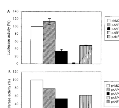
Analysis of 5ⴕ MCP-1 promoter elements involved in Tat-mediated transactivation.The upstream region of the MCP-1 gene contains a number of putative cis-acting elements that contribute to its promoter activity in endothelial and glioblas-toma cell lines (29, 32, 44). To more precisely define the roles of the regulatory elements which are important for Tat-medi-ated transactivation, site-directed mutagenesis of the putative binding sites for the cellular transcription factors AP1 (at nu-cleotides⫺150 and⫺122; hereafter named AP1A and AP1B, respectively),B binding proteins (at nucleotide⫺137), and SP1 (at nucleotide⫺115) were sequentially carried out (Table 1). These binding elements have previously been shown to be important for cytokine (32)- and PMA (29, 44)-mediated in-duction of the MCP-1 gene. The constructs bearing the various mutated elements were first transfected into U-87 MG cells and compared against the wild-type phMCP213 construct.
[image:4.612.55.292.71.283.2]Mu-tation of the first AP1 site (⫺150) did not significantly alter promoter activity (Fig. 6A). In fact, construct p⌬AP1A showed an increase in activity of 13% over that of phMCP213 (Fig. 6A). However, mutation of theB site (⫺137) or the second AP1 site (⫺122) partially inhibited promoter activity by 51 and 66%, respectively (Fig. 6A). This finding suggests that the latter two transcriptional elements, but not the first AP1 bind-ing element, are involved in constitutive MCP-1 gene expres-sion in U-87 MG cells. When construct p⌬SP1, containing a mutated SP1 site, was transfected into U-87 MG cells, we
[image:4.612.311.551.73.176.2]FIG. 4. Tat-mediated induction of hMCP-1 promoter activity. A representa-tive experiment (from three performed) shows luciferase activities of the hMCP-1 deletion constructs following cotransfection in U-87 MG (A) or HeLa (B) cells with the indicated amounts of a Tat expression plasmid. Results are expressed as the induction of luciferase activity of the promoter constructs over pGL2-Basic and were measured 72 h posttransfection. Luciferase activity was normalized to units of-galactosidase activity from cotransfection with a pSV--galactosidase plasmid. The raw luciferase values for pGL2-Basic ranged from 0 to 0.05⫾0.05.
FIG. 5. Constitutive hMCP-1 promoter activity in HeLa-tat-III cells. A rep-resentative experiment (from three performed) shows luciferase activities of the hMCP-1 deletion constructs following transfection in HeLa-tat-III cells. Results are expressed as induction of luciferase activity of the hMCP promoter constructs over pGL2-Basic and were measured 48 h posttransfection. Luciferase activities were normalized to units of-galactosidase activity from cotransfection with a pSV--galactosidase plasmid. The raw luciferase value for pGL2-Basic was 0.09⫾0.01.
FIG. 6. Analysis of transcriptional control elements in the hMCP-1 promoter in U-87 MG cells. Luciferase assays were performed with extracts of U-87 MG cells transfected with hMCP-1 promoter constructs bearing various mutated
cis-acting elements. (A) Results are expressed as the percentage of luciferase activity compared to the wild-type construct phMCP213 and in each case were normalized to units of-galactosidase activity from cotransfection with a pSV--galactosidase plasmid. Luciferase activities from transfected U-87 MG cells were measured 48 h posttransfection, and data shown are the means (⫾standard deviations) of four independent transfection experiments. The average raw lu-ciferase value for pGL2-Basic was 0.31⫾0.1. (B) Representative cotransfection experiment (from three performed) of the various mutated constructs performed with 1g of Tat expression construct. Results are expressed as the percentage of luciferase activity compared to the wild-type construct phMCP213 and in each case were normalized to units of-galactosidase activity from cotransfection with a pSV--galactosidase plasmid. The average raw luciferase values for pGL2-Basic ranged from 0.18 to 0.4⫾0.03.
on November 9, 2019 by guest
http://jvi.asm.org/
[image:4.612.312.551.380.596.2]observed a dramatic decrease in promoter activity (Fig. 6A). Its level of activity was reduced to background values, compa-rable to that of pGL-2 Basic (Fig. 6A). This indicates that the SP1 site is critically involved in constitutive activation of the MCP-1 promoter in U-87 MG cells.
We then repeated the experiments with cotransfection of 1 g of pCMtat-HIV-1. All of the deletion constructs were less active than wild-type construct phMCP213 (Fig. 6B). The ac-tivity of Tat-induced p⌬AP1A was 22% lower, and those of p⌬AP1B and p⌬B were by 47 and 38%, respectively, lower (Fig. 6B). This finding suggests that Tat-mediated induction of the MCP-1 promoter requires theB element and the second AP1 site, AP1B. Deletion of the SP1 binding element (in p⌬SP1) completely abolished the promoter activity of phMCP213 (Fig. 6A). Even in the presence of Tat, p⌬SP1 failed to exhibit detectable luciferase activity (Fig. 6B). We next examined the activities of phMCP213 and its various deletion constructs in HeLa-tat-III cells. Mutation of the AP1A site reduced Tat-mediated transactivation of the MCP-1 promoter by 15% (Fig. 7). Mutation of the B site or the AP1B site reduced Tat-mediated transactivation more markedly, by 50 or 63%, re-spectively (Fig. 7). Similar to the observation in U-87 MG cells, removal of the SP1 site led to a dramatic decrease in MCP-1 promoter activity in HeLa-tat-III cells. Further treatment of the cells with PMA for 8 h failed to stimulate the activity of p⌬SP1 (data not shown). Overall, the data here are consistent with the observation made in U-87 MG cells, where multiple
cis-acting factors are involved in augmentation of MCP pro-moter activity by Tat.
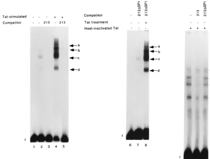
Treatment of U-87 MG cells with purified recombinant Tat increases the DNA binding activities of AP1, NF-B, and SP1 to the MCP-1 promoter. Results from luciferase functional assays indicated that SP1, AP1, andB binding sites are in-volved in basal and Tat-induced activity of the MCP-1 pro-moter. HIV-1 Tat has been reported to increase the binding of cellular factors such as NF-B (9, 39) and NF-IL-6 (9, 42) to their cognate binding elements. Moreover, Tat has also been shown to interact directly with SP1 (25), and both SP1 and NF-B are required for Tat-mediated transactivation of the HIV-1 promoter (11, 48). Hence we examined the binding of cellular factors to the MCP-1 promoter in U-87 MG cells in the absence of and following treatment with soluble recombinant HPLC-purified Tat (rTat). Nuclear extracts prepared from un-stimulated U-87 MG cells shifted with a radiolabeled DNA fragment corresponding to region⫺213 to⫹6 of the MCP-1
promoter (named hMCP213) resulted in the formation of three DNA-protein complexes (Fig. 8 lane 2, bands b, c, and d). All three complexes were disrupted by excess unlabeled hMCP213 (lane 3) but not by excess hMCP213 containing a mutated SP1 site (lane 7). When EMSAs were performed with nuclear extracts prepared from rTat-treated U-87 MG cells, marked induction of the DNA-protein complexes (bands b, c, and d), was detected (lane 4). Furthermore, a band of higher mobility (band a) was induced. This complex was previously not observed in the EMSAs performed with extracts from unstimulated cells (compare lane 2 with lane 4). Formation of complexes a to d was abolished by preincubation with excess unlabeled probe (lanes 5). In contrast, they were not competed out by preincubation with excess hMCP213 containing a mu-tated SP1 site (lane 8). When we repeated the experiments with nuclear extracts prepared from cells treated with the same purified rTat protein preparation subjected to heat inactiva-tion, we observed no similar induction of these bands (lanes 9 to 11).
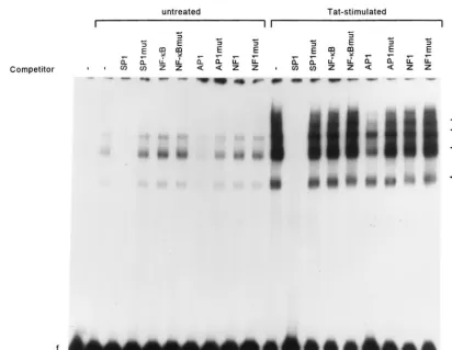
To determine the specificity of interaction between DNA binding proteins and hMCP213, we conducted competition studies using unlabeled oligonucleotides corresponding to con-sensus SP1, AP1, NF-B, and NF1 binding motifs for com-petition (Fig. 9). Binding of nuclear extracts prepared from unstimulated cells or from cells treated with the rTat to hMCP213 was completely inhibited by the presence of excess SP1 oligonucleotides (Fig. 9, lanes 3 and 12). AP1 oligonucle-otides also partially competed out binding (lanes 7 and 16). Thus, the EMSA results corroborate the data from func-tional transfection experiments where SP1 and to a lesser ex-tent AP1 were observed to be important for constitutive MCP-1 promoter activity. Competition experiments performed with NF-B oligonucleotides failed to compete out binding (lanes 5 and 14). One possible explanation for this may be that members of the Rel family other than NF-B are involved in binding to this proximal hMCP-1B site. NF1 oligonucleotides also did not inhibit complex formation (lanes 9 and 18). As expected, the controls using excess unlabeled oligonucleotides with mutant SP1, AP1, NF-B, and NF1 sequences did not compete out binding (lanes 4, 6, 8, 10, 13, 15, 17, and 19). These results suggest that both SP1 and AP1 are involved in Tat-mediated induction and may bind cooperatively.
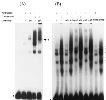
To confirm that the nuclear activator protein SP1 indeed binds to the MCP-1 promoter, nuclear extracts were preincu-bated with SP1-specific antibodies and again tested by EMSA with labeled hMCP213. With extracts prepared from both un-treated and Tat-un-treated U-87 MG cells, the major complex, c, was supershifted (Fig. 10; compare lane 2 with lane 3 and lane 4 with lane 5; the supershift band is denoted by arrow a). Thus, the results indicate that the transcriptional activator SP1 is part of the binding complexes interacting with the MCP-1 promoter and that its binding activities are enhanced following treatment of U-87 MG cells with Tat. We carried out similar experiments using antibodies to AP1 and the two subunits of NF-B, p50 and p65. The mobility of the bands from extracts of untreated cells was not affected by preincubation with AP1 antibodies (Fig. 10, lane 8). Instead, all bands in extracts of Tat-treated cells were supershifted by antibodies to AP1 (lane 9; denoted by arrow b). When antibodies to p50 and p65 subunits were used in the EMSAs all bands in extracts from untreated and Tat-treated cells were supershifted (lanes 10 to 13; denoted by arrows c to f). Similar to the observation with SP1 antibodies, the mobility of the major band, band c, was consistently super-shifted with antibodies to AP1, p50, and p65 (lanes 9, 11, and 13), suggesting that all three factors interact cooperatively, possibly as a multimeric complex. Preincubation with
antibod-FIG. 7. Analysis of transcriptional control elements in the hMCP-1 promoter in tat-III cells. Luciferase assays were performed with extracts of HeLa-tat-III cells transfected with hMCP-1 promoter constructs bearing various mu-tated cis-acting elements and were measured 48 h posttransfection. Results (average of four experiments) are expressed as the percentage of luciferase activity compared to the wild-type construct phMCP213 and in each case were normalized to units of-galactosidase activity from cotransfection with a pSV--galactosidase plasmid. The average raw luciferase value for pGL2-Basic was 4.5⫾2.3.
on November 9, 2019 by guest
http://jvi.asm.org/
[image:5.612.64.287.73.185.2]ies to C/EBP, used as a control, did not affect the mobility of any of the bands (Fig. 10, lanes 14 and 15).
DISCUSSION
The MCP-1 upstream promoter region contains putative
cis-acting elements for the cellular transcription factors SP1, AP1, and members of the Rel family of transactivators. PMA is a strong inducer of MCP-1 expression in various cell types (8, 27, 29, 40, 44, 45, 49). Both AP1 sites of the MCP-1 promoter (at⫺156 to ⫺150 and⫺128 to ⫺122) have previously been shown to be important for PMA induction in human endothe-lial (44) and glioblastoma (29) cells. MCP-1 expression is also inducible by cytokines (22, 32), including TNF-␣, which in-duces the DNA binding activity of NF-B and AP1 in a variety of cell lines (32, 49). Moreover, TNF-␣-induced monocyte chemotactic activity in the culture medium of MC3T3E1 cells was inhibited by antisense oligonucleotides of c-fosand c-jun
genes (22). Recently, the transcription of MCP-1 was also found to be inducible by HIV-1 Tat in primary human astro-cytes (10).
We studied the promoter activity of MCP-1 in a region encompassing 486 nucleotides upstream from the translational start site (Fig. 2). In both the astrocytoma cell line U-87 MG and the fibroblast cell line HeLa, constitutive activity was con-sistently highest in a construct bearing 213 nucleotides up-stream from the translational start site (Fig. 2A and 3A). This region of the promoter was also the most responsive to PMA
induction in both cell lines, albeit with different kinetics (Fig. 2B and 3B). A similar study by Li and Kolattukudy on the human glioblastoma cell line U138MG led to a different find-ing; only the construct equivalent to phMCP213 and not that equivalent to phMCP486 was found to be PMA responsive (29). Recently, the same ⫺213 to ⫹6 region has also been reported to mediate gamma interferon induction of the hMCP-1 gene (53). We show in this study that HIV-1 Tat is also capable of transactivating the hMCP-1 promoter via this region (Fig. 4 and 5). This mechanism of Tat-mediated activa-tion is TAR independent, as no TAR-like sequence (18) could be identified in the MCP-1 5⬘ untranslated region (data not shown). The GC box located between ⫺123 and ⫺115 was found to be critical for maintenance of basal and Tat-stimu-lated transcriptional activity. Mutation of this site completely abolished basal promoter activity (Fig. 6A), and treatment with Tat could not overcome this loss in function (Fig. 6B and 7). The GC box is well characterized as an SP1 binding site (26). In gel shift assays performed with the region from⫺213 to⫹6 of the MCP-1 promoter, all complexes formed from nuclear extracts of untreated U-87 MG cells were readily competed out by excess SP1 oligonucleotides and not by mutant SP1 oligo-nucleotides (Fig. 9). In addition, they were not competed out by excess hMCP213 DNA fragment with a mutated SP1 site (Fig. 8). Treatment with Tat, but not heat-inactivated Tat, further enhanced their binding (Fig. 8). Consistent with these findings, we observed that antibodies to SP1 successfully su-FIG. 8. Tat-enhanced binding of transcriptional factors to the hMCP-1 promoter region. A radiolabeled DNA fragment containing the region from⫺213 to⫹6 bp of the hMCP-1 5⬘flanking region was incubated with nuclear extracts from unstimulated U-87 MG cells (lanes 2, 3, and 7), cells treated with 100 nM Tat (lanes 4, 5 and 8), or cells treated with heat-inactivated Tat (lane 9-11). Competition assays were performed with a 100-fold molar excess of unlabeled probe (lanes 3, 5, and 10) or with hMCP213 containing a mutated SP1 site (lanes 7, 8, and 11). Lanes 1 and 6 consist of the probe alone. Arrows indicate specific DNA-protein complexes.
on November 9, 2019 by guest
http://jvi.asm.org/
[image:6.612.92.512.76.393.2]pershifted the major binding complex formed with both un-stimulated and Tat-induced extracts when the region from ⫺213 to⫹6 was used as a probe (Fig. 10).
SP1 has been shown to be required for the basal and Tat-activated transcription from the HIV-1 LTR (23, 47) and has been shown to bind to HIV-1 Tat proteinin vitroandin vivo
(25). More recently, HIV-1 Tat has been found to augment SP1 phosphorylation, leading to enhanced promoter activity (7). In addition, both Jurkat-Tat and HeLa-Tat cell lines con-tain elevated levels of phosphorylated SP1 compared to their respective parental cell lines (7). Thus, it is possible that the increase in DNA-protein complex formation observed follow-ing Tat treatment of U-87 MG cells is a consequence of in-creased SP1 phosphorylation. The role of this GC box in main-tenance of basal hMCP-1 transcription is also supported by a study by Uedaet al., who found that mutating this element led to a complete loss of reporter promoter activity in A172 glioblastoma cells, HT1080 fibrosarcoma cells, and SKLMS1 leiomyosarcoma cells (50). In a more recent report, disrupt-ing this same site in another human astrocytoma cell line, CRT, reduced promoter-reporter expression by half (53).
HIV-1 Tat protein has been reported to activate the nuclear translocation of NF-B in a HeLa derivative, HL3T1 (11). It has also been observed that treatment of Tat is associated with an increase in both NF-B binding and protein kinase C ac-tivity in primary human astrocytes (9). Both the second AP1 (at ⫺128 to⫺122; AP1B) and theB (at ⫺150 to ⫺137) sites appear to play some role in basal and Tat-stimulated MCP-1
gene expression, as mutations of these two sites reduced pro-moter activity significantly in both U-87 MG and HeLa-tat-III cells (Fig. 6 and 7). Gel shift assays with the⫺213 to⫹6 region of the MCP-1 promoter show that these binding factors are indeed induced by Tat treatment of U-87 MG cells (Fig. 10). Interestingly, Uedaet al. found that mutating AP1B had no effect on reporter gene activity (50). We do not know the reason for this discrepancy with our observations. Yet Tat does stimulate AP1 binding to the MCP-1 promoter, as gel shift experiments clearly showed the induction of AP1 binding fac-tors (Fig. 9 and 10). Since the second AP1 element does not contain a canonical AP1 binding motif (it has a C rather than an A in the last nucleotide of the heptanucleotide recognition sequence), we think that AP1 consequently has a low affinity for it. We envision that in order to stabilize its binding at AP1B, AP1 might require interaction with NF-B binding to its upstreamB site at nucleotides⫺150 to⫺137. In support of this hypothesis, it has been reported that a complex consist-ing of NF-Bp65 and Jun or Fos exhibits enhanced DNA binding and biological functions via theB site of the 5⬘LTR of HIV-1 (47).
[image:7.612.95.507.71.389.2]In the HIV-1 LTR, two NF-B sites and three SP1 sites are located close together. Synergism between NF-B/Rel and SP1 is important for activation of the LTR (28, 35, 36). Coopera-tion between AP1 and SP1 to optimally mediate cellular gene promoter activity, such as in the human CD11c gene (33) and the involucrin gene (3), has also been documented. For the hMCP-1 gene, our data suggest that SP1, AP1, and NF-B FIG. 9. SP1 and AP1 bind to the hMCP-1 promoter region. A radiolabeled DNA fragment containing the region from⫺213 to⫹6 bp of the hMCP-1 5⬘flanking region was incubated with nuclear extracts from U-87 MG cells. Competition assays were performed with a 100-fold molar excess of specific oligonucleotides. Extracts were prepared from unstimulated U-87 MG cells (lanes 2 to 10) or cells treated with 100 nM Tat (lanes 11 to 19). Lane 1 consists of the probe alone. Arrows indicate specific DNA-protein complexes. mut, mutant.
on November 9, 2019 by guest
http://jvi.asm.org/
binding factors are involved in basal and Tat-stimulated tran-scription, most likely in a synergistic manner. It is possible that potential Tat-responsive elements are present within the re-gion spanning ⫺486 to ⫺213 (Fig. 4 and 5). A consensus sequence for the octamer transcription factor is located at ⫺282 (44). This along with other unidentified binding elements could contribute to Tat-mediated stimulation of the MCP-1 genein vivo. In a related study, a functional interaction be-tween proteins binding to the GC box and an upstream gamma interferon-activated site (at⫺212 from the translational start site) on the hMCP-1 promoter was observed in the astrocy-toma cell line CRT (53). This report, together with our data, establishes the importance of the GC box in inducible up-regulation of the hMCP-1 gene following exposure to external stimuli. Our results are consistent with the possibility that HIV-1 Tat augments SP1 binding activities—perhaps through posttranslational modifications (7, 24, 34)—which in turn serve as a platform to recruit and stabilize the interaction of AP1 and NF-B proteins to the hMCP-1 promoter. In this way, HIV-1 Tat may specifically induce overexpression of hMCP-1, a che-mokine whose levels are elevated in HIV-1-associated demen-tia (10).
ACKNOWLEDGMENTS
We thank K. Conant, R. C. Gallo, M. S. Reitz, Jr., and A. L. DeVico for valuable suggestions and discussions and for critical reviews of the manuscript; S. K. Arya for providing the HIV-1 Tat expression vector; B. C. Nair for providing purified Tat protein; and Anna Mazzuca for editorial assistance.
This work was supported by the Institute of Human Virology (Uni-versity of Maryland Biotechnology Institute, Uni(Uni-versity of Maryland, Baltimore) and the National Science and Technology Board (Singa-pore).
REFERENCES
1.Andrews, N. C., and D. V. Faller.1991. A rapid micropreparation technique for extraction of DNA-binding proteins from limiting numbers of mamma-lian cells. Nucleic Acids Res.19:2499.
2.Arya, S. K., C. Guo, S. F. Josephs, and F. Wong-Staal.1985. Trans-activator gene of human T-lymphotropic virus type III (HTLV-III). Science229:
69–73.
3.Banks, E. B., J. F. Crish, J. F. Welter, and R. L. Eckert.1998. Character-ization of human involucrin promoter distal regulatory region transcription activator elements—a role for SP1 and AP1 binding sites. Biochem. J.331:
61–68.
4.Benos, D. J., B. H. Hahn, J. K. Bubein, S. K. Ghosh, N. A. Mashburn, M. A. Chaikin, G. M. Shaw, and E. N. Benveniste.1994. Envelope glycoprotein gp120 of human immunodeficiency virus type 1 alters ion transport in astro-FIG. 10. Supershifts of protein complexes binding to hMCP-1 promoter region. (A) Nuclear extracts from unstimulated U-87 MG cells (lanes 2 and 3) or cells treated with 100 nM Tat (lanes 4 and 5) were preincubated with polyclonal antibodies to SP1 (lanes 3 and 5) and then gel shifted with a radiolabeled DNA fragment containing the⫺213 to⫹6 bp hMCP-1 5⬘flanking region. (B) Nuclear extracts from unstimulated U-87 MG cells (lanes 6, 8, 10, 12, and 14) or 40 nM Tat (lanes 7, 9, 11, 13, and 15) were preincubated with polyclonal antibodies to AP1 (lanes 8 and 9), NF-B p50 (lanes 10 and 11) and p65 (lanes 12 and 13), and C/EBP (lanes 14 and 15) and then gel shifted with a radiolabeled DNA fragment containing the⫺213 to⫹6 bp hMCP-1 5⬘flanking region. Lane 1 consists of the probe alone. Arrows a and b show positions of supershifted bands with the SP1 and AP1 antibodies, respectively; arrows c, d, and e denote positions of supershifted bands from unstimulated U-87 MG cell extracts with the p50 and p65 antibodies; arrows f, b, and g, represent supershifts with the same antibodies from Tat-treated cell extracts.
on November 9, 2019 by guest
http://jvi.asm.org/
[image:8.612.123.481.72.407.2]cytes: implications for AIDS dementia complex. Proc. Natl. Acad. Sci. USA
91:494–498.
5.Buonaguro, L., G. Barillari, H. K. Chang, C. A. Bohan, V. Kao, R. Morgan, R. C. Gallo, and B. Ensoli.1992. Effects of the human immunodeficiency virus type 1 Tat protein on the expression of inflammatory cytokine. J. Virol.
66:7159–7167.
6.Chang, H. C., F. Samaniego, B. C. Nair, L. Buonaguro, and B. Ensoli.1997. HIV-1 Tat protein exits from cells via a leaderless secretory pathway and binds to extracellular matrix-associated heparan sulfate proteoglycans through its basic region. AIDS11:1421–1431.
7.Chun, R. F., O. J. Semmes, C. Neuveut, and K.-T. Jeang.1998. Modulation of SP1 phosphorylation by human immunodeficiency virus type 1 Tat. J. Vi-rol.72:2615–2629.
8.Colotta, F., A. Borre, J. M. Wang, M. Tattanelli, F. Madalena, N. Polenta-rutti, G. Peri, and A. Mantovani.1992. Expression of a monocytic cytokine by mononuclear phagocytes. J. Immunol.148:760–765.
9.Conant, K., M. Ma, A. Nath, and E. O. Major.1996. Extracellular human immunodeficiency virus type 1 Tat protein is associated with an increase in both NF-B binding and protein kinase C activity in primary astrocytes. J. Virol.70:1384–1389.
10. Conant, K., A. Garzino-Demo, A. Nath, J. C. McArthur, W. Halliday, C. Power, R. C. Gallo, and E. O. Major.1998. Induction of moncyte chemoat-tractic protein-1 in HIV-1 Tat-stimulated astrocytes and elevation in AIDS dementia. Proc. Natl. Acad. Sci. USA95:3117–3121.
11. Demarchi, F., F. D’Adda di Fagagna, A. Falaschi, and M. Giacca.1996. Activation of transcription factor NF-B by the Tat protein of human im-munodeficiency virus 1. J. Virol.70:4427–4437.
12. Elder, G. A., and E. O. Major.1988. Early appearance of type II astrocytes in developing human fetal brain. Brain Res.470:146–150.
13. Ensoli, B., G. Barillari, S. Z. Salahuddin, R. C. Gallo, and F. Wong-Staal.
1990. Tat protein of HIV-1 stimulates growth of cells derived from Kaposi’s sarcoma lesions of AIDS patients. Nature345:84–86.
14. Ensoli, B., L. Buonaguro, G. Barillari, V. Fiorelli, R. Gendelman, R. A. Morgan, P. Wingfield, and R. C. Gallo.1992. Release, uptake, and effects of extracellular human immunodeficiency virus type 1 tat protein on cell growth and viral transactivation. J. Virol.67:277–287.
15. Epstein, L. G., and H. E. Gendelman.1993. Human immunodeficiency virus type 1 infection of the nervous system: pathogenetic mechanisms. Ann. Neurol.33:429–436.
16. Faller, D. V., H. Q. Weng, D. T. Graves, and S. Y. Choi.1997. Moloney murine leukemia virus long terminal repeat activates monocyte chemotactic protein-1 expression and chemotactic activity. J. Cell Physiol.172:240–252. 17. Feinberg, M. B., R. F. Jarret, A. Aldovini, R. C. Gallo, and F. Wong-Staal.
1986. HTLV-III expression and production involve complex regulation at the levels of splicing and translation of viral RNA. Cell46:807–817.
18. Feng, S., and E. C. Holland.1988. HIV-1 tat trans-activation requires the loop sequence within TAR. Nature (London)334:165–167.
19. Frankel, A. D., and C. O. Pabo.1988. Cellular uptake of the Tat protein from human immunodeficiency virus. Cell55:1189–1193.
20. Garzino-Demo, A., S. K. Arya, A. L. DeVico, F. Cocchi, P. Lusso, and R. C. Gallo.1997. CC chemokine RANTES and HIV long terminal repeat-driven gene expression. AIDS Res. Hum. Retroviruses13:1367–1371.
21. Glass, J. D., H. S. Fedor, S. L. Wesselingh, and J. C. McArthur.1995. Immunocytochemical quantitation of human immunodeficiency virus in the brain: correlations with dementia. Ann. Neurol.38:755–762.
22. Hanazawa, S., A. Takeshita, S. Amano, T. Semba, T. Nirazuka, H. Katoh, and S. Kitano.1993. Tumour necrosis factor-␣induced expression of mono-cyte chemoattractant JE via fos and jun genes in clonal osteoblastic MC3T3-E1. J. Biol. Chem.268:9526–9532.
23. Harrich, D., J. Garcia, F. Wu, R. Mitsuyasu, J. Gonzalez, and R. Gaynor.
1989. Role of SP1-binding domains in vivo transcriptional regulation of the human immunodeficiency virus type 1 long terminal repeat. J. Virol.63:
2585–2591.
24. Jackson, S. P., and R. Tjian.1988. O-glycosylation of eukaryotic transcrip-tions for mechanisms of transcriptional regulation. Cell55:125–133. 25. Jeang, K. T., R. Chun, N. H. Lin, A. Gatignol, C. G. Glade, and H. Fan.1993.
In vitro and in vivo binding of human immunodeficiency virus type 1 Tat protein and SP1 transcription factor. J. Virol.67:6224–6233.
26. Kadonaga, J. T., K. R. Carner, F. R. Masiarz, and R. Tjian.1987. Isolation of cDNA encoding transcription factor SP1 and functional analysis of the DNA binding domain. Cell51:1079–1090.
27. Leonard, E. J., and T. Yoshmura.1990. Human monocyte chemoattractant protein-1 (MCP-1). Immunol. Today11:97–101.
28. Li, Y., G. Mak, and B. R. Franza, Jr.1994. In vitro study of functional involvement of SP1, NF-B/Rel, and AP1 in phorbol 12-myristate 13-ace-tate-mediated HIV-1 long terminal repeat activation. J. Biol. Chem.269:
30616–30619.
29. Li, Y.-S., and P. E. Kolattukudy.1994. Functional role of the cis-acting elements in human monocyte chemotactic protein-1 gene in the regulation of its expression by phorbol ester in human glioblastoma cells. Mol. Cell. Biochem.141:121–128.
30. Magnuson, D. S., B. E. Knudsen, J. D. Geiger, R. M. Brownstone, and A. Nath.1995. Human immunodeficiency virus type 1 tat activates non-N-methyl-D-aspartate excitatory amino acid receptors and causes neurotoxicity. Ann. Neurol.37:373–380.
31. Mann, D. A., and A. D. Frankel.1991. Endocytosis and targeting of exoge-nous HIV-1 Tat protein. EMBO J.10:1733–1739.
32. Martin, T., P. M. Cardarelli, G. C. N. Parry, K. A. Felts, and R. R. Cobb.
1997. Cytokine induction of monocyte chemoattractant protein-1 gene ex-pression in human endothelial cells depend on the cooperative action of NF-B and AP-1. Eur. J. Immunol.27:1091–1097.
33. Noti, J. D., B. C. Reinemann, and M. N. Petrus.1996. SP1 binds two sites in the CD11c promoter in vivo specifically in myeloid cells and cooperates with AP1 to activate transcription. Mol. Cell. Biol.16:2940–2950.
34. Pecorino, L. T., A. L. Darrow, and S. Strickland.1991. In vitro analysis of the tissue plasminogen activator promoter reveals a GC box-binding activity present in murine brain but undetectable in kidney and liver. Mol. Cell. Biol.
11:3139–3147.
35. Perkins, N., N. L. Edwards, C. S. Duckett, A. B. Agranoff, R. M. Schmid, and G. J. Nabel.1993. A cooperative interaction between NF-B and SP1 is required for HIV-1 enhancer activation. EMBO J.12:3551–3558. 36. Perkins, N. D., A. B. Agranoff, E. Pascal, and G. J. Nabel.1994. An
inter-action between the DNA-binding domains of RelA(p65) and SP1 mediates human immunodeficiency virus gene activation. Mol. Cell. Biol.14:6570– 6583.
37. Price, R. W., B. Brew, J. Sidtis, M. Rosenblum, A. C. Scheck, and P. Cleary.
1988. The brain in AIDS: central nervous system HIV-1 infection and AIDS dementia complex. Science239:586–592.
38. Rosen, C. A., J. G. Sodroski, K. Campbell, and W. A. Haseltine.1986. Construction of recombinant murine retroviruses that express the human T-cell leukemia virus type II and human T-cell lymphotropic virus type III
transactivator genes. J. Virol.57:379–384.
39. Roulston, A., P. Beauparlant, N. Rice, and J. Hiscott.1993. Chronic human immunodeficiency virus type 1 infection stimulates distinct NF-B/rel DNA binding activities in myelo-monoblastic cells. J. Virol.67:5235–5246. 40. Rovin, B. H., T. Yoshimura, and L. Tan.1992. Cytokine-induced production
of monocyte chemoattractant protein-1 by cultured human mesangial cells. J. Immunol.148:2148–2153.
41. Sastry, K. J., M. C. Marin, N. P. Nehete, K. McConnell, A. K. el-Nagger, and T. J. McDonnell.1996. Expression of human immunodeficiency virus type 1 tat results in down-regulation of bcl-2 and induction of apoptosis in hema-topoietic cells. Oncogene13:487–493.
42. Scala, G., M. R. Ruocco, C. Ambrosino, M. Mallardo, V. Giordano, F. Baldassarre, E. Dragonetti, I. Quinto, and S. Venuta.1994. The expression of the interleukin 6 gene is induced by the human immunodeficiency virus 1 tat protein. J. Exp. Med.179:961–971.
43. Schall, T. J.1991. Biology of the RANTES/SIS cytokine family. Cytokine3:
165–183.
44. Shyy, Y.-J., Y.-S. Li, and P. E. Kolattukudy.1990. Structure of human monocyte chemotactic protein gene and its regulation by TPA. Biochem. Biophys. Res. Commun.169:346–351.
45. Shyy, Y. J., Y. Li, and P. E. Kolattukudy.1993. Activation of MCP-1 gene expression is mediated through multiple signaling pathways. Biochem. Bio-phys. Res. Commun.192:693–699.
46. Sodroski, J. G., R. Patarca, C. A. Rosen, F. Wong-Staal, and W. A. Haseltine.
1985. Location of the trans-activating region of the genome of human T-cell lymphotropic virus type III. Science229:74–77.
47. Stein, B., A. S. Baldwin, Jr., D. W. Ballard, W. C. Greene, P. Angel, and P. Herrlich.1993. Cross-coupling of the NF-kB p65 and Fos/Jun transcriptional factor produces potentiated biological function. EMBO J.12:3879–3891. 48. Sune, C., and M. A. Garcia-Blanco.1995. SP1 transcription factor is required
for in vitro basal and Tat-activated transcription from the human immuno-deficiency virus type 1 long terminal repeat. J. Virol.69:6572–6576. 49. Toggas, S. M., E. Masliah, E. M. Rockenstein, G. F. Rall, C. R. Abraham,
and L. Mucke.1994. Central nervous system damage produced by expression of the HIV-1 coat protein gp120 in transgenic mice. Nature367:188–193. 50. Ueda, A., K. Okuda, S. Ohno, A. Shirai, T. Igarashi, K. Matsunaga, J.
Fukushima, S. Kawamoto, Y. Ishigatsubo, and T. Okubo.1994. NF-B and SP1 regulate transcription of the human monocyte chemoattractant pro-tein-1. J. Immunol.153:2052–2063.
51. Viscidi, R. P., K. Mayur, H. M. Lederman, and A. D. Frankel.1989. Inhibi-tion of antigen-induced lymphocyte proliferaInhibi-tion by Tat protein from HIV-1. Science246:1606–1608.
52. Westendorp, M. O., M. Li-Weber, R. W. Frank, and P. H. Krammer.1994. Human immunodeficiency virus type 1 Tat up-regulates interleukin-2 secre-tion in activated T cells. J. Virol.68:4177–4185.
53. Zhou, Z.-H. L., P. Chaturvedi, Y.-L. Han, S. Aras, Y.-S. Li, P. E. Kolat-tukudy, D. Ping, J. M. Boss, and R. M. Ransohoff.1998. IFN-␥induction of the human monocyte chemoattractant protein (hMCP-1) gene in astro-cytoma cells: functional interaction between an IFN-␥activated site and a GC-rich element. J. Immunol.160:3908.