JOURNAL OF VIROLOGY, Jan. 1994,
p.
521-525 Vol. 68,No. 1 0022-538X/94/$04.00+0Copyright C) 1994,American Society for Microbiology
Retention of the
Herpes Simplex Virus Type 1
(HSV-1)
UL37
Protein
on
Single-Stranded DNA Columns Requires
the HSV-1 ICP8 Protein
LISA S. G.
SHELTON,'
ALLEN G.
ALBRIGHT,'
WILLIAM T.
RUYECHAN,2
AND
FRANK J.
JENKINS'*
Department
of Microbiology, Uniformed Services University of the Health Sciences, Bethesda, Maryland
20814,'
and Department of Microbiology, School of Medicine and Biomedical Sciences, State University of New York
atBuffalo, Buffalo, New York 142142
Received 3 June 1993/Accepted 27 September 1993
The
UL37 and ICP8 proteins present in herpes simplex virus type 1 (HSV-1)-infected-cell extracts produced
at 24 h
postinfection coeluted from
single-stranded-DNA-cellulose columns. Experiments carried out with the
UL37 protein expressed by a vaccinia
virus recombinant (V37) revealed that the UL37 protein did not exhibit
DNA-binding activity in the absence of other HSV
proteins. Analysis of extracts derived from cells coinfected
with V37 and an
ICP8-expressing vaccinia virus recombinant (V8) and analysis of extracts prepared from cells
infected with the
HSV-1 ICP8 deletion mutants
d21 and nlO revealed that the retention of the
UL37
protein on
single-stranded DNA columns required a
DNA-binding-competent ICP8 protein.
The herpes simplex virus type 1 (HSV-1) genome is a linear,
double-stranded DNA molecule of 160 kb (9, 15).
Computer-assisted
analysis of the DNA sequence has revealed at least 75
separate open reading frames
(ORFs) (15). The functions of
many
of these genes in viral replication are at least partially
understood. However, a
significant fraction of HSV ORFs
have not yet been characterized with regard to either the
protein produced or the role that the protein plays in the viral
life cycle (21). Advances in molecular techniques, along with
knowledge of the precise locations of the different ORFs, now
permit detailed studies on individual viral proteins with the
ultimate
goal directed towards the determination of individual
protein
function(s).
The
protein product of the UL37 ORF of HSV-1 represents
aviral
protein for
which the precise function is unknown. The
UL37gene
is located in the unique long sequences between
0.527 and 0.522 map
unit
onthe HSV-1 genome.
Inearlier
publications,
wereported
onthe
identification and
character-ization of the UL37 protein in HSV-1-infected cells (1, 22). We
determined that
the UL37 ORF encoded a phosphorylated,
nonstructural
protein with
anapparent molecular
massof
120kDa.
Analysis of the kinetics of production of UL37 places it in
the
-yl class of HSV genes.
Inaddition,
the UL37
protein
waseluted from
single-stranded-DNA-agarose
columns
with
anelution
profile
that
completely overlapped
with that
of the
major HSV-1
DNA-binding protein,
commonly designated
ICP8.
Because
the UL37
protein
has the
sameapparent
molecular
weight
asICP8 and
comigrates
with ICP8
onsodium
dodecyl
sulfate-polyacrylamide gel
electrophoresis (SDS-PAGE),
the
presence
of the UL37
protein
was notdetected in
previous
studies of HSV
DNA-binding proteins
(11,
12,
18-20,
23).
In order tostudy the UL37
protein independently of other HSV-1
proteins and to examine possible interactions between
theUL37
andICP8
proteins,
(i)
fusions of
UL37gene sequences
and the Escherichia coli
maltose-binding protein
weregener-*Correspondingauthor.Mailingaddress:Departmentof
Microbiol-ogy, USUHS, 4301 Jones Bridge Rd., Bethesda, MD 20814-4799. Phone: (301)295-3403. Fax: (301)295-1545. Electronic mail address: FJENKINS@usuhs.usuhs.mil.
ated,
(ii)
arecombinant vaccinia virus which
expressed ICP8
was
constructed, and
(iii) UL37- and
ICP8-specific polyclonal
rabbit antisera
weregenerated. Rabbit polyclonal antisera
were
raised against the wild-type ICP8
protein
andagainst
anICP8
carboxy-terminal
peptide
aspreviously described
(10).
Rabbit polyclonal antibodies
specific
for the UL37
protein
were generated
against
(i) in vitro-translated UL37 protein,
designated
487antiserum
(22); (ii)
amalE fusion
protein
containing the entire UL37 protein,
designated 779 antiserum;
and
(iii)
amalE fusion
protein containing the
carboxy-terminal
one-third of the UL37
protein,
designated 780 antiserum
(Fig.
1B). To test the
specificity of each antiserum,
weused the
vaccinia virus
recombinants V37, V8, and VSC1
1as
well
asHSV-1-infected-cell
extracts.The
construction and characterization of V37,
arecombi-nant
vaccinia
virus that expresses the HSV-1 UL37
protein,
and VSC1 1,
arecombinant vaccinia virus that does
notexpress
any
HSV gene and
serves as avirus
control,
have been
previously described
(22).
Arecombinant vaccinia virus which
expressed ICP8
(V8)
wasconstructed
by using
the
plasmid pV8
essentially
asdescribed
previously
(2,
13, 14).
Theplasmid
pV8
contains the HSV-1 ICP8 gene cloned into the vaccinia virus
shuttle
vectorpSC11
(2).
A1.6-kbp
SalI-KpnI
fragment
from
pNN1
(3) (obtained
from
Mark
Challberg,
National Institutes
of
Health) containing
the 5' end of the ICP8 ORF
wascloned
into
pUC19, creating the
plasmid pJF37.
AnRsrII-HindIII
collapse
of
pJF37
followed
by
the addition of
BglII
linkers
created the
plasmid pJF65.
This
placed
the
BglII
site
approx-imately
6bp upstream of the ICP8 ATG translation
startcodon.
A4.3-kbp KpnI
fragment
from
pNN1
which contained
the
remainder of the ICP8 gene
wasthen inserted
atthe
KpnI
site
of
pJF65,
creating
the
plasmid pJF67.
The
entire ORF of
ICP8
wasexcised from
pJF67
as aBglII-EcoRI fragment
and
cloned into the
vaccinia virus shuttle
vectorpSC11,
creating
the
plasmid pV8
(Fig.
1).
The
expression
of
ICP8by
the
V8recombinant
virus
wasverified
by immunoblot
analysis
of
V8-infected CV-1 cell
proteins by using
antiserum
directedagainst
ICP8(data
notshown).
The
specificity
of the antisera
raised
against
UL37and
ICP8was
demonstrated
by immunoblot
analysis
of
HSV-1(F)-,
V37-,
V8-, and VSC1 1-infected-cell
proteins
harvested
at24 h
521on November 9, 2019 by guest
http://jvi.asm.org/
522 NOTES
U L
4-Icp8
IRL IRS
US
UL4
UL37
C P P PP P P H
I I TRL
A.
_
B.
UL37
779
780TRS
ICP8
C-Term
8
-M
IN
.
V37 V8 VSC HSV HSV VSC V8 V37
J. VIROI.
780
O
-120
HSV VSC V8 V37
487
C.
ICP8
I
DNA BINDING
ZBD DOMAIN NLS
d2l -1-11-;1
-nlO x x x x
D. pV8
TKL
pll
i
*_ IlaczTKCR
ICP8 P7.5
FIG. 1. (A) Sequence arrangement of the HSV-1 DNAgenome,
showing the locations of the unique sequences of the L and S
components (UL and Us) and ofthe terminal (TR, and TRs) and inverted (IRL and
IRs)
repeats and the ICP8 andUL37 genes. (B)Schematicdiagramofthe UL37gene.The hatched boxrepresentsthe
UL37-coding region. The solid boxes represent the domains of the UL37proteininthe fusions between malE andfull-lengthUL37(779)
and betweenmalE and the terminal 1.1kbpof UL37(780).C,Clal; P, PstI; H,HindlIl. (C) Schematicdiagram of the ICP8gene, indicating
the putative DNA-binding domain, potential zinc-binding domain
(ZBD), and nuclear localization signal (NLS). The speckled and
stripedbarsindicate the domains of the ICP8proteincontained in the d21 and nlOmutants,respectively. (D)Schematicdiagram of thepV8 plasmid used to construct the V8 vaccinia virus recombinant as
described in thetext.
postinfection (p.i.). As shown in Fig. 2, the antisera directed against ICP8 and its C-terminal peptide reacted with a
120-kDa protein from cells infected with either HSV-1 orvaccinia
virus V8 butnotwithproteins from cells infected with vaccinia virus V37 or the VSCIl vaccinia virus control. The
UL37-specific antisera 780 and 487 reacted with a 120-kDa protein
from cellsinfectedwith eitherHSV-I orvacciniavirus V37 but notwithproteins from cells infected with vaccinia virus V8or
VSC1 1. Results similartothoseobtained with the 780 and 487 antisera were obtained with the 779 antiserum (data not
shown). These results demonstrate the specificities of the UL37 andICP8 antibodies andshowthat the UL37 and ICP8 proteins comigrate in SDS-PAGE.
Previously we reported that the UL37 and ICP8 proteins
from HSV-1-infected cells coeluted from single-stranded-DNA (ssDNA)-agarose columns (22). The observation that the UL37 protein from HSV-1-infected cells could be
repro-ducibly eluted from ssDNA columns indicated that the UL37
[image:2.612.62.299.74.381.2]HSV VSC Va V37
FIG. 2.
Phosphorimage
ofimmunoblotsofHSV-1(HSV)-,
VSCI1(VSC)-,
V37-, andV8infected-cell
proteins.
Infected-cellproteins
wereharvestedat24 h
p.i., separated
onSDS-9%polyacrylamide gels,
and
probed
withpolyclonal
rabbit antiserum directedagainst
eitherwild-type
ICP8protein
(ICP8),
ICP8carboxy-terminal peptide
(C-Term
8),
malE-I1.1
UL37 fusionprotein (780),
or in vitro-translated UL37(487).
Antigen-antibody binding
was detected with'251I-labeled
protein
A.protein
either bound this DNA substratedirectly
orpossibly
interacted with other
proteins,
suchasICP8,
whichthemselves bound DNA. To determine whether the UL37protein
is able to bind ssDNA in the absence of other HSVproteins,
theDNA-binding capability
of the UL37protein expressed by
the recombinant vacciniavirus, V37,
was assessed. V37-infected-cellprotein
extractswereprepared
from CV-1 cells at48hp.i.
andanalyzed by ssDNA-agarose chromatography
asdescribed
by
Shelton et al.(22).
Individual fractions wereanalyzed by
immunoblot for the presence of the UL37
protein.
The vacciniavirus-expressed
UL37protein (V37
protein)
wasdetected
within
the columnflowthrough
and wash and wasabsent within the
gradient
elution
(Fig. 3A).
Incontrast,
the ICP8protein produced by
the V8 vaccinia virus recombinant bound tossDNA(Fig.
3B3), suggesting
that the lack ofbinding
observed with the V37protein
wasnotcausedby
expression
invaccinia
virus. These results demonstrated theinability
of the
UL37protein
tobind DNA in the absence of ICP8 and other HSVproteins.
To testwhether UL37 could bind DNA in the presence ofICP8,
protein
extracts wereprepared
from cells coinfected with V37 and V8 andanalyzed by ssDNA-agarose
chromatography.
Fractionsfrom the linearKCI elutiongradient
wereanalyzed by
immunoblotfor the UL37 and ICP8proteins.
As shown inFig.
4,
thefull-length
UL37 and ICP8 proteins coeluted in fractions 4through
12,
whichencompassed
KCl
concentrations of 400 to 600mM. Thelower-m-olecular-weight
bands which did notbind tothe column
represent
amixture of ICP8 and UL37degradation
products
as well asbackground
bands
present
in vaccinia virus-infected cells.The above-described data
strongly suggest
that
the HSV-1 ICP8 proteinis
required
for
theapparent
DNA-binding
prop-I I I I
I
on November 9, 2019 by guest
http://jvi.asm.org/
[image:2.612.319.553.76.319.2]NOTES 523
A
FT Wash
L
||O.
I GradientC L F.T. WASH FRACT. FRACTIONS
12 1 2 3 4 5 1 234 56 7 8 9101121314
4-
ICP8
1 2 3 4 5 6 1 2 3 4 5 6 7 89
Gradient
2M StripP I 10
10 111213 14 15 16 17 18
B
150 mM
C
L FT!
D-1 2 3 4 5
19 20 21 22 23 1 2 3 4 c F.T. WASH FRACTIONS
1l 2 1 2 3 1 2 3 4 5 6 78
11VMPlm !flwo-n
150 mM 300 mM
C
l-
i
300 mM 500mM 1 1
C i- 0
C--
1---
Cmon
_M
-if R _ ___ sg ! _-EPP
3 4 5 1 2 3 4 5
7 8 9 10 1 2 M
[image:3.612.61.293.70.335.2]1 2 3 4 5
FIG. 3. Phosphorimage of immunoblots of fractionsfrom
ssDNA-agarosecolumns. (A)V37-infected-cell proteinswereharvestedat47 hp.i.andseparatedon ssDNA-agarose columns. Individual fractions wereseparatedonSDS-9%polyacrylamide gels and probed with 780
(UL37)antiserum.(B)V8-infected-cell proteinswereharvestedat48 hp.i., separatedonssDNA-agarose columnsby usingstepelutions of
150, 300, 500, and 1000 mM KCI, and probed with ICP8 antiserum. Antigen-antibody bindingwasdetected with 125I-labeledproteinA.C,
control; L,load; FT,flowthrough.
erties of UL37. To investigate whether a
DNA-binding-com-petent ICP8 wasrequired for UL37 tobind DNA in HSV-1-infected-cell extracts, the ICP8 deletion mutantsd21 andnlO
wereobtainedfrom Priscilla Schaffer(Harvard University) and
David Knipe (Harvard University), respectively. The d21
mu-tanthas260internal amino acids deletedfrom the ICP8gene,
including the entire potential zinc-binding domain (5, 8) and 23amino acids into theputative DNA-binding region (6, 7, 24) (Fig. IC). The nlOmutanthas 36amino acidsdeleted from the carboxy terminus of the ICP8 gene, which results in the
removal of the nuclear localizationsignal (Fig. IC) (6). When these mutantsare grownin noncomplementing cells,an HSV DNA-negative phenotype results, and the ICP8 molecules remain in the cytoplasm (6, 17).
SinceafunctionalICP8 molecule is essentialforHSVDNA
replication (3, 4, 16, 18, 25, 26) and sinceUL37 demonstrates
yl kinetics, which requires DNA replication for full expression,
we first determined whether d21 and nlO infection of
non-complementing cells would result in detectable UL37
expres-sion, thus allowing us to assess correlations between a
func-tional ICP8 and the DNA-binding properties of UL37. Immunoblot analysis of d21-infected CV-1 (noncomplement-ing) and U-47
(complementing)
cells was performed withUL37- and ICP8-specific antisera. U-47 is an
ICP8-comple-menting cell line which contains three copies of the ICP8gene perhaploid genome (17). The UL37 protein wasdetected in
both CV-1 and U-47 cells infected with d21 or
nlO,
with adiminished butclearly detectable level ofexpression found in
FRACTIONS
9 10 11 12
4-
UL37
FIG. 4. Phosphorimage of immunoblots offractions from
ssDNA-agarose columns. CV-1 cellswere coinfected with V37 and V8, and
protein extracts were harvested at 48 h p.i. and separated on an
ssDNA-agarose column by using a linear KCl gradient. Individual fractionswereseparatedonSDS-9% polyacrylamide gelsandprobed
with either ICP8 (ot8) or 487 (037) antiserum. Antigen-antibody
bindingwas detectedwith '25I-labeled protein A.C, control; L, load;
F.T., flowthrough fractions.
thenoncomplementing (CV-1) cells. In the CV-1 cells, only the faster-migrating ICP8 species encoded by d21 was detected,
while in the U-47 cells, both the shorter ICP8 protein encoded bythe d21 mutantand the full-length wild-type ICP8 protein expressed by the cell linewere observed. Similar resultswere
obtained with the nlO mutant(data not shown).
CV-1 cells infectedwithd21 ornlOwere harvested at24 h p.i., and high-salt extracts were prepared and analyzed by
ssDNA-agarose chromatography. The mutant ICP8 protein
A
L
iFT WashI
2- 3,
1 2 3 1 2 3 4 5
Gradient
10 11 12 13 14 15 16 17 18
Gradient
1 2 3 456 7 8 9
Gradient
2M192021222324 1 2
B Wash Gradient 2M StripC
1 2 1 2 34 5 1 2 34 5 6 789 101112 1 2 3
FIG. 5. Phosphorimage of immunoblots of fractionsfrom
ssDNA-agarosecolumns.d21-infected-cellproteinswereharvested at 24 hp.i.
andseparated on ssDNA-agarosecolumns. Individual fractionswere
separatedonSDS-9%polyacrylamide gelsandprobedwithICP8(A)
or 780 (UL37) (B) antiserum. Panels A and B represent separate
experiments.Antigen-antibody bindingwasdetected with '25I-labeled proteinA. L, load: FT, flowthrough; C,control.
I
Gradient
hh_
VOL.68, 1994
oa8
4.
lot
qdb.l
6 7
on November 9, 2019 by guest
http://jvi.asm.org/
[image:3.612.323.552.72.276.2] [image:3.612.320.557.497.656.2]524 NOTES
C L 150300 500 1000
UL37-_
ocUL37
C L 150 300 500 1000
ICP8 4'
(xICP8
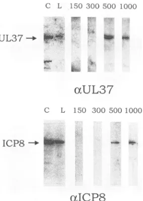
FIG. 6. Phosphorimageof immunoblots of fractions from
ssDNA-agarosecolumn.nlO-infected cellproteinswereharvestedat24hp.i.
andseparatedon anssDNA columnby usingastepgradientof150, 300, 500, and 1,000mM KCI. Individual fractionswere separated on
SDS-9%polyacrylamide gelsandprobedwith either 487(otUL37) or
ICP8 (cICP8)antiserum. Antigen-antibody bindingwasdetected with
'25I-labeledproteinA. For detection with the 487antiserum, individ-ualfractionswereconcentrated priortoloading. C,control; L, load;
numbers indicate KCI concentration (millimolar).
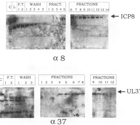
encoded by thed21 virus was unable to bind to ssDNA (Fig. 5A), aswas predicted by Orberg and Schaffer (17). Immuno-blot analysis of thed21 fractions showed the presence of the
UL37 protein in the column wash but not within the elution
gradient (Fig. 5B), indicating the inability of UL37 (in the
absence of a DNA-binding-competentICP8 protein) to bind
ssDNA. In contrast to the results obtained with the d21 mutant,the ICP8protein expressed bythen10 mutantwasable
to bindssDNA,whichagreedwithpreviousresults of Gao and
Knipe (6), and the UL37protein cofractionatedwith the nlO
ICP8 in 500 and 1,000mM KCl stepelutions(Fig. 6).
In conclusion, by comparative studies using HSV-1-, V37-, d21-, and
nIO-infected-cell
proteins, we have demonstrated that theabilityof the UL37proteinto bindssDNA columns isdependent upon the presence of a DNA binding-competent
ICP8 protein.The retention of UL37 may be due eitherto a
direct interaction with ICP8ortoanICP8-induced conforma-tional change of the DNA structure. From the kinetics of
expressionof theseproteins,it ispossiblethatUL37 could bind
tosingle-stranded nucleic acid atornearsites boundbyICP8
in the infected cell. Studies are currently in progress to determine the mechanism ofbinding.
We thankJonathan Hirsch fortechnical assistance. We also thank
Priscilla Schaffer for the d21 mutant and David Knipe for the nIO
mutant.
This investigation was supported by USUHS grant R07396 (to F.J.J.)and PublicHealth ServicegrantA122468(to W.T.R.).
REFERENCES
1. Albright,A.G.,and F.J. Jenkins. 1993. The herpes simplexvirus
UL37proteinisphosphorylatedininfectedcells. J. Virol.
67:4842-4847.
2. Chakrabarti, S., K.Brechling,and B.Moss. 1985. Vaccinia virus
expressionvectors:coexpressionof
0-galactosidase
providesvisualscreening of recombinant virus plaques. Mol. Cell. Biol.
5:3403-3409.
3. Challberg, M. D. 1986. A method foridentifyingthe viralgenes required for herpes virusDNAreplication. Proc. NatI.Acad. Sci. USA83:9094-9098.
4. Conley, A. J., D. M. Knipe, P. C.Jones,and B. Roizman. 1981.
Molecular geneticsofherpes simplexvirus. VII. Characterization
ofatemperature-sensitive mutantproduced by in vitro mutagen-esisand defective in DNAsynthesis and accumulation ofgamma polypeptides.J.Virol.37:191-206.
5. Gao,M., J. Bouchey, K. Curtin,and D. M.Knipe. 1988.Genetic
identification ofaportion of the herpes simplex virus ICP8protein required for DNA-binding. Virology163:319-329.
6. Gao, M., and D. M.Knipe. 1989. Genetic evidence formultiple nuclear functionsof theherpes simplexvirus ICP8DNA-binding protein.J.Virol. 63:5258-5267.
7. Gao, M., and D. M. Knipe. 1991. Potential roleforherpessimplex virus ICP8 DNA replication protein in stimulation of lategene
expression. J. Virol. 65:2666-2675.
8. Gupte, S. S., J. W. Olson, and W. T. Ruyechan. 1991. Themajor herpes simplex virus type-I DNA-bindingproteinisazinc
metal-loprotein.J.Biol. Chem. 266:11413-11416.
9. Kieff, E. D., S. L. Bachenheimer, and B. Roizman. 1971. Size, composition, andstructureof thedeoxyribonucleic acid of herpes simplex virus subtypes I and 2. J. Virol. 8:125-129.
10. Kinchington, P. R.,G. Inchauspe, J. H. Subak-Sharpe, F. Robey, J. Hay, and W. T. Ruyechan. 1988. Identification and character-ization ofavaricella-zoster virus DNA-binding protein by using antisera directed against a predicted synthetic oligopeptide. J. Virol. 62:802-809.
11. Leinbach,S.S.,J. F. Casto,and T. K.Pickett. 1984. Deoxyribo-nucleoprotein complexes and DNA synthesis of herpes simplex virustype 1. Virology 137:287-296.
12. Littler, E., D. Purifoy, A. Minson, and K. L. Powell. 1983.Herpes
simplex virus non-structural proteins. III. Function of the major DNA-binding protein.J. Gen.Virol. 64:983-995.
13. Mackett, M., G.L.Smith,and B. Moss.1984.General method for theproduction and selection of infectious vaccinia virus
recombi-nantsexpressing foreigngenes.J.Virol. 49:857-864.
14. Mackett, M., G. L. Smith, and B. Moss. 1985.The construction and characterization of vaccinia virus recombinants expressing foreigngenes, p. 191-211.In D.M.Glover (ed.),DNAcloning,a
practical approach, vol. II. IRLPress,Oxford.
15. McGeoch,D.J., M. A. Dalrymple, A. J. Davison, A. Dolan, M. C. Frame, D. McNab, L. J. Perry, J. E. Scott, and P. Taylor. 1988. The
completeDNA sequenceofthelong unique region in thegenome
ofherpes simplex virustype 1. J.Gen.Virol. 69:1531-1574. 16. McGeoch, D. J., M. A. Dalrymple, A. Dolan, D. McNab, L. J.
Perry,P. Taylor, and M. D.Challberg. 1988. Structures of herpes simplex virustype1 genesrequired for replication of virusDNA. J.
Virol. 62:444-453.
17. Orberg, P. K., and P. A. Schaffer. 1987. Expression of herpes simplex virus type I major DNA-binding protein, ICP8, in
trans-formed cell lines: complementation of deletion mutants and inhibition ofwild-type virus.J. Virol. 61:1136-1146.
18. Powell,K.L.,E.Littler,and D. J. M. Purifoy.1981. Nonstructural proteins of herpes simplex virus. II. Major virus-specific
DNA-binding protein. J.Virol. 39:894-902.
19. Powell,K.L., and D. J. M. Purifoy. 1976.DNA-binding proteins of cells infected by herpes simplex virustype 1 and type2. Intervi-rology 7:225-239.
20. Purifoy,D.J. M., and K. L. Powell. 1976.DNA-binding proteins induced by herpes simplex virus type 2 in HEp-2cells. J. Virol. 19:717-731.
21. Roizman, B.1990. Whitherherpesviruses,p.285-291. In C. Lopez, M.Mori,B. Roizman,and R.Whitley (ed.), Immunobiology and prophylaxis of human herpesvirus infections. Plenum Press, New
York.
22. Shelton, L. S. G., M. N. Pensiero, and F. J. Jenkins. 1990.
Identification and characterization of the herpes simplex virustype I protein encoded by the UL37 open reading frame. J. Virol. 64:6101-6109.
23. Vaughn,P.J., L. M. Banks, D. J. M. Purifoy, and K. L. Powell. J. VIROL.
on November 9, 2019 by guest
http://jvi.asm.org/
[image:4.612.108.250.70.269.2]VOL.68, 1994 NOTES 525
1984. Interactions between herpes simplex virus DNA-binding proteins. J. Gen. Virol. 65:2033-2041.
24. Wang, Y. S., and J. D. Hall. 1990. Characterization of a major DNA-binding domain in the herpes simplex virus type 1 DNA-binding protein (ICP8). J. Virol. 64:2082-2089.
25. Weller, S. K., K. J. Lee, D. J. Sabourin, and P. A. Schaffer. 1988.
Genetic analysis of temperature-sensitive mutants which define the gene for the major herpes simplex type 1 DNA-binding protein. J. Virol. 62:435-443.
26. Wu, C. A., N. J. Nelson, D. J. McGeoch, and M. D. Challberg. 1988. Identification of herpessimplex virus type 1 genes required fororigin-dependent DNA synthesis. J. Virol. 62:435-443.