PAX
WITH
5 EXPRE
H CLINIC
THE T PSG INSESSION I
CAL FEA
TAMILNA DEPA STITUTE PEELAMIN LYMP
ATURES A
IN LY
DISSE SUBM M.D. INADU DR. M
ARTMEN OF MED MEDU, CO TAMILN AP
PHOMAS
AND OT
YMPHOM
ERTATIO MITTED F PATHOL MGR MENT OF PAT
DICAL SC
OIMBAT
NADU, IN
PRIL 2013
S AND IT
HER PAN
MAS
ON FOR LOGY DICAL U THOLOG IENCES &ORE – 64
CERTIFICATE
This is to certify that the dissertation work entitled “PAX 5 EXPRESSION IN LYMPHOMAS AND ITS CORRELATION WITH CLINICAL
FEATURES AND OTHER PANEL OF MARKERS IN LYMPHOMAS”
submitted by Dr. R.M.LAKSHMI KANTH is the work done by him during the period of study in the department of Pathology, PSG IMS & R from June 2010 to February 2013. This work was done under the guidance of Dr. S.Shanthakumari, Professor, Department of Pathology
.
Dr.S.Shanthakumari, Professor,
Department of pathology, PSGIMSR
Dr. Alamelu Jayaraman, Professor and head of the department,
Department of pathology, PSGIMSR
Dr.S.Ramalingam, Principal,
PSGIMSR
ACKNOWLEDGEMENT
It gives me immense pleasure to express my heartfelt gratitude to my respected teacher and guide, Dr. S. Shanthakumari, Professor, Department of pathology, PSGIMSR for her invaluable guidance and timely advice.
I would like to thank Dr. Alamelu Jayaraman, Professor and Head, Department of pathology, PSGIMSR for allowing me to carry out my study.
I wish to express my thanks to Dr. S. Ramalingam, Principal, PSGIMSR for allowing me to do this project in this esteemed institute.
My humble thanks to all my Professors, Associate Professors, Assistant Professors and tutors for their valuable comments and suggestions.
I am very thankful to our technicians Mrs. Angeline Mary, Mrs. L. Renuka and other technical staff of the histopathology division, secretary staffs and attenders of our department for their support and help.
I take this opportunity to thank all my colleagues, friends and family for their constant support, encouragement and for being my strength all along.
Lastly, I thank God for all he has provided me and for all he has denied me in my life.
CONTENTS
PAGE. NO CERTIFICATE
ETHICAL CLEARANCE CERTIFICATE
ACKNOWLEDGEMENT
1. INTRODUCTION 1
2. AIMS AND OBJECTIVES 4
3. REVIEW OF LITERATURE 5
4. MATERIALS AND METHODS 48
5. RESULTS 59
6. DISCUSSION 71
7. CONCLUSION 76
BIBLIOGRAPHY
INTRODUCTION
“Lymphomas are malignant neoplasms of lymphoreticular system” as quoted by
John K C Chan in the book ‘Diagnostic histopathology of tumors’ [1]. They present as a solid mass generally. The incidence of this tumor is increasing
worldwide as well as in India and is not a rare tumor anymore. The national cancer registry programme in its first all India report in the year 2001-2002 claims an incidence of 5.1 per one lakh population. [2]
Painless lymph node enlargement confined to one lymph node region or
involving multiple lymph node regions is the commonest clinical presentation of a lymphoma. Apart from the nodes, lymphomas can involve skin, gastrointestinal tract, salivary glands, nasopharynx, paranasal sinuses, lung,
thyroid, ocular adnexa, kidneys, adrenals, breast etc. When the lymphomas involve the sites other than the nodes they are called extranodal lymphomas. [1]
Lymphomas when suspected clinically needs to be confirmed histologically. A confirmatory diagnosis of lymphoma is made by a histological examination,
sometimes aided by ancillary techniques like immunohistochemistry, flow cytometry and genetic studies. The lymphomas can be broadly categorised as
• Precursor lymphoid neoplasm
• Mature B cell neoplasms.
• Mature T cell and natural killer (NK) cell neoplasms
• Hodgkin lymphoma
• Histiocytic and dendritic cell neoplasm
• Post transplant lymphoproliferative disorders
With the existence of this vast range of lymphomas it is quite essential to
diagnose the specific type of lymphoma as they all have different clinical outcomes and different modes of treatment. Diagnosing the type of lymphoma and subcategorizing them is challenging as most of them have an overlapping
morphology making it practically difficult to diagnose the type based upon the morphology alone. Hence the aid of ancillary techniques like flow cytometry and
IHC becomes essential. IHC is a reliable and most commonly used technique in these situations. IHC has many roles in a case of lymphoma, but the most important role of IHC is in establishing the cell lineage. PAX5 is one such IHC
marker helpful in establishing the cell lineage and thereby aid in diagnosing the specific subtype of lymphoma.
PAX5 gene is a member of the paired box gene family and is located in chromosome 9p13. It encodes the transcription factor, PAX5, also known as B
pro B cells to mature B cells and is essential for B cell development and
differentiation and is reliably detected by immunohistochemistry.[4] PAX5 expression is specific for B cells especially in the precursor stage where CD20
is negative. Thereby detection of PAX5 expression in lymphomas is a useful tool for diagnosis and in sub classification of lymphomas. [5][6]
Therefore, we propose to do a retrospective study to assess the PAX5 immunoreactivity as a B cell lineage marker in the samples received as
lymphoid malignancies during the period of 2009 to 2010 and to correlate with the other routinely used CD markers in cases of lymphoma.
AIMS AND OBJECTIVES
• To assess the expression of PAX5 in lymphomas and to correlate it with the other pan B cell markers.
• To correlate it with various clinical parameters and other panel of markers for lymphoma
REVIEW OF LITERATURE
Lymphomas are the malignant neoplasms arising from the cells of immune system or the lympho-reticular system.[1] They present as solid masses, unlike
leukemia, which also have the similar origin but involves the bone marrow and/or peripheral blood. A global study on incidence and survival rates of
various cancers across different global areas done by Parkin DM et al estimated 2,80,000 new cases of lymphomas contributing 3.4% of the total cancer
incidence. Lymphomas are responsible for about 2.9% of all cancer related deaths globally. The incidence of lymphomas was very less in the south central Asia when compared to the rest of the world and the maximum number of cases
was reported from western countries, Africa and Australia. [7]
Although the incidence of lymphoma is low in India, the five year survival
rate is markedly less when compared to the developed countries. The incidence of lymphomas is increasing throughout the world as well as in India. As per the
national cancer registry programme: first all India report 2001-2002 the incidence of lymphoma was 5.1 per 1,00,000.[2] Balakrishna B Yeole et al
studied the incidence of Non Hodgkin Lymphoma (NHL) in all the major registries in India and observed a significant increase in the incidence of NHL over the years in all the registries.[8] This increasing incidence rates and poor
Lymphoma registry has been established in Tata Memorial Hospital, Mumbai.[9]
The increasing incidence rate is in part contributed by improving diagnostic methods, evolving criteria for lymphomas, increased incidence in AIDS and
AIDS associated lymphomas. Balakrishna B Yeole suggests an extensive study to ascertain the influence of the above mentioned factors on the increasing
incidence rates. [8]
Lymphomas show a definite male predominance throughout the world.[7]
The age group commonly involved vary with the type of lymphoma. Hodgkin Lymphoma (HL) shows a peak incidence between 11-30 years and between 51-60 years. NHL consists of numerous histologic subtypes and the age groups
involved understandingly vary. In general, NHL are more common after 50 years of age.[1] A study of HL and NHL in rural India done by Sudipta
Chakraborti et al also found a similar age distribution for HL, but NHL seemed to involve younger patients when compared to the rest of the world.[10]
A non tender enlarged lymph node, involving a single or multiple groups is the most common presentation of lymphoma. They can virtually involve any
site, and commonly involved sites other than lymph node are skin, gastro intestinal tract (GIT), salivary glands, spleen, tonsils, sinuses, brain, lung etc. Lymphomas involving sites other than lymph nodes are called extra-nodal
lymphadenopathy they can have other symptoms like fever, weight loss,
anorexia, dyspnoea, abdominal/ chest pain, compressive symptoms, pruritus or even bleeding. There could be an enlarged liver or spleen and anemia associated
with lymphomas.[10] They are prone to infections due to loss of immunity. The patients also have predilection to develop auto immune diseases owing to
immune modulation and the vice versa is also true. [11]
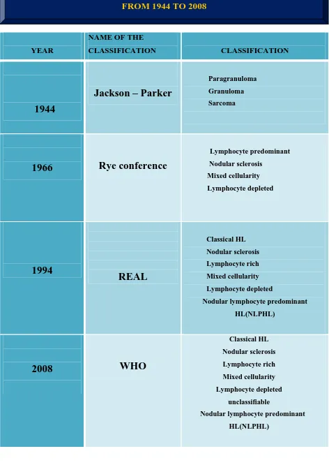
EVOLUTION OF CLASSIFICATION OF LYMPHOMAS:
The first case of lymphoma was identified by Thomas Hodgkin while working at the Guys hospital, London in the year 1832. He published his paper on
lymphatic disease “On some morbid appearances of absorbent glands and spleen”. His observations consented with those of Samuel Wilks, who named this condition as Hodgkin’s disease, paying credit to Thomas Hodgkin. It was in
1898 and 1902, when Carl Sternberg and Dorothy Reed gave the most important diagnostic entity for Hodgkin’s disease by defining the classical
Reed-Sternberg cell (RS cell). Variants of RS cells were later identified. [12]
HD was believed to be a benign process progressing into malignancy. The first
classification of HD introduced in the year 1944 by Jackson and Parker was based upon this concept. Later in 1966, Lukes and Butler introduced a
correlation with the survival and outcome, making it popular among the
clinicians.[12]
The application of immunohistochemical methods for studying various diseases
came into light in 1970s. The advent of the immunologic methods resulted in better understanding of the disease process, recognition of new neoplasms and
eventually influenced the classification of many tumors. The RS cells and its variants showed positivity for CD15 and CD 30 in most of the cases, but there
were a considerable number of cases which were negative for these markers. These cases showed similarity in clinical and morphological features. The Revised European American classification of lymphomas (REAL) introduced in
the year 1994, took into consideration the immunological and genetic features in addition to morphology.[13] They categorized HL into classical HL (CHL),
which are positive for CD15, CD30 and nodular lymphocyte predominant HL (NLPHL), which are negative for the above markers. The CHL was again
categorized into 4 subtypes. The present WHO classification of lymphoma adapts the REAL classification for HL, with a slight modification of including unclassified category under CHL. The following table shows the various
YEAR
NAME OF THE
CLASSIFICATION CLASSIFICATION
1944
Jackson – Parker
Paragranuloma
Granuloma
Sarcoma
1966
Rye conference
Lymphocyte predominant
Nodular sclerosis
Mixed cellularity
Lymphocyte depleted
1994
REAL
Classical HL Nodular sclerosis
Lymphocyte rich
Mixed cellularity
Lymphocyte depleted
Nodular lymphocyte predominant
HL(NLPHL)
2008
WHO
Classical HL
Nodular sclerosis
Lymphocyte rich
Mixed cellularity
Lymphocyte depleted
unclassifiable
Nodular lymphocyte predominant
[image:14.612.89.561.62.728.2]HL(NLPHL)
The evolution of the classification of the NHL is a little more complex than that
of the HL. Ever since the identification of HD, numerous neoplasms arising from the lymphocytes were identified, which were different from the HL in
terms of morphology and clinical picture. These tumors were categorized as Non Hodgkin Lymphoma (NHL), simply implying that these tumors are
lymphomas other than Hodgkin’s lymphoma.
At first, attempts were made to classify NHL based upon the differentiation.
Differentiation refers to the degree of resemblance to the normal counterpart. Hence in 1966, Henry Rappaport classified the NHL into lymphocytic, histiocytic, mixed type and undifferentiated types. The lymphocytic type was
again classified into poorly differentiated and well differentiated subtypes. With improved understanding of lymphomas, the term histiocytic used in this
classification was found to be erroneous as they do not represent the tumors arising from histiocytes. Hence an improved classification became
mandatory.[14]
In 1982, a working formulation of NHL was introduced categorizing the NHLs
into low grade, intermediate grade, high grade and miscellaneous. This classification was welcomed by the clinicians and was in use for a long time since this way of classifying lymphomas also indicates the prognosis.
introduced in 1989 and was followed in many European countries, though the
Americans retained the use of working formulation for classifying lymphomas. It classified NHL into B and T cell lymphomas, which in turn were again
classified into low grade, high grade and rare tumors.[14]
In 1994, an international lymphoma study group proposed the Revised
European American classification of lymphomas (REAL) in order to achieve consensus between the European and American nations. It took into
consideration the morphologic, immunologic and genetic characteristics of the lymphomas.[13] The lymphomas were classified as B cell neoplasms, T cell neoplasms and Hodgkin disease. The B and T cell neoplasms were again
classified into precursor neoplasms and peripheral neoplasms.
WHO classification of the hematopoietic and lymphoid neoplasms proposed in
the year 2003 took into consideration all the classifications used in various parts of the world and sought the opinion of renowned clinicians across the globe.
This was the first classification to be agreed throughout the globe. The current WHO classification is a revision of the 2003 classification. It provides a
separate category for lymphomas which cannot be classified under a specific category, thereby making it easy to recognize the cases which require further study.
Precursor lymphoid neoplasm
Mature B cell neoplasms.
Mature T cell and natural killer (NK) cell neoplasms
Hodgkin lymphoma
Histiocytic and dendritic cell neoplasm
Post transplant lymphoproliferative disorders.
The following table no :2 gives the detailed classification of both NHL and HL adopted by WHO in 2008.
PRECURSOR LYMPHOID NEOPLASMS
• B lymphoblastic lymphoma, NOS
• B lymphoblastic lymphoma with recurrent genetic abnormalities
• B lymphoblastic lymphoma with t(9;22)
• B lymphoblastic lymphoma with MLL rearranged
• B lymphoblastic lymphoma with t(12;21)
• B lymphoblastic lymphoma with hyperdiploidy
• B lymphoblastic lymphoma with hypodiploidy
• B lymphoblastic lymphoma with t(5;14)
• B lymphoblastic lymphoma with t(1;19)
[image:17.612.89.530.353.644.2]• T lymphoblastic lymphoma
MATURE B CELL
NEOPLASMS
• Small lymphocytic lymphoma (SLL/CLL)
• B cell prolymphocytic leukemia
• Splenic B cell marginal zone lymphoma
• Hairy cell leukemia
• Splenic B cell lymphoma
• Lymphoplasmacytic lymphoma (LPL)
• Heavy chain diseases
• Plasma cell myeloma
• Solitary plasmacytoma of bone
• Extra-osseous plasmacytoma
• Extranodal marginal zone lymphoma of mucosa associated lymphoid tissue (MALT lymphoma)
• Nodal marginal zone lymphoma (MZL)
• Follicular lymphoma (FL)
• Primary cutaneous follicle centre lymphoma
• Mantle cell lymphoma
• Diffuse large B cell lymphoma (DLBCL)
• DLBCL associated with chronic inflammation
• Lymphomatoid granulomatosis
• Primary mediastinal large B cell lymphoma
• Burkitt lymphoma
• B cell lymphoma , unclassifiable,with feature intermediate between DLBCL and Burkitt lymphoma
MATURE T CELL AND NK
CELL NEOPLASMS
•
T cell prolymphocytic lymphoma
•
T cell large granular lymphocytic
lymphoma
•
Aggressive NK cell leukemia
•
Systemic EBV positive T cell
lymphoproliferative disease of
childhood
•
Hydroa vacciniforme like
lymphoma
•
Adult T cell lymphoma
•
Extranodal T cell lymphoma, nasal
type
•
Enteropathy associated T cell
lymphoma
•
Hepatosplenic T cell lymphoma
•
Subcutaneous panniculitis like T
cell lymphoma
•
Mycosis fungoides
•
Sezary syndrome
•
Primary cutaneous CD30 positive
T cell lymphoproliferative
disorders
•
Lymphomatoid papulosis
•
Primary cutaneous peripheral T cell
lymphomas, rare types
•
Primary T cell lymphoma, NOS
•
Angioimmunoblastic T cell
lymphoma
•
Anaplastic large cell lymphoma,
ALK positive
•
Anaplastic large cell lymphoma,
ALK negative
HISTIOCYTIC AND DENDRITIC NEOPLASMS
•
Histiocytic sarcoma
•
Langerhans cell histiocytosis
•
Langerhans cell sarcoma
•
Interdigitating dendritic cell
sarcoma
•
Follicular dendritic cell sarcoma
•
Fibroblastic reticular cell tumor
•
Indeterminate dendritic cell tumor
•
Disseminated juvenile
xanthogranuloma
POST TRANSPLANT LYMPHOPROLIFERATIVE
DISORDER (PTLD)
•
Polymorphic PTLD
•
Monomorphic PTLD
•
Classical HL type PTLD
•
Plasmacytic hyperplasia and
infectious mononucleosis like
PTLD
Once a lymphoma is diagnosed and is classified under a specific category, it needs to be staged like any other neoplasm. The Ann-Arbor staging system has been in use for staging both the HL and NHL.[1][11] In addition to the
history of illness, physical examination and radiological data are essential for staging the disease. A minimum of ultra sonogram of abdomen has to be done
in order to stage the disease efficiently. [14] The staging system stratifies lymphomas into four stages, from stage I to stage IV. A letter ‘E’ is added to
this stage in case the primary tumor is in an extra-lymphoid organ, which are the organs other than lymph node, spleen, thymus, Waldeyer’s ring, appendix and Peyer’s patches. These stages are again sub-classified based upon the
Unexplained fever
Unexplained weight loss of more than 10% of body weight in
preceding 6 months
Night sweats
The following table 3 illustrates the Ann-Arbor staging.[1]
STAGE I
Involvment of a single lymph node or a single extralymphoid organ
STAGE II
Involvement of two or more lymph node regions on the same side of the diaphragm or localized involvement of an extralymphatic organ and one or more lymph node regions on the same side of the diaphragm
STAGE III
Involvement of lymph node regions on both side of the diaphragm, which may also be accompanied by localised involvement of an extralymphatic organ or involvement of spleen or both
STAGE IV
Diffuse or disseminated involvement of one or more extralymphatic organs or tissues with or without associated lymph node enlargement
SUBCLASSIFICATION:
A- Without the symptoms listed below B- With systemic symptoms
Unexplained fever
Unexplained weight loss of more than 10% of body weight in preceding 6 months
[image:21.612.86.510.230.660.2] Night sweats
The accurate diagnosis of lymphoma can only be made with adequate clinical
history. Some of the lymphomas have certain clinical findings reserved to them and aid in the diagnosis. The important clinical findings that are
essential in the diagnosis of lymphoma include
Age
Sex
Site
Duration
Presence of B symptoms
Presence of lymphadenopathy
Associated organomegaly
History of treatment
Skin lesions
History of associated infections like HIV, HBV, HCV etc.
The clinical picture not only drives a pathologist towards a diagnosis but also helps the pathologist to assess the optimal ancillary technique that will be required in a particular case.
HISTOLOGY OF A NORMAL LYMPHNODE:
A thorough knowledge about the histology of the lymph node is mandatory
before assessing a case of lymphoma. This is because certain lymphomas have characteristic pattern of involving the lymph node and the lymphomas
lymph node, and hence named after them.[15] For example marginal zone
lymphoma and mantle cell lymphoma are named so because of their resemblance to marginal zone cells and mantle cells of a normal lymph node
respectively. Knowing the histology of lymph node also helps in understanding the pattern of immunohistochemical markers in a normal
lymph node.
Lymph nodes are encapsulated, bean shaped, firm structures, whose size vary with site and activity. The afferent lymphatics enter through the convex
surface and the efferent lymphatics and veins leave through the hilum. Ateries and nerves enter through the hilum. The lymph node is composed
mainly of three regions, which are:[16][17]
Cortex
Medulla
Paracortex
The cortex is identified by the presence of lymphoid follicles with cortical sinuses running between them. The follicles can be primary follicles or secondary follicles. The secondary follicles have a germinal centre,
surrounded by a mantle zone which in turn is surrounded by a marginal zone. Follicular dendritic cells and tingible body macrophages are present in the
germinal centre in addition to the lymphoid cells. The lymphoid cells are predominantly B cells admixed with a few T cells. The B cells in the follicles
and amphophilic cytoplasm whereas centrocytes have a cleaved nuclei, dense
chromatin and scant cytoplasm. The cells of the primary follicles and the mantle zone are small and have dense chromatin and the cells in the marginal
zone are similar but slightly larger and have more cytoplasm. [16][17]
The medulla is made up of cords of cells, mainly plasma cells with
intervening medullary sinuses which contain histiocytes.
The region between the cortex and medulla is called paracortex. This region is highly vascular and is rich in T lymphocytes. [16][17]
We need to bear in mind that the architecture of the lymph node is not static and it changes with different stimuli. The lympnodes from different sites
show different morphology. For example the abdominal nodes show a prominent marginal zone. This should not be confused with a reactive or
neoplastic process. The following figure (fig:1) depicts the structure of a lymph node as in google images site//www.dartmouth.edu/... in the article
Figure 1: STRUCTURE OF A LYMPHNODE
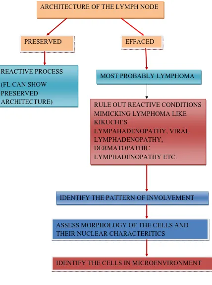
MORPHOLOGICAL APPROACH TO LYMPHOMA:
In almost all the cases of nodal lymphomas, the normal structure of the lymph node is completely or partly replaced with the neoplastic cells. Some of the
lymphomas like follicular lymphoma (FL) do not disturb the normal structure of a lymph node and hence can be confused with a reactive process. FL can
be differentiated from a reactive follicular hyperplasia by the presence of prominent mantle zone. Viral infections can show numerous immunoblasts
differential diagnosis. Therefore we need to bear in mind all the reactive
conditions that simulate a lymphoma before making a diagnosis of lymphoma.[1]
Once all the reactive conditions have been ruled out, the pattern of the lymphoma needs to be assessed. Different lymphomas have a different
pattern of involvement and are characteristic to the type of lymphoma. The commonly seen patterns in a lymphoma include
Nodular
Mantle zone
Marginal zone
Sinusoidal
Starry sky
Interfollicular
Vascular
Diffuse
Nodular pattern of involvement is seen in cases like follicular lymphoma and nodular type of mantle cell lymphoma or marginal zone lymphoma. NLPHL
and nodular sclerosis type of classical HL also show a nodular pattern. These nodules are separated by dense fibrous tissue. Sometimes ALCL also has this
rarely by lymphoplasmacytoid lymphoma are vague and are also called
pseudonodular or pseudofollicular pattern.[1]
Mantle zone and marginal zone pattern are seen in mantle cell lymphoma and
Marginal zone lymphoma repectively. Starry sky pattern, called so due to the presence of numerous tingible body macrophages interspersed with them is
characteristic feature in Burkitt’s lymphoma. This pattern is also noted in lymphoblastic lymphoma.[1]
Lymphomas like ALCL, mycosis fungoides etc can be seen involving only
the sinuses and spread along the sinuses. Carcinomas metastasizing to the lymphnodes also involve the sinuses first. Hence care should be taken to
avoid misdiagnosis. An interfollicular pattern of involvement is seen commonly in the T cell lymphomas. Some lymphomas like have a prominent
vascular proliferation. This vascular pattern in commonly seen in angioimmunoblastic T cell lymphomas and peripheral T cell lymphomas. A
PATTERN LYMPHOMA
Nodular
•
Follicular lymphoma
•
Mantle cell lymphoma
•
Marginal zone lymphoma
•
NLPHL
•
Nodular sclerosis HL
•
Small lymphocytic lymphoma
•
Lymphoplasmacytoid lymphoma
•
Anaplastic large cell lymphoma
Mantle zone
Mantle cell lymphoma
Marginal zone
Marginal zone lymphoma
Starry sky pattern
Burkitt’s lymphoma
Lymphoblastic lymphoma
Sinus pattern
Anaplastic large cell lymphoma
Mycoises fungoides
Marginal zone lymphoma
Interfollicular
pattern
T cell lymphomas
Vascular pattern
Angioimmunoblastic T cell lymphoma
Peripheral T cell lymphoma NOS
After identifying the pattern, the morphology of individual cells needs to be examined, starting from the size of the cells. The cells can be divided into
three types:
Small cells
Medium sized cells
Large cells
The cells are compared with the size of the endothelial cells. The cells
smaller than endothelial cells are called small cells and those larger than them are called large cells. The cells which are just about the size of the endothelial
cells are called medium sized cells. Other fine details like the shape of the nucleus and chromatin pattern are also observed. The small cells are seen commonly in follicular lymphoma and mantle cell lymphoma while the large
cell morphology is seen in ALCL, DLBCL and plasmablastic lymphoma. The neoplastic cells can be admixed with a reactive cell population.
Identifying the reactive population helps in the diagnosis. These cells in the background form the basis for classifying HL.[1]
FIGURE 2: ALGORITHM OF APPROACH TO A CASE OF LYMPHOMA
ARCHITECTURE OF THE LYMPH NODE
PRESERVED EFFACED
MOST PROBABLY LYMPHOMA
IDENTIFY THE PATTERN OF INVOLVEMENT
ASSESS MORPHOLOGY OF THE CELLS AND THEIR NUCLEAR CHARACTERITICS
IDENTIFY THE CELLS IN MICROENVIRONMENT REACTIVE PROCESS
(FL CAN SHOW PRESERVED
ARCHITECTURE) RULE OUT REACTIVE CONDITIONS MIMICKING LYMPHOMA LIKE KIKUCHI’S
LYMPAHADENOPATHY, VIRAL LYMPHADENOPATHY,
DERMATOPATHIC
IMMUNOHISTOCHEMISTRY:
Immunohistochemistry is a method of identification of the cellular constituents with the help of antigen antibody reaction.[19] It is based on the
principles used for immunofluorescence, but does not require a specialized microscope for assessing the antigen antibody reaction. Antigens labelled
with enzymes which produce a colourful reaction on addition of a chromogen is used in place of a fluorescent dye labelled antibodies in immunofluorescence.
Flow cytometry is another technique that can be used for immunophenotypic analysis. But it requires sophisticated machinery and fresh specimen. Though
IHC was initially practised on fresh and frozen specimens, with the advent of antigen reviving techniques and development of antibodies which can be used
on paraffin embedded tissues, IHC can now be reliably practised on routine paraffin sections.[20][21][22] The morphology of the cells cannot be assessed in
flow cytometry. IHC has the added advantage of morphological analysis and immunological analysis simultaneously. Hence IHC is superior to flow cytometry in certain conditions, especially lymphomas.[20] In addition, IHC
facilitates the identification of the antigen antibody reaction at the sub cellular level i.e. the membrane, nucleus, Golgi apparatus etc.[19]
The IHC markers can be categorized into three types, based upon their use as
Diagnostic markers
Predictive markers
Prognostic markers
Diagnostic markers are those which help in making diagnosis, like in case of a poorly differentiated tumour and in differentiating various other neoplasms
from the normal or reactive process. Predictive markers are those which help in assessing the response to an available treatment. They predict the outcome of a therapeutic method and hence they are called so. Prognostic markers are
those markers which help in determining the aggressiveness of the neoplasm and patient’s survival.[23]
The majority of IHC markers used in lymphomas are for the leucocyte differentiation antigen and have an acronym CD which stands for ‘cluster of
differentiation’. It was standardised to use this acronym followed by a number that corresponds to the small region, called epitope on the antigen, in
order to overcome the confusion arising due to the different names used for the same marker by different investigators. The monoclonal antibodies are directed against the epitopes.[20]
VALUE OF IHC IN LYMPHOMAS:
Diagnosing a case of lymphoma and subtyping the lymphoma is one of the
most challenging tasks for a pathologist. Lymphoma can be easily confused with other nonlymphoid neoplasms like poorly differentiated carcinomas,
(PNET), desmoplastic small round cell tumor (DSRCT),
rhabdomyosarcoma, Wilms tumor etc. A positive reaction with CD45, also known as leucocyte common antigen helps in establishing the diagnosis as
lymphoma and rules out the other possibilities.[19]
There is a myriad of reactive conditions which mimic the lymphomas. IHC
plays an important role in differentiating reactive process from a lymphoma. Even cases of HL can be confused with reactive node when the diagnostic Reed-Sternberg cells are scarce. IHC staining with CD15 and CD30 can help
in establishing the diagnosis. The pattern of IHC staining is also helpful to differentiate the lymphomas from a reactive process. For example in a case
of follicular lymphoma, Bcl-2 is negative in the follicular centre, whereas it is positive in a reactive lymph node.
Once a diagnosis of lymphoma is made, it needs to be categorised as HL or NHL, followed by placing it under a particular subtype. Morphology in
adjunct with IHC is sufficient to type most of the lymphomas. The Non Hodgkin’s lymphoma project observed a significant increase in accurate diagnosis and sub-classification of lymphoma using IHC.[24] Some of the
lymphomas have characteristic immunophenotypes that the presence of such immunophenotype is essential for making that diagnosis.
Though HL can be diagnosed with the presence of Reed-Sternberg cell, it can be confused with diffuse large B cell lymphoma (DLBCL) when they are
distinguishing them from HL. Nodular lymphocyte predominant Hodgkins
lymphoma (NLPHL) with a predominant T cell population in the reactive component brings T cell rich B cell lymphoma into the differential diagnosis.
In such cases, CD57+ve, CD4+ve T cell rimming around the Lymphohistiocytic cell (L H cell) helps in recognising them as NLPHL.
Anaplastic large cell lymphoma (ALCL) is another lymphoma having cell that resemble the RS cells, but these cases are positive for ALK, clusterin and fascin. Certain antigens are preserved even in some necrotic tissues and IHC
can be helpful in detecting these antigens in biopsies containing large areas of necrosis.[25]
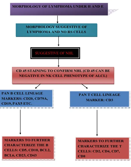
In case of NHL, the lineage of the lymphoma needs to be recognised. IHC markers are essential for establishing the lineage. CD20, CD79a, CD19 etc
are commonly used for determining B cell lineage. All these markers can be used on paraffin embedded sections except CD19. The clonality of the B cell
neoplasm can be established with the use of immunohistochemical staining with kappa and lambda light chains. CD3 is the frequently used pan T cell marker. [25]
Further characterisation of the neoplastic cells may be required after the recognition of the lineage. In case of B cell lymphomas CD5, CD10, CD23,
BCL2 and BCL6 have been used and in case of T cell lymphomas CD2.CD4, CD5, CD7 and CD8 are used for this purpose. The absence of staining with
The following figures (figure3 and 4) shows the use of IHC markers in
[image:35.612.71.556.153.754.2]diagnosing lymphomas and sub-categorizing them.
FIGURE 3: Role of IHC markers in subtyping Hodgkin lymphomas
MORPHOLOGY OF LYMPHOMA UNDER H AND E
PRESENCE OF CLASSICAL RS CELLS OR ITS VARIANTS
SUGGESTIVE OF HL
STAINING WITH CD15 AND CD30
POSITIVE
HL CONFIRMED
NEAGTIVE
CONSIDER NLPHL
IDENTIFICATION OF THE REACTIVE POPULATION TO CLASSIFY HL
CD57+VE CD4+VE CELLS RIMMING THE LH CELLS CONFIRM
NO RIMMING WITH CD57+VE , CD4+VE CELLS
FIGURE 4: Role of IHC markers in subtyping non Hodgkin lymphomas
MORPHOLOGY OF LYMPHOMA UNDER H AND E
MORPHOLOGY SUGGESTIVE OF LYMPHOMA AND NO RS CELLS
SUGGESTIVE OF NHL
CD 45 STAINING TO CONFIRM NHL (CD 45 CAN BE NEGATIVE IN NK CELL PHENOTYPE OF ALCL)
PAN B CELL LINEAGE MARKERS: CD20, CD79A, CD19, PAX5 ETC.
PAN T CELL LINEAGE MARKER: CD3
MARKERS TO FURTHER CHARACTERIZE THE B CELLS: CD5, CD10, BCL2, BCL6, CD23, CD43
IHC markers can also be used to assess the prognosis in lymphomas. DLBCL
cases showing positive reaction with Bcl2, MUM1, FOXP1 and negative reaction with CD10 were found to have a bad prognosis. ALK positive cases of
ALCL carry a good prognosis. Ki67/ MIB1 staining shows the proliferation index.[23] Lymphomas with high proliferation index have a bad prognosis.
Reinhard Von Wasilewski et al observed that cases of HL that lack the expression of CD15 showed a bad prognosis.[26]
The identification of the cell antigens has become more important ever since the successful introduction of Rituximab in the treatment of lymphomas. Rituximab is an anti CD20 therapy targeted against the cells having this surface antigen.
The success of Rituximab has driven the investigators to identify other therapeutic targets. The drugs targeting other cell antigens like CD22, CD30,
CD40, CD80 are still in the research process.[23]
Thus immunophenotypic analysis in addition to being an integral part of WHO
classification is also essential in making the correct diagnosis and assessing the prognosis. Though IHC plays a vital role in diagnosing lymphomas,
morphological assessment still remains essential. IHC markers should be used cautiously and judiciously. A single marker is never sufficient in a case of lymphoma. Hence a panel of markers is almost always necessary.[25] The panel
CD45 – to confirm the diagnosis of lymphoma (CD 45 is negative in
classical HL and NK cell phenotype of ALCL)
CD3 – pan T cell marker
CD20- pan B cell marker
CD15 and CD30 – for diagnosing HL.
CD45:
CD45, also referred to as leucocyte common antigen (LCA) is a tyrosine phosphatase present on the surface of the leucocytes. CD45 is lost in plasma cells.[27] This antigen is rarely present in the Golgi apparatus in addition to the
cell membrane. Some isoforms of CD45 are specific to certain cells. For example CD45RO is specific for T cell, histiocytes and CD45RA is seen
predominantly in T cells.[1][27] CD45RB is the commonly used isoform as it is present in all leucocytes. Its primary role is to identify a lymphoma and to
differentiate it from other tumors resembling lymphoma. CD45 expression is lost in classical HL and in cases of ALCL with NK cell phenotype. [1]
CD3:
CD3 is also referred by different names like Leu 4, T3 etc... [21] It is the T cell
seen in some NK cells. [15] CD 3 is preferred over other T cell markers because
of its specificity and easy, reliable detection on paraffin embedded sections.[27] The following figure 5 illustrates the various CD markers expressed during
[image:39.612.71.549.264.720.2]various stages of maturation of T cells.
Figure 5: T cell development with the respective immune profile
CD20:
PROTHYMOCYTE:
CD7
CORTICAL THYMOCYTE:
• CD7, CD2, CD5
• BOTH CD4 AND CD8
• CD3(CYTOPLASM)
MEDULLARY THYMOCYTE:
• CD7,CD2,CD5
• EITHER CD4 OR CD8
• CD3(MEMBRANE)
PERIPHERAL T CELLS
• CD7, CD2, CD5
• EITHER CD4 OR CD8
• CD3(MEMBRANE)
CD20 is also known by other names like Leu26 and B1.[21] It is a nonglycosylated phosphoprotein seen on cell membrane surface of all the
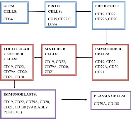
mature B cells. Its expression is seen from the pre B cell stage of maturation and is lost in plasma cell stage. The following flow chart shows the expression of
[image:40.612.76.523.308.738.2]various CD markers during the development of B cells.
Figure 6: B cell development with the respective immuomarker profile
STEM CELLS: CD34 PRO B CELLS: CD19,CD22,C D79A
PRE B CELL:
CD19, CD22, CD79A,CD20 IMMATURE B CELLS: CD19, CD22, CD79A, CD20, CD21 MATURE B CELLS: CD19, CD22, CD79A, CD20, CD21 FOLLICULAR CENTRE B CELLS: CD19, CD22, CD79A, CD20, CD21, CD10 IMMUNOBLASTS:
CD19, CD22, CD79A, CD20, CD21, CD138 (VARIABLY POSITIVE)
PLASMA CELLS:
CD20 is expressed in all the mature B cell lymphomas. It is expressed in a part of classical HL and is seen in all NLPHL.[12][28] Rare CD20 expression is seen in some T cell lymphomas
CD15:
It is also referred to as Lewis X antigen, X hapten, Leu M1 and myelomonocytic marker.[21][27] It is a cell adhesion molecule and its expression is seen on the cell membrane of RS cells of classical HL with or without dot like
golgi localization.[27] CD15 is positive only in a part of HL and its expression is believed to have a better prognosis. Certain B and T cell lymphomas may also
be positive for CD15 and ALCL is always negative for CD15. Its expression is confined to the lymphomas and is noted in other cells like granulocytes,
histiocytes and other non hematolymphoid tissues like breast, proximal convoluted tubules of kidney, lung and Paneth cells in GI tract.[29]
CD30:
CD30 is also referred to as Kil, Ber H2 and lymphocyte activation antigen.[21] It
belongs to TNF receptor superfamily and is always positive in classical HL and ALCL. CD30 mediated NFkB activation is believed to be the pathophysiology behind classical HL.[30][31] It is seen as a membranous positivity with or without
expression of the commonly used CD markers in various situations and the
subcellular site of positivity.
TABLE 5: CD MARKERS WITH THE STAINING PATTERN AND
POSITIVE CELLS
MARKERS SITE OF POSITIVITY COMMONLY
POSITIVE CELLS
CD45 Membrane All leucocytes except
plasma cells
CD3 Membrane T cells
CD20 Membrane B cells
CD15 Membrane with or
without golgi positivity
RS cells, activated T lymphocytes , proximal convoluted tubules of kidney, Paneth cells, tumours of lung, breast , thyroid and GIT
CD30 Membrane with or
without golgi positivity
RS cells, ALCL,
In addition to the available wide range of CD markers for determining the lineage of lymphomas, there are other markers which are specific for the
lineage. PAX5 is one such marker which is expressed exclusively in B cells.
PAX5:
PAX stands for paired box. The PAX proteins are all transcription factors that usually determine the fate of the cells during the early stages of development
and maturation mostly during embryogenesis and sometimes even in the adult life. They are closely related proteins and are called paired box as they have a
paired domain for binding to the DNA .Genes encoding the PAX proteins are called PAX genes. There are about nine PAX genes identified till date.[4]
PAX5 is otherwise called B cell specific activator protein (BSAP). It is named so because of its exclusive expression in B lymphoid lineage. It is a
transcription protein like other PAX proteins and its expression is noted as early as pro B cell stage and is lost during the plasma cell stage.[4] (Figure 7) PAX5 drives the cells into B cell lineage and hence plays an essential part in B cell
FIGUR
The rol ability t
in the tr CD19 is the imm regulate activate C B
RE 7: B CE
e of PAX5 to regulate
ransduction s involved
mune syst es the gene ed by PAX
CD79a
B lymphoid
ELL MAT
5 in B cell e the CD19
n of signa d in B cell
em.[32] Th eration and
5 include:
d kinase (B
TURATIO
l differenti 9 gene. CD
als from B proliferatio
hus by reg d maturatio
Blk)
ON AND P
iation and D19 is a tr
cell recep on in addit
gulating th on of the B
PAX 5 EXP
maturation rans-memb
tors that a tion to its
he express B cells. Th
PRESSIO
n is mainl brane prote
are specific functions
ion of CD he other ge
N
y due to it ein involve
c to B cell in regard t
D19, PAX enes that ar
PAX5 suppresses the J chain and XBP1, thereby preventing the cells from
maturing into plasma cells. Thus BSAP is not only essential in driving the cells towards B cell lineage, but also retaining its identity. [33] PAX5 along with
PAX2 and PAX8 has been demonstrated to influence the apoptosis and aid in survival of the cell in nematodes. Hence they are believed to have a role in
survival of the B cells. [34]
The suppression of PAX5 is mandatory for plasma cell differentiation. This is
achieved through increased expression of B lymphocyte induced maturation protein 1 (Blimp 1), which is induced by various cytokines. Blimp1 suppress the expression of PAX5 in addition to other genes like C-myc. [33] Following
diagram (fig 8) illustrates the role of PAX5 in B cell development and the mechanism by which BLIMP 1 drives the cells towards plasma cell
Figure 8: The role of PAX5 in B cell development and the mechanism by
which BLIMP 1 drives the cells towards plasma cell differentiation
Though immunohistochemical expression of PAX5 is noted in all stages of
maturation the intensity of staining varies in subsets of B cells. Krenacs et al observed a strong PAX5 immunoreactivity in the cells of the marginal zone as
opposed to the follicular centre cells and monocytoid B cells. [35]
PAX5, in addition to the B lymphocytes is also observed in CNS during the
early stages of maturation and in adult testis. [4] In contrast to these findings, Tarlakovic et al observed an absent immunohistochemical reaction of PAX5 in
oblongata and a few cells in the caudal nucleus. PAX5 is strongly expressed in
mesonephric rests of epididymis and smooth muscle cells of uterus. Rare cells in prostatic glands and endocervical glands express PAX5 weakly. [36] Thus in
addition to B cell development and CNS development, PAX5 is thought to play a role in urogenital development.
The various types of B cell lymphomas are believed to represent the cells of different stages of maturation.[15][23] PAX 5 expression is noted in majority of
mature B cell lymphomas, premature B cell lymphomas and HL. They show a sharp nuclear reactivity making the assessment of immunoreactivity easy since they are devoid of background staining. Being a nuclear antigen, their
immunoexpression is altered with poor tissue preservation. [5]
The main utility of PAX5 is its expression in B cell lymphomas which lack or
show equivocal immunoreactions with the commonly used IHC markers in paraffin embedded sections like CD20. CD20 expression is usually absent in
classic HL and precursor lymphoid neoplasms. The surface CD20 expression is lost in patients following Rituximab therapy as it is a targeted therapy against
CD20. In case of relapse in these patients, the cells may remain to be negative for CD20. PAX 5 helps in situations like these to establish the cell lineage.
classical HL. This reduced expression is due to the down regulation of PAX5
along with other transcription factors and surface antigens. Blimp1 has been observed to be expressed in certain classical HL. This is thought to be the
reason behind the reduced expression of PAX5 in some cases and even for the absence of PAX5 reaction in some cases.[5]
The expression of PAX5 is absent in a few but a significant number of cases of lymphomas with large cell morphology like DLBCL. A study by Torlakovic et
al showed PAX5 was positive in about 96% of cases, of which 90% showed strong positivity. These findings correlated with the immunoexpression of CD20. [38] Therefore though, PAX5 is negative in few cases of DLBCL, it is
equivalent to CD20 in its ability to recognize DLBCL. The weak or absent immunoreativity with PAX5 in DLBCL may be because they represent the post
follicular cells in normal B cell development. [35]
T cell lymphomas are consistently negative for PAX5. Its expression in T cell
lymphomas was observed very rarely by different authors.[39][40][41] These cases are thought to represent the aberrant or over expression of PAX5 in these
tumors and Feldman et al observed extra copies of the PAX5 genes in such cases. Over expression of PAX5 is observed to induce T cell lymphomas in experimental animals. Hence PAX5 was postulated in tumorigenesis of these
Problems can arise in differentiating between DLBCL, ALCL and HL as all
these lymphomas can show large cells closely resembling RS cells. The routinely used IHC markers can add on to the confusion as these tumors can
have similar immunophenotypes. Browne et al used PAX5 along with other B cell transcription factors oct2, BOB1 and other pan B cell antigens in known
cases of ALCL, DLBCL and HL. PAX5 was observed to be negative in all the cases of ALCL and positive in all cases of DLBCL and NLPHL. It was positive in about 91% of classical HL.[42] The negative reaction of PAX5 in T cell
lymphomas can be exploited in situations like these.
The expression of PAX5 has also been observed in acute leukemia, both acute
myeloid leukemia (AML) and acute lymphoid leukemia (ALL). PAX5 positive cases of AML mostly belonged to AML with t (8: 12) subtype, but cases of
AML belonging to other subtypes also showed positive reaction in a few instances. AML with t (8:12) is known to express CD19, which is regulated by
PAX5 and hence its expression is these cases. The number of PAX5 positive cases in this subtype of AML varied among different studies. Sarah E Gibson et al observed PAX5 being expressed in about 44% of this subtype of AML, [43]
whereas Joe R Vulbueva et al observed PAX5 expression in all the cases of this subtype included in his study. [44] In both the studies the blasts showed weak
PAX5 expression is noted in leukemia, their value in classifying these leukemia
was minimal.[39]
Though the common understanding is that the PAX5 expression is lost in
plasma cells differentiation, Pein Lim et al demonstrated PAX5 immunoexpression in a few cases of multiple myeloma. They were then
suggested to be retained products of early stages of maturation. [45]
In addition to the lymphoid cells, neuroendocrine tumors also show positive
PAX5 expression. [5][36] It is negative in case of carcinoid tumors but is positive in a high proportion of Merkel cell carcinomas and small cell carcinomas. The
other non hematopoietic tumor that shows PAX5 expression is transitional cell carcinoma of urinary bladder.[46][47] The expression of PAX5 in these cases was found to be inversely proportional to the level of differentiation and directly
proportional to the stage.[47] This increased expression of PAX5 in more aggressive forms suggests they have a prognostic value. PAX5 expression is
noted in CNS tumors like medulloblastomas, astrocytomas, neuroblastomas etc.
[48][49]
Rare expression of PAX5 was noted by Tarlakovic et al and Paulette et
al.[36][50]
The aberrant expression of PAX5 in tumors like neuroendocrine carcinomas,
suppresses the transcription of p53, a tumor suppressor gene and thereby
leading to development of the tumor.[48]
The translocation t (9;14) involving the PAX5 region is found to be linked to
lymphoplasmacytic lymphoma, further implying the role of PAX5 in tumorigenesis.[51] Balasenthil et al postulated that regulation of PAX5 by
metastasis associated protein (MTA1) lead to its over expression and lymphoma genesis.[52]
Gilles A Roubischad et al identified about five isoforms of PAX5 from normal and lymphoma cells. They observed that all these isoforms had different
sequence and transactivation properties. PAX5 FL isoform was noted predominantly in the lymphomas. Thus this altered form of PAX5 may be the reason behind the development of lymphoma in these cases. Moreover the
oncogenic role of PAX5 in certain T cell lymphomas has been demonstrated in mice.[53]
The high expression of PAX5 in lymphomas, certain non haematolymphoid malignancies and its possible role in the development of these tumours have
encouraged the investigators to develop a targeted therapy against PAX5. Mengyong et al have managed to generate a cytotoxic T lymphocyte lines
poses to be an ideal target for immunotherapy in patients having tumors with
aberrant expression of PAX5.[54]
In spite of its expression in various non haematolymphoid tissues and tumors
PAX5 is still an excellent marker for B cell lineage. Paulette et al and Kirsten et al observed a highly specific immunoexpression of PAX5 in B cell
lymphomas.[50][6] Neuroendocrine carcinomas were the only other tumours that which showed high reactivity with PAX5 next to lymphomas. Less than 1% of
carcinomas showed immunoreactivity with PAX5.
MATERIALS AND METHODS
All the cases diagnosed as lymphomas from January 2009 to December 2010 in
the department of pathology, PSG institute of medical science and research, Coimbatore were considered in the study. The cases with inadequate tissue for
performing Immunohistochemistry were then excluded from the study.
The clinical data required for the study like the age, sex, site and stage of the tumor were retrieved from the medical records department of PSG institute of
medical science and research after obtaining permission from the concerned authorities and institute human ethics committee clearance. The hemotoxylin
and eosin slides of these cases were analysed for assessing the morphology and also typing.
The immunohistochemical slides which were used for classification of the lymphomas were also retrieved and analyzed. The common panel of markers
included was CD45, CD3, CD20, CD15 and CD30. These were run for routine diagnostic reasons in the department. A few cases were the panel was not
necrosis were excluded. The following flowchart (figure 9) depicts the study
[image:54.612.56.540.164.636.2]plan .
Fig 9: THE STUDY PLAN
CASES DIAGNOSED AS LYMPHOMAS
H &E SLIDES FOR ASESSING THE MORPHOLOGY CASES WITH COMPLETE
LYMPHOMA PANEL CASES WITH
NO/INCOMPLETE LYMPHOMA PANEL
LYMPHOMA PANEL WAS COMPLETED
PAX5
IMMUNOHISTOCHEMISTRY WAS PERFORMED
Immunohistochemistry was done using the supersensitive HRP detecting
[image:55.612.91.524.212.564.2]system. The following table (table 6) shows the clones of the various primary antibodies used during the study
TABLE 6: The markers, clone of the primary and controls used
MARKERS
COMPANY
CLONE
CONTROL
CD45
Biogenex
PD7/26/16
Lymphnode
CD3
Biogenex
PSI
Lymphnode
CD20
Biogenex
L-26
Lymphnode
CD15
Biogenex
BRA4F1
Kidney
CD30
Biogenex
HRS-4
Hodgkin’s
lymphoma
The procedure for Immunohistochemistry was similar for all the markers and it
is as follows.[19]
PRINCIPLE: In an immunohistochemical reaction , the specific antigen present
in the cells and tissues was detected in a two stage process, (Figure 10) that includes:
1) The binding of the primary antibody to its specific epitope in the tissues.
2) Detection of this bound antibody using a dextran polymer bound secondary
antibody in a calorimetric reaction that involves a chromogen.
In this method, primary antibody to the specific antigen is first added and is then
followed by the addition of a dextran polymer linked to multiple conjugated secondary antibodies and horse raddish peroxidase enzyme. This multiple
STEP 1:
Antigen retrieval:
This is a process to unmask the epitopes of the specific antigens that are masked by cross linking action of formalin during routine processing. There are various
methods for antigen retrieval. The methods include,
1) Pressure cooker method
2) Microwave method
3) Proteolytic digestion method
Of the above, the pressure cooker method is used for the present study. Here, the tissue is exposed to the additive effects of both heat and pressure thereby
bringing out the full antigenicity. After dewaxing and dehydrating in graded alcohols, the slides were subjected to antigen retrieval in a pressure cooker for
10 minutes with EDTA buffer at pH 9.
Reagents used:
¾ EDTA buffer at pH 9
¾ 3% hydrogen peroxide ( H2O2) in distilled water - To block endogenous
¾ 0.01M Phosphate buffered saline (PBS) with a pH value of 7.6. It was prepared
by dissolving the following substances in 1000 ml of distilled water.
1.Na2HPO4 Dibasic sodium phosphate, anhydrate 17.5g
2.KH2PO4 Monobasic potassium phosphate, anhydrous 2.5g
3.NaCl Sodium chloride 17.0g
¾ Blocking reagent- casein in PBS with 15mM sodium azide. This was used to
blocks non specific protein binding.
¾ Step 2:
¾ Primary antibodies. All the primary antibodies used in the study were in ready
to use formulation
¾ Step 3:
¾ Poly HRP reagent- anti-mouse and anti-rabbit IgG complex linked to Horse
radish peroxidase enzyme.
¾ Step 4:
¾ DAB (3, 3’Diamino Benzidine tetra hydrochloride) - Chromogen.
It offers great sensitivity as an HRP calorimetric chromogen and provides
Step 5
¾ Harris hematoxylin as counter stain.
¾ DPX (Distrene dibutyl phthalate Xylene) - Mountant.
PROCEDURE:
Immunohistochemical Staining with the two specific antibodies were done as follows
• Slides were deparaffinised
• deparaffinised slides were hydrated using graded alcohol.
• Antigen retrieval: using EDTA buffer at pH 9.0 in a pressure cooker for 10
minutes.
• Fast cooling under tap water.
• Washed in PBS buffer at pH 7.6 for for 5 minutes
• After wiping off excess PBS buffer the slides were immersed in 0.3% H2O2 for
20 minutes to block endogenous peroxidase activity.
• Washed in PBS buffer thrice, each 5 minutes
• Slides were incubated in blocking solution for 10 minutes to block non-specific
• Washed in PBS buffer thrice , each 5 minutes.
• Slides were incubated with the primary antibody for 1 Hr.
• To enhance the signal intensity, the sections were put in superenhancer for 30
minutes.
• Washed in PBS buffer thrice, each 5 minutes.
• Horse radish peroxidase polymer reagent was added to the slide and incubated
for 30 minutes.
• Washed in PBS buffer thrice, each 5 minutes.
• Chromogen Diamino Benzidine (DAB) was applied for 8 minutes.
• Washed in PBS buffer thrice, each 5 minutes.
• Sections were counter stained with Harris hematoxylin for 1 minute.
• Washed in tap water.
Sections were cleared in Xylene and mounted with DPX mountant.
These sections were than assessed for the immunoreactivity. All the CD markers
were considered positive when they showed a membranous positivity in the neoplastic cells. Slides stained with PAX5 immunohistochemistry were
cells showing positivity were considered negative. The intensity of the staining
was also considered.
Figure 11
:
Algorithm of the steps involved in running Immunohistochemistry
Tissue Preparation and antigen retrieval
Step 2
Primary antibody
Step 3
Secondary antibody
Step 4 DAB – chromogen application
Step 5 Counter stain and mount
RESULTS
The department of pathology, PSG institute of medical science and research has
received 8207 biopsy specimens over a period of two years from 1st January 2009 to 31st December 2010. The total number of malignancies reported during the study period was
922. Out of these 922 cases, 59 cases were reported as malignant lymphomas giving an overall incidence of 6.3%. Table 7 shows the no of malignancies reported each year and
the incidence of lymphomas for the year 2009 and 2010.
YEAR NO: OF
MALIGNANCIES
NO:OF LYMPHOMAS
INCIDENCE RATES
2009 443 30 6.7
2010 479 29 6.0
TOTAL 922 59 6.3
Of the 59 cases reported as lymphomas during the study period, 19 cases were not
included in the study owing to inadequate tissue samples and unavailability of tissue blocks. The remaining 40 cases include 28 nodal lymphomas and 12 extra-nodal
[image:64.612.87.528.328.528.2]lymphomas of which 33 were non Hodgkin’s lymphoma and 7 were Hodgkin’s lymphoma. Most of the non Hodgkin’s lymphomas were of B cell type. 4 out of the 33 non Hodgkin’s lymphomas were not classified under a specific cell type and were
rep
inc
ported as u
cluded in th
HO
CHART T
unclassifia
he study an
T
ODGKINS
NO EXTRA
TO
T 1 – Dist
[image:65.612.86.526.160.381.2]30%
TABLE 8
able. Table
nd its % in
TYPE S LYMPH NON ODAL A NODAL OTAL ribution o
C
– Distribue 8and Cha
ncidence di OMA NHOGKIN L of lymphom 17% 53%
Chart
Tit
ution of ly
art 1 show
istribution.
NS LYMP
mas with %
tle
ymphomas
w the vario
. NO: O PHOMA % inciden HL NODALN EXTRANO s included ous types OF CASES 7 21 12 40 nce HL ODAL NHL
in the stu
of lympho
S udy
The age group ranged from 3 years to 75 years, with a mean age of 43.5 years. 5 out of
7 cases of Hodgkin’s lymphoma were in the second and the fifth decades. 18 out of the 33 cases of non hodgkins lymphoma were in the sixth and seventh decade of life.
[image:66.612.78.521.260.588.2]Table 9 shows the age wise distribution of various lymphomas.
Table 9- Distribution of various lymphomas age wise
AGE HL NHL
B CELL T CELL UNCLASSIFIED
0-10 1 0 1 1
11-20 3 1 1 0
21-30 0 0 2 0
31-40 0 0 0 1
41-50 2 5 1 0
51-60 0 6 1 0
61-70 1 8 2 1
>70 0 1 0 1
TOTAL 7 21 8 4
Of all the 40 cases, 28 were males and 12 were females, giving rise to a male: female ration of 2.3:1. All the cases of Hodgkin’s lymphoma were males. B cell type non
in var pre T both the rious lym edilection
Table 10 –
CHAR 4 N O . O F C A S E S AGE MALE FEMAL sexes. Cha
mphomas a for males.
Sex wise d
RT 2 - Sex
0 1 2 3 4 5 6 7 8 9 1 0-10 2 LES 1
art 2, char
across diff distributio wise distr 2 0 11-20 2 3 4 1
rt 3, table
ferent age
on of vario
ribution of 3 4 21-30 31-40 1 0 1 1
10 and ta
e groups a
ous lymph f lymphom 5 - 41-50 4 4
able 11 de
and sex, homas acro mas across 6 51-60 6 7 7 9 0 3
epicts the
which cle
oss variou
s various a
7 8 61-70 >70 9 1 3 1 distributio early show
us age grou
SEX MALE FEMALE 0 5 10 15 20 25 Ta H 7
E 0
C
HL
able 11 – D
HL
Chart 3‐ Di
B CELL NHL
Distributio
B CEL
15
6
istribution
L T CEL
on of lymp
LL NHL
n of lymph
LL NHL U
phomas se
T CELL NH
5
3
omas sex w
UNCLASSIFIED
x wise
4
lym III
acr
1
out of 40
mphomas w I and stage
ross differe T STAGE STAGE STAGE STAGE STAGE 0 1 2 3 4 5 6 7 8 9 0 I cases cou were stage e IV. Tabl
[image:69.612.88.530.238.414.2] [image:69.612.68.463.495.738.2]ent stages.
Table 12–
E HL
E I 1
E II 1
E III 1
E IV 4
Chart 4 -
uld not be
ed based u e 12 and c
Distribut B NH 6 6 4 3 Distributi II
e staged d
upon the A chart 4 de
tion of lym
CELL HL T N 3 1 3 0
ion of lym
III
due to unav
Ann-Arbor epicts the d
mphomas a T CELL NHL 3 1 3 0 mphomas a IV vailability staging int distribution across diff UNCLA 0 1 2 0 across diffe U T B H of necess
to stage I, n of variou
ferent stag
ASSIFIED
erent stag
UNCLASSIFIED CELL NHL CELL NHL HL
sary data.
Sections of lymphomas were stained with the immunohistochemical markers,
commonly used in the diagnosis of lymphomas which includes CD45, CD3, CD20,CD15,CD30. They were studied and determined whether positive or negative.
Areas staining the reactive lymphoid cells were excluded.
All the cases except one were positive for CD45. This case had the following
immunoprofile – CD45-ve, CD3-ve, CD20-ve, CD15-ve, CD30+ve and EMA+ve. Based on the morphology and the immunoprofile, it was diagnosed as anaplastic large
cell lymphoma, a T/NK cell non Hodgkin’s lymphoma.
The Reed-Sternberg cells of all the cases of Hodgkin’s lymphoma were positive for
CD15, CD30 (figure 13 to 15) and negative for CD3, CD20. This is the immunoprofile for classical Hodgkin’s lymphoma.
Among the non Hodgkin’s lymphoma, 7 were positive for CD3, 21 were positive for
CD 20 and 4 were negative for both CD3 and CD20. Lymphomas positive for CD20 were classified as B cell non Hodgkin’s lymphoma, those positive for CD3 were
Table 13 – IHC analysis of commonly used antibodies in the diagnosis of
lymphomas
IMMUNOPROFILE NO OF CASES HL (CD 15 +VE, CD 30 +VE) 7
NHL(CD 15 –VE, CD 30 –VE)
-CD 3 +VE 7
-CD20 +VE 21
-CD 45 +VE, CD 3 –VE, CD 20 –VE 4
-CD 45 –VE 1
All the cases were stained with PAX5 antibody and its expression in different age
groups, sex, site, stage and types of lymphomas. A strong nuclear staining was considered positive.
23 out of the 40 cases were positive for PAX5. PAX5 was negative in all T cell non Hodgkin’s lymphoma and the cases under the unclassified category. Out of 21 cases of B cell non Hodgkin’s lymphoma, 18 were positive. Reed-Sternberg cells of Hodgkin’s
Table 14 – expression of PAX5in various lymphomas
TYPE OF LYMPHOMA
NO OF CASES PAX 5 +VE PAX 5 –VE
HL 7 5 2
NHL 33 18 15
-B CELL TYPE NHL
21 18 3
-T CELL TYPE NHL
8 0 8
-UNCLASSIFIED 4 0 4
PAX5 was positive in 17 out of 28 nodal lymphomas. This 17 cases consists of 5 cases of Hodgkins lymphoma and 12 cases of B cell type non Hodgkins lymphoma. Among
the 12 extranodal lymphomas, 7 were of B cell type non Hodgkins lymphoma. PAX5 was positive in 6 of them. (figure 17 to20)Table 15 shows PAX5 expression in relation