ON INSTRUMENTED ROOT CANAL DENTIN – AN INVITRO STUDY
Dissertation submitted to
The Tamil Nadu Dr. M.G.R. Medical University
In the partial fulfillment of the degree of
MASTER OF DENTAL SURGERY
Branch IV
thanks to my guide Dr. Vijay Mathai; M.D.S for his incessant encouragement, unwavering support, brilliant advice, boundless patience, practical approach and constructive criticism.
I would like to thank Dr. Rajesh.S; M.D.S, Head of the Department of Conservative Dentistry and Endodontics, for the everlasting inspiration along with valuable suggestions for improvement in every aspect of this study as well as during the entire period of my post graduation.
I deem it a pleasure to express my thanks to my beloved teachers
Dr.Mariamma K. C, Dr. Mano Christaine Angelo, Dr. Jothish Ravi and Dr.T.S Manoj Kumar for their valuable inspiration and support right from the start of this study.
It’s my utmost privilege to acknowledge the debt I owe to Dr. Prabhakar Rao, Dr.U.T.S Pillai, Senior Scientists and Shri.M.R Chandran, Technical expert, Materials and Minerals Division, National Institute for Interdisciplinary Science & Technology (NIIST), CSIR, Thiruvananthapuram for their technical help, constant encouragement and liberty they provided me during the course of this study.
which I wouldn’t have been where I am today. I am eternally grateful to them for all
that they have done for me.
I gratefully acknowledge my batch mate Dr Santhini. G. Nair and my fellow post graduates, Dr Suji Susan, Dr Rajalakshmy, Dr Sarah Christopher and Dr Rahul S for their motivation and encouraging words.
This small endeavour of mine would not have be possible without the help and effort from Print Land Printers,Nagercovil. I thank them for their sincere support.
Above all this study was possible by the blessings and grace of Almighty God.
SL NO: INDEX PAGE NO:
1 List of Tables i
2 List of Figures ii - iii
3 Abstract iv - v
4 Introduction 1-5
5 Aims & Objectives 6
6 Review of Literature 7-43
7 Materials & Methods 44-47
8 Results and Observations 48-52
9 Discussion 53-65
10 Summary & Conclusion 66-67
11 Tables vi - xv
12 Figures xvi- xxiv
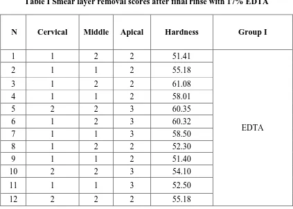
i Table II Smear layer removal scores after final rinse with 17% EGTA
Table III Smear layer removal scores after final rinse with MTAD
Table IV Smear layer removal scores after final rinse with 10% citric acid
Table V
Smear layer removal scores after final rinse with 5.25% NaOCl & 17% EDTA
Table VI
Mean, Median and Standard deviation of smear layer removal scores and one way analysis using Kruskal-Wallis test
Table VII
Multiple comparisons of smear layer removal scores using Mann-Whitney U Test of Cervical third of root canal
Table VIII
Multiple comparisons of smear layer removal scores using Mann-Whitney U Test of Middle third of root canal
Table IX
Multiple comparisons of smear layer removal scores using Mann-Whitney U Test of Apical third of root canal
Table X
Parametric one way analysis of microhardness values using ANOVA test
Table XI
Multiple comparison of microhardness values using Post Hoc test by Tuckey HSD analysis
ii Figure 2 Irrigants Used in the Study – 10% citric acid,5.25% NaOCl, 17%
EDTA, 17% EGTA, MTAD
Figure 3 Longitudinal sections using chisel and mallet
Figure 4 JEOL JSM-5600LV Scanning electron microscope
Figure 5 Specimen embedded on cold cure acrylic resin
Figure 6 Vickers microhardness testing machine
Figure 7 Photo micrograph of cervical portion of root canal treated with Citric acid (X3000)
Figure 8 Photo micrograph of middle portion of root canal treated with Citric acid (X3000)
Figure 9 Photo micrograph of apical portion of root canal treated with Citric acid (X3000)
Figure 10 Photo micrograph of cervical portion of root canal treated with EDTA (X1000)
Figure 11 Photo micrograph of middle portion of root canal treated with EDTA (X3000)
Figure 12 Photo micrograph of apical portion of root canal treated with EDTA (X3000)
Figure 13 Photo micrograph of cervical portion of root canal treated with EGTA (X1000)
[image:7.595.102.533.92.757.2]iii Figure 16 Photo micrograph of cervical portion of root canal treated with
MTAD (X1000)
Figure 17 Photo micrograph of middle portion of root canal treated with MTAD (X1000)
Figure 18 Photo micrograph of apical portion of root canal treated with MTAD (X1000)
Figure 19 Photo micrograph of cervical portion of root canal treated with NaOCl & EDTA (X3000)
Figure 20 Photo micrograph of middle portion of root canal treated with NaOCl & EDTA (X3000)
Figure 21 Photo micrograph of apical portion of root canal treated with NaOCl & EDTA (X3000)
iv
ABSTRACT
Introduction – The cleaning and shaping of root canal system has undergone paradigm shift from one fulfilling a prime debridement function to one more on gaining radicular access for action of irrigant and three dimensional obturation of prepared root canal. Chemomechanical preparation of root canal system helps in removing organic as well as inorganic debris and microorganisms from the infected root canals. One desirable property of root canal irrigants is to remove smear layer from instrumented root canals with no adverse effects on remaining tooth structure.
Aims and Objectives- This study was done to evaluate and compare the smear layer removal property and dentin microhardness by the use of 17 % EDTA, 17% EGTA, 10 % citric acid, MTAD, and alternating use of 5.25% sodium hypochlorite and 17% EDTA as a final rinse for 5 minutes on extracted mandibular single rooted premolars after rotary instrumentation.
v
Results and Observations– The sequential use of 5.25% sodium hypochlorite and 17% EDTA as final rinse was found to be a more effective irrigation regimen for smear layer removal and this protocol when followed resulted in lesser reduction in dentin microhardness. 17 % EDTA when applied for 5 minutes was more effective in smear layer removal from cervical and middle portions of root canal but created dentinal erosion more in cervical and middle third of root canal. . 17 % EGTA caused least reduction in dentin microhardness but was less effective in removing smear layer particularly the smear plugs. 10 % citric acid was effective in removing smear layer from cervical, middle and apical portions. Application of higher volumes and time was required to effectively remove smear layer and moreover citric acid caused substantial reduction in dentin microhardness. MTAD was effective in removing smear layer effectively from cervical and middle portions but least effective in apical third of instrumented root canals.
Conclusion – The alternating regimen of irrigation with 10ml of 5.25 % sodium hypochlorite and 17% EDTA for 5 minutes was found to be more effective in smear layer removal and produced less dentinal erosion and thus lesser reduction in dentin microhardness when compared with the other irrigants.
Clinical Significance – Cleaning and shaping of root canals supplemented by
1 The phase of preparing and debriding the root canal is undoubtedly the most important, the most complex, and the most delicate part of endodontic treatment. It is difficult to imagine how one can completely obturate a canal that has not been adequately cleaned and disinfected. Over the years, canal preparation has been described by a variety of names including “enlargement”, mechanical preparation” and “instrumentation”.In modern Endodontics the emphasis is on biological and
anatomical problem, hence cleaning and shaping are more correct terms. Schilder introduced these terms to endodontics in 1974. [1]
The shaping of root canal system has undergone a paradigm shift from one fulfilling a prime debriding function to one more regarded as gaining a radicular access to complex root canal systems, for the irrigant and root filling material. It has been found by using high resolution computed tomography that nearly 35 -53 % of the root canal surface remained uninstrumented, showing that instrumentation alone is inadequate. [2]
Thus when “preparing” a root canal system , it is infact cleaned of all
inorganic debris, organic substances, and microorganisms and it is shaped to facilitate placement of a permanent three dimensional filling. When files produce shaping, it is essential that irrigants clean a root canal system. [1]
2 The instrumentation, depending upon the design of instruments used, remove some of the residual tissue by engaging it, some will be compacted and burnished against the root canal walls. Whenever dentine is being cut using hand or rotary instruments, the mineralized tissues does not get shredded or cleaved but are shattered to produce considerable quantities of debris. The nature of smear layer created with current nickel – titanium rotary techniques may considerably vary from that formed using stainless steel instrumentation because of the different mechanical and chemical forces that comes into play. [2]
The identification of the smear layer using scanning electron microscope SEM was first reported by Eick. Scanning electron microscope studies of cavity preparations by Brannstrom and Johnson demonstrated a thin layer of grinding debris. They estimated it to be 2-5 µm thick. The first researchers to report the smear layer on the surface of instrumented root canals were McComb and Smith in [3]
It has been observed that bacteria could remain in the smear layer and in the
dentinal tubules despite instrumentation of the root canal and thus they may survive
and multiply and can grow into dentinal tubules. The chemomechanical cleansing is
often supported by the use of disinfectants. Few others believe in the fact that the
presence of the smear layer may block the antimicrobial effects of intracanal
disinfectants into the tubules. [4]
3 good surface wetting, have no adverse effects on remaining tooth structure, and be easy to use and effective within clinical parameters.[2]
The current methods of smear layer removal includes chemical, ultrasonic and laser techniques. The quantity of smear layer removed by a material is related to its pH and time of exposure. A number of chemicals have been investigated as irrigants to remove smear layer. According to Castelluci a working solution is the one which is used to clean the canal, and an irrigation solution is one which is essential to remove debris and smear layer created by instrumentation process.[3]
Removal of the smear layer is accomplished with acids or other chelating agents such as ethylenediamine tetracetic acid (EDTA) following cleaning and shaping. Irrigation with 17% EDTA for one minute followed by a final rinse with sodium hypochlorite is a recommended method.[5]
Chelators remove the inorganic components leaving the organic tissue elements intact. Sodium hypochlorite is then necessary for removal of the remaining organic components. Citric acid has also been shown to be an effective method for removing the smear layer as has tetracycline. [6]
4 Ethylene glycol-bis (β-aminoethyl ether) – N,N,N,N – tetra acetic acid (EGTA) has been used by researchers at low concentration when a medium free of calcium is needed. A lot of research in medicine on chelating action to heavy metals that contaminate patients had been done. EGTA has been used for this purpose. Schmid & Reilley reported that EGTA can bind calcium more specifically. [8]
In this study 5.25 % sodium hypochlorite is used as the common irrigant during instrumentation in all the groups. Alternating use of 17 % EDTA and 5.25 % sodium hypochlorite is used as positive control. Distilled water is used as negative control. In Group Ι, 10 ml of 17% EDTA was used as the final rinse, in Group II, 10 ml of EGTA was used as the final rinse, in Group III, 10 ml of MTAD was used as the final rinse and in Group IV, 10 ml of 10% citric acid was used as the final rinse. The irrigating solutions when in contact on dentine cause alterations on dentine and enamel. The studies on modes of action and efficiency of various chemical irrigating solutions have shown to have effect on both organic and inorganic components of root canal dentin.
5 methods. The suitability and practicality of Vickers hardness test for evaluating surface changes is adopted in this study.[9]
6
Aims -This study was done to examine the smear layer removal property and dentin
microhardness by the use of 17 % EDTA, 17% EGTA , 10% citric acid, MTAD and alternating use of 5.25 % sodium hypochlorite and 17 % EDTA as a final rinse for 5 minutes on extracted human mandibular single rooted pre molars after rotary instrumentation.7
Jay.K.Jaylor et al. (1997) [10] examined the effect of obturation technique, sealer and presence of smear layer on coronal microleakage. The comparison between smear layer removal and smear layer not removed was done. Results showed less leakage when smear layer was removed. AH 26 displayed less leakage than Roth’s 811 sealer. Vertical compaction reduced leakage when compared to lateral condensation, ultrafil and thermafil obturations. The study shows that removal of the smear layer resulted in decreased coronal leakage regardless of obturation technique used. Wennberg and Ostavik reported that when compared to zinc oxide eugenol sealers, the resin sealers had increased adhesion to root canal dentine. The Ketac – Endo and AH-26 showed less dye leakage when smear layer was removed. Thermafil technique without vertical condensation was susceptible to coronal leakage. This study reported that leakage significantly decreased with vertical condensation around the plastic carrier.
8
F.H.Takeda et al.(1999)[12] stated that 17% EDTA when used as final flush was not effective in smear layer removal whereas acidic solutions like 6% phosphoric acid and 6% citric acid produced demineralization of dentin. CO2 and Er.YAG laser were more efficient in smear layer removal than EDTA or acidic solutions. The phosphoric acid and citric acid removed smear layer from the root canal , but it decalcified and caused softening of dentine to a depth of 10 to 15µm. The low pH (1.5) could cause adverse effects on the periapical tissues. The dentine demineralization caused by 17% EDTA was less compared acidic solutions. The smear layer removal produced with citric acid were similar when compared to EDTA. Nd:YAG laser irradiation created very clean root canals with smear layer removed or melted and recrystallized. The root canals showed charred, melted, recrystallized, or glazed smear layer. The Er:YAG laser showed clean surfaces, free of a smear layer having open dentinal tubules without any melting.
9
M.F. Bertrand et al.(1999)[14] found that the middle and apical thirds of canals were cleaner using the complete Quantac sequence when compared using K.files. The Quantac system is designed to collect debris and smear layer and carry it out of canal system. The conventional manual instrumentation with K files and handpiece driven Quantac system both were effective in removing smear layer from coronal third of the canals.
V.Kytridou et al. (1999)[15] using the thermafil obturation technique showed extrusion of material beyond apical foramen in 84% of the specimens where as only 43% of specimens obturated with system B technique showed extrusion. In this study, the removal of smear layer was not a factor influencing the adaptation of obturation and was not a factor contributing to apical leakage. Both the Themafil and System B techniques showed acceptable toot canal fills in the coronal,middle and apical third.
Miriam.F Zaccaro Scelza et al.(2000)[16] used three different solutions as final irrigation to assess the degree of smear layer removal. The final 4 minute irrigation included three groups
Group I – 10ml 1% of NaOCl + 10ml of 10% citric acid + 10ml of distilled water.
Group II – 15ml of 0.5% of NaOCl + 15ml of EDTA-T , Tergentol (sodium lauryl ether sulfate)
10 Scanning electron microscopic photomicrographs evaluation for mean number of visible open dentinal tubules showed largest number of visible tubules in cervical third followed by middle and apical thirds. The volume of solution as well as chemical properties of irrigating solutions were important determining factors which aids in removal of debris. Adding Tergentol (sodium lauryl ether sulfate ) to EDTA caused significant reduction in surface tension, resulting in deeper penetration of solution for removal of smear layer.
11
Von Fraunhofer et al.(2000) [18] evaluated effects of canal instrumentation and effects of smear layer on leakage in filled teeths. Results indicated less microleakage when smear layer was removed. The canals obturated using thermoplasticized gutta percha showed less leakage than lateral condensation. Rotary driven instrumented canals had less leakage than hand instrumented canals. Nickel- titanium instruments has superiority with regard to elasticity and resistance to torsional fracture when compared to stainless steel endodontic files. The conservation of original path of curved root canals and accuracy of final apical diameter produced by rotary – instrumentation resulted in less leakage.
12
J.T.Marais et al.(2000)[20] evaluated a new product named electro-chemically activated water and compared it to NaOCl for its cleaning effect on root canal walls. The results showed that electro chemically activated water produced markedly clean surfaces and removed smear layer in large areas. The cleaning efficacy of electro chemically activated water was superior to NaOCl. The physical and chemical nature of electro-chemically activated water is not known. Russian scientists developed a process through which electro-chemically activated water (ECA) was produced with new anode – cathode system. Elecro-chemically activated water is produced from tap water, salt and electricity. This solution cleaned root canal wall in remarkable way, removing the smear layer. Exposure of collagen fibres and fibrils indicate that dentine was decalcified. Anolyte used was of a neutral pH, the catholyte of pH 9.8.
13 11.56 and 5.39 times more likely to leak than vertical compaction. Teeth obturated by lateral compaction was 2.14 times more likely to leak than Thermafil.
Hata et al.(2001)[22] evaluated effectiveness of oxidative potential water (O.P.W) on its ability to remove smear layer and debris from instrumented root canals. Oxidative potential water (O.P.W) was used extensively in Japan for household and agricultural disinfection because of the safety and bactericidal effectiveness. Scientific basis for development of the Oxidative potential water was that microorganisms cannot survive in aqueous environment with low pH (less than 3) and high oxidation – reduction potential. The study indicated that ultrasonic irrigation with oxidative potential water was less effective in removing smear layer than syringe irrigation with oxidative potential water. The study demonstrated the ability of oxidative potential water to remove the smear layer after root canal instrumentation and found that irrigation with Oxidative potential water and a syringe was deemed useful for root canal irrigation. Study showed that ultrasonic irrigation with Oxidative potential water was less effective in smear layer removal than syringe irrigation with oxidative potential water.
14 treated specimens were higher than those treated with 3% sodium hypochlorite. The results were that dentine bars exposed to Ca(OH)2 had no effect on modulous of elasticity but reduction in flexural strength whereas dentine bars treated with 3% and 5% NaOCl solutions had significant decrease in modulous of elasticity and flexural strength.
J.T.Marais et al.(2001)[24] stated that there are two types of electro chemically activated water used as irrigants. The Anolyte and Catholyte. The Anolyte has high oxidative potential. Catholyte is an alkaline solution with high reduction potential. The antimicrobial activity of anolyte has been reported. The study evaluated the effect of electro chemically activated water on selected group of anaerobic bacteria. The result showed that electrochemically activated water failed to destroy all of the bacteria within the root canals. Explanation for failure of Electro-chemically activated water to destroy bacteria is that in this study only anolyte was used , as opposed to the use of catholyte followed by anolyte. Catholyte is claimed to have cleaning or detergent effect. Another reason may be at the time of preparation of the Electro- chemically activated solutions , a technical failure of the unit may have caused an altered Electro-chemically activated solution.
15 physical properties was explained by the loss of the organic matrix within the dentine. During two 30 minute period irrigation with 5.25% sodium hypochlorite, the organic portion of the dentine was removed to an extent that further removal of organic matrix affect the residual strength of the tooth. An aggressive irrigation regimen was selected in this study in order that the effect was guaranteed. Study demonstrated that 5.25% sodium hypochlorite reduces flexural strength and elastic modulus of dentin.
G.E.Evans et al.(2001)[26] evaluated the effectiveness of preparation technique and sodium hypochlorite in removal of pulp and predentine from root canals of posterior teeth. The importance of preparation technique had a more dominant influence than the irrigant. The isthmus areas were untouched by both preparation techniques (Step back preparation and Ni Ti rotary preparation technique). The residual pulp tissue was more at apical portion for step back preparation than for rotary preparation. A higher number of no debris score was found in the Quantac preparations . Inference is both the preparation technique and irrigant play an important role in debridement . Frequency of remaining pulp tissue scores for all groups were at 1 mm level. Overall finding was one of pulp tissue being compacted against apical stop and some extruded. If patency filing procedure were used , the pattern might been different.
16 dentin were 42.26 to 45.84 for water, 36.10 to 41.02 for 15% EDTAC , 35.22 to 41.72 for 1% CDTA , and 33.54 to 38.62 for 1% EGTA. Test showed statistically significant difference for the control solution compared with EDTAC,CDTA, and EGTA, which were similar. Yamamoto et al. stated that EDTA chelates calcium and magnesium ions and that EGTA chelates calcium ions only.The results showed that the three chelating agents significantly reduced microhardness when compared with water. There was no statistically significant difference among the three solutions.
17
Ahmet Serper et al.( 2002)[28] studied the effects of concentration and pH of EDTA on the dentinal demineralization was studied, Demineralizing effects of EDTA solutions at 10% and 17% concentrations at pH 7.5 and 9.0 was determined by measuring the liberated phosphorous at 1,3,5,10 and 15 min after exposure. The phosphorous liberated from dentine was greater with increased concentration of EDTA and increased time of exposure. Cury et al. reported that the effect of EDTA solutions on the demineralization of dentin is influenced by pH , the greatest demineralizing effect of EDTA solution was achieved at pH between 5.0 and 6.0. EDTA was found to be effective at a neutral pH than when it is at pH 9.0.
Balagi.T.S et al.(2002)[29] evaluated when 17% EDTA and 4% NaOCl were used as alternative irrigants and 17% EDTA and 5% ethylene diamine were used as a single mixture. Both were effective in complete removal of smear layer and debris at coronal, middle and apical thirds of root canal. When 17% EDTA or 4% NaOCl was used alone it could remove inorganic and organic components of smear layer only. Specimens that were irrigated with alternate use of 17% EDTA and 4% sodium hypochlorite showed more removal of smear layer. Specimens treated with single mixture of 17% EDTA and 5% Ethylenediamine showed maximum removal of smear layer and debris Sodium hypochlorite is a reducing agent and has ability to remove loose superficial debris and dissolve organic debris by release of hypochlorous acid that reacts with insoluble protein to form soluble peptides.
18 transmitters of ultrasonic energy to activate irrigants. The activation of irrigants did not significantly reduce smear layer and debris, but anti bacterial effects of ultrasonically activated irrigants was more effective. The larger apical shapes improves debridement and disinfection of canals as stated by Abou-Rass and Paccinino. Two tips of different instrument design and material , a cutting stainless steel K file and blunt Ni-Ti wire were compared.
M.Hulsmann et al.(2002)[31], M.Heckendorff and F.Schafers studied the effects of three chelating agents namely - Calcinase slide, Glyde file and R.C.prep on root dentine was evaluated. No differences in changes in microhardness were found regardless of time of application (2.5, 5 and 10 min) with regard to canal cleanliness for all pastes. The cleanliness decreased from coronal to apical third and the specimens treated with calcinase slide showed higher weight loss than R.C.prep after 6 and 9 min working time. Glyde file was significantly superior to R.C prep after 6 min. To enhance the degree of cleanliness copious irrigation of canal using irrigants has been suggested including chelating agents as stated by Harrison et al . The chemical and pharmacological properties of EDTA preparations were evaluated. The EDTA containing agents should be used between 1 and 5 min. Apical extrusion of chelators should be avoided.
19 down of protein structure caused by the alkalinity of the materials used. MTA samples showed greatest range in force required to fracture.
.Mahmoud Torabinejad et al.(2003)[33] studied the effect of various concentrations of sodium hypochlorite as an intracanal irrigant before use of MTAD as a final rinse to remove smear layer. The canals were treated for 2 minutes with 5ml of irrigating solutions as final rinse. The solutions used were 5.25% NaOCl, sterile distilled water, 17% EDTA, MTAD. The results showed the use of MTAD as irrigant left some odontoblastic process in the dentinal tubules and some organic debris. As high concentrations of NaOCl are more toxic, the difference in ability to dissolve necrotic tissue was not significant between 1.3%, 2.6% and 5.25% NaOCl. It seems precedent to use lowest concentration of NaOCl (1.3%) followed by MTAD as final rinse to remove efficiently the smear layer. MTAD is an acidic solution with a pH of 2.15. Yamada et al. found a final flush with EDTA followed by sodium hypochlorite was effective method to remove smear layer. A study using application of various concentrations of sodium hypochlorite and final rinse with MTAD, a chemical reaction occurs between sodium hypochlorite and residual MTAD, resulted in brown solution in root canals caused by release of doxycycline present in MTAD solution.
20 spectrum antibiotic that is effective against wide range of microorganisms. Tetracycline is bacteriostatic in nature , because in the absence of bacterial cell lysis, antigenic by-products are not released, It has low pH and act as a calcium chelator and cause enamel and root surface demineralization. In addition it has substantive property. Tetracycline shown to enhance healing after periodontal therapy.
Deborah Clark-Holke et al. (2003) [35] evaluated if the smear layer affects the passage of bacteria through obturating material. Smear layer forms an intermediate barrier that interferes the adhesion and penetration of sealers into dentinal tubules. Cergneux et al. found less dye leakage when smear layer was removed with either EDTA or ultrasound. In the in vitro study where lateral condensation and AH 26 sealer were applied, removal of smear layer resulted in no bacterial leakage.
A.Khademi et al.(2004)[36] stated thatboth EDTA 17% and citric acid 7% removed the smear layer but 17% EDTA was more effective and the superiority was observed in middle and apical regions. In the cervical region both solutions showed no significant differences. The middle third region had highest degree of cleanliness compared to other areas and apical third had the least. Results of this study states that using both EDTA and citric acid in smear layer removal provide satisfactory results.
21 microhardness after Ca(OH)2 glycerine treatment was significantly greater than after Ca(OH)2 distilled water combination. The greater reduction in dentin microhardness found after calcium hydroxide-glycerine combination was explained by different penetration ability of the two combinations into dentinal tubules.
Brent.J.Crumpton et al.(2005)[38] stated that 1ml of EDTA with a contact time of 1 minute was effective in removal of smear layer as 10ml. The use of EDTA over 1ml resulted in no further debris removal. The study result showed that 1ml of EDTA was as effective in removing the smear layer as 10ml. It may be hypothesized that the effects of EDTA are a function of contact time with no relation to volume of irrigation.
Hale Ari et al. (2005)[39] evaluated the mineral contents of root canal dentin after irrigating with 0.2% chlorhexidine gluconate for 15min, 3% H2O2 for 15min, 17% EDTA for 15min, 5.25% NaOCl for 15min, 2.5% NaOCl for 15min and distilled water as control. The levels of five elements (calcium, phosphorus, magnesium, potassium, sulphur) were analyzed using inductively coupled plasma-atomic emission spectrometry (ICP-AES) technique. Result showed that a significant decrease in Ca level with all irrigation solutions except 5.25% sodium hypochlorite. This study found that sodium hypochlorite, hydrogen peroxide, or combination of sodium hypochlorite and hydrogen peroxide have negative effects on bond strength of adhesive cement to root canal dentin.
22 of organic element of dentin (collagenous component) and mineral component is left relatively intact. When irrigation is alternated with EDTA, the degradation of hydroxyapatite occurs and results in change in visco elastic properties. The aggressive irrigation with NaOCl and EDTA cause surface erosions in dentine.
C.S.Teixeira et al.(2005)[40] studied the combined use of EDTA and NaOCl and found to be effective in removing smear layer from cervical and middle third for all times of application for 1,3 and 5 minutes. In the apical third the effective removal of smear layer was decreased when irrigated for 1 minute. The entrapment of air bubbles prevent the total filling of irrigant in the apical third. The mechanical stirring with lentulospiral removes air bubbles and favors improved contact of EDTA to canal walls. Irrigating canals for 5 minutes, Lopes et al. reported that the mechanical stirring of EDTA for 2 minutes using lentilo spiral allowed for near removal of smear layer from apical third. On account of reduced dimension of root canal, air bubbles remain trapped and prevent total filling with the irrigant. Mechanical stirring with lentilo spiral removes the air bubbles and favours improved contact of EDTA with the canal walls.
23 around dentinal tubules. The thickness of these demineralized zones were comparable to those formed on crown dentin after phosphoric acid etching for 15 seconds. The acidity of Biopure MTAD is similar to mild self-etching primers used in dentin bonding. A 5 minute irrigation is long compared with 30 -60 seconds etching time recommended for self-etching primers. Mechanical agitation for enhanced efficacy of smear layer removal and replenishing of fresh irrigants during subsequent rinsing may have also contributed to formation of thick demineralized dentin zones. Because demineralized collagen matrices are susceptible to collapse after air drying, their presence after endodontic irrigation challenge the concept of success of hydrophobic sealers. Concomitantly these collagen matrices offer an opportunity for dentin hybridization with hydrophilic methacrylate-based resins/sealers without the adjunctive use of phosphoric acid etching or self etching primers.
24
Matthias Zehnder (2006)[43] reviewed the requirement for irrigating solutions. Sodium hypochlorite due to its broad anti microbial spectrum as well as capacity to dissolve necrotic tissue remnants is recommended as main irrigant. During canal instrumentation canals should always be filled with sodium hypochlorite. This increases working time of the irrigant. The cutting efficacy of hand instruments is improved and torsional load on rotary nickel-titanium instruments is reduced when canals are filled with sodium hypochlorite solution. A 5.25% sodium hypochlorite solution significantly decreases the elastic modulus and flexural strength of human dentin compared to physiologic saline, while a 0.5% solution does not decrease the elastic modulus and flexural strength. Alternative approach to improve effectiveness of hypochlorite in root canal system could be to increase the temperature of low – concentration sodium hypochlorite solutions. 1 mol of hypochlorite contains 1 mol of available chlorine. State of available chlorine is depending on the pH of the solution. Above pH of 7.6, the predominant form is hypochlorite, below this value is hypochlorous acid. Both forms are extremely reactive oxidizing agents. Pure hypochlorite solutions used in endodontics have a pH of 12, thus entire available chlorine is in the form of hypochlorite. At identical levels of available chlorine, hypochlorous acid is more bactericidal than hypochlorite . One way to increase the efficacy of hypochlorite solution could be to lower their pH. Such solutions was found to be less toxic to vital tissues than nonbuffered solutions.Buffered hypochlorite with bicarbonate renders the solution unstable with a decreased shelf life to less than 1 week.
25 hypochlorite with the surface tension of MTAD and Tetraclean. Results showed that MTAD had a low surface tension (34.5 mJ/m2) , similar to Cetrexidin one (31.1 mJ/m2). The lowest surface tension was shown by Tetraclean (29.1 mJ/m2). The surface tencion vaue for Smear Clean was 33 mJ/m2).
Mercedes Perez-Heredia et al.(2006)[45] evaluated the cleaning ability of three acid irrigating solutions after hand and rotary instrumentation. The irrigating solutions were 1) 15% citric acid plus 2.5% NaOCl 2) 15% EDTA plus 2.5% NaOCl 3) 5% orthophosphoric acid plus 2.5% NaCl and 4) 2.5% NaCl alone as control. Neither the smear layer nor debris was removed combining 15% EDTA with 2% NaOCl with both instrumentation technique in the coronal, middle and apical thirds. With hand instrumentation, 15% EDTA showed the best effectiveness in debris removal in coronal, middle and apical thirds. With regard to smear layer removal 15% EDTA showed major effectiveness in apical and middle third. With rotary instrumentation technique 15% citric acid plus 2.5% sodium hypochlorite removes smear layer in apical, middle and cervical thirds. Cervino et al. found that 17 % EDTA and 15% citric acid alternated with 5% sodium hypochlorite were equally effective in smear layer removal with the hand instrumentation. Orthophosphoric acid is a universal conditioner shown to have smear layer removal capability. Partial removal was obtained with 10 second application of 10% phosphoric acid or 10% citric acid and complete elimination achieved after similar treatment using 32% phosphoric acid. In debris removal orthophosphoric had the had the least behavior.
26 The effect of 1% chlorhexidine has been studied. The specimens were immersed in 25ml of each of solutions at 3, 10 and 15minutes of immersion. The concentration of Ca2+ was measured by atomic absorption spectrometry. Results showed no significant differences in the amount of Ca2+ extracted by 10% or 20% citric acid or by 17% EDTA. EDTA extracted a significantly higher amount during first three minutes compared with other solutions.The decalcifying action was time dependent. The amount of Ca2+ extracted in the citric acid and EDTA solutions increased with longer immersion time. Study shows that 17 % EDTA and the two citric acid concentrations have the same decalcifying power but the effect of EDTA is more rapid. A higher amount of Ca2+ was obtained in the first 3 minutes with 17 % EDTA than with other solutions. Application of 17 % EDTA for 10 minutes causes excessive erosion of peritubular and intertubular dentin. A short periods are recommended for its application.
Stevens et al.(2006)[47] conducted a study in which the maxillary incisors were decoronated, prepared in crown down fashion and smear layer removed with 17% EDTA followed by 5.25% NaOCl (Group N) and again rinsed with NaOCl before obturation and Roth 801 sealer was used. The roots in a group were rinsed with 95% ethyl alcohol instead of NaOCl as final rinse. Leakage was determined using fluid flow model. Result showed that final rinse with 95% ethyl alcohol increased sealer penetration and decreased leakage.
27 markedly enhanced with exposure to 1% NaOCl and more with 5% NaOCl solution. A concentration dependent reduction of elastic modulus and flexural strength of root dentin resulted. 5% sodium hypochlorite severely altered the peripheral dentin matrix..Stoward and Davies reported that sodium hypochlorite fragments long peptide chains and to chlorinate protein terminal groups; the resulting N-chloramines are broken down into other species. Thus sodium hypochlorite solutions may affect mechanical dentin properties via the degradation of organic dentin components. The study showed a concentration dependent effect of sodium hypochlorite on mechanical dentin properties resulting from the disintegration of organic dentin matrix.
N.Nisha Soumithran et al.(2007)[49] studied the demineralizing effect on radicular dentin by 17% EDTA and MTAD at different time intervals. The assessment of amount of phosphorous liberated at different time intervals was done using random access automated bio chemistry analyzer. The results of the study concluded the use of MTAD as a chelating agent followed by a final flush with saline as irrigant is better than 17% EDTA. The irrigants have to be flushed out of the canal within 10 minutes. Care must be taken to avoid the use of MTAD in children and pregnant women for its deleterious action on dental structures.
28 irrigants showed that microhardness of root canal dentin was reduced for all. Bio Pure MTAD was least effective in reducing microhardness of root canal dentine and 17% EDTA had maximum effect. Panighi and G’Sell reported positive correlation between hardness and mineral content of the tooth. The microhardness determination provides indirect evidence of mineral loss or gain in dental hard tissues. There is a variable increase in microhardness from coronal to apical third of the root canal dentin irrespective of any treatment with test agent. This was attributed to the histology of root canal dentin. Carrigan et al. reported that tubular density decreased from cervical to apical dentin. Pashley et al. showed that an inverse correlation between dentin microhardness and tubular density.
29 efficiently removed with a final rinse of 1 ml of 17 % EDTA for 1 minute followed by 3 ml of 5.25 % sodium hypochlorite . This study reported that this protocol was not efficient to remove completely smear layer from the apical third.
I.M.Saleh, et al.(2008)[52] studied the effect of smear layer on penetration of bacteria along different root canal filling materials and the presence of bacteria at the interface of dentine and sealer and sealer and core material. The results showed the bacterial penetration along root filling with Gutta percha and AH plus sealer occurred more slowly in the presence of the smear layer than in its absence. Differences in leakage among these sealers using AH plus, Apexit sealer and Real seal cones and sealer was not significant when smear layer was present. The results support the view that retaining the smear layer on the root canals may be beneficial in preventing bacterial penetration and colonization. Smear layer may also act as a filler for the sealer, reducing the contraction stress that lead to pulling out of the sealer tags from the dentinal tubules.
30
Emboava Spano et al.(2009)[54] evaluated the concentration of calcium ions and smear layer removal using 15% EDTA, 10% citric acid, 10% sodium citrate, apple vinegar, 5% acetic acid, 5% malic acid and sodium hypochlorite. Results showed that use of 15% EDTA resulted in greatest concentration of calcium ions, followed by 10% citric acid. 15% EDTA and 10% citric acid were the most efficient solutions for removal of smear layer. Zehnder et al. studied the effect of reducing surface tension of 15.5 % EDTA, 10% citric acid, or 18% hydroxyethylidene-1, 1-bisphosphonate [HEBP] prepared with and without 1% polysorbate (Tween) 80 and 9% propylene glycol on their ability to remove calcium from instrumented root canals. The results showed no increase in calcium chelating ability when surface tension of chelator solutions was lowered with wetting agents. Citric acid removed more calcium than EDTA or HEBP solutions.
31
Punit Bansal and Hitesh Gupta (2009)[4] reviewed on smear layer in endodontics. The article explains the importance of smear layer. The presence of smear layer on root canal walls acts as an intermediate physical barrier and may interfere with adhesion and penetration of sealers into dentinal tubules. When smear layer is not removed, the durability of apical seal has to be evaluated over a long period, since this layer is non homogenous and weakly adherent structure, dissolving around a leakage filling material and thus creating a void between the root canal wall and the sealer.
M.A.Mozayeni et al.( 2009)[5] studied the effectiveness of MTAD as the final irrigant to remove the smear layer, compared with that of 17% EDTA, both following root canal irrigation with 5.25% sodium hypochlorite. When EDTA is alternately used with 5.25% NaOCl, the smear layer is removed in middle and coronal thirds of canal preparation, and is less effective in the apical third. In this study placement of MTAD with wrapped cotton in broach allows intimate contact of solution even in apical region of the canals and improves debridement of the entire root canal wall.
32 swimming pools because of its compatibility with hypochlorite to prevent stains from metal ions. Irrigation protocols using 1% sodium hypochlorite and then 17 % EDTA, 1% sodium hypochlorite and then 2.25% peracetic acid or a combined solution containing 1% sodium hypochlorite and 9% etidronic acid produced similar efficiency in smear layer removal.
Mancini et al.(2009)[57] compared the efficacy of 17 % EDTA, Bio Pure MTAD and 42% citric acid in endodontic smear layer removal and degree of erosion in the apical third of instrumented root canals. The study showed Bio Pure MTAD did not remove the smear layer from apical third of the canals. The study using citric acid 42% did not remove smear layer from the apical third of the canals. 5.25% Sodium hypochlorite at 37o C did not remove smear layer from the apical third of the canals. In addition to sodium hypochlorite, the application of chelating agents to remove smear layer has been suggested. Calt and Sepper and O’Connell et al. found the combination of 17% EDTA and 5 % sodium hypochlorite is an effective irrigation solution in removing smear layer in the apical third of instrumented canals.
33 inducing erosive action and allowed sealers to penetrate dentinal tubules completely. The sealing ability of Ketac-Endo was higher in comparison with groups using Acroseal.
D.R.Violich and N.P Chandler( 2010)[3] explained the current methods to smear layer removal - chemical, ultrasonic and laser techniques and none of which are totally effective throughout the length of all canals or are universally accepted. Smear layer is removed for thorough disinfection of the root canal system and for better adaptation of materials to the canal walls. The adaptation of root canal materials to canal walls were studied. White et al. found pHEMA, silicone, Roth 801, and AH 26 sealers extended into the tubules when smear layer was removed.
Vishal A.Mahajan et al.(2010)[59] evaluated the effect of MTAD and EDTA in removing the smear layer and its effect on peritubular and intertubular structures using SEM examination. The results indicated that MTAD is an efficient solution for removal of smear layer, especially in the apical third of root canals, and does not cause structural change in dentinal tubules. The smear layer removal action of EDTA is attributed to chelation action. The moderate smear layer removal by EDTA in the apical third of root canal attributed to poor penetration of EDTA in the apical area of root canal. When comparing the degree of erosion from cervical and middle third areas in MTAD and EDTA treated groups , a statistically significant difference was found. MTAD group showing no erosion. Erosion of dentinal tubules can be due to hyperdecalcification induced by EDTA on dentin.
34 The study compares the effect of mechanical agitation using a fully tapered and apically trimmed non standardized gutta percha master cone, Rins endo irrigation system and Endoactivator system in removing the smear layer. The study found activations resulted in cleaner canals compared with no activation. In this study the access cavity provided a strategic reservoir to hold a more effective volume of irrigant for exchange during activation. The irrigating solutions are apically exchanged each time the activator system is inserted into the canal. When the activator tip moves toward length, reagent id displaced. When activator is partially withdrawn there is exchange of solution into the apical third. Efficiency of this hydrodynamic circuit is further enhanced when combined with sonic oscillating movements. A pumping action synergistically combined with mechanical agitation explains better results achieved with the EndoActivator.
35
Xiaoli Hu et al. (2010) [62] evaluated the effect of 17% EDTA, 5.25% NaOCl, 3% H2O2 on dentin wettability and roughness . The wettability is a crucial factor for adhesion and is dependent on chemical composition, roughness and hydration state and is influenced by tubule density. The study found 5.25% NaOCl increased the wettability of dentin. The roughness of dentin increased with NaOCl application because of organic dissolving properties of NaOCl on collagen components. The dissolution of inorganic components by EDTA by its chelating action softens root canal wall and tubules become patent, surface roughness is increased due to smear layer removal. The chelating action of EDTA softens root canal wall by dissolution of inorganic components. Dentin tubules become patent and surface roughness increased.
Rodig et al(2010)[63] evaluated the cleaning efficacy of different irrigant agitation techniques on debris and smear layer removal in curved root canals. The effectiveness of ultrasonic activation, endo activator, and canal brush was evaluated. The agitation devices were introduced 2mm short of working length, activation time for each irrigant was 1 minute. The results showed that endo activator was superior in smear layer removal compared with ultrasonic agitation and canal brush. The removal of smear layer was more effective in coronal than in apical region. The activation of irrigant improved smear layer removal, but no benefit of irrigant activation was observed in apical portions.
36 and 17% EDTA, and found 7% maleic acid significantly better than EDTA in removing smear layer from the apical third of root canal system. Microhardness determination provide indirect evidence of mineral loss or gain in dental hard tissues. Changes in mineral content may adversely affect the sealing ability and adhesion of dental materials such as resin-based cements and root canal sealers. The degree of mineral content and amount of hydroxyapatite in intertubular substance are factors determining the intrinsic hardness of dentin structure. 7% maleic acid is highly acidic and a pH of 1.05. This acidic pH caused demineralization of dentin and reduction in microhardness. There was no statistically significant difference between 7% maleic acid and 17% EDTA.
De Deus et al.(2011)[65] evaluated the effect of the exposure time and concentration of peracetic acid (PAA) on removal of smear layer. The study showed that concentration of 2.25% PAA can dissolve an experimental smear layer as quickly as a standard 17% EDTA solution. After 60s of contact, the 0.5% peracetic acid solution dissolved smear layer as well as 2.25% PAA and 17% EDTA. 17% EDTA, 2.25 % peracetic acid and 9% etidronic acid removed smear layer and demineralized the root canal wall. After 60 seconds of contact, the 0.5% peracetic acid solution dissolved smear layer as well as 2.25% peracetic acid and 17% EDTA.
37 layer removal of 85%, 60% and 50% and of debris was 95%, 90%, 85%. SAF a newly developed file system, when inserted into a root canal adapts itself to the canal’s original shape. The surface of the lattice is slightly abrasive, and the system
removes dentin with a back-and-forth grinding motion with vibration. An irrigation device (Vatea) is connected to the silicon tube and provides continuous flow of the preferred irrigation solution. This motion creates turbulence in the root canal allowing continuous and fresh irrigant present in the canal. The use of the SAF in combination with a dual-irrigation regimen of 3% sodium hypochlorite and 17% EDTA has been reported to create in clean dentin surface especially in the apical third. Successful removal of smear layer for both EDTA and MTAD from the entire root may be due to vibrating motion of SAF within the continuously replaced fluid. SAF system vibrating at 5000 vibration /minute induces sonic activation of the chosen irrigant.
38 of dentin microhardness and increase in surface roughness. This effect were related to the demineralizing effect on root canal dentin.
Prado et al.(2011)[67] compared the effectiveness of 37% phosphoric acid with that of 17% EDTA and 10% citric acid in the removal of smear layer. The results showed that at 3 minutes of application, phosphoric acid solution was more effective, in the apical third, followed by citric acid, EDTA and phosphoric acid gel. When comparing the solutions in cervical, middle, and apical thirds, EDTA and citric acid were more effective in cervical third than middle and apical thirds. Phosphoric acid solution was equally effective in cervical, middle and apical thirds. Phosphoric acid gel was more efficient in the cervical and middle thirds than in the apical third. 37% phosphoric acid showed dentin erosion related to time of exposure. At 1 minute or longer, the erosion was present in middle and cervical thirds and no erosion found in the apical third.
Rossi-Fedele et al.(2011)[68] reviewed the influence of pH changes on the efficacy of chlorine containing endodontic solutions. PH value of sodium hypochlorite between 6 and 7.5 would lead to improved antibacterial efficacy. The tissue dissolution activity of sodium hypochlorite decreased when pH reached values between 6 and 7.5. Zehnder explained the property of sodium hypochlorite used widely as main root canal irrigant because of its broad antibacterial activity, its function to prevent formation and to dissolve the smear layer and its ability to dissolve tissue remnants. Chlorine has a strong tendency to acquire electrons in order to achieve greater stability, and this translates into chlorine’s oxidizing activity. Oxidizing capacity is
39 acid is responsible for the disinfectant action of chlorine solutions. The relative amount of hypochlorite ion and HOCl present in chlorine solutions at a given pH and temperature is constant, as HOCl in water undergoes an instantaneous and reversible ionization into hypochlorite (OCl-) and hydrogen (H+) ions. Subsequently, pH changes will reflect the relative amounts of hypochlorite ion and HOCl present in the solution. If HOCl is consumed, then the balance will shift, new HOCl will form at the expense of OCl-. The OCl- in the aqueous solution can work as reservoir for the formation of new HOCl and vice versa. Lowering the pH to values below 4 and 5 diminishes the relative amount of HOCl and chlorine gas (Cl2) dissolved in water increases at the same rate. Chlorine gas form is unstable because of volatility and has noxious odour and irritant to the respiratory tract, eyes, and mucous membrane and at higher concentrations can have fatal effects.
40 scanning probe microscopy family and is possible to reconstruct three-dimensional surface topography images in real time. Topographic surface changes in ProTaper instruments immersed in sodium hypochlorite and ethylenediaminetetraacetic acid solutions were evaluated. Significant deterioration of instrument surfaces resulting in an increase in root mean square value and roughness average were caused by both irrigants. Localized pitting and cracks that modifies the integrity and resistance to fracture of NiTi instruments were seen on immersion in 5.25 % sodium hypochlorite for 5 minutes.
De-Deus et al.(2011)[70] made critical appraisal of published smear layer removal studies. In 2007 De-Deus et al. successfully developed an optical microscopy method, called cosite optical microscopy (CSOM) that allows longitudinal observation of a predetermined dentin area. CSOM produces a set of images from a large number of x-y positions of dentin sample at different experimental times. Different from the traditional score analysis, CSOM provides operator-independent quantitative results with more reliable statistics for comparison. For each sample, at each experimental time, the 15 analysed image fields contained between 6000 and 7000 tubules. For each substance evaluated, approximately 20,000 tubules were automatically measured at each experimental time without operator influence. The procedure is fast taking about 25 minutes for image acquisition for all experimental times, and 3 minutes for image analysis of the full set of images using a common personal computer.
41 reported that Q Mix has better antibacterial property and contains chlorhexidine and EDTA and that smear layer removing ability was comparable to EDTA solution. Irrigants play an important role in accessing areas not instrumented such as lateral and accessory canals as well as fins and webs throughout the canal. None of present irrigants meets all requirements of an ideal endodontic irrigant. Sodium hypochlorite in concentrations from 0.5 % to 6 % is the most accepted irrigating solution. Sodium hypochlorite has antibacterial, tissue dissolving properties. It is toxic to periapical tissue and suggested to decrease micromechanical characteristics of dentin. Has no action on inorganic part of smear layer and EDTA used in 17 % concentration to dissolve inorganic portion of dentin and smear layer by chelation. EDTA is recommended for use after sodium hypochlorite to remove smear layer. Quin et al reported that if NaOCl is used again after EDTA or citric acid as final antibacterial rinse , it cause marked erosion of root canal dentin. Considerable efforts made on developing new irrigants and/or establishing new irrigation protocols. Bio Pure MTAD were introduced in 2003. It was suggested to be more effective than NaOCl and EDTA against Enterococcus faecalis and mixed bacteria. QMiX is a novel endodontic irrigant for smear layer removal with added antimicrobial agents. It contains EDTA, CHX and a detergent, It is a clear solution ready to use with no chair side mixing. Mixing EDTA and CHX is known to produce a white precipitate. In QMiX this is avoided because of its chemical design.
42 initial rinse is significantly reduced. The MTAD protocol for irrigation is using 1.3% sodium hypochlorite as initial rinse followed by MTAD solution as final rinse, Torabinejad studied the effect of MTAD by using 1.3 % sodium hypochlorite as initial rinse for a cumulative period of 18 -20 minutes during instrumentation and left MTAD solution for 2 minutes after instrumentation . Smear layer removal was obtained using the above irrigation protocol. Lofti et al. evaluated the effect of MTAD on removal of smear layer as final rinse when 1.3% sodium hypochlorite was used during 10 – minute instrumentation time. In the study MTAD could not eliminate the smear layer from root canal walls. It was concluded that the irrigation with 1.3% NaOCl during instrumentation for 10 minutes was not effective to remove organic materials from the smear layer and consequently MTAD could not remove smear layer effectively.
44
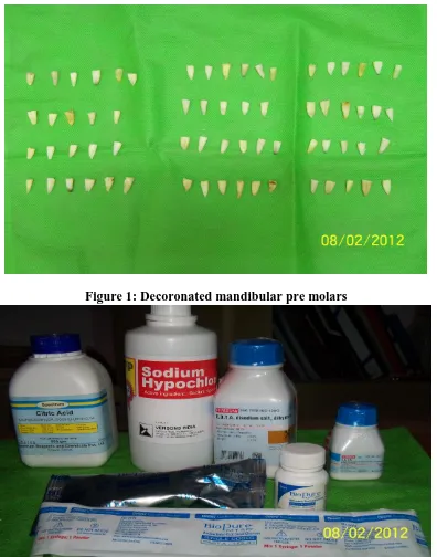
Materials used in the study
–a) 5.25 % Sodium hypochlorite ( Vensons India)
b) 17 % EDTA ( Himedia Laboratories)
c) 17% EGTA (Himedia Laboratories)
d) 10% Citric acid (Spectrum Reagents and Chemicals Pvt Ltd)
e) BioPure MTAD ( Dentsply, Tulsa dental specialities)
f) Distilled water
g) Paper Points ( Suredent Corporation)
h) 7% Glutardehyde ( SDFCI Fine Chem Ltd)
i) Ethanol
j) Self cure acrylic resin (DPI)
k) Plastic ring
l) Silicon carbide polishing paper
Equipment used in this study –
a) 10 number K –File ( Mani)
b) 28 gauge side vented irrigation needle
c) X Smart Endo rotary system ( Dentsply)
45 e) Micromotor and diamond disc
f) Chisel and mallet
g) JEOL JSM-5600LV Scanning electron microscope
h) Vickers microhardness testing machine ( CLEMEX)
Methodology -
Sixty non-carious freshly extracted mandibular pre-molars for orthodontic reasons were used in this study. After cleaning they were immersed in isotonic saline solution. Then the teeth were decoronated and a 10 number K- file was inserted into the canal until just visible at apex to determine patency. One millimeter was subtracted from this measurement and this was the working length.
The teeth were divided randomly into five experimental groups of 12 each.
Group Ι – 10 ml of 17 % EDTA was used as a final rinse for 5 minutes
Group ΙI – 10 ml of 17% EGTA was used as a final rinse for 5 minutes
Group III – 10 ml of MTAD was used as a final rinse for 5 minutes
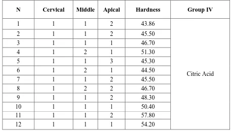
Group IV – 10 ml of 10% citric acid was used as a final rinse for 5 minutes
Group V – 10 ml of 17 % and 5.25 % sodium hypochlorite. (fig. 2)
46 and dried with paper points. A total of 10 ml irrigant at the rate of 1ml per 30 sec was used as final irrigant for each root canal.
The total exposure time to final solution was approximately 5 minutes. Irrigants were passively delivered using side vented 28 guage needle to within 1-2 mm from the working length in each canal. The specimens were fixed using glutaraldehyde and the fixed specimens were rinsed three times with a sodium cacodylate – buffered solution (Concentration 0.1, pH 7.2), incubated in osmium tetraoxide for 2 hours, dehydrated with ascending concentrations of ethyl alcohol and placed in a desiccator for 24 hours. The specimens were split into two halves using chisel and mallet along the prepared groove (fig.3).Each specimen was mounted on a aluminum stub and coated with 30 µm of gold palladium and examined under scanning electron microscope (fig.4).The smear layer on the surface of the root canal or in the dentinal tubules at the cervical, middle and the apical portion of each canal was evaluated according to the following criteria used by Torabinejad et al.
1- No smear layer: no smear layer on the surface of the root canals, all tubules were clean and open.
2- Moderate smear layer: no smear layer on the surface of the root canal, but tubules contained debris.
3- Heavy smear layer: smear layer covered the root canal surface and the tubules.[34]
47 0.05 µm size aluminum oxide powder mixed with distilled water.A plastic ring was then taken and poured with a mixture of cold cure resin. Specimens were embedded on the resin with polished surface facing outside. After curing of the resin, the ring was removed and repolishing of specimens was done to remove excess material present on the tooth surface (fig.5).Microhardness testing was done by mounting specimens on the stage of Vickers microhardness tester (fig.6). The midroot portion halfway from the outer surface was focused for testing. Indentations were made with Vickers diamond indenter using 300 gm load with a dwell time of 15 seconds. These indentations were measured and converted into Vickers hardness number (VHN).
48
Table – I shows the smear layer scores and microhardness values obtained after final rinse with 17% EDTA
Table – II shows the smear layer scores and microhardness values obtained after final rinse with 17% EGTA
Table – III shows the smear layer scores and microhardness values obtained after final rinse with MTAD solution
Table – IV shows the smear layer scores and microhardness values obtained after final rinse with 10% Citric acid
Table – V shows the smear layer scores and microhardness values obtained after final rinse with alternating use of 5.25% Sodium Hypochlorite and 17% EDTA
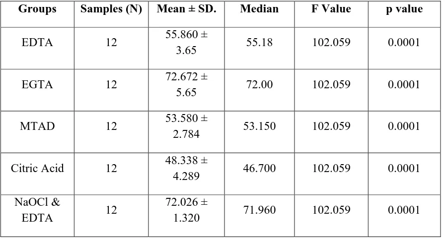
Table – VI shows the data obtained for smear layer removal were discrete variables in scores, hence single comparisons taking groups together was performed with Kruskal – Wallis one way analysis for 5 groups and Chi square values were obtained Mean, Median and standard deviation values for smear layer removal from cervical middle and apical portions for five groups were obtained.
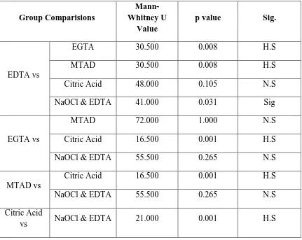
Multiple comparisons of two groups taken at a time were performed using Mann – Whitney U Test and p values for all comparisons were fixed as less than 5 %.
Table VII shows the group comparison for cervical portions of root canal treated by comparing two groups taken at a time.
No significant difference was shown between 17 % EDTA and 10 % citric acid in cervical portions with p value > 0.05 (0.070).
49 more effective than MTAD in cervical portion of root canal. (p value 0.0001)
A statistically significant difference between 10% citric acid and combined use of 5.25 % sodium hypochlorite and 17 % EDTA was found in cervical portion with a p value < 0.05 (0.014)
Table VIII shows the group comparison for middle portions of root canal treated by comparing two groups taken at a time.
17 % EDTA was found to have no statistically significant difference with 10 % citric acid in middle portion of root canal (p value > 0.05, ( 0.105)) . 17 % EDTA was found to be highly significant when compared with 17 % EGTA and MTAD in middle portion (p value < 0.05 ((0.008)). 10 % citric acid also shows statistically significant difference to MTAD and 17 % EGTA in middle portion (p value < 0.05 (0.001)) . 10 % citric acid shows statistically significant difference to 5.25 % sodium hypochlorite and EDTA (p value < 0.05 ( 0.001)).
Table IX shows group comparison for apical portion of root canal by comparing two groups taken at a time.
10 % citric acid shows statistically significant difference between 17 % EGTA and MTAD at apical portion of root canal (p value < 0.05 (0.0001)). 10 % citric acid shows statistically significance with combined use of 5.25 % sodium hypochlorite and 17 % EDTA at apical portion (p value = 0.005 ( 0.005)).
50
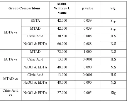
Table –X shows the data obtained were statistically analyzed using parametric one way analysis of variance ANOVA and significance was tested using f test. The p value was fixed at 5 % .
The mean, median and standard deviation values for five groups were obtained .
Table –XI shows the multiple comparison of microhardness values between groups were evaluated using Post hoc tuckey analysis.
10 % citric acid showed statistically high significance in reduction in microhardness with 5.25% sodium hypochlorite and 17 % EDTA indicating (p value < 0.05 (0.0001)).
10 % citric acid showed statistically significant difference in microhardness with 17 % EDTA (p value < 0.05 (0.0001))
17 % EGTA and 5.25 5% sodium hypochlorite combined with 17 % EDTA showed statistically significant difference between 17 % EDTA, 10 % citric acid and MTAD.