JOURNAL OFVIROLOGY,
0022-538X/00/$04.00⫹0 Nov. 2000, p. 9858–9867 Vol. 74, No. 21
Copyright © 2000, American Society for Microbiology. All Rights Reserved.
Structural Phosphoprotein M2-1 of the Human Respiratory
Syncytial Virus Is an RNA Binding Protein
ISABEL CUESTA, XUEHUI GENG, ANA ASENJO,ANDNIEVES VILLANUEVA* Centro Nacional de Microbiologı´a, Instituto de Salud Carlos III, Majadahonda, Madrid 28220, Spain
Received 20 March 2000/Accepted 28 July 2000
The structural phosphoprotein M2-1 of human respiratory syncytial virus (HRSV) Long strain shows RNA binding capacity in three different assays that detect RNA-protein complexes: cross-linking, gel retardation, and Northern-Western assays. It is able to bind HRSV leader RNA specifically with cooperative kinetics, with
an apparentKdof at least 90 nM. It also binds to long RNAs with no sequence specificity. The RNA binding
domain has been located between amino acid residues 59 and 85, at the NH2terminus of the protein. This
region contains the phosphorylatable amino acid residues threonine 56 and serine 58, whose modification decreases the binding capacity of M2-1 protein to long RNAs.
Human respiratory syncytial virus(HRSV), a member of the
Pneumovirusgenus of theParamyxoviridaefamily, is the most common infectious agent in bronchiolitis and pneumonia re-quiring hospitalization of infants and young children. Immu-nocompromised and elderly people are also severely affected. No vaccine or specific antiviral treatment is available at present (11). The best way to control these infections, according to the characteristics of the human populations mainly affected, is the use of different agents as live attenuated vaccines (38), human-ized neutralizing monoclonal antibodies (21), and antiviral compounds (27).
The HRSV nucleocapsids are, as in all paramyxoviruses, structural components and functional units for replication and transcription processes (11). The nucleocapsid proteins are thus multifunctional, serving as ideal targets for the action of antiviral compounds and for conveying attenuation mutations. The design of both types of reagents would be aided by un-derstanding nucleocapsid protein functions.
HRSV nucleocapsids are composed of the negative-sense RNA genome (15,222 nucleotides long), tightly bound to the N protein, the large polymerase protein (L), the phosphoprotein (P), and probably the M2-1 protein (11, 16). The RNA poly-merizing activity of the viral nucleocapsids is due to L protein, for which activity P protein is essential (41). N protein plays a histone-like function, since synthesis of viral RNA always oc-curs on ribonucleoprotein templates. P protein also acts as a chaperone of N protein, maintaining it competent for encap-sidation (30). M2-1 protein enhances the processivity of the viral polymerase (9, 10) and its readthrough of intergenic junc-tions (17). These properties identified M2-1 protein as an elongation and antiterminator transcription factor, a constitu-ent of the nucleocapsids. The protein is encoded by the M2 gene, present only in the pneumoviruses, which codes for an mRNA containing two open reading frames (ORFs). ORF1 encodes the 22K structural protein, also known as M2-ORF1 or M2-1 protein (194 amino acids). ORF2 encodes M2-2 pro-tein (90 amino acids), detected in HRSV-infected cells, which has a regulatory function in the balance between viral tran-scription and replication (1, 6, 8).
The M2-1 protein was long ago described as a structural
membrane phosphoprotein that could be present as different species in HRSV-infected cells (20, 23). These species could differ in the number of intramolecular disulfide bonds formed among its four cysteine residues (23). Three of the four cys-teine residues are at the amino-terminal end, forming a C3H1
motif that is highly conserved among members of the genus. In other proteins, this motif has been shown to bind Zn ions (40). Mutation at the predicted Zn coordinating residues of the C3H1motif (C7, C15, and H25) abolished M2-1 enhancement
of transcriptional readthrough (18). Interaction of M2-1 with the N protein has been also described in coexpression experi-ments (16).
Here we present evidence that M2-1 is an RNA binding protein, able to bind the RNA leader specifically, whose bind-ing capacity is modulated by phosphorylation. The amino acid residues in contact with the RNA and those modified by phos-phorylation have been located.
MATERIALS AND METHODS
Cells and viruses.HEp-2 cells, COS1 cells (CV1 cells containing the simian virus 40 T antigen), HRSV Long strain, and the vaccinia virus recombinant vTF-3 were used throughout this study in experimental conditions previously described (35, 37).
Transfection.cDNAs of different viral proteins, all cloned in pGEM3, were transiently expressed in the vaccinia virus-based expression system under the control of the T7 RNA polymerase promoter (vTF-3). The corresponding solu-ble proteins were prepared as described elsewhere (37). Briefly, cell cultures were recovered by low-speed centrifugation, twice washed with phosphate-buff-ered, and then homogenized in 10 mM Tris-HCl (pH 7.5)–140 mM NaCl–5 mM EDTA–1% Triton X-100 and–1% deoxycholate. The supernatant fraction ob-tained after centrifugation in a minicentrifuge (15 min, 4°C) was considered the soluble protein fraction. For some variants, insoluble proteins were obtained after removal of soluble proteins; they were further solubilized in sodium dodecyl sulfate-polyacrylamide gel electrophoresis (SDS-PAGE) sample buffer.
Plasmids.The cDNAs corresponding to N, P, and M2 genes of HRSV Long strain, cloned in the corresponding pBSV9 recombinant plasmids (24), were subcloned asHpaI-StuI fragments in theSmaI site of the plasmid vector pGEM3. Plasmid pL was obtained by cloning, in theEcoRI site of vector pUC19, a cDNA fragment obtained by reverse transcriptase-PCR containing the promoter sequences of the T7 RNA polymerase and the 674 nucleotides of the HRSV Long strain genomic 3⬘end. This fragment contains the leader (44 nucleotides plus three extra G’s, which increase transcription by the T7 RNA polymerase [9]), together with the NS1 gene and the first 80 nucleotides of the NS2 gene.
The pGEM3 recombinant plasmids containing the cDNAs corresponding to the M2-1 variants were obtained by directed mutagenesis (19) or after cutting the M2 cDNA with restriction enzymesPstI,DpnI,EcoRV, andEcoRV plusNdeI, creating the variants PstI, DpnI, EcoRV, and EcoRV-NdeI, respectively. Variant EcoRVts is similar to EcoRV, but with T56 and S58 replaced by alanine. In the cases of variants PstI and EcoRV-NdeI, after cutting with the corresponding restriction enzyme, the DNA ends generated were blunted by the action of the
* Corresponding author. Mailing address: Centro Nacional de Mi-crobiologı´a, Instituto de Salud Carlos III, Carretera de Majadahonda a Pozuelo Km 2, Majadahonda, Madrid 28220, Spain. Phone: (34) 91/ 509-7901, ext. 3662. Fax: (34) 91/ 509-7966. E-mail: nvilla@isciii.es.
9858
on November 9, 2019 by guest
http://jvi.asm.org/
3⬘exonuclease or polymerase activities of T4 DNA polymerase. In this way, the structure of the mRNA was preserved to facilitate stability and translation.
M2-1 protein purification.HEp-2 cells were infected with HRSV, and the microfilament-containing protein fraction and extracellular viral particles were obtained as previously described (32). The proteins contained in the different fractions were separated and characterized in SDS-gels (12% acrylamide) con-taining 0.4% Coomassie blue in the electrophoresis buffer or after negative staining (14). The piece of gel containing the M2-1 protein was excised, and the protein was electroeluted. The protein was then precipitated with acetone and resuspended in TE buffer (10 mM Tris-HCl, 1 mM EDTA [pH 7.5]) containing 6 M guanidine chloride. The protein was renatured by chromatography through an acrylamide column using TE as the running buffer. The fractions containing the M2-1 protein were detected after SDS-PAGE, pooled, and densitometrically quantified by comparison to known amounts of bovine serum albumin (BSA), separated in the same gel.
Preparation of labeled riboprobes. The32P-labeled riboprobes used were
prepared by in vitro transcription of the corresponding pGEM3 recombinant plasmids, using T7 or SP6 RNA polymerase and adding [␣-32P]CTP to the
reaction (25). In this way,32P-labeled plus- or minus-polarity RNAs were
ob-tained, corresponding to genes P (1,000 nucleotides) and N (1,200 nucleotides). A plus-polarity RNA (700 nucleotides) containing the leader sequence plus three extra G’s at the 5⬘end was obtained using plasmid pL digested withNcoI in the in vitro transcription reaction.
The short riboprobe (80 nucleotides) containing the leader sequence was obtained using plasmid pL digested withStyI. This short probe contains three extra G’s, the 44 nucleotides of the leader, and 33 nucleotides of the NS1 gene at its 5⬘end. The nonspecific RNA control was obtained using pGEM4 digested withEcoRI (58 nucleotides).
UV cross-linking assay.M2-1 protein (10 to 180 ng) was mixed with 1 to 5 ng of the indicated riboprobe (specific activity, 30,000 Cerenkov cpm/ng) and incu-bated in binding buffer (10 mM Tris-HCl [pH 7.5], 100 mM NaCl, 100M ZnCl2,
4 mM dithiothreitol). The RNA-protein complex was UV cross-linked as previ-ously described (3). After treatment with bovine pancreas RNase A (60g/ml), SDS-PAGE (12% acrylamide gel) was carried out. The gels were stained with Coomassie blue and autoradiographed.
Gel retardation assay.The protein and riboprobes were incubated as indicated above and then electrophoresed in a gel containing 3.5% acrylamide and 0.5⫻ Tris-borate-EDTA buffer at 100 V. The gels were fixed with 95% ethanol, dried, and autoradiographed. The amount of radioactivity present in the riboprobe position was quantified by a PhosphorImager. This procedure was used for kinetic studies to measure the formation of the protein-RNA complex. The protein concentration at which 50% of the labeled RNA formed an RNA-protein complex was defined as the apparentKd(dissociation constant).
Northern-Western assay.Protein (30 to 40g) obtained after transfection experiments was separated by SDS-PAGE and electrotransferred to nitrocellu-lose paper. The paper was washed with 100 mM NaCl–40 mM Tris-HCl (pH 8.0), and the proteins were renatured by incubation in the same buffer containing 0.02% PVP-40 (polyvinylpyrrolidone), 0.02% BSA, 0.02% Ficoll 400, 0.1% Tri-ton X-100, and 50M ZnSO4for 3 h at 20°C. The32P-labeled riboprobe was
added in the same buffer without ZnSO4and containing yeast RNA (1g/ml).
After 2 h of incubation, the probe was removed and the paper was washed several times with incubation buffer. The dried paper was exposed in a Phosphor Imager.
Chemical and enzymatic treatments of the M2-1 protein.The cross-linking reaction described above was carried out with 80 ng of purified M2-1 protein and 5 ng of32P-labeled riboprobe. After RNase digestion and precipitation with
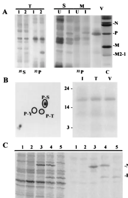
FIG. 1. Purified and renatured M2-1 protein binds to RNA, as shown by cross-linking with UV and in electrophoresis gel retardation experiments. (A) SDS-polyacrylamide gel stained by Coomassie blue showing the protein composition of 20g of protein contained in extracellular viral particles (V) and soluble (S), microfilament-bound (M), and insoluble (I) protein fractions from HRSV-infected HEp-2 cells. (B) Purity of viral proteins after renaturation and quantification. The BSA slots correspond to 92, 231, 462, 693, and 926 ng of pure BSA. The following slots correspond to different volumes (2 and 4l) of purified and renatured N, P, M, and M2-1 proteins. The calculated protein concentrations varied between 0.1 and 0.2 mg/ml. The electrophoretic mobility of HRSV proteins and of molecular weight markers (MW; expressed in kilodaltons) are indicated at the left and right, respectively. (C and D) The indicated amounts (nanograms) of purified M2-1 protein were incubated with 2 ng of RNA (1,000 nucleotides long) labeled with32P (35,000 Cerenkov cpm/ng) under the indicated conditions. Samples were then UV cross-linked
and analyzed by SDS-PAGE following RNase digestion (C) or run in a 5% acrylamide gel (D). Asterisks indicate the electrophoretic mobility of M2-1 and its dimers.
on November 9, 2019 by guest
http://jvi.asm.org/
acetone, the labeled M2-1 protein with associated radioactivity was mixed with 100 to 300 ng of electroeluted M2-1 protein. The mixture was then treated with 14 or 28 mg of cyanogen bromide (CNBr) per ml in 50% formic acid at 37°C overnight or for 4 h (15). After incubation, the sample was vacuum dried, neutralized by several cycles of resuspension in 0.1 M NH4HCO3(pH 8.3), and
again vacuum dried. When the sample was at neutral pH, it was resuspended in 50l of electrophoresis sample buffer. In other cases, the protein was treated withN-bromosuccinimide or with V8 protease as previously described (15, 39). The peptides generated after treatment were separated by SDS-PAGE as described elsewhere (31) and transferred to an Inmobilon membrane. After peptide staining with amido black, labeled peptides were detected by autora-diography using a PhosphorImager, and their amino-terminal ends were deter-mined by partial sequencing (34).
RESULTS
M2-1 is an RNA binding protein. To determine whether
HRSV nucleocapsid proteins are able to interact with viral RNA, protein fractions from HRSV-infected HEp-2 cells were chromatographed through Sepharose 4B containing covalently bound monoclonal antibodies specific for the different viral proteins. The viral proteins purified in this way were unable to bind32P-labeled RNAs by cross-linking and by retardation in
gel electrophoresis assays. These results could be due to the denaturation of the purified proteins, to the fact that RNA is already bound to them, or to both possibilities. To avoid this difficulty, we first purified the nucleocapsid proteins by SDS-PAGE and then renatured them. As a nucleocapsid source, a microfilament fraction and extracellular viral particles from infected HEp-2 cells were used, because nucleocapsid proteins are in a higher proportion in these fractions than in other subcellular fractions. The protein composition of the protein fractions obtained from HRSV-infected HEp-2 cells and the purity of the viral nucleocapsid proteins after purification and renaturation are shown in Fig. 1A and B.
To determine the capacity of M2-1 protein to bind RNA, the purified protein was incubated with32P-labeled riboprobes and
binding was assayed by UV cross-linking (Fig. 1C) and by retardation of the electrophoretic mobility of32P-labeled
ribo-probes in gel electrophoresis (Fig. 1D). In both assays, M2-1 protein binding to RNA was detected, but the cross-linking assay did not always yield a linear response with increased amounts of protein. For this reason, kinetic studies of the binding reaction were carried out using gel electrophoresis retardation assays.
The appearance in the cross-linking assays of a labeled pro-tein with an electrophoretic mobility corresponding approxi-mately to a 44-kDa protein, the predicted position for M2-1 dimers, is noteworthy. In addition, N and M but not P proteins were able to bind RNA in assays similar to those described above. The RNA binding capacity of N protein was modulated by P protein, as determined when both proteins were rena-tured together. This supports the suggested chaperone func-tion of N as described for P protein (30) (data not shown).
M2-1 protein specifically binds to short RNAs containing
the 5ⴕ-end leader sequence.Different amounts of the renatured
M2-1 protein were assayed by mobility retardation in gel elec-trophoresis, using 1 ng of32P-labeled riboprobes
correspond-ing to plus (mRNA) and to minus (viral RNA) polarities of HRSV P and N genes (1,000 and 1,300 nucleotides, respec-tively) and to plus-polarity RNA containing the HRSV leader plus three extra G’s at the 5⬘end (700 nucleotides) and there-fore with the structure at the 5⬘end of the viral cRNA. Similar kinetics were obtained in all cases tested (Fig. 2A). The protein is also able to bind to other long RNAs unrelated to HRSV (data not shown); M2-1 protein therefore binds without se-quence specificity to long RNAs. The apparentKd, calculated
from quantification of the results, is 30 nM for these long
RNAs. The retardation of the probes in all cases is due to binding of the protein to a long RNA since binding can be competed for by different unlabeled long RNAs but not by short yeast RNA, which has an average size of 100 nucleotides (data not shown).
To determine the specificity of M2-1 binding to any RNA sequence, shorter RNAs were assayed. A specific 80-nucleo-tide cRNA, containing three extra G’s and the leader se-quences (44 nucleotides) at its 5⬘end, was also tested. In this case, M2-1 specifically binds to 0.1 ng of the cRNA leader in the presence of 25 ng of yeast RNA (Fig. 2B). The apparentKd
calculated for this RNA binding was 90 nM. There is no bind-ing to the 58-nucleotide nonspecific probe or to 20- to 100-nucleotide-long yeast RNA, as indicated above. This result indicates that M2-1 protein binds specifically to the leader RNA contained in short (80-nucleotide) but not long (700-nucleotide) RNAs.
FIG. 2. M2-1 protein binds specifically to HRSV leader RNA. Different quantities of purified M2-1 protein were incubated with 1 ng of32P-labeled
(30,000 Cerenkov cpm/ng) long riboprobes (vRNA [viral RNA] and mRNA) corresponding to the RNAs for the P gene in minus and plus polarities, respec-tively (cRNA corresponds to 700-nucleotide positive-polarity long RNA contain-ing the leader sequence at its 5⬘end) (A) or with 0.1 ng of32P-labeled (100,000
Cerenkov cpm) short RNAs (leRNA, 90 nucleotides long, containing the leader sequence at its 5⬘end; sRNA, a pGEM4-derived RNA, 50 nucleotides long) (B) with increasing amounts of M2-1 protein. Lanes labeled 0, 1, 2, 3, 4, and 5 correspond to the absence of protein or to the addition of M2-1 protein at final concentrations of 18, 36, 54, 109, and 218 (A) and 18, 36, 72, 145, and 290 (B) nM. The amount of radioactivity was quantified by PhosphorImager. The results represent the mean of several experiments with each riboprobe.
9860 CUESTA ET AL. J. VIROL.
on November 9, 2019 by guest
http://jvi.asm.org/
Mapping of the M2-1 RNA binding domain.After prepara-tive cross-linking and RNase digestion, the labeled protein was treated with CNBr,N-bromosuccinimide, or V8 protease. The peptides generated after the different treatments were sepa-rated by electrophoresis and visualized by staining with amido black; those labeled with32P were detected by
autoradiogra-phy. Figure 3A shows the result obtained after treatment with CNBr and V8 protease digestion. The amino-terminal se-quence of the peptides generated was determined. The data from several experiments (summarized in Fig. 3B) indicate the calculated sizes for the fragments and the amino acid residues located at their NH2-terminal ends, although the calculated
fragment sizes may be overestimated due to their smallness and to the possible presence of oligoribonucleotides bound to them. The approximate locations of these fragments on the M2-1 molecule are shown, although their C-terminal ends are unknown (Fig. 3C). These results indicate that at least some of the residues in contact with the RNA are between positions 55 and 75 but do not rule out the existence of others around this region. The RNA binding region was thus considered to be located between amino acid residues 40 and 80. It also appears that the zinc finger domain does not contain the residues in direct contact with the RNA since the V3 fragment, which starts at the residue at position 10, contains no associated radioactivity.
Deletion variants of M2-1 protein and their RNA binding
capacities.To confirm that between amino acids 40 and 80 of
M2-1 protein are those that contact directly RNA and to pin-point the residues essential for RNA binding, we tested the RNA binding capacity of M2-1 protein and different deletion variants by Northern-Western blotting using M2-1 proteins transiently expressed in the vaccinia virus-based expression system. The cDNA corresponding to the HRSV M2 gene was
expressed in this system, and a protein with a mobility slower than that of M2-1 present in the purified virions was observed. This protein has been identified as phosphorylated M2-1 pro-tein (see below).
We made sequential and C-terminal amino acid sequence deletions of M2-1 protein. The sequential deletions (del5,
del10,del16, anddel26) were those in which 5, 10, 16, and 26 amino acids were eliminated between positions 32 and 58. The C-terminal deletions correspond to M2-1 variants PstI, DpnI, EcoRV, EcoRVts, and EcoRV-NdeI. The amino acid residues deleted and/or added up to the first termination codon in each M2-1 variant are indicated in Fig. 4A. All nucleotide sequences of the M2 gene variants were determined by automatic se-quencing.
Assay of the M2-1 protein variants for RNA binding capacity in Northern-Western assays showed that all were able to bind long RNAs (Fig. 4B to D). Comparison of RNA binding ca-pacities of different amounts of unphosphorylated and phos-phorylated M2-1 protein indicated that the phosphos-phorylated form has a decreased RNA binding capacity (data not shown). The data from the distinct variants suggested that the RNA binding region is located between amino acid residues 59 and 85. The RNA binding of variant PstI, which contains only the first 62 M2-1 protein amino acid residues, and the fact that the protein containing deletions between residues 32 and 58 does bind to RNA indicate that residues 59 to 62 (EISG) participate in and may be sufficient for RNA binding. The del5 variant (Fig. 4B, lane 3), which is devoid of the zinc finger, is not essential for binding, as suggested by the preparative cross-linking experiments. In this variant, two G insertions occurred at nucleotide 100 of the 5⬘end of the mRNA; this produces a frameshift that results in the appearance of a stop codon 24 nucleotides after the insertion. The protein synthesized by this
FIG. 3. Location of M2-1 protein amino acids residues in contact with RNA. (A) Preparative RNA binding assays were carried out with 150 ng of M2-1 protein and 8 ng of32P-labeled RNA (1,000 nucleotides long). After UV cross-linking, RNase A digestion, and acetone precipitation, the covalently bound RNA–M2-1 protein
was resuspended in TE buffer containing 300 ng of purified M2-1 protein. The mixture was treated with 28 mg of CNBr per ml in 50% formic acid [CNBr(1)] or with 1g of protease V8 (V8) in 50 mM NH4HCO3for 4 or 2 h, respectively, at 37°C. The peptides generated were separated in SDS-polyacrylamide gels containing 6%
urea and Tricine in the electrophoresis buffer (31). After electrophoresis, peptides were transferred to Inmobilon membranes and detected by amido black staining (S) or by PhosphorImager (32P). (B) NH
2-terminal sequences for the peptides. Sizes of molecular weight (MW) markers are indicated in kilodaltons. (C) Representation
of the M2-1 protein molecule and the mapping of unphosphorylated and phosphorylated peptides to it. The dashed lines at the C-terminal ends of fragments indicate that their exact end positions are unknown. The amino acid sequence in the M2-1 protein RNA binding region is indicated. The residues starting the different phosphorylated peptides are also indicated in that sequence.
on November 9, 2019 by guest
http://jvi.asm.org/
variant probably starts at the third methionine of the M2-1 protein that has a G at position⫹4, according to the Kozak rules (22), and has a C-terminal truncation, according to its observed size. These results are in agreement with those ob-tained in the preparative cross-linking assays, which locate the RNA binding region between amino acids 40 and 80, with some of the residues in contact with RNA located between positions 55 and 75. It is also clear that the amino acid residues located between positions 85 and 194 are unnecessary for RNA binding.
M2-1 protein is phosphorylated mainly when expressed in
the absence of other viral proteins.HEp-2 cells infected with
the vaccinia virus recombinant vTF-3 were transfected with plasmid pGEM3 or pGEM3-M2. After labeling with [35
S]me-thionine, cell extracts were prepared, and the proteins were analyzed by SDS-PAGE. In pGEM3-M2-transfected cells, we detected a protein that is absent in the cells transfected with pGEM3. This protein showed a mobility slower than that of M2-1 protein present in purified virions (Fig. 5A, lane 2,35S).
Previous results (23) described a 24-kDa structural phospho-protein, in addition to P phospho-protein, which may be M2-1 protein. The change in electrophoretic mobility observed for the tran-siently expressed M2-1 protein may thus be due to phosphor-ylation. To test this, the above experiment was repeated but with [32P]orthophosphate labeling. 32P labeling was found in
the protein expressed by the recombinant pGEM3-M2 plasmid (Fig. 5A, lane 2,32P).
When 32P-labeled mock- or HRSV-infected HEp-2 cells
were fractionated (Fig. 5A) to obtain soluble and microfila-ment-bound protein fractions and extracellular viral particles, a phosphorylated protein was found with the same electro-phoretic mobility as that detected after transfection with the pGEM3 recombinant M2, mainly in the microfilament fraction and extracellular viral particles. The piece of gel containing M2-1 protein, transiently expressed in transfected cells, was excised, and its phosphoamino acid residues were determined (Fig. 5B, left). It was found that M2-1 protein is phosphory-lated on serine and to a lesser extent on threonine residues. Similar results were obtained using phosphorylated M2-1 pro-tein from HRSV-infected HEp-2 cells (data not shown). Phos-phorylated M2-1 protein from transfected cells, present in the microfilament fraction of HRSV-infected HEp-2 cells and in extracellular viral particles, was treated with CNBr, and the peptides generated were separated by SDS-PAGE; phosphor-ylated peptides were detected by autoradiography (Fig. 5B, right). The same phosphopeptides were detected in all cases. Also, the two species of M2-1 have been detected at early and late times in HRSV-infected HEp-2 cells (13).
[image:5.612.102.502.69.362.2]These results, like others recently published (18), indicated that M2-1 protein is phosphorylated and that phosphorylation
FIG. 4. M2-1 protein deletion variants and their RNA binding capacities determined by Northern-Western assays. (A) Diagram representation of M2-1 protein indicating the amino acid residues deleted in the different variants as a broken lines. The residues unrelated to M2-1 protein and added as a consequence of the cloning procedures are also shown. Right and left parts of panels B to D correspond to Northern-Western and Western assays, respectively, of 50g of total protein. The different M2-1 protein variants are indicated by asterisks in both assays. In panel B, lane V corresponds to extracellular viral particles, and lanes 1, 2, 3, 4, 5, and 6 correspond to soluble proteins from transfections with pGEM3 and pGEM3 recombinants M2-1,del5,del10,del16, anddel26, respectively. In panel C, lanes, 2, 3, 7, 8, 9, and 10 correspond to insoluble proteins from transfection with pGEM3 recombinant plasmid M2-1,del5, EcoRV, EcoRVts, EcoRV-NdeI, and DpnI constructs. In panel D, lane 11 corresponds to soluble protein from transfection with the pGEM3 recombinant plasmid PstI. The center portion corresponds to a longer exposure of the Northern-Western blot; the asterisk indicates the PstI variant M2-1 protein position. The Western blots were developed with monospecific anti-M2-1 protein serum except for the insets containing lanes 9 and 3 in panel C, which were developed with anti-HRSV serum.
9862 CUESTA ET AL. J. VIROL.
on November 9, 2019 by guest
http://jvi.asm.org/
modifies its electrophoretic mobility. When it is expressed alone, without other viral proteins, all molecules are modified; in the presence of the remaining viral proteins, the bulk of M2-1 protein remains mainly in an unphosphorylated form. It appears that when M2-1 is expressed alone, its phosphoryla-tion sites are exposed to protein kinases and the acphosphoryla-tion of cellular protein phosphatases is prevented. In contrast, in the
presence of the rest of viral proteins, M2-1 phosphorylation sites are partially hidden from the protein kinases or the phos-phorylated residues are exposed to phosphatase action, prob-ably due to conformational changes in the protein caused by protein-protein interactions.
Coexpression experiments with viral proteins N and P indi-cate that M2-1 coexpression with P protein results in the lack of phosphorylation of a part of the M2-1 molecules (Fig. 5C). Direct or indirect interactions between P and M2-1 proteins thus result in the prevention of exposure of phosphorylatable M2-1 protein residues to the action of cellular protein kinases or phosphatases, respectively.
Deletions variants of M2-1 protein and their phosphorylation
capacity.When M2-1 protein and its variants were transiently
expressed, differences in solubility and in the presence of two molecular species were observed (Fig. 6A). To determine whether the presence of a second protein species is due to phosphorylation, transfected cells were labeled with [32
P]ortho-phosphate (Fig. 6B), their specific activities were determined, and the results obtained were compared to those for the nor-mal M2-1 protein (Fig. 6; Table 1).
M2-1 proteins with deletions between amino acids 32 and 42, 32 and 48, and 32 and 58 (del10,del16, anddel26) were phos-phorylated 30, 9, and 7% with respect to the phosphorylation level of the whole M2-1 protein. Only one protein species was expressed for these variants, except fordel10. For the M2-1 variants containing large deletions (EcoRV [amino acids 110 to 166 deleted], DpnI [amino acids 85 to 166 deleted], and EcoRV-NdeI [amino acids 110 to 194 deleted]), higher specific activities (2.0, 2.6, and 1.7, respectively) than those found for the control M2-1 protein were observed. In these variants, two proteins species were detected. These results indicated that the absence of amino acids 1 to 59 or 110 to 162 regulates M2-1 protein phosphorylation in a negative or positive manner.
Identification and localization of phosphorylated residues
on the M2-1 molecule.M2-1 protein transiently expressed in
HEp-2 cells with the vaccinia virus-based system was labeled with [32P]orthophosphate or [35S]methionine, fractionated by
gel electrophoresis, transferred to Inmobilon membranes, and excised. Proteins were then treated with CNBr andN -bromo-succinimide. Labeled peptides were separated and further identified by autoradiography. In accordance with the cleavage specificity of the chemical agents used, peptide size, and me-thionine residue distribution in the M2-1 molecule, the phos-phorylated residues were tentatively located at the NH2
-termi-nal region between amino acid residues 40 and 100 (data not shown). Concurring with this location, the deletion variants lacking amino acid residues between positions 85 and 166, 110 and 166, and 110 and 194 were phosphorylated at a higher level than the normal protein. The slight phosphorylation fordel10 and the lack of phosphorylation found for del16 and del26 indicated that the phosphorylated residues could be located between residues 48 and 85, taking into account that there are no serine and threonine residues between amino acid residues 32 and 48 of M2-1 protein. Previous experiments indicated (36) that purified HRSV particles contain casein kinase II (CKII)-like activity, able to phosphorylate their P protein among others proteins present in the purified viral particles. One of these proteins has the same electrophoretic mobility as the phosphorylated M2-1 protein. The phosphorylated amino acids of M2-1 may thus be modified by CKII protein.
[image:6.612.64.280.71.404.2]We looked for S and T residues between amino acids 48 and 85 containing the signal recognition sequence for CKII (29) (an acidic or phosphorylated residue at position⫹3). T56 has this recognition sequence; if it is phosphorylated, S53 could also be a suitable target for phosphorylation. In addition, S58
FIG. 5. M2-1 protein is phosphorylated at serine and threonine residues when is expressed in transfected and HRSV-infected HEp-2 cells. Direct or indirect interactions with P protein modulate M2-1 protein phosphorylation. (A) HEp-2 cells were transfected (T) with plasmid pGEM3 (lane 1) or pGem3-M2 (lane 2), mock (U) or HRSV (I) infected, and labeled with [32P]orthophosphate.
In the transfection experiment, the protein was also labeled with [35
S]methi-onine. Soluble (S) and microfilament-associated (M) protein fractions were obtained. The proteins were separated by SDS-PAGE and visualized in a Phos-phorImager. The viral proteins in extracellular viral particles (V) stained by Coomassie blue (C) are shown at the right. (B) (Left) The piece of gel containing the phosphorylated protein, with electrophoretic mobility slower than that of M2-1 protein of extracellular viral particles expressed by the recombinant plas-mid in the transfection experiment (T), was excised from the gel and hydrolyzed with 6 N HCl for 2 h at 110°C. The resulting amino acid residues were separated by two-dimensional electrophoresis in thin layer and characterized by their32P
labels. Unlabeled phosphothreonine (P-T), phosphoserine (P-S), and phospho-tyrosine (P-Y) were included as markers and detected with ninhydrin. (Right) Phosphorylated protein expressed by pGEM2-M2 (T), and polypeptides with the same electrophoretic mobility in the microfilament fraction of infected cells (I) and in virions (V), after excision from the gel and CNBr treatment. After renaturation, the peptides were separated in SDS–20% urea acrylamide gels, and those in phosphorylated form were detected by a PhosphorImager. Sizes are indicated in kilodaltons. (C) HEp-2 cells were infected with recombinant vaccinia virus vTF-3 and transfected with pGEM3 (lane 1), with pGEM3-M2 (lanes 2 to 5), and with pGEM3-M2 plus pGEM3-N (lane 3), pGEM3-N and pGEM3-P (lane 4), and pGEM-P (lane 5). The cultures were [35S]methionine labeled, and
the soluble proteins were analyzed by SDS-PAGE before (left) or after (right) immunoprecipitation with rabbit anti-HRSV serum.
on November 9, 2019 by guest
http://jvi.asm.org/
has the CKI consensus sequence (an acidic or phosphorylated residue at position⫺3), and if S58 is phosphorylated, S61 also can be phosphorylated by the same protein kinase. This ten-tative location is in agreement with the lack of phosphorylation observed fordel16 anddel26, although onlydel26 has lost the S and T residues present at positions 58 and 56. Changes in protein structure may explain the lack of phosphorylation in the other M2-1 protein variants. We thus replaced T56 and S58 with alanine in deletion variant EcoRV and confirmed the resulting nucleotide sequence by automated sequencing.
This new M2-1 protein variant, EcoRVts, was transiently expressed and labeled with [35S]methionine and [32
P]ortho-phosphate. The protein in the cell extracts was fractionated by SDS-PAGE and visualized by autoradiography (Fig. 6A, lane 8). The EcoRVts variant protein was expressed but was poorly phosphorylated. Calculation of its specific activity showed that it is phosphorylated only to 2 to 4% of the level of the EcoRV variant protein. The same phosphorylation level was obtained in relation to M2-1 when the T56A and S58A substitutions were incorporated into wild-type M2-1 protein (data not shown). Thus, it seems that one or both of the substituted residues are responsible for 96 to 98% of the M2-1 phosphor-ylation, although it cannot be excluded that substitutions T56A and S58A avoid M2-1 phosphorylation by inducing conforma-tional changes. It is noteworthy that phosphorylatable residues precede amino acid residues 59 to 62, which may be essential for M2-1 protein RNA binding.
DISCUSSION
M2-1 protein has been described as a putative nucleocapsid structural component that acts as a transcription factor by increasing the processivity and readthrough of intergenic se-quences by the viral polymerase constituted by the L and P
proteins (9, 10, 17). Nevertheless, the increased readthrough of the viral polymerase in the presence of the M2-1 protein does not increase the replication products of HRSV RNA analogs (13). M2-1 incorporation into the viral polymerase complex composed of L and P proteins is thus not related to a switch of polymerase activity from the transcription to the replication mode (10).
In its NH2-terminal sequence, the protein has a Zn finger
formed by three cysteine residues and one histidine residue, like several other transcription factors (40). Like them, M2-1 has RNA binding capacity, shown here using three different assays.
By testing long (700- to 1,300-nucleotide), RNAs we found that M2-1 protein may bind to different RNAs with the same affinity, suggesting a histone-like activity for this protein similar to that ascribed to the N protein in paramyxoviruses (12). M2-1 shows also specificity for certain RNA sequences. To deter-mine sequence specificity, we tested shorter RNA molecules and found M2-1 to specifically recognize the leader RNA (44 nucleotides) at the 5⬘end of short RNAs (80 nucleotides). The distinct affinities of the M2-1 protein for long and leader RNAs could be due to the binding of different M2-1 oligomers (monomers and trimers or dimers and hexamers) for each type of RNA. It may also be due to the increased number of non-specific binding sites in the long RNAs. On the other hand, the phosphorylation state of M2-1 may change the affinity and perhaps the specificity of the protein for the different RNAs. The M2-1 RNA binding domain has been mapped outside the zinc finger by preparative cross-linking of the protein to la-beled riboprobes. In addition, an M2-1 protein variant (del5) without the Zn finger (located in the first 30 amino acid resi-dues) binds to RNA with greater affinity than does the intact M2-1 protein, according to their expression levels of both
pro-FIG. 6. Phosphorylation of M2-1 deletion variant proteins. HEp2-cells were infected with vaccinia virus recombinant vTF-3 and transfected with the following or with pGEM3 recombinant plasmids: M2 (lane 1),del5 (lane 2),del10 (lane 3),del16 (lane 4),del26 (lane 5), EcoRV (lane 6), EcoRV-NdeI (lane 7), EcoRVts (lane 8), and DpnI (lane 9). The cultures were labeled with [32P]orthophosphate, and soluble (S) and insoluble (I) protein fractions were isolated, analyzed in SDS–15%
polyacrylamide gels, and visualized by Coomassie blue staining (A, left) or by autoradiography (A, right), [35S]methionine-labeled cultures; (B, right and left;
[32P]orthophosphate-labeled cultures). M, size markers; V, viral proteins contained in extracellular viral particles. Table 1 shows specific activity (phosphorylations),
relative to that of control M2-1, for protein variants.
9864 CUESTA ET AL. J. VIROL.
on November 9, 2019 by guest
http://jvi.asm.org/
teins (Fig. 4C), indicating the the Zn finger is not essential for M2-1 RNA binding. The amino acid residues in contact with the RNA are located between residues 59 and 85, as indicated by binding capacities of variants lacking residues 32 to 58. In addition, residues at positions 59 to 62 may be sufficient for RNA binding, as indicated by positive RNA binding by the PstI deletion mutant. Nevertheless, the RNA binding capacity of the residues added in the cloning procedure involved in ob-taining variant PstI cannot be excluded. Other residues in contact with the RNA, between residues 62 and 85, cannot be ruled out. In addition, the binding capacity of the protein variants devoid of residues 85 to 194 suggests that this part of the molecule is not important for M2-1 binding to RNA. The affinities of the different variants remain to be determined to ensure the importance of different residues in RNA binding.
In contrast, the retroviral nucleoprotein interacts with viral RNA through a Zn finger. In human immunodeficiency virus, basic residues in the first and second Zn fingers (33) are in-volved in electrostatic contacts with RNA and may be respon-sible for the specificity of RNA encapsidation. Other Zn fin-gers are not involved in the binding to nucleic acids; replication protein A (26), for example, binds to single-stranded DNA through molecular regions located outside the Zn finger. These metal binding domains have been identified in several proteins of different origins (5); although they are able to bind nucleic acids in some cases, they also facilitate interactions between proteins and other macromolecules. The metal bind-ing domain can thus also be involved in interactions between protein monomers (28). One can therefore speculate that the metal binding domain of M2-1 plays a role in the dimer for-mation detected in the UV cross-linking experiments. Its
pres-ence could also stabilize the native protein conformation. It appears that the native conformation of the RNA binding domain is facilitated by the presence of Zn ions during the renaturation process, in accordance with the importance of this domain in global structure of the protein. Thus, substitution of residues C7, C15, and H25 predicted at those that coordinate Zn and prevent M2-1 readthrough transcription activity, phos-phorylation, and interaction with N protein indicates that the C3H1motif is essential for maintaining the functional integrity
of the protein (18).
In the M2-1 proteins of human, bovine, and ovine virus origin, amino acid conservation between residues 59 and 80 is 95% (2). When the comparison includes the mouse pneumo-virus M2-1 protein (1), there is only 45% similarity between residues 59 and 70 (residues S58, E/D59, I/V60, S61, G62, R68, and T69). Between residues 70 and 80, the conservation is 90% among all strains. The majority of these residues have an ar-omatic and hydrophobic nature (Fig. 7), similar to those in contact with the RNA in other RNA binding domains (5). The secondary structure of these binding region is asheet, ac-cording to Chou and Fasman predictions (7). For the amino acid residues at positions 50 to 62, an ␣-helix structure is predicted (7). These secondary structures have been found in the RNA binding domains of other RNA binding proteins (5). It is noteworthy that the phosphorylatable residues, whose modification changes the protein RNA binding capacity, are also located in the M2-1 protein RNA binding domain. M2-1 appears to be phosphorylated at T56 and S58, with modifica-tion at these residues responsible for 96 to 98% of M2-1 pro-tein phosphorylation.
[image:8.612.75.526.79.348.2]T56 has the recognition consensus sequence described for FIG. 7. Comparison of pneumovirus M2-1 protein primary structures, showing conservation of the RNA binding domain and phosphorylatable residues. (A) Amino acid sequences of pneumovirus M2-1 proteins of human (A and B subgroup), ovine, bovine, and murine origins (1, 2). The RNA binding domain is boxed; phosphorylated residues are underlined; the last residue from each sequence is indicated in boldface. (B) Secondary structure prediction of M2-1 protein by the Chou and Fasman method (7). The rectangles represent blowups of the M2-1 protein NH2-terminal region showing the effect of substitutions T56E and S58E on its secondary
structure.
on November 9, 2019 by guest
http://jvi.asm.org/
the cellular protein kinase CKII (29), and its modification allows the same protein kinase to phosphorylate S53. T56 is not conserved in the mouse pneumovirus M2-1; in this case there is a threonine at position 52, with a consensus recogni-tion sequence for CKII. In this protein, no addirecogni-tional residues could be modified after phosphorylation at T52. The amount of phosphorylated serine is higher than that of phosphorylated threonine; thus, additional serines seem to be modified. We therefore prepared the mutant T56A, S58A, which lacks 96 to 98% of the corresponding phosphorylation found in the EcoRV variant. This indicates that simultaneous phosphoryla-tion at T56 and S58 is responsible for 96 to 98% of M2-1 phosphorylation. Other possibilities nonetheless remain open, as phosphorylation at these residues may be essential for fur-ther phosphorylation at a distant site located between amino acid residues 40 and 100, or replacement of these phosphory-latable residues by alanines, like the presence of del10 and
del16, may disrupt the protein conformation essential for phos-phorylation.
It is nevertheless clear that after removal of these residues, other threonine or serine residues are phosphorylated, since 2 to 4% of the phosphorylation level of the EcoRV mutant was observed. In the M2-1 protein sequence, S and T residues contained in the EcoRV variant with a signal recognition quence for known kinases are S2 (a residue present in a se-quence similar to that of the serine phosphorylated in the nucleoprotein of influenza virus [4]), S108 (CKI), and T180 (CKII). It is likely that this threonine residue or the serines at positions 2 and 108 are also modified, although further exper-iments are needed to probe this point.
M2-1 protein phosphorylation decreases its RNA binding capacity at least 5- to 10-fold in Northern-Western assays using long RNAs (data not shown). According to this result, modi-fication in the predicted secondary structure of the M2-1 pro-tein may occur when T56 and S58 are replaced by glutamic acid (Fig. 7).
M2-1 protein increases the processitivity of the viral poly-merase intragenic (9, 10) and its readthrough capacity on all gene junctions, except at the junction leader-NS1 gene (13). It is tempting to speculate that the M2-1 protein capacity to bind leader RNA, present in short RNAs, should be related to the RNA polymerase ability to start RNA synthesis at the bound-ary leader-NS1 gene during viral transcription. In this point, it would be important to test the ability of M2-1 variants, mainly new ones with deletions in the RNA binding domain, for tran-scription processivity and readthrough. These and other in-triguing possibilities are now being followed up. Because the direct or mediated interaction of M2-1 with P protein may determine the M2-1 phosphorylation level, the formation of these complexes could also modulate M2-1 protein RNA bind-ing capacity.
ACKNOWLEDGMENTS
We are grateful to R. Serra for preliminary experiments, to J. Avila for many comments and suggestions, to G. Wertz for critical reading of the manuscript, to M. I. Garcı´a-Albert and R. Martinez for excellent technical assistance, and to K. Mark for editing.
X. Geng and A. Asenjo were fellows of the Agencia Espan˜ola de Cooperacio´n Internacional and Instituto de Salud Carlos III, respec-tively. This work was supported by FIS 00/0204.
REFERENCES
1.Ahmadian, G., P. Chambers, and A. J. Easton.1999. Detection and charac-terization of proteins encoded by the second ORF of the M2 gene of pneumovirus. J. Gen. Virol.80:2011–2016.
2.Alansari, H., and L. N. D. Potgieter.1994. Molecular cloning and sequence analysis of the phosphoprotein, nucleocapsid protein, matrix protein and
22K (M2) protein of the ovine respiratory syncytial virus. J. Gen. Virol.
75:3597–3601.
3.Albo´, C., A. Valencia, and A. Portela.1995. Identification of an RNA binding region within the N-terminal third of the influenza A virus nucleoprotein. J. Virol.69:3799–3806.
4.Arrese, M., and A. Portela.1996. Serine 3 is critical for phosphorylation at the N-terminal end of the nucleoprotein of influenza virus A/Victoria/3/75. J. Virol.70:3385–3391.
5.Berg, J. M.1990. Zinc fingers and other metal-binding domains. J. Biol. Chem.265:6513–6516.
6.Bermingham, A., and P. L. Collins.1999. The M2-2 protein of human respiratory syncytial virus is a regulatory factor involved in the balance between RNA replication and transcription. Proc. Natl. Acad. Sci. USA
96:11259–11264.
7.Chou, P. Y., and G. D. Fasman.1974. Prediction of protein conformation. Biochemistry13:222–245.
8.Collins, P. L., M. Hill, and P. R. Johnson.1990. The two open reading frames of the 22K mRNA of human respiratory syncytial virus: sequence comparison of antigenic subgroups A and B and expression in vitro. J. Gen. Virol.71:3015–3020.
9.Collins, P. L., M. G. Hill, E. Camargo, H. Grosfeld, R. M. Chanock, and B. R. Murphy.1995. Production of infectious human respiratory syncytial virus from cloned cDNA confirms an essential role for the transcription elongation factor from the 5⬘proximal open reading frame of the M2 mRNA in gene expression and provides a capability for vaccine development. Proc. Natl. Acad. Sci. USA92:11563–11567.
10. Collins, P. L., M. G. Hill, J. Cristina, and H. Grosfeld.1996. Transcription elongation factor of respiratory syncytial virus, a nonsegmented negative strand RNA virus. Proc. Natl. Acad. Sci. USA93:81–85.
11. Collins, P. L., K. McIntosh, and R. M. Chanock.1996. Respiratory syncytial virus, p. 1313–1351.InB. N. Fields, D. M. Knipe, P. M. Howley, et al. (ed.), Fields virology, 3rd ed. Lippincott-Raven Publishers, Philadelphia, Pa. 12. Fearns, R., M. E. Peeples, and P. L. Collins.1997. Increased expression of
the N protein of respiratory syncytial virus stimulates minigenome replica-tion but does not alter the balance between the synthesis of mRNA and antigenome. Virology236:188–201.
13. Fearns, R., and P. L. Collins.1999. Role of the M2-1 transcription antiter-minator protein of respiratory syncytial virus in sequential transcription. J. Virol.73:5852–5864.
14. Ferna´ndez-Patro´n, C., M. Calero, P. Rodrı´guez Collazo, J. R. Garcı´a, J. Madrazo, A. Musachio, F. Soriano, R. Estrada, R. Frank, L. R. Castellano-Serra, and E. Me´ndez.1995. Protein reverse staining: high efficiency micro-analysis of unmodified proteins detected on electrophoresis gels. Anal. Bio-chem.224:203–211.
15. Fontana, A., and E. Gross.1986. Fragmentation of polypeptides by chemical methods, p. 67–120.InA. Darbre (ed.), Practical protein chemistry—a hand-book. Plenum Press, New York, N.Y.
16. Garcı´a, J., B. Garcı´a-Barreno, A. Vivo, and J. A. Melero.1993. Cytoplasmic inclusions of respiratory syncytial virus infected cells: formation of inclusion bodies in transfected cells that coexpress the nucleoprotein, the phospho-protein and the 22K phospho-protein. Virology195:243–247.
17. Hardy, R., and G. Wertz.1998. The product of the respiratory syncytial virus M2 gene ORF1 enhances readthrough of intergenic junctions during viral transcription. J. Virol.72:520–526.
18. Hardy, R., and G. Wertz.2000. The Cys3-Hys1motif of the respiratory
syncytial virus M2-1 protein is essential for protein function. J. Virol.74:
5880–5885.
19.Higuchi, R., B. Krummel, and R. K. Saiki.1988. A general method ofin vitro
preparation and specific mutagenesis of DNA fragments: study of protein and DNA interactions. Nucleic Acids Res.16:7351–7367.
20. Huang, Y. T., P. L. Collins, and G. Wertz.1985. Characterisation of 10 proteins of human respiratory syncytial virus: identification of a four enve-lope associated protein. Virus Res.2:157–173.
21. Johnson, S., S. D. Griego, D. S. Pfarr, M. L. Doyle, R. Woods, D. Carlin, G. A. Prince, S. Koenig, J. F. Young, and S. B. Dillon.1999. A direct comparison of the activities of two humanised respiratory syncytial virus monoclonal antibodies: MEDI-493 and RSHZ19. J. Infect. Dis.180:35–40. 22. Kozak, M.1987. Effects of intercistronic length on efficiency of reinitiation
by eukaryotic ribosomes. Mol. Cell. Biol.7:3438–3445.
23. Lambert, D. M., J. Hambor, M. Diebold, and B. Galiski.1988. Kinetics studies and phosphorylation of respiratory syncytial virus polypeptides. J. Gen. Virol.69:313–323.
24. Lo´pez, J. A., N. Villanueva, J. A. Melero, and A. Portela.1988. Nucleotide sequences of the fusion and phosphoprotein genes of the human respiratory syncytial (RS) virus Long strain: evidence of subtype genetic heterogeneity. Virus Res.10:249–262.
25. Melton, D. A., P. A. Krieg, M. R. Rebagliati, T. Maniatis, K. Zinn, and M. R. Green.1984. Efficient in vitro synthesis of biologically active RNA and RNA hybridization probes from plasmids containing a bacteriophage SP6 pro-moter. Nucleic Acids Res.12:7035–7056.
26. Park, J. S., M. Wang, S. J. Park, and S. H. Lee.1999. Zinc finger of replication protein A, a non-DNA binding element, regulates its binding
9866 CUESTA ET AL. J. VIROL.
on November 9, 2019 by guest
http://jvi.asm.org/
activity through redox. J. Biol. Chem.274:29075–29080.
27.Pastey, M. K., T. L. Gower, P. W. Spearman, J. E. Crowe, Jr., and B. S. Graham.2000. A rho A-derived peptide inhibits syncytium formation in-duced by respiratory syncytial virus and parainfluenza virus type 3. Nat. Med.
6:35–40.
28.Pan, T., D. P. Giedroc, and J. E. Coleman.1989. 1H NMR studies of T4 gene 32 protein: effects of zinc removal and reconstitution. Biochemistry28:8828– 8832.
29.Pearson, R. B., and B. E. Kemp.1991. Protein kinase phosphorylation site sequences and consensus specificity motifs: tabulations. Methods Enzymol.
200:62–81.
30.Peeples, M., and P. L. Collins.2000. Mutations in the 5⬘trailer region of respiratory syncytial virus minigenome which limit RNA replication to one step. J. Virol.74:146–155.
31. Scha¨gger, H., and G. von Jagow. 1987. Tricine-sodium dodecyl sulfate-polyacrylamide gel electrophoresis for the separation of proteins in the range from 1 to 100 kDa. Anal. Biochem.166:368–379.
32. Ulloa, L., R. Serra, A. Asenjo, and N. Villanueva.1998. Interactions between cellular actin and human respiratory syncytial virus (HRSV). Virus Res.
53:13–25.
33. Urbaneja, M. A., B. P. Kane, D. G. Johnson, R. J. Gorelick, L. E. Henderson, and J. R. Casas-Finet.1999. Binding properties of the human immunodefi-ciency virus type 1 nucleocapsid protein p7 to a model RNA: elucidation of the structural determinants for function. J. Mol. Biol.287:59–75. 34. Vanderkerkhove, J., G. Bauw, M. Puype, J. Van Damme, and M. Montagu.
1985. Protein-blotting on polyprene-coated glass-fiber sheets. A basis for acid hydrolysis and gas-phase sequencing of picomole quantities of protein
previously separated on sodium dodecyl sulfate polyacrylamide gel. Eur. J. Biochem.152:9–19.
35. Villanueva, N., J. Navarro, and E. Cubero.1991. Antiviral effects of xanthate D609 on the human respiratory syncytial virus growth cycle. Virology181:
101–108.
36. Villanueva, N., J. Navarro, E. Me´ndez, and M. I. Garcı´a-Albert.1994. Iden-tification of a protein kinase involved in the phosphorylation of the C-terminal region of human respiratory syncytial virus P protein. J. Gen. Virol.
75:555–565.
37. Villanueva, N., R. Hardy, A. Asenjo, Y. Qinghong, and G. Wertz.2000. The bulk of human respiratory syncytial virus (HRSV) P phosphoprotein (P protein) is not essential but modulates RNA viral transcription and replica-tion. J. Gen. Virol.81:123–133.
38. Whitehead, S. S., A. Burreyev, M. N. Teng, C. Y. Firestone, M. St. Claire, W. R. Elkins, P. L. Collins, and B. R. Murphy.1999. Recombinant respira-tory syncytial virus bearing a deletion of either the NS2 or SH gene is attenuated in chimpanzees. J. Virol.73:3438–3442.
39. Wilkinson, J. M.1986. Fragmentation of polypeptides by enzymatic meth-ods, p. 121–148.InA. Darbre (ed.), Practical protein chemistry—a hand-book. Plenum Press, New York, N.Y.
40. Worthington, M., B. T. Amann, D. Nathans, and J. M. Berg.1996. Metal binding properties and secondary structure of the Zn binding domain of Nup475. Proc. Natl. Acad. Sci. USA93:13754–13759.
41. Yu, Q., R. W. Hardy, and G. Wertz.1995. Functional cDNA clones of the human respiratory syncytial (RS) virus N, P, and L proteins support repli-cation of RS virus genomic RNA analogs and define the minimaltrans-acting requirements for RNA replication. J. Virol.69:2412–2419.