NATIONAL INSTITUTE OF SIDDHA
Chennai – 47
THE TAMIL NADU Dr.M.G.R. MEDICAL UNIVERSITY, CHENNAI – 32.
A STUDY ON
KUMBAVATHAM
(DISSERTATION SUBJECT)
For the partial fulfillment of the
requirements to the Degree of
DOCTOR OF MEDICINE (SIDDHA)
ACKNOWLEDGEMENT
The author extends her sincere thanks to Prof. Dr. S. Boopathiraj M.D(S), Director, National Institute of Siddha, Tambaram Sanatorium, Chennai – 47,for granting permission to undertake a study in this dissertation topic and also for providing all the basic facilities in order to carry out this work.
The author is very grateful to Prof.Dr. R.S.Ramaswamy M.D.,(S), Dean i/c and Head of the Department of Sirappu Maruthuvam, National Institute of Siddha, Tambaram Sanatorium, Chennai – 47, for his encouragement, suggestions and valuable guidance in this dissertation work.
The author is very grateful to Dr. N.J.Muthukumar M.D.,(S), Lecturer, Department of Sirappu Maruthuvam, National Institute of Siddha, Tambaram Sanatorium, Chennai – 47, for his encouragement, suggestions and valuable guidance in this dissertation work.
The author is very grateful to Dr.V. Mahalakshmi M.D.,(S), Lecturer, Department of Sirappu Maruthuvam, National Institute of Siddha, Tambaram Sanatorium, Chennai – 47, for his encouragement, suggestions and valuable guidance in this dissertation work.
The author is grateful to Prof. Dr. G.Thiagarajan M.D.,(S), Head of the Department of Sirappu Maruthuvam, Govt. Siddha Medical College, Arumbakkam, Chennai – 106, for his encouragement in this dissertation work.
The author owes special thanks to Dr.P.Rathnavelu M.B.B.S., M.S.Ortho, Head of the Department of Orthopedics, Chengalpattu Medical College hospital, Chengalpattu.
The author expresses her thanks to Mr.M.Subramanian M.Sc., Senior Research
Officer (statistics), National Institute of Siddha, Tambaram Sanatorium, Chennai – 47.
The author expresses her thanks to Dr. S. Venkatraman Ph.D, Director, C.L.Baid Metha College of Pharmacy, Thoraippaakkam, Chennai – 96.
The author expresses her thanks to Dr.B.Ravindran S.M.P., D.Y.N., Agaram hospital, Madhuranthagam.
The author thanks her colleagues and other staff members who helped her in this dissertation
CONTENTS
PAGE NO
1. INTRODUCTION 1
2. AIM AND OBJECTIVES 3
3. REVIEW OF LITERATURE
A. SIDDHA ASPECTS 5
B. MODERN ASPECTS 19
4. TREATMENT 40
5. MATERIALS AND METHODS 45
6. RESULTS AND OBSERVATIONS 49
7. DISCUSSION 69
8. SUMMARY 75
9. CONCLUSION 76
10.ANNEXURES
I.DRUG REVIEW 77
II.PRECLINICAL STUDIES 86
III.PROFORMA 103
INTRODUCTION
“capu;f;FWjp vy;yhk; clk;gpd; gaNd
maph;g;gpd;wp Mjpia ehL.”
- xsitf;Fws;
“clk;ghu; mopapy; capuhu; moptu;
jplk;gl nka;Qhdk; NruTk; khl;lhu; clk;ig tsu;f;Fk; cghak; mwpe;Nj clk;ig tsu;j;Njd; capH tsu;j;NjNd”
-jpU%yu;
This Quotations given above stress the importance of maintaining a healthy body. Thirumoolar says that when one’s health deteriorates, the life force also deteriorates, preventing the person from attaining divine wisdom. He also says that by knowing the techniques of protecting the body from deterioration one can preserve his body and extend his life time.
This reminds us of the famous saying “Health is wealth”
A healthy body is the real wealth of a person. Health is defined as a state of complete physical, mental and social wellbeing and not merely an absence of disease or
infirminity.
Siddhars have explained the basic functions and constituents of the body in a beautiful way. All the things including man are made up of five basic elements (Pancha
Boothas) -Earth, Water, Fire, Air, and Space.
“Uyir thaathukkal” or humours namely Vatham, Pitham and Kabam are formed by the combination of these pancha boothas. Any derangement in these thathus will result in disease.
“kpfpDk; FiwapDk; Neha;nra;Ak; EhNyhu;
tspKjyh vz;zpa %d;W.’
Food habits and daily activities of an individual play a major role in causing disease because the abnormal physical activities may disturb the levels of the three basic humours leading to disease. Emotion and stress also influence the Uyir thaathukkal ending up in a disease.
Siddhars have classified diseases into 4448 types. In this modern mechanical world, people are suffering from various diseases. Females form the majority of sufferers owing to their dual role both in family and in the society.
AIM AND OBJECTIVES
Kumbavatham is one of the Vaatha diseases with most of the signs and symptoms comparable to Cervical spondylosis. It is a painful and a distressing one involving shoulder girdle and upper limbs. It affects the people in their active period of life and causes embarrassment both physically and mentally. The clinical study of
Kumbavatham was done in 20 cases admitted and treated in inpatient ward and 20 cases in outpatient Department of Sirappu Maruthuvam at Ayothidoss Pandithar Hospital of National Institute of Siddha, Chennai - 47.
i) The author has attempted to study in this clinical trial the action of Vithurasa Mezhugu as internal medicine(Reference: Anuboga Vaithiya Navaneetham part - 5) and Vaatha Ennai as external medicine (Reference: Theraiyar Vaagadam)
ii) The author has explained the clinical course of Kumbavatham and its various aspects such as aetiology, signs, sympotoms, pathology and complications on the basis of both Siddha and Modern Science.
iii) The author has attempted to do a complete study of this disease under the following topics:
Mukkutra verupadugal - Imbalance or abnormalities of three thodams. Udal Thaathukkal - Seven physical constituents
Poriyaal arithal - Examination by sense organs Ennvagai thervugal - Eight types of examination
v) To evaluate the drugs by Qualitative, Pharmacological and Toxicological analysis.
vi) To use modern parameters to confirm the diagnosis and to assess the prognosis of the disease.
vii) Finally to produce an awareness among the patients about the preventive
KUMBA VATHAM
DEFINITION
Kumbavatham is one of the varieties of Vaatha diseases. It is a condition involving the Shoulder, presenting with the symptoms of pain in the Shoulder, radiating pain in the upper limbs, especially arms, burning sensation, tingling sensation in the eyes and cheeks, giddiness, pain below the umbilicus, burning sensation in the tongue.
- Yugi Vaidhya Chinthaamani – 800
DESCRIPTION OF THE NOMENCLATURE
Kumbavatham - Kumbam + Vatham
Kumbam - The upper portion of the Shoulder, Scapula or Shoulder blade
Vatham - One of the three humours
AETIOLOGY
The common aetiological factors for all types of Vaatha diseases including Kumbavatham have been described generally in Yugi Vaidhya Chinthaamani – 800 and Agasthiyar Gunavaagadam.
1. In Yugi Vaidhya Chinthaamani, the following causes have been given. Äkioee<x!gsh<OhiM!Kui<h<!Hjxh<H!
sikglib<!lqR<SgqZl<!sjlk<k!u{<{l<! Noee<x!uixqeK!Hsqk<k!ziZl<!
Ngibk<!OkxzK!Gck<kziZl<! hioee<x!hgZxg<g!lqviuqpqh<H!
hm<ceqOb!lqgUXkz<!hiv!olb<kz<! Okoee<x!olipqbii<!Olx<sqf<jk!bikz<!
sQg<gqvlib<!uiklK!oseqg<Gf<!kiOe}/!
“hgvOu!uiklK!Ogihqk<!kh<Ohi!
h{<hig!oh{<Ohig!lKkie<!osb<bqz<! fgvOu!ouGK~vupq!fmg<gqz<!
ftqvie!gix<XOl!heqOlx<!hm<miz<! lqgvOu!gib<gt<!geqgqpr<G!ke<je!
lqguVf<kq!lQxqOb!kbqi<kie<!ogi{<miz<! LgvOu!LKogZl<jh!LXg<gq!ofif<K!
Lpr<giZl<!gj{g<giZl<!gMh<H!d{<miOl”/!
.!himz<!396?!hg<gl<!9:! !
1. Consumption of bitter, astringent and pungent food items excessively. 2. Eating food cooked the previous day
3. Drinking polluted water 4. Changing Sleep rhythm 5. Excessive starvation 6. Lifting heavy objects 7. Excessive Lust 8. Sexual indulgence 9. Walking long distance 10. Living in chill environment
11. Excessive consumption of tubers, fruits, curd etc.
2. In Agasthiyar Gunavaagadam
Äokiz<jz!osb<b!-e<El<!ouG!uikOfib<gt<! okiz<Zzgqz<!lif<kVg<Gg<!gi{<hK{<M! wz<jzbqz<jz!uikOfib<!Ofi<jlke<je!
-bz<hig!nxqf<kqmOu!uqhvr<!OgOt}! Äuquvlmi!nskqse<eq!&jt!OfiU!
uqiquie!&jtbK!lqVKuigq! nueqkeqz<!kqmligh<!OhiukiZl<!
nh<hOe!&k<kqvg<!G{<cg<gib<!uqbikqbiZl<! kuLequi<!kQi<gig<jg!OlgOvigl<!
n[Glmi!uikOfib<!NGl<hiOv}! Än[Glmi!lilqsk<kqe<!uqbikqbiZl<!
nh<hOe!$kk<kqe<!ohVg<giZl<! G{lqz<zi!-vsl<!ur<gl<!kqe<eziZl<!
GcogMk<k!uiklK!d{<milh<hi}!
.!ngk<kqbi<!G{uigml<!
1. Tiredness 2. Brain diseases 3. Renal disorders 4. Convulsions
5. Sexually transmitted diseases
6. Diseases of the vertebral column and Spinal cord 7. Menorrhagia
8. Intake of improperly prepared medicines of mercury and lead will cause Vaatha disease.
Kanmavinai is also indicated in the aetiology of Vaatha including Kumbavatham.
The aetiological factors are as follows:
!
“F~oze<x!uikl<!uf<kujg!kiOeK!
K{<jlbib<g<!ge<lk<kqe<!ujgjbg<!OgT! gizqOz!Okie<xqbK!gMh<h!OkK!
jggizqz<!Lmg<gqbK!uQg<gOlK! OgizqOz!hMgqe<x!uqVm<s!lie!
Gpf<jk!lvf<ke<je!oum<mz<Olz<!Okiz<sQuz<! fizqOz!sQuosf<K!giz<!Lxqk<kz<!
fz<z!ogi{<H!kjp!Lxqk<kz<!fzqk<kz<!kiOe”!
“we<eOu!uikf<ki!oe{<hkiGl<!
-gk<kqOz!leqki<gTg<!ogb<BliX! hqe<eOu!ohie<ekjeOb!OsivR<osb<K!
ohiqObii<gt<!hqvil{jvk<!K~meqk<Kl<! ue<e!Okus<!osik<kqz<!OsivR<osb<K!
likihqki!GVju!lxf<k!Ohi<g<Gl<! ge<eOu!fqf<jk!osb<kiz<!
gibk<kqx<!gzf<kqMOl!uikf<kiOe”!
.!himz<!354?!b,gq!juk<kqb!sqf<kil{q!911?!hg<gl<!87! !
“Neie!uve<xjeOb!lkqbilif<ki<! ngkq!hvOksqbi<gm<!ge<e!lQbii<! Ogieie!GVolipqjb!lxf<k!Ohi<gt<!
ogijz!gtU!ohib<gilr<!Gxqk<k!Ohi<g<G! Deie!smf<ke<eqz<!uikl<!uf<K!
dx<huqg<Gl<!Oukk<kqe<!d{<jlkiOe”!
.!himz<!364?!b,gq!juk<kqb!sqf<kil{q!911?!hg<gl<!89!
1. Cutting trees, peeling of tree bark, cutting tender leaves 2. Breaking legs of animals
3. Abusing the elderly people and priests 4. Exploitation of charitable properties 5. Ingratitude to Mother, Father and Gurus 6. Disrespectful attitude towards God 7. Refusing food for destitute and refugees
8. Involvement in murder, theft, uttering lies and lustful activities.
PATHOPHYSIOLOGY
elemental composition is altered Uyir Thaathukkal or the three humours which are made up of these elements naturally get deranged. This simultaneously leads to derangement of seven Udal Thaathukal, which produces symptoms of Kumbavatham. Another theory explains that the aetiological factors for Kumbavatham are diet that produces excessive Vaayu and other agents which cause vitiation of Vaayu, leading to derangement of Pitham and Kabam.
Here –
Vali + Aahaayam - Vaatham Earth + Water - Kabam
Fire - Pitham
So Vaatham, Pitham and Kabam are deranged and the Udal Thaathukkal get deranged. These changes give rise to clinical features of Kumbavatham.
UYIR THAATHUKKAL
Vaatham – Commonly affected Vaatham are Viyaanan, Samaanan, Naagan,
Korrman, and Kirukaran.
• Derangement of Viyaanan (Vaayu + Earth) leads to pain in the Shoulder and arms, pain along the upper limbs, pain below the umbilicus.
• Involvement of Samaanan (Vaayu + Aahayam) leads to imbalance of functions of other Vaayus.
• Involvement of Naagan leads to pain in the eyes.
• Involvement of Koorman leads to burning and tingling sensation in eyes.
• Involvement of Kirukaran leads to burning sensation in the tongue.
Pitham – Commonly affected Pitham is Saathaga Pitham.
Kabam – Avalambagam, Tharpagam and Santhigam are affected.
• Involvement of Avalambagam leads to imbalance of functions of other Vaayus.
• Derangement of Tharpagam produces burning sensation in eyes.
• Derangement of Santhigam produces pain and stiffness in joints
UDAL THAATHUKKAL
Panchaboothas forming the basic constituents of these Thaathukkal get deranged. Commonly affected Udal Thaathukkal are Saaram, Oon, Kozhuppu and Enbu. Nerves and skin are also affected.
GNANENTHIRIYAM
Panchaboothas forming the basic constituents of these Gnanenthiriyams are deranged. Commonly affected Gnanenthiriyams are Mei, Kan.
KANMENTHIRIYAM
Panchaboothas forming the basic constituents of these Kanmenthiriyams are deranged. Commonly affected Kanmenthiriyams (Organs of action) are Kai.
CLINICAL FEATURES
The signs and symptoms of Kumbavatham are described in the following verses.
In Yugi Vaithya Chinthaamani and Yugi Muni Vaithiya Kaaviam !
“fuqzOu!Oki{<lQKr<!gvk<kqe<!lQKl<! fzqf<kolk<k!uigqOb!fsU{<miGl<! guqzOu!ge<eOliM!fbef<!kiEr<!
KuqzOu!Kch<hiGR<!sqvS!ke<eqx<!
Spx<xqOb!fihqg<gQp<!uzqB!L{<mil<! nuqzOu!nc!fig<gqz<!npe<X!gi[l<!
lzVOl!uVGl<h!uikf<!kiOe”!
.!himz<!375?!b,gq!juk<kqb!sqf<kil{q!hg<gl<!93! !
“db<bOujgjbh<!hx<xq!BkxqOb!uzqk<Kg<ogiz<Zl<! olb<bOukqlqVL{<Omi!OleqBl<!uijkgi[f<!! Kb<bkif<Okitqz<olk<kk<!K{<ck<Kk<K{<ck<Kg<gi[! jlbuqpqliOkjgbqS!uikole<xqbl<hziOl”!
.!himz<!85?!b,gq!Leq!juk<kqb!giuqbl<?!hg<gl<!32!
“Okitkqz<!uQg<gl<!g{<M!! Kmi<f<kqMl<!hqmiq!g{<[l<! fitK!&e<xqe<!OlOz!
fz<zOkii<!gMh<Hl<!gim<Ml<! NtK!sgqk<kqmiK!hiiqz<!
nMk<kqMl<!Okitqz<!uikl<! StqK!sqvk<Kt<!fQi<!kiEl<!
$p<f<kqcz<!n[Gl<!kiOe”!
.!uikOfib<!lVk<Kul<?!hg<gl<!:4! !
1. Pain in the Shoulder and arm
2. Muscle weakness in Shoulder and Arm
3. Burning sensation and tingling sensation in the eyes and cheeks. 4. Giddiness and twitching over the scalp
DIAGNOSIS
Diagnosis of Kumbavatham in Siddha is based on
Ennvagai Thervu (eight types of examination)
and also on other factors like
• Uyir Thaathukkal
• Udal Thaathukkal
• Gnanenthiriyam
• Kanmenthiriyam
Ennvagai Thervu (Eight types of Examination)
“fich<!hiqsl<!fi!fqxl<!olipq!uqpq! lzl<!&k<kqvlqju!lVk<KuviBkl<”!
“olb<g<Gxq!fqxf<okieq!uqpq!fiuqVlzl<!jgg<Gxq”!
! ! ! ! .!Okjvbi<!)Ofib<!fimz<!higl<!2?!hg<gl<!364*/!
The Eight types of Examination
1. Naadi (Pulse reading) 2. Sparism (Tactile sensation 3. Naa (Tongue)
4. Niram (Color)
5. Mozhi (Speech or Voice) 6. Vizhi (Eyes)
GENERAL DEFINITION OF EACH TYPE FEATURES IN KUMBAVATHAM
1. Naadi:
Naadi means a vital force responsible for birth
– Agathiyar
This vital force is divided into three humours – Vaatham,
Pitham and Kabam. It can be assessed in 10 sites. The
commonest site is radial artery.
In Kumbavatham the Naadi felt are,
Vaatha Pitham
Pitha Vaatham
“osiz<zOu!uiklK!lQxqx<!xieiz<!
olz<zOu!jggiz<g!tskq!B{<mil<”!!
!
“ohiVtie!uikk<kqz<!hqk<kR<!Osi<f<K!
jggiz<!kxqh<H!fig<gsg<G!le<el<<”/!
.!Ofib<fimz<!hg<gl<!2,!285,!293
2. Sparism:
By Sparism the temperature of the body, smoothness or
roughness, dryness, hard patches, abnormal growth, sweating,
swelling, tenderness and nourishment can be felt.
In Kumbavatham Patients
General body temperature – slight warmth
Tenderness present in neck and Shoulders Girdle, upper
extremities
3. Naa
Colour, Coating, Dryness, Movement, Deviation, Sensory
changes, Ulcer, Conditions of the Tooth and Gums are noted.
In Kumbavatham Patients
The tongue shows sensory changes like burning.
4. Niram
Colour of the Skin, Mucous Membranes, Hair and Nail are
examined.
In Kumbavatham
General colour – mixed colour (Thonthaniram due to mixed
5. Mozhi
Disturbances in voice, hoarseness of voice are assessed.
In Kumbavatham
No change or disturbance of voice are found
6. Vizhi
Testing for – Acuity of vision, Colour – redness, pallor,
whiteness any burning sensation, excessive lacrimation
In Kumbavatham
Burning sensation of eyes is present. In aged patients acuity of
vision is diminished.
7. Malam
The waste and excretory products of body are called as Malam.
The faeces should be semi-solid without hardness and
looseness.
Nature, Quantity, Colour, Odour, Froth, Presence of blood and
mucus are noted.
In Kumbavatham
8. Moothiram
The Urine is examined by two methods.
• Neerkuri
• Neikkuri
Neerkuri
Urine is collected after taking a well balanced diet (appetite corrected, seasonally correlated), which do not alter the three thodams. It should be examined within 3-3/4 Nazhigai. (90 minutes)
ÄÄuf<k!fQi<g<giqwjm!l{l<!Fjv!wR<soze<! jxf<kqbZtuju!bjxGK!LjxOb/}}!
.!Okvi<!fQi<g<Gxq!ofb<g<Gxq!F~z<!)sqk<k!lVk<Kuir<gs<!SVg<gl<?!hg<gl<!445*!
In Neerkuri the Niram (Colour), Manam (Odour), Nurai (froth), Eadai (specific gravity) and Enjal (Quantity) are noted. Apart from these, the frequency of urination, presence of abnormal constituents such as sugar, Protein etc., and sediments are also noted.
Neikkuri:
The collected urine is kept in a glass bowl and is placed under direct Sunlight. A drop of Gingelly oil is added and nature of Neikkuri is noted. If the drop of oil lengthens like a snake it indicates Vaatham, if it spreads like a ring it indicates Pittham, if it appears like a pearl it indicates Kabham.
ÄÄnvoue!fQ{<c!e0Ok!uikl<}}!.!Ofib<fimz<!higl<!2?!hg<gl<!38:! ÄÄNpq!Ohix<hvuqe<!n0Ok!hqk<kl<}}!.!Ofib<himz<!higl<!2?!hg<gl<!38:!
When the drop of oil shows two shapes enclosed within one another it indicates Thondha Neer.
Noi Kanippu Vivaadham (Differential Diagnosis)
Some other types of Vaatha disease mimicking Kumbavatham are mentioned. Careful and clear history taking and examination will reveal the diagnosis.
They are
1. Pei Vaatham
2. Sirakkamba Vaatham 3. Paanikkamba Vaatham 4. Atshebaga Vaatham
1. Pei Vaatham (Ohb<!uikl<*! !
ÄÄohx<xqbil<!ohVjlbir<!giZr<!jgBl<! ohVubqX!ofMR<OsiM!uqvZ!&g<G! Wx<xqbi!olxqgPk<K!olr<Gl<!hx<xq!
Wg<glib<!ofif<KUml<!ohr<Gl<!uQr<gq! dx<xqbi!&{Ou!fqlqi<k<ok!Mk<K!
dXkqbib<h<!hqcg<gU!ol{il!ziGl<! sg<kqbib<!uib<gsf<K!lbg<g!liGl<!
kiqk<kqmoui{<!{iKOhb<!uikf<!kiOe}}/! !
.!himz<!387?!b,gq!juk<kqb!sqf<kil{q!.!911?!hg<gl<!97!
The clinical features are
1. Pain and swelling in neck, abdomen, upper and lower limbs
4. Giddiness
5. Swelling all over the body
2. Sirakkamba Vaatham )sqvg<gl<h!uikl<*! !
ÄÄkl<hlib<!dkqi<g{<m!fvl<hqx<!Hg<gqk<!
kjzObiM!siQvolz<zif<!kig<gqh<!Hg<Gl<! gl<hlir<!gikqv{<M!lqgUr<!Ogti!
jgObiM!gizqv{<Ml<!usg<Og!miGl<! fql<hlib<!fqjeOukie<!gzr<gqg<!gi[l<!
ofM&s<Sr<!ogim<miuqfqk<!kqjvB!liGl<! sql<hlib<k<!kjzfMr<gqg<!geh<H!L{<mil<!
sqvg<gl<h!uikole<Ox!osh<h!ziOl}}/!
.!himz<!411?!b,gq!juk<kqb!sqf<kil{q!.!911?!hg<gl<!:5
The Clinical features are 1. Stiffness of neck 2. Deafness
3. Difficulty in using lower and upper limbs 4. Confused thinking/impaired memory 5. Difficulty in breathing
6. Yawning, excessive sleeping 7. Tremor in the head and neck
3. Paanikkamba Vaatham (hi{qg<gl<h!uikl<)
ÄÄlii<g<glib<!uib<U!lib<!olb<fq!jxf<K! ubqXkeqx<!hsqbqzi!K~[!lx<X! fii<g<glib<!Rizk<K!fmg<jg!bx<X!
fMg<glir<!jgbqv{<Mf<!kqlqV!L{<mil<! Di<g<gli!Bxg<glqz<zi!K{i<s<sq!bx<X!
dkxqOb!siQvolr<G!Lzi<f<K!gi[l<! hii<g<glib<!uib<uqm<M!nzk<k!ziGl<!
.!himz<!377?!b,gq!juk<kqb!sqf<kil{q!.!911?!hg<gl<!94!
The Clinical features are
1. Gaseous accumulation and anorexia
2. Tingling sensation and numbness of upper limbs 3. Tremor of upper limbs
4. Sleeplessness
5. Dryness all over the body
4. Atshebaga Vaatham )nm<Oshg!uikl<*! !
ÄÄuikOl!Ogihqk<Ks<!siQvf<!ke<eqz<!
ubqXuqzih<!hg<gr<gt<!uic!Obic! fikOl!Bm<HGf<K!-Vhk<!kR<S!
fzlxqb!=vjzk<kie<!hx<xq!ofif<K! gikOl!jggiZ!Olib<k<K!OhiGr<!
geg<gOu!Ofiuigqs<!SVt!ofif<K! NkOl!nr<golzi!ljsk!ziGl<!
nO]uglil<!uikk<kq!ei{<jl!kiOe}}!
.!himz<!27?!b,gq!juk<kqb!sqf<kil{q?!hg<gl<!672!
1. Gaseous accumulation in abdomen and flanks 2. Pain in liver
THE ANATOMY
The Vertebral Column :-
The Vertebral column which lodges and protects the spinal cord, its meninges and the continuation of the central nervous system lies in the dorsum of the body. It forms a pillar which contains 33 segments and lengths about 70cm in an average male and 60cm in a female. It supports the body weight and transmits it to the ground through the lower limbs. The segments can be divided into cervical, thoracic, lumbar, sarcal and coccygeal segments. The cervical segment has seven vertebral bones, thoracic tweleve, lumbar five, sarcal five and coccygeal four. All are separate bones except the sacrum and coccyx.
The Curvatures of the Spine:-
There are four curvatures in the vertebral column. They are two primary and two secondary curvatures.
The primary curvatures are the thoracic and the sacral. They are convex posteriorly. The secondary curvatures are the cervical and lumbar. They are anteriorly convex. The cervical curvature becomes prominent when the child is able to hold its head up and fit upright. The lumbar curvature appears by 12-18 months after the child starts walking. A slight lateral curvature is seen in the upper thoracic region. It is curved to the right in right handed persons and vice versa.
The General features of the Vertebrae:-
The articular processes are four in number, bearing the articular facets and articulate with the adjacent vertebrae. Transverse processes project laterally from the junction of pedicle and laminae. In thoracic region they articulate with ribs.
The Cervical Vertebrae
The Cervical segment of Vertebral column contains 7 Vertebrae. The Cervical spine is divided into anterior and posterior columns. The component parts of anterior columns are
• Anterior longitudinal ligament (ALL)
• Annulus fibrosus (ANN)
• Unco vertebral Joint
Posterior Column consists of
• Nerve root (NR)
• Facet
• Superior ligament
• Posterior Longitudinal Ligament
Typical Cervical Vertebrae:-
1. Body :-
It is small and oval. Its superior surface is concave transversely with upward projecting lips on each side and its inferior surface is saddle shaped, convex from side to side and concave from before backwards.
2. Vertebral Foramen :-
Its larger than the body and triangular.
3. Vertebral Arch :-
i. Pedicles
These are short and directed outwards and backwards from the middle of postero lateral parts of the body and they form the postero medial wall of the foramen transversarium.
ii. Laminae
These are long and narrow, being thinner above than below.
iii. Articular Facets
The superior and inferior articular processes form the articular pillars which project laterally at the junction of the pedicle and the laminae. The superior articular facets are flat and directed backwards and upwards. The inferior articular facets are also flat but directed forwards and downwards.
iv. Transverse Processes :-
Each Transverse process is short and pierced by foramen transversarium. Each process has an anterior and posterior root which ends in tubercles joined by the costotransverse bar. The anterior tubercle of the sixth cervical vertebra is large and is called carotid tubercle.
v. The Spine :-
Foramen Transversarium
It transmits the vertebral artery, vertebral veins and sympathetic plexus.
The Atypical Cervical Vertebrae :-
1. Atlas :-
It is the first cervical vertebrae which lodges the skull. It has no body and spine. It has anterior and posterior arch, right and left lateral masses and transverse processes. The anterior arch bears an anterior tubercle in the anterior aspect. Its posterior aspect bears an oval facet which articulates with dens. The posterior surface of the posterior arch has a median posterior tubercle. The two lateral masses bear an elongated superior articular facet for atlanto-occipital joint and an inferior articular facet for atlanto axial joint.
The transverse process of atlas is long and thick. It is pierced by the foramen transversarium.
2. The Axis :-
The Axis has a peg like projection in its upper part of the body known as the dens (or) odontoid process. It has a circular facet anteriorly articulating with atlas. There are two articular facets on either side of the dens on the upper surface of the body. The laminae are thick. The spine is large and bifid. The traverse process is small and possess a tubercle in its tip.
3. The seventh cervical vertebrae :-
It is also known as “Vertebral Prominent”. The transverse process does not posses anterior tubercle. The foramen transversarium is small (or) absent. It transmits accessory vertebral vein only. The spine is long.
Palpable parts of cervical vertebrae :-
1. The spine of C2 is in the nape 5 cm below the external occipital protuberance.
3. The transverse process of C1 through the anterior border of sternocleidomastoid,
immediately below the tip of the mastoid process.
Inter-Vertebral Discs
They are fibro cartilaginous discs interposed between the adjacent surfaces of the vertebral bodies. They are thicker in lumbar region than in thoracic. Their peripheral parts are supplied by the adjacent blood vessels but the central parts are avascular.
They receive their nutrients by diffusion from spongy bone of adjacent vertebrae. The central portion of disc is known as Nucleus Pulposus and the peripheral zone is known as Annulus Fibrosus. The central portion is made up of gelatinous mucoid material and it is composed of around 80 – 90% of water. On aging it is converted into fibro cartilagenous material and its water binding capacity is reduced. Annulus Fibrosus (outer ligamentous ring) which hydraulically seals the nucleus and this annulus fibrosus contains collagen bundle in periphery and fibro cartilaginous tissue in the inner part.
The annulus has overlapping radial bands, not unlike the piles of a radial artery. The thickness of the disc varies daily. In the morning it is thick due to absorption of fluids in lying posture and its is thin at night.
Intervertebral Discs – Physiology
1. As a spacer :
Proper spacing of intervertebral disc allows the intervertebral foramen to maintain its height, which allows the segmental nerve roots to exit spinal level without compression.
2. As a shock absorber :
3. As a motion unit :
4. As a hydraulic cylinder
The annulus interacts with the nucleus. As the nucleus is pressurized the annular fibres serve a containment function to prevent the nucleus from bulging or herniating. The gelatinous nuclear material directs the forces of axial loading outward and the hoops of annular fibres help to distribute that force without injury.
Joints of the Vertebral Column
The vertebrae from the 2nd cervical to 1st sacral are articulated to one another by a series of cartilaginous joints between vertebral bodies and a series of synovial joints between the vertebral arches. The vertebral bodies are united by anterior posterior longitudinal ligaments and by intervertebral disc of fibro cartilage.
1. Atlanto Occipital Joint
It is a synovial condyloid variety
Articular ends :-
Superiorly - Occipital condyles
Inferiorly - Superior articular facet of the atlas
Adjacent structures - Ligaments, capsule, anterior and posterior occipital membranes Blood supply - Vertebral artery
Nerve supply - First cervical nerve
Ligaments :-
1. Capsular ligament
2. The Anterior Atlanto-occipital membrane 3. The Posterior Atlanto-occipital membrane
Movements :-
2. Atlanto Axial Joints :-
Consists of
a. A pair of lateral atlanto-axial joints b. Median atlanto axial joint
a. Lateral atlanto-axial joints :-
Synovial joint - Plane variety
Articular ends - Inferior facets of atlas and the superior facets of axis Ligaments - Ant. longitudinal ligament and ligamentum flavum
b. Medial atlanto-axial joint :-
Synovial joint - Pivot variety
Articular ends - Between the dens of axis, anterior arch of atlas Ligaments - Transverse ligament
Movement - Rotatory movements around a vertical axis
Ligaments between axis and occipital bone :-
1. Membrane tectoria 2. Cruciate ligament 3. Apical ligament of dens 4. Linear ligament
The Unco Vertebral (Luschka’s) joints :-
Luschka’s joints are not true synovial joints; which develop as a result of degenarative changes in the edges of the disc in early adult.
Luschka’s joints are important, because
ii. Vertebral artery lies lateral to the joints intruding on the canal and can cause distortion of the artery and leads to Vertebro Basilar insufficiency in atherosclerotic vessels.
Movements of the Vertebral Column :-
The greater thickness of the discs in the cervical and lumbar regions as compared with the thoracic region is associated with the greater individual range of movements occurring in thoracic regions.
Flexion (or) forward bending, extension (or) backward bending, lateral flexion and rotation are possible in vertebral column. Numerous muscles are attached directly on the vertebrae.
Movements of the Head and Neck :-
Movements Muscles Nerve Supply
Sternocleidomastoid Accessory ventral rami of cervical spinal nerves C2, C3, C4
Longus Coli Cervical Ventral rami C2 – C6
Longus Capitis Cervical Ventral rami C1 – C3
Flexion
Rectus Capitis Anterior C1 Ventral ramus Splenius Cervicis and Capitis Dorsal cervical nerve Erector Spinae Dorsal rami
Rectus capitis posterior major and minor
Dorsal rami C1
Obliques capitis superior C1 – Dorsal ramus
Extension
Structures passing through : a. Foramen Transversarium
Vertebral artery, Vertebral vein, Plexus of sympathetic nerve
b. Intervertebral foramen
Spinal nerves form dorsal medulla
Blood supply of Vertebral Column :-
The vertebrae and longitudinal muscles attached to them are supplied by segmental arteries. The arteries give multiple small branches to the vertebral bodies. The extensor muscles of neck are supplied by the occipital, deep cervial and transverse cervical arteries.
Venous Drinage
The Internal vertebral venous plexus lies within the vertebral canal, but outside the spinal dura. It receives tributaries from
i. The vertebrae through the basilo vertebral veins. ii. The meninges and spinal cord
The internal vertebral venous plexus is drained by the intervertebral veins, which pass out through the intervertebral foramen. Here they are joined by the tributaries from the external vertebral and sacral veins. The internal venous plexus communicates with the occipital and basilar veins through the forament magnum.
Sternocleido mastoid Accessory, Ventral rami of Cervial spinal nerves C2, C3, C4
Scalene Cervical Ventral rami C3 – C8
Longus Coli Cervical Ventral rami C3 – C8
Levator Scapulae Cervical Ventral rami C3 C4, C5
Rectus Capitis C1 – Ventral ramus
Splenius Cervical dorsal ramus
Lateral flexion and rotation
Longismus obliques capitis superior and inferior
CERVICAL SPONDYLOSIS
Nomenclature
Cervical - Neck region
Spondylosis - Vertebral ankylosis
Synonyms and related keywords
Cervical Degenerative Joint Disease, Cervical Degenerative Disk Disease, Cervical Osteoarthritis, Cervical Spondylotic Myelopathy, Disk Degeneration, Degenerative Cervical Disease, Osteophytic Bars, Cervical Radiculopathy.
Definition :-
Cervical spondylosis is a disorder characterised by increasing degeneration of the intervertebral disc, with subsequent changes in the bones and soft tissues. Spondylosis is usually asymptomatic. Symptoms are usually manifested by encroachment on local neural elements such as cervical nerve roots, spinal cord, vertebral artery (or) sympathetic nerves. The symptoms and signs appear to be related to the cause and time course of compression as well as the structures being compressed.
Epidemiology
Age : Cervical spondylosis is present in 5 – 10% over the age of 20 to 30 years,
20 – 25% by the age of 50 years and 70 – 85% by the age of 65 years.
Location
C5-C6 levels are commonly involved due to maximal movements occurring at this cervical spine.
However C6-C7 and C4-C5 can also be affected at times.
AETIOLOGY
I. Degenerative Causes
They are primary and secondary
Primary - Senility, genetic factors, metabolic factors and manual labour Secondary - Osteoarthritis, rheumatoid arthritis, metastatic carcinoma or
lymphomas of the spine and TB spine.
II. Injury
Automobile accidents with “Whiplash” injury, athletic injury Sudden jerks on the arms during fall down
Previous injury with fracture or disc prolapse
III. Occupational causes
The physical discomfort, which arises through an occupation is occupational stress. The physical strain, intensity of work and duration of working hours all constitutes the occupational strain.
IV. Hereditary factors
Congenital narrowing of the cervical spinal canal (myelopathy is often seen when canal’s sagital diameter is 12mm or loss)
Segmental defects – Hemi vertebra, fused vertebra.
V. Aquired narrowing of cervical spinal canal due to Osteophytes
Ossified Posterior Longitudinal Ligament (OPLL)
Hypertrophied Ligamentum Flavum (Compress the cord during extension). Outgrowths of bone that sometimes occur with aging.
Inter vertebral disc protrusions are commonest in the Cervial region which is due to degeneration of the intervertebral disc and if it involves several discs with Osteoarthrosis liable to interfere with blood supply of the cord and thus leads to further damage.
Pathology
In Cervical spondylosis, involvement of following structures has to be considered.
• Intervertebral disc
• Unconvertebral joints
• Apophyseal joints
• The foramina (intervertebral) and
• The transverse foramina
Intervertebral disc
All the three parts of disc cartilage plate, nucleus and annulus are involved.
Cartilage plate
First the cartilage plate thins out and cracks. Fissuring and erosion is common. The whole plate is replaced by fibrous tissue.
Nucleus
Annulus
Annulus undergoes some changes as in nucleus. Focal necrosis and calcification is common. They form hard ridge within the cervical canal. Osteophytes are formed as a result of instability producing stress on the periosteum
Uncovertebral joints
The unconvertebral joints are most affected as C5-C6 and C6-C7 levels. Progressive decrease in disc height the uncinate process approximates against the vertebral body
undergoes erosion and formation of osteophytes.
Apophyseal Joints
They may remain unaffected for long time. When they are subjected to heavy weight pathological changes like erosion, degeneration, lipping and osteophyte formation occurs.
The foramina (intervertebral and transverse)
Foramina are narrowed by fibrosis and posterior longitudinal ligament thickening which is not obviously seen on radiography.
Vertebral Artery
PATHOGENESIS
1.Cervical spondylosis is very common and histological evidence of degenerative changes is present virtually, even present over the age of 70.
2.The disc degeneration, the primary event which is a progressive decrease in the degree of hydration. Glycoproteins diminish in size and number and their ability to retain water diminishes. This results in loss of disc height, disc fibrosis and annular weakening. Adjacent vertebral bodies approximate each other and uneven abnormal movement in the affected areas probably results in oseteophyte formation. These occur at all the joints, namely the disc, zygoapophyseal joints and the neurocentral joints of luschka. Though ostephyte formation may be the body’s attempt to stabilize the joints their growth.
3.Ostephytes may form posteriorly with osteoarthritis of the apophyseal joints and also anteriorly in relation to degenerative changes and narrowing of the intervertebral disc with sclerosis of the bony end plates. The osteophytes may cause symptoms by encroaching on the spinal nerve foramina or in narrowing of the spinal canal and cord compression or in the cervical regions on the vertebral artery foramen. In the cervical region intermittent pain and discomfort may be followed eventually by stiffness and limitation of movements.
4.The predisposing factors which may accelerate of these changes viz.
Occupation requiring repetitive motion and chronic flexion of the Previous injury with fracture or disc prolapse.
Segmentation defects like hemivertebrae or fused vertebrae. May be a hereditary predisposition to intervertebral disc disease.
5.Factors responsible for Myelopathy in cervical spondylosis:
Uncovertebral osteophytes cause anterior compression of cord
Bony ridges on the posterior vertebral bodies cause central compression on the cord. Zygapophyseal osteophytes causing posterior compression
Dynamic effect of narrowing of the cervical canal Calcification of the posterior longitudinal ligament Teethering of the roots to the osteophytes
Arachnoiditis, postoperative scar Interference of blood supply to cord.
CLINICAL FEATURES
Symptoms and Signs:
Symptoms and signs of cervical spondylosis can be acute, subacute or chronic occasionally acute exacerbation of chronic symptoms can occur.
Symptoms:
Symptoms can be described as 1) Neck pain
2) Radiculopathy 3) Headache 4) Myelopathy
5) VBI [Vertebro Basilar Insufficiency] 6) Autonomic symptoms
1) Neck pain:
Pain is present in nape and its nature is aching or boring quality, which radiates to the shoulder blades top of the shoulder, upper arm and hands or back of the head. Clinically it is very difficult to decide, which disc is responsible to pain. Patients feel of crunching sounds with the movement of the neck or shoulder muscles.
2) Radiculopathy:
may be muscle spasm. Unlike sciatica it is not made worse as a rule by coughing or sneezing. In one type of acute radiculopathy, pain in the neck and muscle spasm may be absent. A frozen shoulder is not an uncommon complication of this type of brachial neuritis.
While in these acute attacks, there may be slight impairment of sensation in the affected segment, obvious motor weakness is rare, though when it occurs, it may be quite marked with rapid wasting of the muscles.
In chronic radiculopathy, symptoms may come on insiduously or it may be the grumbling on of an acute attack.
Neurological deficit specially the sensory disturbance may be more marked. In these too, a frozen shoulder is often seen incidentally.
3) Headache:
Headache is a common symptom; its pathogenesis is not fully understood. It is more a pain than a headache usually located in occipit on bothsides. It spreads to the temple or eyes. It is described as a tight band round the head.
4) Myelopathy:
Myelopathy can be classified in various ways and depends on the involvement of the lateral or medial cord or vascular involvement. The signs may be a mixure of upper motor neuron signs in the lower limbs and lower motor neuron signs in the upper limbs.
Generally myelopathy may be predicated by central disc herniation, but is more commonly the result of spondylytic change superimposed on a congenitally narrow canal. Motor weakness is rare. If they occur there may be marked wasting of the muscles.
5) VBI [Vertebro Basilar Insufficiency]:
6)
Autonomic symptoms:
Vertigo, flushing, tinnitus & visual blurring are the autonomic symptoms produced by cervical disc diseases. These may be mediated by sympathetic distribution to the sinuvertebral nerves from stellate ganglion.
Signs:
1. Motor
Atrophy of the hand musculature (Intrinsic muscle atrophy) Muscle weakness
2. Sensory
Loss of vibratory sense or proprioception in the extremites especially in the feet, superficial sensory loss.
3. Reflexes
Hyper reflexia Ankle clonus
4. Babinski’s sign
5. Lhermitte’s sign positive (Electric shock like sensations down to the center of the
Summary of the site of lesion
Cervical spondylosis can produce cord compression (upper motor neuron signs) or root compressions (lower motor neuron signs)
Motor Raised elbows (axillary n.) Reflex Biceps (musculocutaneous n.)
Sensory Upper, Lateral arm, near/over deltoid (axillary n.)
C5
Pain Upper, Lateral arm, never below elbow
Motor Elbow supination (radial n.) / pronation (median n.) Reflex Brachioradialis (radial n.)
Sensory Lateral forearm (musculocutaneous n.)
C6
Pain Lateral forearm, possibly into thumb Motor Elbow extension (radial n.)
Reflex Triceps (radial n.)
Sensory Over triceps, mid-forearm and middle finger
C7
Pain Deep pain in triceps, front and back of forearm and into middle finger
Motor Thumb index pinch (ant. interosseus n. off median n. at the elbow)
Sensory Medial forearm (antebrachial cutaneous n.)
C8
Pain Medial forearm, into the 2 medial fingers Motor Finger abduction (ulnar n.)
Sensory Medial arm (brachial cutaneous n.)
T1
DIAGNOSIS
Mainly based on X-ray
1. X-ray cervical spine
Anteroposterior (AP) view
• Lateral view
• Right Oblique
• Left Oblique
• AP Odontoid view
Plain X-rays can demonstrate loss of disc space height, anterior and posterior end-plate osteophytes, fusion or instability. A lateral view will also show the anteroposterior diameter of the spinal canal; and if this is less than 14 mm then cord compression is a real possibility.
2. CT Scan
A CT Scan of your spine uses X-ray technology, but produces a more detailed image than X-ray can.
3. Myelogram
This test involves generating images using X-rays or CT scans after dye is injected into the spinal canal. The dye makes areas of your spine more visible.
4. Electromyography (EMG) and Nerve conduction study.
5. MRI (Magnetic Resonance Imaging)
MRI uses a magnetic field and radio waves and can produce detailed, cross-sectional images of your spine. These tests may help your doctor determine the extent of damage to your cervical spine.
DIFFERENTIAL DIAGNOSIS
1. Motor neuron disease 2. Multiple sclerosis 3. Syringomyelia 4. Spinal cord tumors
5. Tropical spastic paresis from HTLV – 1 infection 6. Amyotrophic lateral sclerosis
7. Carcinomatous infiltration or radiotheraphy
8. Peripheral nerve lesions (distal ulnar or median nerve) 9. Reffered pain
Cardiac ischaemia
• Sub-diaphragmatic lesions
• Gall bladder lesions
COMPLICATIONS
1. Cord compression-Quadriplegia, spastic gait, affecting the bladder. 2. Nerve root compression – Neurological injury, Brachialgia
TREATMENT OF KUMBAVATHAM
Treatment – Directed towards relief of symptoms like
Ofib<fic!Ofib<Lkz<!fic!nKk{qg<Gl<! uib<fic!uib<h<hs<!osbz</! ! ! !
.!kqVg<Gxt<! !
dx<xie<!ntUl<!hq{qbtUl<!gizLl<! gx<xie<!gVkqs<!osbz</!! ! ! !
.!kqVg<Gxt<
According to Thiruvalluvar, after diagnosis and finding the etiology, a physician prescribes the line of treatment based on patient’s condition, condition of the disease and climatic condition.
For Kumbavatham patients, the following line of treatment is given,
• Purgation
• Internal Medicine
• External Medicine
• Paththiyam
• Thokkanam
• Exercise
• Kanmaneekkam (Expiation)
Purgation:
“uqOvsek<kiz<!uikl<!kiPl<”!-!Ofib<fimz<!higl<!2,!hg<gl<!359!
“nxqf<kqMl<!uikl<!nmr<G!lzk<kqeqz<” – sqk<k!lVk<Kuir<gs<!SVg<gl<!hg<gl<<!97!
!!!!!!!!!!!!!!
Purgative medicine:
Agasthiar Kuzhambu – 130mg with hot water at early morning on first day only.
Internal medicine:
Vithurasa Mezhugu – 130mg, twice a day with butter.
External medicine:
Vaatha Ennai – 30ml
Patthiyam:
Dietary regimen (or) regulation of diet.
The sort of diet to be observed either simple or rigorous depends on various factors such as patient’s strength and nature, nature of the disease, quality of medicine, time, climate etc.
Patthiyam supports the treatment and produce successful result. Substances that should not be consumed are:
“HtqKuv<!uqR<Sr<gxq!bix<H,vqg<!Gl<uikl<!
ytq!Buv<jgh<!Ohxqz<!hqk<Ks<!sQXl<!.!gqtqolipqOb! giv<h<hqeqh<H!uqR<sqx<!ghl<uqR<S!R<sm<cvks<!
Osvh<!H{v<!Ofib[giOk”
hkiv<k<k!G{sqf<kil{q!)Ofib<fimz<!higl<!2, hg<gl<!33*!
“-zu{l<!HtqgMou{<!{iZ!Lkzig!
ouiouiV!G{li!obipquiOb!-!fuqzqjxs<sq!
Salt, tamarind, mustard, gingelly - any of them should be prohibited as warranted by the medicine taken. Further flesh, fish ash, pumpkin, tobacco, horse gram, and lustful activities should be avoided.
!
hikvs!hk<kqbl<! !
lx<sLh<H!sQOkim<ce!lf<kikq!uk<ok{<o{b<! Kx<slk<kq!br<jgh<H!okir<GHtq!–!jbs<sx<Xr<!
%m<milx<!S,kr<!ogiMh<hV{<mi!jvs<sle<hix<! gm<mil<!xlqVk<kg<!gi{<!
sqgqs<sivk<e!kQhl<,!hg<gl<!42:
In Siddha System of Medicine “Sirappu Maruthuvam” deals with cure and prevention of diseases especially with special treatment techniques such as Yogam, Thokkanam,
Varmam, in addition to internal medicine and external medicine. The author has explained here the methods of Thokkanam and also some exercise for treating [Kumbavatham] patients
Thokkanam:
Thokkanam is one of the oldest and simplest forms of siddha treatment. It improves muscle tone, stimulates blood circulation and helps elimination of waste products throughout the body. At its best thokkanam has the potential to restore the individual physically, mentally and spiritually.
okig<g{l<! .! okig<G – n{l<!
okig<G! .! Okiz<!
n{l<!! .! nj{f<K!osb<kz<!
Thokkanam - A process consisting in striking with fist and then pressing the body or its part of a person suffering from some ailment. On account of this treatment the body grows strong, the skin gets luster and the person gets sound sleep.
okig<g{k<kq!eizqvk<kf<!Okiz<!Deqjugm<G! lqg<G!sUg<gqbR<s!lQvEl<Ohi!.!olb<g<gkqg! Hm<cbxg<gl<!H{i<s<sq!bqjugkqg<Gl<!
hm<m!njzs<szXl<!hii<! ! ! ! ! )slQvl<!.!uiB*!
.!ohiVm<h{<H!F~z<!)sqk<ki<!nXju!lVk<Kul<?!hg<gl<!41*! !
“li<k<keligqb!okig<g{k<kqe<!osbz<!uGh<OhOe!.!ski!
fqf<kLl<!uikl<!hq{qk<k!hq{qh<jhs<!osGh<OhOe! lz<zgvie!hqmgi<jg!obe<gqx!uitiOz!.!hq{q! uz<zqjb!olb<bqeqx<!Oskqh<hvif<!kqxjl!uitiOz! km<mzqXg<gz<!hqck<kz<!LXgz<jg!jkuf<K!.!gvr<! gm<mzPk<k!zqPk<kz<!lzik<Kkz<!jguf<K!
The nine techniques mentioned in the verse above are the thokkanam procedures described in our Siddha system. In our inpatient ward and outpatient department, the author treated the patients of Kumbavatham with the following thokkanam methods.
1. Azhutthal 2. Piditthal
Exercise:
Simple movements of the Shoulder, head, neck and upperlimbs were precribed to patients of Kumbavatham.
Expiation: (Kanmaneekkam)
As per Siddha literature, poorvakanmam is one of the reasons for diseases among mankind. It should be expiated.
“fzqbiOz!uf<kge<lf<!kQvoue<xiz<!
fe<lvr<gt<!Okih<H!fjmsijz!juk<kz<! oktquie!gq{Xoum<mz<!Gtr<gt<!oum<mz<!
okb<ukzr<!Ogibqz<!gm<mk<kQVl<!hiV! wtqkie!hizgi<g<gi!hv{lQkz<!
we<hoke<x!uikolz<zi!lqjmk<K!OhiGl<! hpqbieOfib<!uf<kiuqh<hcOb!osb<K!
hkquig!juk<kqbk<jkh<!hqxG!osb<Ob”!
-!himz<!68?!ngk<kqbi<!ge<l!gi{<ml<!.!411?!hg<gl<!34/! !
PROTOCOL
AN OPEN TRIAL OF “VITHU RASA MEZHUGU” AND “VAATHA ENNAI” FOR THE TREATMENT OF “KUMBA VATHAM (CERVICAL SPONDYLOSIS)”
BY
Dr. S. SENTHIL KUMARI, Dept.of SIRAPPU MARUTHUVAM, NIS. 1.BACKGROUND:
According to the literature of ‘Anuboga Vaithiya Navaneetham - Part 5’ and ‘Theraiyar Vaagadam’, ‘Vithu Rasa Mezhugu’ and ‘Vaatha Ennai’ are the preparations given for ‘Kumba Vatham’. Vithu Rasa Mezhugu contains Rasam and Seraankottai which are very effective anti – vathaa drugs as described in Pathartha Guna Chinthamani.
2. AIMS:
a) Primary aim:
To find out the efficacy of Vithu Rasa Mezhugu and Vaatha Ennai for Kumba
Vatham .
b) Secondary aim:
To evaluate any adverse effects of the trial drug (if any).
3. POPULATION:
A) About the disease:-
In siddha literature the clinical features of Kumba vatham according to Yugi
muni is given below,
“fuqzOu!Oki{<lQKr<!gvk<kqe<!lQKl<!
! ! ! fzqf<kolk<k!uigqOb!fsU{<miGl<! ! ! guqzOu!ge<eOliM!fbef<!kiEr<!
! ! ! gMk<KOl!uqXuqXh<H!oliqUr<!gi[l<! ! ! KuqzOu!Kch<hiGR<!sqvS!ke<eqx<!
! we<hkiz<?!-f<Ofibqz<!Okit<hm<jm?!jg?!Lkzqb!-mr<<gtqz<!lqg<g!OfiB{<migq!nju! fQm<mUl<! Lmg<gUl<! ym<milz<! OfiGl<?! ge<eLl<?! g{<[l<! gMk<K! uqXuqXk<K! wiqU{<miGl<! dmz<! Kck<K?! kjz! Sx<xq! lqG! SvL{<mib<! fihqbqe<! gQp<! uzqBl<! nc! fig<gqz<! npx<sqBL{<miGl</!
B) ACCORDING TO MODERN MEDICINE, CLINICAL FEATURES OF CERVICAL SPONDYLOSIS ARE:-
1. Pain in the neck radiating down to arm.
2. Pain radiates to shoulder, arms and hands with restriction of movements. 3. The neck is held rigidly and neck movements may exacerbate pain. 4. Paresthesia and sensory loss may be found in affected segment.
5. Headache in the occipital region, on one or both sides and may spread into the temple or into one eye.
6. Burning and tingling sensation in the upper limbs especially extension of the neck. 7. Giddiness (or) drop attacks precipitated by neck movements.
8. Muscle weakness.
4. SAMPLE:
Kumba Vaatham Patients reporting at OPD - Ayothidoss Pandithar Hospital in
National Institute of Siddha, Tambaram Sanatorium, Chennai- 47.
SAMPLE SIZE:
It is proposed to study a sample of 40 patients (20 In patients and 20 Outpatients)
STUDY DESIGN:
Clinical trial of cases of Kumba vatham (Cervical spondylosis) treated with Vithu Rasa Mezhugu and Vathaa Ennai for a period of 20 days.
a) INCLUSION CRITERIA:
1. Age: between 25 years and 60 years. 2. Sex: Both Male and Female
3. Pain in the neck radiating to shoulder and arm.
b) EXCLUSION CRITERIA:
1. History of trauma 2. Hypertension 3. Cardiac diseases 4. Narcotic addicts 5. Pregnancy 6. Lactation
c) TERMINATION CRITERIA:
1. Development of any adverse reaction (ADR). 2. Occurrence of any other serious illness.
d) TRIAL DRUG AND DURATION:
1. PURGATIVE : Agasthiyar Kulambu - 130mg at early morning on the
first day only.
2. INTERNAL DRUG : Vithu Rasa Mezhugu, 130 mg twice a day with butter
for 9 days
3. EXTERNAL DRUG(Along with internal drug): Vaatha ennai, 30-50 ml for
external application twice a day for 20 days. 4. TRIAL PERIOD : 20 days for each patients.
5. STUDY PERIOD: 6 months
6. RECRUITMENT: As and when patients with inclusion criteria are reporting, they
will be included in the study with their consent. The recruitment will take a period of 5 months. Kumba Vatham patients satisfying inclusion and exclusion criteria will be eligible for admission to the trial. Informed consent will be obtained from the patients.
7. TESTS AND ASSESSMENTS:
RADIOLOGICAL TESTS: X-ray cervical spine ROUTINE INVESTIGATIONS:
BLOOD - TC, DC, ESR, Hb, Blood sugar, Serum cholesterol, Blood Urea,
Serum creatinine
URINE - Albumin, Sugar, Deposit
8. ASSESSSMENT BY SIDDHA ASPECTS:
En vagai thervugal and Mukkutra assessment.
SIDDHA ASPECTS (According to Yugi Vaithiya Chinthamani): • Pain in the shoulder and arm
• Muscle weakness in shoulder and arm
• Burning sensation and tingling sensation in the eyes and cheeks
• Giddiness
• Pain below the umbilicus
• Burning sensation in the tongue
9. METHODOLOGY OF TREATMENT:
A day before starting trial treatment, Purgation will be given for balancing the mukkutras.
For In patients, the trial drugs will be given daily in the IP Department of Sirappu Maruthuvam of APH. The clinical assessment will be made daily and laboratory investigations will be done on the first day, 10th day and 20th day of the treatment. The radiological investigations will be done before and after the treatment.
For out patients, the trial drugs will be given in the Outpatiet Department of our APHospital. The patients will be asked to follow regular check up in the OP Department. In each visit, the clinical assessment will be made regularly. The laboratory investigations and radiological investigations will be done before and after treatment.
10. DATA COLLECTION FORMS:
Required information will be collected from each patient by using forms I, II.
FORM I – SELECTION PROFORMA – At the time of admission of the patient to the study.
Form II – CLINICAL ASSESSMENT PROFORMA – during study period.
11. ANALYSIS:
Normal proportion test for determining the significance of treatment
RESULTS AND OBSERVATION
Results of the study were observed with respect to the following criteria.
1. Sex distribution 2. Age distribution 3. Kaalam (Life span) 4. Paruvakaalam 5. Diet
6. Thinai
7. Socio economic status 8. Derangement of Mukkutram 9. Envagai thervugal
10.Derangement of Udalthaathukkal 11.Derangement of Kanmenthriam 12.Duration of illness
13.Associated history 14.Clinical features 15.Precipitating factors 16.Occupational status 17.Radiological findings
18.Degree of Shoulder movements 19.Preclinical studies
RESULTS AND OBSERVATION
Table 1 Gender distribution
Cases
Gender
No. Percentage
Male 17 42.5
Female 23 57.5
Total 40 100.0
Observation:
Out of 40 patients recruited for the study, 57.5% were females.
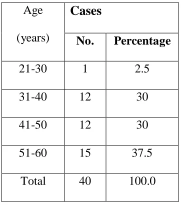
Table 2 Age distribution
Cases
Age
(years) No. Percentage
21-30 1 2.5
31-40 12 30
41-50 12 30
51-60 15 37.5
Total 40 100.0
Observation:
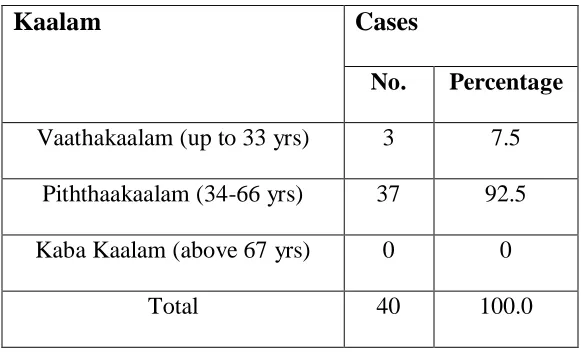
[image:54.612.182.363.433.638.2]Table :3 Kaalam (Life span)
Cases Kaalam
No. Percentage
Vaathakaalam (up to 33 yrs) 3 7.5
Piththaakaalam (34-66 yrs) 37 92.5
Kaba Kaalam (above 67 yrs) 0 0
Total 40 100.0
Observation:
Out of 40 cases, 92.5% of the cases were found to be in Piththakaalam i.e. between 34 –66 years.
4. Paruvakaalam
Among 40 patients, 36(90%) cases were admitted to the trial in kaarkaalam and the remaining 4 (10%) cases were admitted in koothirkaalam
No case was admitted in Munpanikaalam, Pinpanikaalam Elavenil Kaalam, Muthuvenil Kaalam.
5. Diet
Cases Diet
No. Percentage
Vegetarian 3 7.5
Non-Vegetarian 37 92.5
Observation:
Out of 40 cases 92.5% of cases were non-vegetarians and 7.5% of cases vegetarians.
6. Thinai:
Most of the cases (100%) were reported from Neithal thinai.
Neithal thinai, which is responsible for vaatha diseases, may be the reason for the higher incidence.
6. Socio economic status:
Cases
Socia Economic Status
No. Percentage
Poor 10 25 Middle class 25 62.5
Rich 5 12.5 Total 40 100.0
Observation:
62.5% of patients belong to middle class income group.
7. Disturbances in Vaatham:
Out of 40 cases observed viyaanan and samanan were affected in almost all the cases while abanan affected in 5 cases, koorman affected in 3 cases
8. Disturbances in Piththam:
Out of 40 cases, Saathagam was affected in almost all cases.
9.Disturbances in Kabam:
Only Santhigam was affected in all the 40 cases.
10. Envagai Thervugal (Siddha Diagnostic Parameters)
11. Naadi
Cases
Naadi
No. Percentage
Vaatham 0 0
Vaathapitham 40 100 Piththavaatham 0 0 Piththakabam 0 0 Total 40 100
All the cases were diagnosed as having Vathapitham
12. Neikuri
Cases
Spreading Pattern
No. Percentage
Aravenaneendathu 11 18.3
Aazhipolparaviathu 0 0.0
Muththupol Ninrathu 29 81.7
Total 40 100.0
Out of 40 patients, Neerkuri revealed 81.7% were Kaba neer and 18.3% were vaatha neer.
13. Udal Thathukkal:
Enbu was affected in all 40 cases (100%) .
14. Disturbances in Kanmenthiriyam:
Kai was affected in all 40 cases (100%)
15. Duration of Illness:
Cases
Duration of Illness
(month) No. Percentage
Up to 6 months 22 55
7 – 12 months 8 20
13 –18 months 0 0
19 – 24 months 5 12.5
25 – 30 months 0 0.0
Above 30 months 5 12.5
Total 40 100.0
55% of patients reported with 6 months duration of illness
16. Involvement of Shoulder Joints
Cases
Shoulder Joints
No. Percentage
Both Shoulder Joints 8 20
Right Shoulder joint only 12 37.5
Left Shoulder Joint only 20 42.5
17. Clinical Features
Cases
Clinical Feature
No. Percentage
Pain in shoulder 40 100.0
Pain in upper limbs 40 100.0
Numbness 23 57.5
Tenderness 21 52.5
Stiffness 30 75
Headache 15 37.5
Burning sensation of the eyes 10 25
Burning sensation of the tongue 1 2.5
Pain below the umbilicus 5 5
18. Precipitating Factors:
Cases
Factors
No. Percentage
Menopause 11 27.5
Heavy household works 10 25
Occupational related 20 50
History of trauma 2 10
Diabetis Mellitus 7 17.5
19. Occupation:
Cases
Occupation
No. Percentage
Clerk 13 32.5
Teacher 3 7.5
Watchman 2 5
Tailor 2 5
Farmer 10 25
Household works 10 25
Total 40 100.0
20. Results:
Cases
Results
No. Percentage
Clinically Releived 6 15
Good Improvement 27 67.5
Moderate Improvement 5 12.5
No Improvement 2 5
RESULT OF TREATMENT
Clinically Releived Good Improvement Moderate Improvement No Improvement
67.5% 15% 5%
21. Degree of Shoulder Movements:
Degree of Shoulder Movements SI.NO PATIENT NO
BT AT 1. 1515 60 120 2. 1525 50 110 3. AL 6409 110 170 4. AI 5794 120 160 5. AM 3183 60 130 6. 1127 80 180 7. 1141 90 180 8. 1133 70 100 9. AM 7096 120 160 10. AM 7162 120 180 11. 1156 50 120 12. 1640 90 90 13. 1152 110 130 14. 1214 50 70 15. 1231 30 50 0 20 40 60 80 100 120 140 160 180 AFTER TREATMENT
SI.NO 2 4 6 8 10 12 14
BEFORE TREATMENT
DEGREE OF SHOULDER MOVEMENTS BT
Paired t test - comparision of Degree of shoulder movement before and after treatment
---
Variable | Obs Mean Std. Err. Std. Dev. [95% Conf. Interval]
---+---
debt | 40 142.75 8.219641 51.98557 126.1242 159.3758
deat | 40 161.25 5.514404 34.87615 150.0961 172.4039
---+---
diff | 40 18.5 4.662315 29.48707 27.93042 -9.069578
---
Ho: mean(debt - deat) = mean(diff) = 0
Ha: mean(diff) < 0 Ha: mean(diff) ~= 0 Ha: mean(diff) >0 t = -3.9680 t = -3.9680 t = -3.9680 P < t = 0.0002 P > |t| = 0.0003 P > t = 0.9998
22. Genderwise prognosis
RESULT FEMALE MALE TOTAL
C.R 3 3 6
G.I 16 11 27
M.I 3 2 5
N.I 1 1 2
TOTAL 23 17 40
ROUTINE HAEMATOLOGICAL INVESTIGATIONS (OP cases)
BT = Before Treatment AT = After Treatment
TC (Cumm)
DC(Cumm) Hb (mg/dl) Blood Sugar(mg%) Blood Urea(mg %) Blood Cholester ol (mg%) ESR(mm)
BT(%) AT (%) BT AT BT BT AT
P L E M P L E M
BT AT
F PP F PP
BT AT BT AT 1/2h r
1h r
AL6409 6900 7200 50 45 5 0 53 42 5 0 12.5 9.2 198 300 120 192 25 20 252 253 13 40
7300 7100 52 43 5 0 50 46 4 0 12.1 12.8 85 123 79 110 18 20 187 192 4 8
6800 7000 54 42 6 0 51 47 2 0 13.2 12.8 80 107 70 108 22 24 231 235 10 20
8900 6800 56 42 2 0 56 39 5 0 12.5 14.1 108 118 20 19 155 191 2 4
AM3183 7000 7100 50 46 4 0 52 43 5 0 9.7 10.8 85 104 90 107 19 20 244 238 30 63
6400 8000 58 40 2 0 53 44 3 0 9.7 10.8 247 216 18 18 158 163 4 10
AL7832 7000 8000 55 42 3 0 53 45 2 0 9.2 10.5 82 74 22 15 167 147 2 4
AM5387 7000 7200 54 43 3 0 55 42 3 0 11.8 12.1 89 112 85 114 23 21 234 245 4 8
AM7096 6900 7800 55 43 2 0 52 45 3 0 11.1 11.8 125 96 112 31 32 245 236 2 4
AM7162 7000 7400 52 46 4 0 51 46 3 0 10.2 11.4 81 81 24 28 194 155 6 12
7200 8100 58 38 4 0 53 45 2 0 12 13 70 78 32 34 198 187 4 8
7200 7500 56 40 3 1 53 42 5 0 9.2 10.2 113 109 35 28 222 231 10 22
AM9152 8600 7800 56 41 3 0 57 39 3 1 14.6 12.5 99 81 44 23 203 219 2 4
7900 8200 60 37 3 0 53 45 2 0 9.8 12 85 104 16 19 - 240 22 44
AM8976 6800 7500 52 45 3 0 53 42 5 0 9.7 11 95 88 27 18 170 165 8 16
G8071 6200 8000 50 48 2 0 42 48 4 0 13 14 171 - 256 16 31 - 210 4 10
AN7073 7800 6900 53 42 3 2 52 46 2 0 12.5 12 70 78 17 16 154 166 4 8
AN6125 8200 8900 54 42 4 0 51 46 3 0 13.1 12.5 112 124 27 29 189 198 2 4
AO1510 8100 8200 53 44 3 0 52 45 2 1 8.7 10.8 119 110 20 25 268 257 18 36