Copyright © 1998, American Society for Microbiology
Interaction of the Bovine Papillomavirus E6 Protein with the
Clathrin Adaptor Complex AP-1
XIAO TONG,1WERNER BOLL,2TOMAS KIRCHHAUSEN,2ANDPETER M. HOWLEY1*
Department of Pathology1and Department of Cell Biology and Center for Blood Research,2
Harvard Medical School, Boston, Massachusetts 02115 Received 18 August 1997/Accepted 7 October 1997
The E6 gene of the bovine papillomavirus type 1 (BPV-1) is expressed in fibropapillomas caused by BPV-1 and in tissue culture cells transformed by BPV-1. It encodes one of the two major oncoproteins of BPV-1. In this study, we demonstrate an interaction between the BPV-1 E6 protein and AP-1, the TGN (trans-Golgi network)-specific clathrin adaptor complex. AP-1 is a four-subunit protein complex required for clathrin-mediated cellular transport from the TGN. The AP-1/E6 interaction was observed in vitro and in cells. The E6 binding site on AP-1 was mapped to the N-terminal trunk domain of thegsubunit. BPV-1 E6 preferentially associated with membrane-bound AP-1 in cells but not with free cytosolic AP-1. BPV-1 E6 was further shown to be recruited to isolated Golgi membranes and to copurify with clathrin-coated vesicles. The recruitment of BPV-1 E6 to Golgi membranes was AP-1 independent, but the E6 interaction with AP-1 was required for its association with clathrin-coated vesicles. Furthermore, AP-1 proteins could compete with BPV-1 E6 for binding to Golgi membranes, suggesting that the recruitment of BPV-1 E6 and AP-1 to Golgi membranes involves a common factor. Taken together, our results suggest that cytosolic BPV-1 E6 is first recruited to the TGN, where it is then recognized by membrane-bound AP-1 and subsequently recruited into TGN-derived clathrin-coated vesicles. We propose that BPV-1 E6, through its interaction with AP-1, can affect cellular processes involving clathrin-mediated trafficking pathway.
The bovine papillomavirus type 1 (BPV-1) is a small DNA virus that can induce proliferation of dermal fibroblasts and squamous epithelial cells of the skin, causing fibropapillomas in cattle. It can also induce fibroblastic tumors in hamsters, rabbits, and mice and neoplastically transform a variety of rodent cells in tissue culture (for a review, see reference 16). BPV-1 has served as the prototype for the studies of various aspects of papillomavirus biology, such as cellular transforma-tion, viral transcriptional regulatransforma-tion, and viral DNA replica-tion. Genetic studies have mapped the BPV-1 transforming genes to two regions of the viral genome: the E5 gene (14, 35) and the E6 and E7 genes (34, 44). The transforming activity of the BPV-1 E5 protein is principally mediated through the constitutive activation of growth factor receptors. It can di-rectly bind and activate the platelet-derived growth factor b
receptor (11, 27) and the epidermal growth factor receptor (6, 23). BPV-1 E5 also interacts with the 16-kDa subunit of the vacuolar H1-ATPase (12, 13), suggesting that it can also
reg-ulate the acidification of intracellular compartments such as endosomes, lysosomes, and the Golgi apparatus, thereby af-fecting the trafficking of cellular proteins.
The BPV-1 E6 gene product is a relatively basic, 137-amino-acid protein. It contains four Cys-X-X-Cys motifs which are conserved among all papillomavirus E6 proteins (Fig. 1A). Expression of BPV-1 E6 by itself can lead to transformation of mouse C127 cells (25, 34). Unlike the E6 proteins of human papillomavirus types 16 and 18, BPV-1 E6 does not bind to the tumor suppressor protein p53 (43) or stimulate E6AP-medi-ated ubquitination and degradation of p53 (39). BPV-1 E6 has been shown to bind in vitro to a 55-kDa putative calcium-binding protein (ERC-55) (4), although the biological
signifi-cance of this interaction remains to be established. Recently, we have provided evidence that BPV-1 E6 transformation is mediated through its interaction with the focal adhesion pro-tein paxillin (38).
So far, the studies of BPV-1 E6 have been largely focused on its transforming mechanism, and little is known about other potential functions of E6 in the papillomavirus life cycle. In this report, we identify the TGN (trans-Golgi network)-specific clathrin adaptor (AP-1) as a cellular target of BPV-1 E6. Clathrin-coated pits and clathrin-coated vesicles (CCV) are important both in endocytosis and in regulated secretion via the TGN. The assembly of clathrin into coated pits on the TGN and the plasma membrane requires the interaction with a heterotetrameric protein complex called clathrin adaptor (AP). Two types of APs have been identified based on their localization: those localized on the TGN (AP-1) and those localized on the plasma membrane (AP-2). AP-1 and AP-2 are related heterotetramers; AP-1 consists of b1, g, m1, and s1 subunits, whereas AP-2 consists ofb2,a,m2, ands2 subunits. Theb1 andb2 chains are closely related (89% similarity) (20). AP-2 is directly involved in endocytosis. AP-1, which is local-ized at the TGN, facilitates the transport of newly syntheslocal-ized proteins to intracellular compartments such as endosomes and lysosomes (for reviews see references 18 and 32). In addition to promoting clathrin assembly, AP-1 and AP-2 are involved in the sorting step where cargo proteins are recruited into coated pits which lead to their directed vesicular traffic.
The BPV-1 E6–AP-1 interaction described here is the first example of a viral protein directly interacting with a compo-nent of the clathrin-dependent sorting machinery. Our data suggest a model of how BPV-1 E6 proceeds in the TGN-derived trafficking pathway. BPV-1 E6 is first recruited to the TGN membrane; it is then recognized by membrane-bound AP-1 and subsequently recruited into TGN-derived CCV. We propose that the interaction of E6 with AP-1 may affect a vesicular trafficking pathway that could be important to E6
* Corresponding author. Mailing address: Department of Pathology, Harvard Medical School, 200 Longwood Ave., Boston, MA 02115. Phone: (617) 432-2884. Fax: (617) 432-2882. E-mail: phowley@warren .med.harvard.edu.
476
on November 9, 2019 by guest
http://jvi.asm.org/
transformation as well as to other aspects of viral pathogenesis such as evasion of the host immune system.
MATERIALS AND METHODS
GST fusion protein affinity binding.Wild-type and mutant glutathione S-transferase (GST)–E6 fusion proteins were constructed by PCR using pXH800-E6 plasmids (42) as templates and cloned into pGEX-2TK, which contains a cyclic AMP-dependent protein kinase site for in vitro phosphorylation (Pharmacia). To identify BPV-1 E6-associated proteins, C127 cells were labeled with [35S]cysteine-methionine overnight and lysed in lysis buffer (20 mM HEPES [pH 8.0], 1% Nonidet p-40 [NP-40], 150 mM NaCl, 2 mM CaCl2, 10% glycerol, 1 mM phenylmethylsulfonyl fluoride, 2mg of aprotinin per ml). Lysates were centrifuged at 14,000 rpm for 15 min, and the supernantants were precipitated with about 2mg of GST-E6 fusion proteins for 1 h at 4°C. Bound proteins were washed in lysis buffer plus 0.1% sodium dodecyl sulfate (SDS) and analyzed by SDS-polyacrylamide gel electrophoresis (PAGE) and autoradiography. To ob-tain p110, lysates from 50- by 150-mm dishes of mouse L cells were purified by GST-E6 affinity column. Bound proteins were eluted with 20 mM glutathione and further purified by a source 15Q column (Pharmacia). Proteins were eluted with 250 mM NaCl and separated by SDS-PAGE. After transfer to a polyvinyli-dene difluoride (PVDF) membrane (Bio-Rad), about 5mg of p110 was excised and subjected to microsequencing at the Harvard microchemistry facility.
Binding of E6 to AP-1 in vitro and in cells.To confirm E6 binding to the AP-1 complex, 0.5-mg aliquots of AP-1 and AP-2 proteins purified from bovine brain (24) were incubated with 2mg of various GST-E6 fusion proteins. Bound AP-1 and AP-2 were detected by immunoblotting for thebsubunit, using antibody 9A (5). The primary antibody was visualized by enhanced chemiluminescence as instructed by the manufacturer (DuPont NEN). The efficiency of binding was quantitated by using NIH image 1.5 software. To immunoprecipitate E6, Cos-7
cells were transfected with pSGFLAG-E6 plasmids encoding wild-type or mutant E6 proteins (38) by electroporation. Cells were labeled by [35 S]cysteine-methi-onine and harvested 40 h after transfection and lysed in lysis buffer. E6 was precipitated with monoclonal antibody M2 against the FLAG epitope (IBI), and the AP-1 and AP-2 complexes were precipitated with antibody 9A against theb
subunit (5).
In vitro translation of AP subunits.All four subunits were in vitro transcribed and translated by using the TNT T7 polymerase as instructed by the manufac-turer (Promega). Theb1 subunit was generated from pRSET (10), thegandm1 subunits were from BSK (2) (1, 31), and thes1 subunit was from pET5a, which was derived from its cDNA clone (19).
Protein binding of immobilized AP complexes.Aliquots of 5mg of AP-1 and AP-2 complexes purified from bovine brain were digested with serial dilutions of 0.25% trypsin (GIBCO/BRL) for 15 min at room temperature, separated by SDS-PAGE, and transferred to a PVDF membrane. The blots were hybridized with32P-labeled GST-E6 fusion protein in 20 mM HEPES (pH 7.7)–75 mM KCl–0.1 mM EDTA–2.5 mM MgCl2–1% bovine serum albumin–0.05% NP-40 as described previously (17).
Cell fractionation.Cell fractionation was carried out as previously described (2), with the following modifications. Cells were washed once with cold phos-phate-buffered saline and lysed on plate in STM buffer (20 mM HEPES [pH 8.0], 0.25 M sucrose, 10 mM MgCl2, 1 mM phenylmethylsulfonyl fluoride, 2mg of aprotinin per ml, 1 mM dithiothreitol). The cells were disrupted with 30 strokes in a glass Wheaton Dounce homogenizer (VWR). Fraction a (crude cytosol) was prepared by centrifuging the homogenate at 14,000 rpm for 20 min in a micro-centrifuge. The membrane fraction (fraction b) was prepared by extracting the pellet from fraction a with STM buffer plus 0.05% NP-40 and centrifuging at 14,000 rpm. The nuclear fraction (fraction c) was prepared by extracting the insoluble material from the membrane fraction with lysis buffer plus 0.1% SDS. The high-speed cytosol was made by further centrifuging the crude cytosol at 100,0003g for 1 h in a TLA 100.4 rotor (Beckman). All fractions were adjusted
to lysis buffer plus 0.1% SDS before immunoprecipitation. The E6 protein and the AP-1 complex were detected by immunoblot analysis using antibody M2 against the FLAG tag and antibody 100/3 against thegsubunit of AP-1 (Sigma) (1).
Isolation of CCV.CCV were isolated as described previously (24). Briefly, cells were lysed on plate in buffer A (100 mM morpholine ethanesulfonic acid [pH 6.5], 1 mM EGTA, 0.5 mM MgCl2, 0.05% Triton X-100) and sonicated. The lysates were centrifuged at 15,000 rpm in TLA 100.4 rotor (Beckman) for 10 min. The supernatants were further centrifuged at 85,000 rpm for 15 min. The speed supernatants (HSS) were saved for immunoblot analysis, and the high-speed pellet was resuspended in buffer A, mixed with Ficoll-sucrose (12.5% each in buffer A), and centrifuged at 30,000 rpm for 12 min. The supernatants were diluted with 63volume of buffer A and centrifuged at 85,000 rpm for 20 min. The final pellet contained partially purified CCV and was resuspended in buffer A followed by SDS-PAGE and immunoblot analysis.
Golgi membrane binding assay.Crude Cos cell cytosol was made by freezing-thawing cells in binding buffer (20 mM HEPES [pH 8.0], 5 mM MgCl2, 1 mM dithiothreitol, 125 mM potassium acetate) and centrifuging at 14,000 rpm for 15 min. The high-speed cytosol was made by further centrifuging the crude cytosol at 100,0003g for 1 h in a TLA 100.4 rotor (Beckman). For each binding
reaction, 200ml of cytosol (3 mg/ml), 2ml of Golgi membrane from rat liver (7.5 mg/ml) (40), and 100mM GTPgS or 100 mg of brefeldin A per ml, when indicated, were added and incubated at 37°C for 15 min. The membrane-bound proteins were retrieved by centrifuging at 10,000 rpm for 10 min and subjected to SDS-PAGE and immunoblot analysis.
RESULTS
The AP-1 complex is a cellular target for BPV-1 E6. To identify potential BPV-1 E6 cellular targets, BPV-1 E6 was fused to GST and used to bind cellular proteins from 35
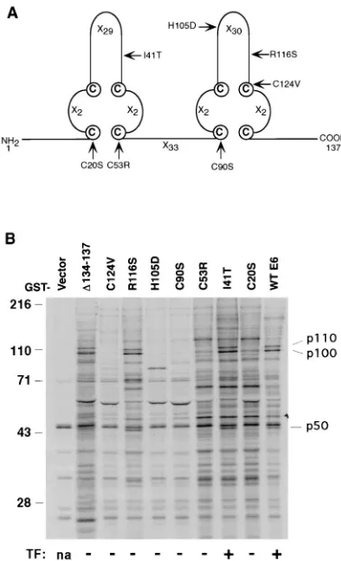
S-labeled cell extracts of C127 cells. E6 mutants which had been tested for the ability to transform C127 cells (42) were included in the experiment as controls of specificity (Fig. 1A). In the GST-E6 affinity binding assay (Fig. 1B), three cellular proteins with apparent molecular masses of 50 kDa (p50), 100 kDa (p100), and 110 kDa (p110) bound to wild-type GST-E6. A transformation-competent E6 mutant (I41T) also bound to the three cellular proteins. Five of the seven nontransforming E6 mutants (C20S, C53R, C90S, H105D, and C124V) did not bind to p50, p100, or p110, whereas the remaining two mutants (R116S andD134-137) did bind. Although the ability of BPV-1 E6 to bind to these three cellular proteins did not correlate with the ability of E6 to transform, these interactions could be necessary though not sufficient for the E6 transformation func-tion, since each of the transformation-competent E6 proteins
FIG. 1. (A) Schematic of the BPV-1 E6 protein structure. The E6 protein has four Cys-X-X-Cys motifs and is predicted to form two zinc-binding sites (41). Xn
indicates the number of residues between the cysteines. The E6 mutants used in this study are also indicated. (B) Identification of BPV-1 E6-associated proteins. 35S-labeled cell lysates from mouse C127 cells were incubated with wild-type (wt) and various mutant GST-E6 fusion proteins. The bound cellular proteins were separated by SDS-PAGE and visualized by autoradiography. The transforming activity (TF) of each E6 mutant and the positions of size standards (in kilodal-tons) and of p110, p100, and p50 are indicated. na, not applicable.
on November 9, 2019 by guest
http://jvi.asm.org/
[image:2.612.74.263.69.380.2]did bind to p50, p100, and p110. Alternatively, these interac-tions could be important for other funcinterac-tions of E6 in the papillomavirus life cycle that are not related to its transforming activity. Therefore, we pursued the identification of these three cellular proteins.
The sequences of two tryptic peptides obtained from puri-fied p110 (148LHDINAQLVEDQGFLDTLK166 and 821RN
VEGQDMLYQSLK834, according to the rat sequence) (20)
were found to match the sequence of theb1 subunit of AP-1. The p50 and p100 polypeptides were further shown by immu-noblotting to be them1 andgsubunits, respectively (data not shown; thes1 subunit is a small protein of 19 kDa which ran out of the gel in Fig. 1B). An antibody against theasubunit of AP-2 (29) failed to detect any band in the same experiment (data not shown). We therefore conclude that GST-E6 can interact with the intact AP-1 complex but not with AP-2 from cell lysates.
BPV-1 E6 interacts with AP-1 in vitro and in cells. To confirm the interaction between BPV-1 E6 and the AP-1 com-plex, equal amounts of AP-1 and AP-2 purified from bovine brain coated vesicles (24) were incubated with GST-E6, and the bound proteins were analyzed by immunoblotting with a monoclonal antibody which recognizes theb1 andb2 subunits of AP-1 and AP-2 (5). Wild-type BPV-1 E6 bound to about 30% of input AP-1 and about 10% of input AP-2 (Fig. 2). Consistent with the results shown in Fig. 1B, mutant GST-E6 (H105D), which did not precipitate p110, p100, or p50, failed to bind to either AP-1 or AP-2 (Fig. 2). Similarly, GST-E6 (D134-137), which did interact with p110, p100, and p50 in Fig. 1B, bound to about 10% of input AP-1 but not to AP-2. Although some interaction between GST-E6 and AP-2 was detected in this assay, the significance of this observation is not clear, since the AP-2 complex was not present in the cellular proteins retrieved by GST-E6 or by E6 coimmunoprecipitation (Fig. 1 and 3).
To test whether the BPV-1 E6–AP-1 interaction occurs in mammalian cells, wild-type and mutant E6 proteins with an N-terminal FLAG epitope tag were expressed in Cos cells by transient transfection and immunoprecipitated with an anti-body against the FLAG epitope. The AP-1 and AP-2 com-plexes were also immunoprecipitated from vector-transfected Cos cells by using the antibody against theb1 andb2 subunits (5). AP-1 and AP-2 proteins purified from bovine brain were run on the same gel and stained to serve as standards in order to identify the position of each subunit (data not shown). Wild-type E6 and mutant E6 (D134-137), which had been shown to bind to AP-1 in the GST affinity assay, specifically coprecipitated the AP-1 but not the AP-2 complex from Cos cells (Fig. 3). Thes1 subunit comigrated with wild-type E6 but was readily visible when the smaller mutant E6 (D134-137) was used. Paxillin, a previously identified E6-binding protein (38), was also coprecipitated by wild-type E6 (Fig. 3). Mutant E6
(H105D), which did not bind AP-1 in the in vitro assays (Fig. 1 and 2), failed to coprecipitate the AP subunits (Fig. 3).
The AP-1–E6 interaction is mediated through the N-termi-nal trunk of the g subunit of AP-1. We next used several approaches to determine which one of the AP-1 subunits was responsible for the interaction with BPV-1 E6. In the first experiment, each subunit of the AP-1 complex was35S labeled
by in vitro translation and tested in the GST-E6 binding assay. Wild-type E6 bound to about 20% of inputgandb1 subunit but did not bind to them1 or thes1 subunit. As a control for specificity, we used mutant E6 (H105D), which does not inter-act with AP-1. We found that E6 (H105D) did not bind to any of the AP-1 subunits in this assay (Fig. 4).
[image:3.612.108.231.70.132.2]The interaction between AP-1 and E6 was further studied by probing immobilized AP-1 subunits with radiolabeled GST-E6. The AP-1 and AP-2 complexes purified from bovine brain were separated by SDS-PAGE and transferred to a PVDF mem-brane. Purified wild-type GST-E6 protein was phosphorylated in vitro on the protein kinase site constructed into the fusion protein and used to probe the AP subunits immobilized on the
[image:3.612.371.487.514.682.2]FIG. 2. In vitro interaction between BPV-1 E6 and the AP-1 complex. AP-1 and AP-2 complexes purified from bovine brain were incubated with various GST-E6 fusion proteins. The AP complexes were detected by immunoblotting with monoclonal antibody 9A against thebsubunit. wt, wild type.
FIG. 3. Interaction between BPV-1 E6 and AP-1 in cells. Cos cells were transiently transfected with vector, FLAG-tagged wild-type (wt) E6, mutant E6 (D134-137), or mutant E6 (H105D) and labeled with [35S]methionine-cysteine. Cell lysates were immunoprecipitated for the AP-1 and AP-2 complexes by using monoclonal antibody 9A against thebsubunit and for E6 by using the FLAG antibody. The positions of each AP subunit, paxillin, and E6 are indicated.
FIG. 4. Interaction between BPV-1 E6 and the AP-1 subunits. Each of the four subunit of AP-1 was35S labeled by in vitro translation and incubated with wild-type GST-E6 (wt E6) and mutant GST-E6 (H105D) fusion proteins. The bound proteins were separated by SDS-PAGE and visualized by autoradiogra-phy. Sizes are indicated in kilodaltons.
on November 9, 2019 by guest
http://jvi.asm.org/
PVDF membrane. As shown in Fig. 5A, GST-E6 bound strongly to thegsubunit of AP-1 and weakly to theb1 subunit (control lane). In contrast, GST-E6 bound only weakly to the
b2 subunit of AP-2 and no other subunits (Fig. 5B, control lane). No binding to any AP subunits was detected when the nonbinding mutant GST-E6 (H105D) was used instead (data not shown). These results indicate that thegsubunit is able to directly mediate the interaction between the AP-1 complex and BPV-1 E6. Although thebsubunit could also bind E6 in vitro (Fig. 4 and 5), it is unlikely to mediate the AP-1–E6 interaction in vivo, since it could not account for the specificity of E6 for AP-1 in cells (Fig. 1 and 3). The observed binding of BPV-1 E6 to thebsubunit could be due to improper folding or denaturation of the polypeptide in vitro, which may be respon-sible for the binding of AP-2 to E6 in vitro as shown in Fig. 2. However, the significance of the in vitro binding observed between AP-2 and BPV-1 E6 is unclear since we did not detect it in the cells (Fig. 1 and 3).
The AP complexes can be proteolytically cleaved into two portions: the “head” and the “ear.” The head is composed of intactmandssubunits and the 70-kDa N-terminal trunk ofb1 and g(for AP-1) or b2 and a (for AP-2); the ear is to the 30-kDa C-terminal portion of each of the large subunits. To determine which domain of thegsubunit mediated E6 bind-ing, purified AP complexes were partially digested with a serial dilution of trypsin, immobilized on a PVDF membrane, and probed with32P-labeled GST-E6. As shown in Fig. 5, GST-E6
bound to a 70-kDa proteolytic polypeptide corresponding to the N-terminal trunk of the g subunit. The identity of the 70-kDa band was confirmed by immunoblot analysis using an antibody against the gsubunit (data not shown) (1). In con-trast, GST-E6 failed to bind to any proteolytic products of either theb1 orb2 subunit (Fig. 5), suggesting that the binding site on these subunits may be destroyed by the protease treat-ment.
BPV-1 E6 coimmunoprecipitates with the membrane-asso-ciated AP-1 complex.In cells, the AP-1 complex cycles between two pools: the crude cytosolic AP-1 (which consists of free AP-1 and AP-1 assembled into CCV) and the TGN mem-brane-bound AP-1 (30). To determine with which cellular AP-1 fraction E6 is associated, Cos cells were transfected with FLAG-tagged E6 and fractionated into a crude cytosolic frac-tion (fracfrac-tion a, obtained by centrifuging cell lysates at 14,000 rpm for 30 min, containing free AP-1 and AP-1 in CCV), a membrane fraction (fraction b, obtained by extracting the pel-let from fraction a in 0.05% Triton X-100, containing TGN membrane-bound AP-1), and a nuclear fraction (fraction c, obtained by extracting the pellet from fraction b in 1% Triton X-100 plus 0.1% SDS) (2). Extracts from each fraction were
subjected to immunoprecipitation and immunoblot analysis. The majority of AP-1 complex was present in the crude cyto-solic and membrane-bound fractions, as judged by immuno-precipitation of AP-1 (Fig. 6A, upper panel). The results were confirmed by direct immunoblot of the lysates from each frac-tion, using an antibody against thegsubunit (data not shown). Furthermore, transfection of either wild-type E6 or mutant E6 does not change AP-1 levels or distribution in the cell (Fig. 7A and data not shown). The distribution of E6 was similar to what has been previously reported in E6-transformed C127 cells (2) (Fig. 6A, middle panel), although we did observe an increased amount of cytosolic E6, perhaps due to the high level of E6 expression in transfected Cos cells. Importantly, AP-1 in the membrane fraction coprecipitated with wild-type E6 but not with the nonbinding mutant E6 (C90S) (Fig. 6A, lower panel, lane b), further demonstrating an specific in vivo inter-action between AP-1 and E6. In contrast, immunoprecipitation of cytosolic wild-type E6 brought down very little of the cyto-solic AP-1 complex (Fig. 6A, lower panel, lane a). This was unexpected since similar amounts of AP-1 and E6 were present in the cytosolic fraction and the membrane fraction. Since the crude cytosol used in this experiment contained free cytosolic AP-1 as well as AP-1 assembled into CCV, we next tested E6–AP-1 interaction in high-speed cytosols which had been centrifuged at 100,0003g for 1 h to obtain free cytosolic AP-1.
As shown in Fig. 6B, AP-1 was readily immunoprecipitated from the high-speed cytosol, using an antibody against its b
[image:4.612.316.542.68.159.2]subunit. However, E6 failed to coprecipitate with AP-1 under such conditions. These results suggest that in cells E6 cannot
[image:4.612.93.244.72.172.2]FIG. 5. Mapping of the E6 binding domain on AP-1. AP-1 and AP-2 proteins purified from bovine brain were partially digested by serially diluted trypsin, separated by SDS-PAGE, immobilized on a PVDF membrane, and probed by 32P labeled GST-E6.
FIG. 6. Study of the interaction between BPV-1 E6 and AP-1 by cell frac-tionation. (A) Lysates from Cos cells transiently transfected with wild-type (wt) E6 or mutant E6 (C90S) were fractionated into crude cytosolic (a), membrane (b), and nuclear (c) fractions. Each fraction was analyzed by immunoprecipita-tion (IP) and immunoblotting as indicated. (B) The crude cytosolic fracimmunoprecipita-tion from panel A was further subjected to centrifugation at 100,0003g for 1 h to generate
a high-speed cytosol which was immunoprecipitated for AP-1 by using an anti-body against thebsubunit and for E6 by using the anti-FLAG antibody M2. The immunocomplexes were then assayed by immunoblotting with an antibody against thegsubunit of AP-1 and the anti-FLAG antibody M2 for E6.
FIG. 7. Assembly of BPV-1 E6 into CCV. (A) CCV were isolated from Cos cells transfected with wild-type (wt) or mutant E6 proteins (C90S). The distri-bution of AP-1 and E6 was determined by immunoblotting. (B) CCV purified from bovine brain were incubated with35S-labeled, in vitro-translated E6 and immunoprecipitated for clathrin heavy chain by using monoclonal antibody X-22 (3). AP-1 was detected by immunoblotting against thegsubunit, and E6 was detected by autoradiography.
on November 9, 2019 by guest
http://jvi.asm.org/
[image:4.612.357.499.590.666.2]bind efficiently to free cytosolic AP-1 and that their interaction probably occurs more favorably when both proteins are on the membranes.
BPV-1 E6 copurifies with clathrin-coated vesicles. Even though the E6 antibody failed to retrieve any cytosolic AP-1, it was possible that E6 was associated with AP-1 in CCV but that the presence of the clathrin coats masked the accessibility of the antibody to epitopes localized within the coats. To test this possibility, CCV were purified from Cos cells transfected with either wild-type E6 or mutant E6 (C90S) following standard procedures (24). Fractions containing purified CCV were an-alyzed by immunoblotting for the AP-1gsubunit and the E6 protein. As shown in Fig. 7A, AP-1 was found in the HSS, which contains free cytosolic AP proteins, and in the pellet, which is enriched for CCV (24). The enrichment was estimated to be 15-fold by comparing the amount of AP-1 found in the HSS to that found in CCV fractions after normalizing the total protein concentration (data not shown). The distributions of AP-1 were similar in cells transfected with wild-type E6 and mutant E6 (C90S). A large proportion of wild-type E6 was also found in the CCV fraction. In contrast, very little of the non-binding mutant E6 (C90S) was detected in the CCV fraction (Fig. 7A). From these data, we conclude that BPV-1 E6 copu-rifies with CCV and that interaction with AP-1 is probably required for its assembly into CCV.
We next investigated whether soluble E6 could interact with AP-1 which has been incorporated into CCV. CCV purified from bovine brain (24) were incubated with in vitro-translated
35S-labeled BPV-1 E6. After incubation, the mixture was
im-munoprecipitated with a monoclonal antibody against the clathrin heavy chain (3) to isolate CCV. The immunoprecipi-tates were analyzed by immunoblotting for the gsubunit of AP-1 and autoradiography for E6. The clathrin antibody co-precipitated the AP-1 complex but not the E6 protein (Fig. 7B). Therefore, under these conditions, BPV-1 E6 is unable to bind to AP-1 when it is enclosed in the CCV, suggesting that E6 interacts with AP-1 before it is assembled into CCV.
BPV-1 E6 can be recruited to isolated Golgi membranes. One possible location for the AP-1–E6 interaction to occur is the TGN membrane. AP-1 is abundant on the TGN membrane and is required for the formation of CCV derived from the TGN. Therefore, we investigated whether BPV-1 E6 and AP-1 were colocalized at the TGN by immunofluorescence. Unfor-tunately, due to the low level of E6 protein expressed in mouse C127 cells stably transformed by FLAG-tagged E6, the anti-body did not detect any E6 signal by immunofluorescence. Transient overexpression of FLAG-tagged BPV-1 E6 in Cos cells and in C127 cells resulted in diffused cytoplasmic staining even after mild detergent treatment (reference 38 and data not shown), which made it difficult to assess its colocalization with AP-1. We also attempted to study E6 localization by immuno-electron microscopy, but the antibody failed to recognize the E6 protein in the electron microscopic experiments.
As an alternative approach, we tested biochemically whether BPV-1 E6 can be recruited onto the TGN membrane in vitro. It has been shown that AP-1 complex can bind to TGN mem-branes in the presence of cytosol and GTPgS. One character-istic of this reaction is that it can be inhibited by the fungal metabolite brefeldin A, indicative of an ADP-ribosylation fac-tor (ARF)-dependent process (33, 37, 40). Thus, purified Golgi membrane from rat liver was incubated with crude cytosol made from Cos cells expressing E6. The crude cytosol (Fig. 6A) was used as a source for both AP-1 and E6 proteins. After incubation at 37°C, the Golgi membranes were pelleted by centrifugation, and the AP-1 and E6 recruited to the mem-brane were detected by immunoblotting. Incubation of cytosol
at 37°C caused a small amount of AP-1 to precipitate out of the solution as previously reported (40) (Fig. 8A, lane 1). Addition of GTPgS and Golgi membrane to cytosol resulted in an in-crease of AP-1 in the pellet, presumably due to AP-1 binding to the Golgi membrane (Fig. 8A, lane 2). As expected, the binding was blocked by brefeldin A (Fig. 8A, lane 3). In the same experiment, wild-type E6 was also recovered from the Golgi membrane upon addition of GTPgS, and treatment with brefeldin A prevented its association with the membrane. These results suggest that the wild-type BPV-1 E6 protein can be recruited to the Golgi membrane.
The crude cytosol used in Fig. 8A contained both free cyto-solic E6 and E6 recruited into CCV. We next tested E6 pro-teins from high-speed cytosol which was free of CCV in the following experiments to confirm that free cytosolic E6 could bind to Golgi membranes. High-speed cytosol free of CCV was made as described for Fig. 6B. Both AP-1 and wild-type E6 from the high-speed cytosol could bind to the Golgi mem-branes, similar to the results obtained using crude cytosol (Fig. 8B, lanes 1 and 2).
Addition of excess purified AP-1 to the assay led to an increase in the amount of membrane recruited AP-1 (Fig. 8B, upper panel, lane 3). In contrast, the amount of E6 bound to the Golgi membranes was decreased concomitantly (Fig. 8B, lower panel, lane 3). These results indicate that E6 and AP-1 may compete for a common factor required for their binding to the Golgi membrane. Furthermore, recruitment of E6 to the Golgi membrane does not seem to require interaction with the AP-1 complex since mutant E6 (C90S), which does not interact with AP-1, bound to Golgi membranes as efficiently as the wild-type E6 (Fig. 8B, lanes 4 and 5). As for wild-type E6, the binding of E6 (C90S) to Golgi membranes was competed off by excess AP-1.
DISCUSSION
Here we demonstrated an interaction between the BPV-1 E6 protein and the AP-1 complex both in vitro and in cells. The interaction is highly specific in that certain single-point muta-tions in BPV-1 E6 greatly impair the binding. BPV-1 E6 pref-erentially interacts with the AP-1 complex but not with the AP-2 complex despite the extensive homology between the subunits of the two protein complexes. Several lines of evi-dence suggest a model of how BPV-1 E6 may proceed in the TGN-derived trafficking pathway. We have found that (i) BPV-1 E6 preferentially interacts with membrane-bound AP-1 but does not bind to free cytosolic AP-1 in cells, (ii) it can bind to isolated Golgi membrane independent of AP-1, and (iii) it
FIG. 8. Recruitment of BPV-1 E6 to Golgi membranes. (A) Isolated rat Golgi membrane (mem), GTPgS, brefeldin A (BFA), and crude cytosol from Cos cells transfected with wild-type (wt) E6 were mixed as indicated. The mem-brane-bound AP-1 and E6 proteins were pelleted by centrifugation and detected by immunoblotting. (B) High-speed cytosols from Cos cells expressing wild-type E6 or mutant E6 (C90S) were used in the membrane binding assay; 2mg of AP-1 purified from bovine brain was added to the reaction mixture where indicated.
on November 9, 2019 by guest
http://jvi.asm.org/
copurifies with CCV but is unable to bind to AP-1 once AP-1 is incorporated into CCV. Based on these observations, we propose the following sequence of events. BPV-1 E6 is first recruited to the TGN membrane in an AP-1-independent man-ner; it is then recognized by the membrane-bound AP-1. Upon assembly of the clathrin coat, it is recruited into TGN-derived CCV together with AP-1. It should be noted that although E6 preferentially interacts with membrane-bound AP-1 in cells, GST-E6 can bind to purified AP-1 in vitro. Thus, the absence of the interaction of E6 with cytosolic AP-1 is likely due to the function of an inhibitory factor or the relative low concentra-tion of the two proteins in the cytosol, and the enrichment of AP-1 and E6 on the Golgi membrane is a prerequisite for their interaction. It is unclear how BPV-1 E6 is recruited to the Golgi membrane. The fact that its recruitment is sensitive to brefeldin A suggests that like AP-1 (37), an ARF may be involved in the process. Even though AP-1 and E6 bind to Golgi membranes independently, they may require a common factor for their membrane recruitment, since excess of AP-1 can compete E6 in the Golgi membrane binding assay. The ARFs, as mentioned above, could be limiting factors; another possibility is that AP-1 and E6 compete for the same binding sites on the Golgi membranes.
The E6–AP-1 interaction is the first example of a viral pro-tein interacting with a component of the clathrin-dependent sorting machinery, and this interaction could have important functional consequences. Genetic evidence has indicated that the AP-1 complex may play an important role in the regulation of cell proliferation and differentiation. In Caenorhabditis
el-egans, mutation in the TGNm1-chain gene (UNC-101) results in poor viability and uncoordinated movements. UNC-101/m1 is proposed to be involved in the regulation of LET-23 (21), a tyrosine kinase related to the epidermal growth factor recep-tor. In humans, a member of theb-subunit gene family local-ized on chromosome 22q12 is deleted in a number of menin-giomas (28). Inactivation of these genes may lead to a loss of growth control due to disruption of growth hormone receptor internalization and/or degradation. In this context, BPV-1 E6 could affect signal transduction by disrupting AP-1 function and thus interfering with the AP-1-dependent delivery of lyso-somal enzymes that are required for receptor downregulation. Alternatively, BPV-1 E6 could affect the biosynthesis and delivery of cell proteins which utilize the vesicular trafficking pathway. One candidate could be the major histocompatibility complex (MHC) class II complex. Natural infection of papil-lomavirus is poorly immunogenic, possibly reflecting the ability of the virus to somehow evade the host immune system (9). Furthermore, human warts are more prevalent in conditions that depress T-cell functions, and regression of warts is asso-ciated with infiltration by CD41T cells (7, 8). One possible
mechanism for BPV-1 to downregulate the CD41T-cell
re-sponse could involve interference with the MHC class II-re-stricted antigen presentation. In fact, the biosynthesis of class II molecules includes many components of the endocytic path-way. The cytoplasmic tail of the class II Ii chain and HLA-DM, an accessory molecule required for the formation of antigen-presenting class II molecules, each contain sorting signals which could mediate their efficient delivery to the endocytic pathway (22, 26, 36). Similar signals have been shown to me-diate AP-1-dependent endocytic transport (15). Therefore, it is possible that BPV-1 E6 affects the antigen presentation of virus-infected cells by impairing the maturation of MHC class II molecules through its interaction with the AP-1 complex.
ACKNOWLEDGMENTS
We are grateful to H. Ploegh and T. Rapoport for helpful sugges-tions and critically reading of the manuscript.
X.T. is supported by the cancer research fund of the Damon Run-yon-Walter Winchell Foundation. This research was supported by NIH grants P01CA50661-08 (P.M.H.) and GM36548 (T.K.).
REFERENCES
1. Ahle, S., A. Mann, U. Eichelsbacher, and E. Ungewickell. 1988. Structural relationships between clathrin assembly proteins from the Golgi and the plasma membrane. EMBO J. 7:919–929.
2. Androphy, E. J., J. T. Schiller, and D. R. Lowy. 1985. Identification of the protein encoded by the E6 transforming gene of bovine papillomavirus. Science 230:442–445.
3. Brodsky, F. M. 1985. Clathrin structure characterized with monoclonal an-tibodies. I. Analysis of multiple antigenic sites. J. Cell Biol. 101:2047–2054. 4. Chen, J. J., C. E. Reid, V. Band, and E. J. Androphy. 1995. Interaction of papillomavirus E6 oncoproteins with a putative calcium-binding protein. Science 269:529–531.
5. Clairmont, K. B., W. Boll, M. Ericsson, and T. Kirchhausen. The hinge-ear domain of theb-chains is required for incorporation into clathrin-coated pits and coated vesicles in cells. Submitted for publication.
6. Cohen, B. D., D. J. Goldstein, L. Rutledge, W. C. Vass, D. R. Lowy, R. Schlegel, and J. T. Schiller.1993. Transformation-specific interaction of the bovine papillomavirus E5 oncoprotein with the platelet-derived growth fac-tor recepfac-tor transmembrane domain and epidermal growth facfac-tor recepfac-tor cytoplasmic domain. J. Virol. 67:5303–5311.
7. Coleman, N., H. D. L. Birley, A. M. Renton, N. F. Hanna, B. K. Ryatt, M. Byrne, D. Taylor-Robinson, and M. A. Stanley.1994. Immunological events in regressing genital warts. Am. J. Clin. Pathol. 102:768–774.
8. Coleman, N., and M. A. Stanley. 1994. Analysis of HLA-DR expression on keratinocytes in cervical neoplasia. Int. J. Cancer 56:314–319.
9. Frazer, I. H. 1996. Immunology of papillomavirus infection. Curr. Opin. Immunol. 8:484–491.
10. Gallusser, A., and T. Kirchhausen. 1993. Theb1 andb2 subunits of the AP complexes are the clathrin coat assembly components. EMBO J. 12:5237– 5244.
11. Goldstein, D. J., T. Andresson, J. J. Sparkowski, and R. Schlegel. 1992. The BPV-1 E5 protein, the 16 kDa membrane pore-forming protein and the PDGF receptor exist in a complex that is dependent on hydrophobic trans-membrane interactions. EMBO J. 11:4851–4859.
12. Goldstein, D. J., M. E. Finbow, and T. Andersson. 1991. Bovine papilloma-virus E5 oncoprotein binds to the 16K component of the vacuolar H-AT-Pase. Nature (London) 352:347–349.
13. Goldstein, D. J., and R. Schlegel. 1990. The E5 oncoprotein of bovine papillomavirus binds to a 16 kd cellular protein. EMBO J. 9:137–145. 14. Groff, D. E., and W. D. Lancaster. 1986. Genetic analysis of the 39early
region transformation and replication functions of bovine papillomavirus type 1. Virology 150:221–230.
15. Honing, S., J. Griffith, H. J. Geuze, and W. Hunziker. 1996. The tyrosine-based lysosomal targeting signal in lamp-1 mediates sorting into Golgi-derived clathrin-coated vesicles. EMBO J. 15:5230–5239.
16. Howley, P. M. 1996. Papillomavirinae: the viruses and their replication, p. 2045–2076. In B. N. Fields, P. M. Howley, and D. M. Knipe (ed.), Virology, 3rd ed. Lippincott-Raven Publishers, Philadelphia, Pa.
17. Kaelin, W. J., D. C. Pallas, J. A. DeCaprio, F. J. Kaye, and D. M. Livingston. 1991. Identification of cellular proteins that can interact specifically with the T/E1A-binding region of the retinoblastoma gene product. Cell 64:521–532. 18. Kirchhausen, T. 1993. Coated pits and coated vesicles—sorting it all out.
Curr. Opin. Struct. Biol. 3:182–188.
19. Kirchhausen, T., A. Davis, S. Frucht, B. Greco, G. Payne, and B. Tubb. 1991. AP17 and AP19, the mammalian small chains of the clathrin-associated protein complexes show homology to Yap17p, their putative homolog in yeast. J. Biol. Chem. 266:11153–11157.
20. Kirchhausen, T., K. L. Nathanson, W. Matsui, A. Vaisberg, E. P. Chow, C. Burne, J. H. Keen, and A. E. Davis.1989. Structural and functional division into two domains of the (100- to 115-kDa) chains of the clathrin-associated protein complex AP-2. Proc. Natl. Acad. Sci. USA 86:2612–2616. 21. Lee, J., G. D. Jongeward, and P. W. Sternberg. 1994. unc-101, a gene
required for many aspects of Caenorhabditis elegans development and be-havior, encodes a clathrin-associated protein. Genes Dev. 8:60–73. 22. Marks, M. S., P. A. Roche, E. V. Donselaar, L. Woodruff, P. J. Peters, and
J. S. Bonifacino.1995. A lysosomal targeting signal in the cytoplasmic tail of thebchain directs HLA-DM to MHC class II compartments. J. Cell Biol. 131:351–369.
23. Martin, P., W. C. Vass, J. T. Schiller, D. R. Lowy, and T. J. Velu. 1989. The bovine papillomavirus E5 transforming protein can stimulate the transform-ing activity of EGF and CSF-1 receptors. Cell 59:21–32.
24. Matsui, W., and T. Kirchhausen. 1990. Stabilization of clathrin coats by the core of the clathrin-associated protein complex AP-2. Biochemistry 29:10791–10798.
on November 9, 2019 by guest
http://jvi.asm.org/
25. Neary, K., and D. DiMaio. 1989. Open reading frames E6 and E7 of bovine papillomavirus type 1 are both required for full transformation of mouse C127 cells. J. Virol. 63:259–266.
26. Odorizzi, C. G., I. S. Trowbridge, L. Xue, C. R. Hopkins, C. D. Davis, and J. F. Collawn.1994. Sorting signals in the MHC class II invariant chain cytoplasmic tail and transmembrane region determine trafficking to an en-docytic processing compartment. J. Cell Biol. 126:317–330.
27. Petti, L., and D. DiMaio. 1992. Stable association between the BPV E5 transforming protein and activated platelet-derived growth factor in trans-formed mouse cells. Proc. Natl. Acad. Sci. USA 89:6736–6740.
28. Peyrard, M., I. Fransson, Y. G. Xie, F. Y. Han, M. H. Ruttledge, S. Swahn, J. E. Colins, I. Dunham, V. P. Collins, and J. P. Dumanski.1994. Charac-terization of a new member of the human b-adaptin gene family from chromosome 22q12, a candidate meningioma gene. Hum. Mol. Genet. 3: 1393–1399.
29. Robinson, M. S. 1987. 100-kD coated vesicle proteins: molecular heteroge-neity and intracellular distribution studied with monoclonal antibodies. J. Cell Biol. 104:887–895.
30. Robinson, M. S. 1992. Adaptins. Trends Cell Biol. 2:293–297.
31. Robinson, M. S. 1993. Assembly and targeting of adaptin chimeras in trans-fected cells. J. Cell Biol. 123:67–77.
32. Robinson, M. S. 1994. The role of clathrin, adaptors and dynamin in endo-cytosis. Curr. Opin. Cell Biol. 6:538–544.
33. Robinson, M. S., and T. E. Kreis. 1992. Recruitment of coat proteins onto Golgi membranes in intact and permeabilized cells: effects of brefeldin A and G protein activators. Cell 69:129–138.
34. Schiller, J. T., W. C. Vass, and D. R. Lowy. 1984. Identification of a second
transforming region in bovine papillomavirus DNA. Proc. Natl. Acad. Sci. USA 81:7880–7884.
35. Schiller, J. T., W. C. Vass, K. H. Vousden, and D. R. Lowy. 1986. E5 open reading frame of bovine papillomavirus type 1 encodes a transforming gene. J. Virol. 57:1–6.
36. Schmid, S. L., and M. R. Jackson. 1994. Making class II presentable. Nature (London) 369:103–104.
37. Stamnes, M. A., and J. E. Rothman. 1993. The binding of AP-1 clathrin adaptor particles to Golgi membranes requires ADP-ribosylation factor, a small GTP-binding protein. Cell 73:999–1005.
38. Tong, X., and P. M. Howley. 1997. The bovine papillomavirus E6 oncopro-tein interacts with paxillin and disrupts the actin cytoskeleton. Proc. Natl. Acad. Sci. USA 94:4412–4417.
39. Tong, X. 1997. Unpublished data.
40. Traub, L. M., J. A. Ostrom, and S. Kornfeld. 1993. Biochemical dissection of AP-1 recruitment onto Golgi membranes. J. Cell Biol. 123:561–573. 41. Ullman, C. G., P. I. Haris, D. A. Galloway, V. C. Emery, and S. J. Perkins.
1996. Predicteda-helix/b-sheet secondary structures for the zinc-binding motifs of human papillomavirus E7 and E6 proteins by consensus prediction averaging and spectroscopic studies of E7. Biochem. J. 319:229–239. 42. Vousden, K. H., E. J. Androphy, J. T. Schiller, and D. R. Lowy. 1989.
Mutational analysis of bovine papillomavirus E6 gene. J. Virol. 63:2340– 2342.
43. Werness, B. A., A. J. Levine, and P. M. Howley. 1990. Association of human papillomavirus types 16 and 18 E6 proteins with p53. Science 248:76–79. 44. Yang, Y.-C., H. Okayama, and P. M. Howley. 1985. Bovine papillomavirus
contains multiple transforming genes. Proc. Natl. Acad. Sci. USA 82:1030– 1034.