DESIGN AND EVALUATION OF MICROSPONGE
DRUG DELIVERY OF PSORALEN - ISOLATED FROM
Psoralea corylifolia
Dissertation submitted to
THE TAMILNADU Dr. M.G.R MEDICAL UNIVERSITY, CHENNAI
in partial fulfillment of the requirements
for the award of the degree of
MASTER OF PHARMACY IN
PHARMACEUTICS
DEPARTMENT OF PHARMACEUTICS
PERIYAR COLLEGE OF PHARMACEUTICAL SCIENCES FOR GIRLS
THIRUCHIRAPPALLI – 620021
MARCH – 2008
Mrs.K.Reeta Vijaya Rani, M.Pharm. (Ph.D),
Assistant Professor,
Department of Pharmaceutics,
Periyar College of Pharmaceutical Sciences for Girls, Trichy – 620021.
CERTIFICATE
This is to certify that the dissertation entitled
“DESIGN AND EVALUATION OF MICROSPONGE DRUG DELIVERY OF PSORALEN – ISOLATED FROM Psoralea corylifolia”
submitted by S. Eugine Leo Prakash, B.Pharm to The Tamilnadu Dr.M.G.R Medical University, Chennai in partial fulfillment for the award of the degree of MASTER OF PHARMACY is an independent bonafied work of the candidate carried out in the Department of Pharmaceutics, Periyar College of Pharmaceutical Sciences for Girls, Trichy-21, during the academic year 2007 - 2008 under my direct guidance and supervision and to my full satisfaction.
Place: Trichy-21.
Dr. R.Senthamarai, M.Pharm, Ph.D., Principal,
Periyar College of Pharmaceutical Sciences for Girls, Trichy – 620021
CERTIFICATE This is to certify that the dissertation entitled
“DESIGN AND EVALUATION OF MICROSPONGE DRUG DELIVERY OF PSORALEN – ISOLATED FROM Psoralea corylifolia”
submitted by S. Eugine Leo Prakash B.Pharm to The Tamilnadu Dr.M.G.R Medical University, Chennai in partial fulfillment for the award of the degree of Master of pharmacy is an independent bonafied work of the candidate carried out under the guidance of Mrs.K.Reeta Vijaya Rani M.Pharm, (Ph.D) Asst. Professor in the Department of Pharmaceutics, Periyar College of Pharmaceutical Sciences for Girls, Trichy-21, during the academic year 2007- 2008 under my direct guidance and supervision.
I recommend this Research Work for acceptance as Project for the partial fulfillment of the degree of “MASTER OF PHARMACY” of the Department of Pharmaceutics, Periyar College of Pharmaceutical Science for Girls, for the year March 2008.
Place: Trichy-21.
ACKNOWLEDGEMENT
Though worlds are seldom sufficient to express gratitude and feelings, it somehow gives me an opportunity to acknowledge those who helped me during the tenure of my study. The work of dissertation preparation was a daunting task and fascinating experience.
I deem it is a matter of great privilege to express my deepest sense of gratitude and inobtrusive to my guide Mrs. K. Reeta Vijaya Rani , my foremost duty in expressing my sincere indebtness to her constant help, affection and valuable guidance during the course of present investigation.
I wish to thank Dr. R. Senthamarai, Principal, Periyar College of Pharmaceutical Sciences for Girls, Trichy for providing me support with constant encouragement.
I extent my sincere thanks to our honorable chairman Dr. K. Veeramani, Chancellor, Periyar Maniyammai University and Thiru. Gnana Sebastian, Correspondent, for all the facilities provided to us at institution and enabling to do this work.
I offer my warmest acknowledgement to my respected Head, Prof T.N.K. Suriya Prakash, Department of Pharmaceutics, his continuous guidance and moral support from date of admission to complete my project work and have always propelled me to perform better.
I wish to express my sincere thanks to The Tamilnadu State Council for Science & Technology, Chennai for granting me scholarship to carry out the work.
I extend heart full of thanks to my Department staff members, Mrs. R. Lathaeswari and
Ms. N. Pavala Rani for their constant help to make my project successful.
It is my privilege and honor to extend my sincere gratitude to Dr. S. Akilandeswari., Head, Dept of pharmaceutical chemistry, Mr. K. Nagarajan, Asst. Prof Dept of pharmaceutical chemistry, for performing the isolation of Psoralen through column chromatography and TLC. Dr. S. Karpagam Kumara Sundhari, Head, Dept of Pharmacology, Mr. K.A.S Mohammed Shafeeq for performing skin irritation test.
First and fore most I dedicate my oceans of thanks to my family members for their whole hearted confidence to me to attain this destiny.
Words seem to be inadequate to express the sense of gratitude in me towards my most affectionate and beloved my Classmates and Juniors.
I offer my warmest acknowledgement to the Non – Teaching staff Department of Pharmaceutics for helping me in the project to sharp in proper way.
I express my sincere thanks to Library Staff for their kind co- operation and help during my references.
S. EUGINE LEO PRAKASH.
C O N T E N T S
Chapter Title Pages
1 Introduction 1 – 34
2 Literature Surveyed 35 – 40
3 Objective and Plan of Work 41 – 43
4 Drug and Excipient Profile 44 – 47
5 Extraction and Isolation 48 – 63
6 Preformulation Studies 64 – 67
7 Fabrication and Characterization 68 – 75
8 In Vitro Drug Release 76 – 80
9 Skin Irritation studies 81 – 83
10 Stability Studies 84 – 85
11 Result and Discussion 86 – 89
12 Conclusion 90
LIST OF TABLES
Table No Title Page No
1 Medicinal plants used in Leucoderma 18
2 Commercial Microsponge Formulations 28
3 Different techniques of chromatography 50
4 Solvent System of Column Chromatography 54
5 Solvent System of TLC 56
6 Interpretation of IR Spectrum 59
7 Interpretation of NMR Eu II 60
8 Calibration curve of Psoralen 67
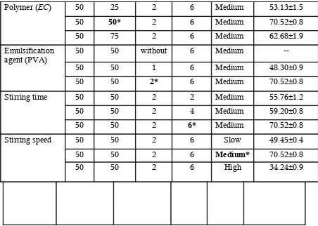
9 Optimization of various parameter of the formulation 69
10 Optimized formula for MDS 69
11 Evaluation parameter of P-MDS in Gel base 75
12 Diffusion Profile of P – MDS 1G 76
13 Diffusion Profile of P – MDS 2G 77
14 Diffusion Profile of P – MDS 3G 77
15 Comparative diffusion profile of P – MDS 78
16 Skin irritation results 81
17 Stability study data of formulation stored at 25±2 °C 84
18 Stability study data of formulation at 4±1°C 85
19 Stability study data of formulation stored at 37±5°C RH
75%
LIST OF FIGURES
Fig No Title Page No
1 Structure of Skin 31
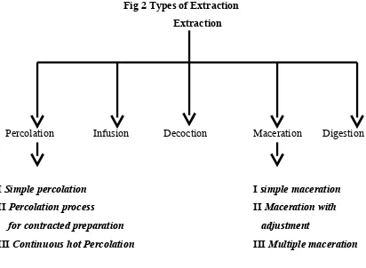
2 Types of Extraction 48
3 TLC Analysis of Different Fractions (III & IV) 58
4 TLC Analysis of Different Fractions (V, X & XI) 58
5 Infra Red spectrum of Crude Extract of Psoralen 61
6 Infra Red spectrum of sample II 61
7 Infra Red spectrum of sample IV 62
8 Infra Red spectrum of Ethyl Cellulose 62
9 NMR Spectrum of isolated compound Eu IV 63
10 NMR Spectrum of isolated compound Eu II 63
11 Absorption maxima of Psoralen 65
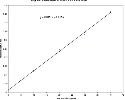
12 Calibration Curve of Psoralen 67
13 DSC of Psoralen 70
14 DSC of Ethyl Cellulose 70
15 DSC of P – MDS 2 71
16 SEM PHOTOGRAPHY OF P – MDS 1 72
17 SEM PHOTOGRAPHY OF P – MDS 2 72
18 SEM PHOTOGRAPHY OF P – MDS 3 73
19 Shape of the Microsponge 73
20 Diffusion Profile of P – MDS 1G 78
21 Diffusion Profile of P – MDS 2G 79
22 Diffusion Profile of P – MDS 3G 79
23 Comparative diffusion profile of P – MDS 80
24 Preparation of skin irritation test 82
25 Control for Skin irritation test (USP adhesive tap) 82
26 After 24 hours study with P – MDS 2 in Gel base 83
INTRODUCTION
India is perhaps the largest producer of medicinal herbs and is rightly called the ‘Botanical Garden of the World’ there are very few medicinal herbs of commercial importance, which are not collected or cultivated in this country. Medicinal herbs have been in use for thousands of years, in one form or another, under the indigenous system of medicine like Ayurveda, Siddha, Unani, since independence in 1947, India has made tremendous progress in agro technology, process technology, standardization, quality control, research and development etc.
Out now 2,25,000 species of plants known, about 35,000 are claimed to have medicinal properties. Today’s estimation indicate that about 80% of people in developing countries still on traditional medicine based largely on species of plants and animals for their primary health care. About 30% of the world wide sales of drugs are based on natural products.
India is one of the world’s 12th leading biodiversity centers with the presence
of over 45,000 different plant species. India’s diversity is unmatched due to the presence of sixteen different agro climatic zones to vegetation zones, 25 biotic provinces & 426 biomes (habitat of specific species).
India has 15,000-18,000 species of flowering plants, 2500 algae, 23000 fungi, 1600 types of lichen, 1800 varieties of bryophytes and an estimated 30 million types of microorganisms.
Of these about 15,000 - 20,000 plants have good medicinal value. However, traditional communities use only about 7,000 - 7,500 for their medicinal values.
According to 2004 United Nations Development Project (UNDP) report, the annual value of medicinal plants derived from developing countries is about $ 60 billion. There are 78 major modern plant based drugs on world market & the predicted 450 more potential drugs have an estimated of $ 180 billion.
Out of the large number of plants with known medicinal value and history of centuries of use in traditional medicine very few have been exploited in modern medicine. The main reason being lack of standardization, isolation and characterization of active principle. Recently World Health Organization (WHO) has taken an official interest on indigenous system of plant used particularly in natural
sources like plants and animals1.
USEFULNESS OF PLANTS IN THERAPY
Many of the early plant based drugs such as curare as a muscle relaxant,
Quinine from cinchona for malaria, Reserpine from Rauwolfia serpentina for
hypertension, Digitoxin from digitalis for heart ailments, the whole range of steroids
from diosgenin which is obtained from Discorea tubers, Vincristence/vinblastine
from Catharanthus roseus, the toxoids from Taxus brevifolia and Taxus baccata as
antimalarial are all still in use. A number of new products are in the market or under development for indications as wide ranging as flue, herpes, dermatitis, wound
healing, cancer, diabetes and Leucoderma.
APPROACH TO USE OF PLANTS IN MEDICINES
PLANTS AS SOURCES OF ACTIVE MOLECULES
An approach comparable with the current new discovery process is to use plants as source of active molecules this approach has led to successful development
of many molecules. Many of them in common use even today Psoralen, Strychnine,
Brucine, Reserpine, Quinine, Quinidine, Ajmaline, Vinca alkaloids, Taxol, Artemesinin & a host of other products have come through this route. A number of semi-synthetic drugs have become major drugs for many disease conditions. Thus the entire fields of hormonal steroids have come out chemical modification of
diasgenia, a product isolated from Dioscorea tubers or the Mexican yam.
The Chinese product Artemesinin has been converted to less toxic artmether & artether. In all these cases, the discovery of new derivatives was made possible only due to the fact that original plants have shown activity, even if it was low & hence was only a lead.
PLANTS AS LEADS TO DISCOVERY OF NEW DRUGS
Based on the strong and traditional knowledge based on the use of plants as therapeutic agents a rational approach is being developed to use medicinal plants as a lead for the discovery of active molecules.
Established drugs are also used as leads to synthesize new derivatives, which led to the well known and often unreasonably maligned me-too category of drugs. They are also termed as molecular manipulations or molecules roulette of existing
drugs2.
INDIAN SYSTEM OF MEDICINE
drink, salve or inhalant; bloodletting; bone-setting; cauterization; the utterance or writing of special prayers for curing purposes; exorcism of spirits said to possess the body; and the use of holy water and other sanctified substances such as soil, ash, or sand. Different indigenous healers specialize in one or more of these types of healing.
Indian system of Medicine and Homoeopathic (AYUSH) cover both the systems which originated in India and outside but got adopted in India in course of time. These systems are Ayurveda, Siddha, Unani, Yoga, Naturopathy and Homoeopathy. People are getting inclined for treatment through these systems due to lesser side effect in comparison to the modern medicines.
Ayurveda System of Medicine
Ayurveda means the “Science of Life”. The knowledge Ayurveda was comprehensively documented in Charak Samhita and Sushruta Samhita. Ayurveda takes an integrated view of the physical, mental and spiritual and social aspects of human beings, each impinging on the others. The philosophy of Ayurveda is based on the theory of Panchmahabhutas (five-element theory) of which all the objects and living bodies are composed of. The combination of the five elements are represented in the form of Tridosha e.g., Vata (Ether+Air), Pitta (Fire), Kaph (Water+Earth). These three ‘Doshas’ are physiological entities in living beings.
Siddha System of Medicine
Siddha means achievements and Siddhars were saintly persons who achieved results in medicine. Siddha is largely therapeutic in nature.
The diagnosis of disease involves identifying its causes. Identification of causative factors is through the examination of pulse, urine, eyes, and study of voice, color of the body, tongue and the status of the digestive system.
The Siddha system is effective in treating chronic cases of liver, skin diseases especially “Psoriasis”. The Siddha medicine which contains mercury, silver, arsenic, lead and sulphur have been found to be effective treating certain infectious diseases including venereal diseases.
Unani System of Medicine
The Unani System of Medicine is based on its well established knowledge and practices relating to promotion of positive health and prevention of diseases. It is very rich, time tested with its therapies having no side effects.
The Unani System emphasizes the use of naturally occurring, most herbal medicine and a few of animal, marine, and mineral origin. HK. Ajmal Khan has institutionalized the Ayurveda & Unani system of medicine and advocated the integration with science in understanding the Tibe-Unani and its application in treatment. He discovered Asrol (Rawolfia serpentina) which established its efficacy in the treatment of high blood pressure.
Its efficacy in the treatment and management of cardiac diseases is being researched. Treatment is carried out in four forms i.e. Pharmacotherapy, Dietotherapy, Regimental Therapy and Surgery.
Homoeopathy
Homoeopathy is a specialized method of drug therapy of curing natural diseases by administration of drugs which has been experimentally proved to possess the power of producing similar artificial symptoms on healthy human beings.
Physicians from the time of Hippocrates (around 400 B.C.) observed that certain substances could produce symptoms that they were used to treat. However, it was a German doctor Dr. Christian Friedrich Samuel Hahnemann (1755-1843) who examined this observation more thoroughly and the emergence of Homoeopathy took place thereafter.
The first principle similar similibus curentur, states that a medicine, which can induce a set of symptoms in healthy human beings, would be capable of curing the similar set of symptoms in disease state. The second principle in single medicine at a time for a particular patient during the treatment on the basis of totality of symptoms. The third principle is minimum dose to be administered to a patient.
Medicines
In Homoeopathy, medicines are prepared from natural sources viz. Vegetable, Mineral, and Animal etc. There is no toxic or poisonous effect in the finished products of Homeopathic Medicines.
Strength of the system
infections. Many Surgical, Gynecological & Obstetrical conditions, ailments affecting eyes, nose, ear, teeth, skin, sexual organs etc are curable through the homoeopathic treatment. Behavioral disorders, neurological problems, metabolic disorders successfully treated by the physicians of this system.
Homoeopathy has effective answer to addiction to drugs, tobacco and alcohol and is highly efficacious in ridding of addicts and their craving for these noxious substances.
Yoga and naturopathy
Yoga is primarily a way of life propounded by patanjali in a systemic form. It consists of eight components, namely, restraint, observance of austerity, physical postures, breathing exercise, restraining of sense organs, contemplation, meditation and Samadhi. These steps in the practice of yoga have potential for improvement of social and personal behaviour, improvement of physical health by encouraging better circulation of oxygenated blood in the body, restraining the sense organs and thereby inducing tranquility and serenity of mind.
Meditation can stabilize emotional changes and prevent abnormal functions of vital organs of the body. Studies have shown that meditation not only restrains the sense organs but also controls the nervous system.
Naturopathy is a system of medicine widely practiced, globally accepted and recognized by WHO. Naturopathy is a system of man building in harmony with constructive principles of Nature on physical, mental, moral and spiritual planes of living. It has great health promotive disease preventive and curative as well as restorative potential.
Amchi system of medicine
The system traces its origin to Ayurvedic system of India. The medical system since its delivery by Lord Buddha, while meditating near Bodh Gaya, in course of time, had accumulated a huge literature and Amchi of great fame and repute were produced. Therapy under the system is divided into treatment by herbs, minerals, animal organs, spring and mineral water, moxibustion and spiritual powder.
The Central Council for Research in Ayurveda and Siddha under the Department of AYUSH is having an Amchi Research Unit at Leh to carry our
research and to conduct survey of local drugs in Leh & Ladakh3.
LEUCODERMA AND ITS MANAGEMENT
Give me a blue sky, brown mountain, silver falls and green around, nothing more I want is a popular saying. Nature is always wondered and admired at for its colorful beauty is being the main attraction.
But the whiteness offered by skin disorder-Leucoderma is often depressing
for all the sufferers. Likewise in white skin people (even for leucoderma patient) hot sun UV rays penetrate more, putting them at risk for sunburn, skin cancer, etc.
Leucoderma is a miserable acquired skin disorder, making skin white due to
loss of the melanin pigment. It is a non contagious disease. It is otherwise termed as
Vitiligo.
Incidences and risk personalities
Causes
Till now researchers have not identified any causative factor for leucoderma. It can also be familiar i.e. hereditary factor also has some role in the prevalence of leucoderma. Being emotionally upset may also precipitate or aggravate the complaint in certain circumstances. Some suspect skin, lack of sun exposure, infection, etc. to be the reason for the problem. All these risk factors only, but the real cause is still obscure or unknown.
The main causes of leucoderma are said to be excessive mental worry, chronic or acute gastric disorder, and impaired hepatic function such as jaundice, worms or other parasites in the alimentary canal, typhoid, a defective perspiratory mechanism, and burn injuries. Heredity is also a well-recognized causative factor.
Symptoms
Leucoderma appears as odd, harmless, white spots or patches disturbing the appearance of the sufferer. Mostly no other symptoms can be noted except whiteness of the skin. The whiteness usually starts as a small discolored white or pale or brown spot which spreads and becomes whiter day by day and becomes milky white in the course of time.
Prevention of spread
Check for any infective focus, worms, chemical, contact, etc if noted, they should be treated or eliminated start treatment early.
Do’s
• Take medicines regularly in the early days itself so that complete cure can be
achieved.
• Don’t worry about leucoderma, since effective treatment can arrest the course
of the disease and cure it.
• Track all your eatables and habits to streamline all irritants or chemicals or sun exposure to arrest the spread of the disease.
Don’ts
• Avoid working or roaming in the sun.
• Avoid fast food, citrus fruits, coffee, tea, cold drinks, alcohol, beer, non-veg,
egg ect, It can causes spread of the disease.
• Avoid multiple drugs or drug cocktails.
• Avoid chemical soaps
Pathologically:
A defect in enzyme Tyrosinase is held responsible for vitiligo. According to some Dermatologists, it is a Trophoneurosis and Melatonin, a substance secreted at nerve endings inhibits Tyrosinase, thus interfering in pigment formation.
Clinical Presentation of Vitiligo
Localized Type:
a) Focal - One or more macules in two single areas but not segmented. b) Segmental - One or more macules in a dermatomal pattern.
c) Mucosal - Involvement of mucous membrane alone.
Generalised Type:
d) Acrofacial - Involvement of face and distal extremities.
e) Vulgaris - Scattered mascules in symmetrical or asymmetrical distribution.
Universalis
Who Is Affected by Vitiligo?
About 1 to 2 percent of the world's population, or 40 to 50 million people, have vitiligo. In the United States, 2 to 5 million people have the disorder.
Ninety-five percent of people who have vitiligo develop it before their 40th birthday. The
disorder affects all races and both sexes equally. Vitiligo seems to be more common in people with certain autoimmune diseases (diseases in which a person's immune system reacts against the body's own organs or tissues).
These autoimmune diseases include hyperthyroidism (an overactive thyroid gland), adrenocortical insufficiency (the adrenal gland does not produce enough of the hormone called corticosteroid), alopecia areata (patches of baldness), and pernicious anemia (a low level of red blood cells caused by failure of the body to absorb vitamin B12). Scientists do not know the reason for the association between vitiligo and these autoimmune diseases. However, most people with vitiligo have no other autoimmune disease. Vitiligo may also be hereditary, that is, it can run in families. Children whose parents have the disorder are more likely to develop vitiligo. However, most children will not get vitiligo even if a parent has it, and most people with vitiligo do not have a family history of the disorder.
How Is Vitiligo Diagnosed?
The doctor may take a small sample (biopsy) of the affected skin. He or she may also take a blood sample to check the blood-cell count and thyroid function. For some patients, the doctor may recommend an eye examination to check for uveitis (inflammation of part of the eye). A blood test to look for the presence of antinuclear antibodies (a type of autoantibody) may also be done. This test helps to determine if the patient has another autoimmune disease.
General Treatment
There is no scheduled, conservative treatment at all with rudimentary knowledge, everyone speaks more, but none of the internal or external claims guarantee for solid cure. No single therapy for leucoderma produces good result in all patients.
What Treatment Options Are Available?
The goal of treating vitiligo is to restore the function of the skin and to improve the patient's appearance. Therapy for vitiligo takes a long time. It usually must be continued for 6 to 18 months. The choice of therapy depends on the number of white patches and how widespread they are and on the patient’s preference for treatment. Each patient responds differently to therapy and a particular treatment may not work in everyone.
TREATMENT OPTIONS FOR VITILIGO
Medical Treatments
Topical steroid therapy
• Topical Psoralen photo chemotherapy
• Oral Psoralen photo chemotherapy
Surgical Therapies
• Autologous skin grafts
• Skin grafts using blisters
• Micropigmentation (tattooing) and Autologous melanocyte transplants.
Adjunctive Therapies
• Sunscreens
• Cosmetics
Counseling and support
Current treatment options for vitiligo include medical, surgical, and adjunctive therapies (therapies that can be used along with surgical or medical treatments).
MEDICAL TREATMENTS
TOPICAL STEROID THERAPY
Psoralen Photochemotherapy
Psoralen photochemotherapy (Psoralen and ultraviolet A therapy, or PUVA) is probably the most beneficial treatment for vitiligo available in the United States.
However, it is time-consuming and care must be taken to avoid side effects, which can sometimes be severe. Psoralens are drugs that contain chemicals that react with ultraviolet light to cause darkening of the skin. The treatment involves taking Psoralen by mouth (orally) or applying it to the skin (topically). This is followed by carefully timed exposure to ultraviolet A (UVA) light from a special lamp or to sunlight. Patients usually receive treatments in their doctor's offices so that they can be carefully watched for any side effects. Patients must minimize exposure to sunlight at other times. The goal of PUVA therapy is to repigment the white patches.
Topical Psoralen Photochemotherapy
Topical Psoralen photochemotherapy often is used for people with a small number of depigmented patches (affecting less than 20 percent of the body). It is also used for children 2 years old and over who have localized patches of vitiligo. Treatments are done in a doctor's office under artificial UVA light once or twice a week. The doctor or nurse applies a thin coat of Psoralen to the patient's depigmented patches about 30 minutes before UVA light exposure. The patient is then exposed to an amount of UVA light that turns the affected area pink. The doctor usually increases the dose of UVA light slowly over many weeks. Eventually, the pink areas fade and a more normal skin color appears.
After each treatment, the patient washes his or her skin with soap and water and applies a sunscreen before leaving the doctor's office.
(1) Severe sunburn and blistering and (2) too much repigmentation or darkening of the treated patches or the normal skin surrounding the vitiligo (hyperpigmentation). Patients can minimize their chances of sunburn if they avoid exposure to direct sunlight after each treatment. Hyperpigmentation is usually a temporary problem and eventually disappears when treatment is stopped.
Oral Psoralen Photohemotherapy
Oral PUVA therapy is used for people with more extensive vitiligo (affecting more than 20 percent of the body) or for people who do not respond to topical PUVA
therapy. Oral Psoralen is not recommended for children under 10 years of age
because of an increased risk of damage to the eyes, such as cataracts. For oral PUVA therapy, the patient takes a prescribed dose of Psoralen by mouth about 2 hours before exposure to artificial UVA light or sunlight. The doctor adjusts the dose of light until the skin areas being treated become pink. Treatments are usually given two or three times a week, but never on two days in a row. For patients who cannot go to a PUVA facility Psoralen may be used with natural sunlight exposure, with careful instruction and frequent monitoring by the treating physician.
Known side effects of oral Psoralen include sunburn, nausea and vomiting,
itching, abnormal hair growth, and hyperpigmentation. Oral Psoralen
Depigmentation
Depigmentation involves fading the rest of the skin on the body to match the already white areas.
For people who have vitiligo on more than 50 percent of their body, depigmentation may be the best treatment option. Patients apply the drug monobenzylether of hydroquinone (monobenzone or Benoquin) twice a day to pigmented areas until they match the already depigmented areas. Patients must avoid direct skin-to-skin contact with other people for at least 2 hours after applying the drug.
The major side effect of depigmentation therapy is inflammation (redness
and swelling) of the skin. Patients may experience itching, dry skin or abnormal darkening of the membrane that covers the white of the eye. Depigmentation is permanent and cannot be reversed. In addition, a person who undergoes depigmentation will always be abnormally sensitive to sunlight.
SURGICAL THERAPY
All surgical therapies must be viewed as experimental because their effectiveness
and side effects remain to be fully defined.
Autologous Skin Grafts
In autologous (use of a person’s own tissues) skin graft, the doctors removes skin from one area of a patient's body and attaches it to another area. This type of skin grafting is sometimes used for patients with small patches of vitiligo. The doctor removes sections of the normal, pigmented skin (donor sites) and places them on the depigmented areas (recipient sites). There are several possible complications of
The recipient and donor sites may develop scarring, a cobblestone appearance or a spotty pigmentation, or may fail to repigment at all. Treatment with grafting takes time and is costly, and most people find it neither acceptable nor affordable.
Skin Grafts Using Blisters
In this procedure, the doctor creates blisters on the patient's pigmented skin by using heat, suction, or freezing cold. The tops of the blisters are then cut out and transplanted to a depigmented skin area.
The risks of blister grafting include the development of a cobblestone
appearance, scarring, and lack of repigmentation. However, there is less risk of scarring with this procedure than with other types of grafting.
Micropigmentation (Tattooing)
Tattooing implants pigment into the skin with a special surgical instrument. This procedure works best for the lip area, particularly in people with dark skin; however, it is difficult for the doctor to match perfectly the color of the skin of the
surrounding area. Tattooing tends to fade over time. In addition, tattooing of the
lips may lead to episodes of blister outbreaks caused by the Herpes Simplex Virus.
Autologous Melanocyte Transplants
In this procedure, the doctor takes a sample of the patient's normally pigmented skin and places it in a laboratory dish containing a special cell culture solution to grow melanocytes.
When the melanocytes in the culture solution have multiplied, the doctor transplants them to the patient's depigmented skin patches. This procedure is currently
ADJUNCTIVE THERAPY
Sunscreens
People, who have vitiligo, particularly those with fair skin, should use a sunscreen that provides protection from both the UVA and UVB forms of ultraviolet light. Sunscreen helps protect the skin from sunburn and long-term damage. Sunscreen also minimizes tanning, which makes the contrast between normal and depigmented skin less noticeable.
Cosmetics
Some patients with vitiligo camouflage depigmented patches with stains, makeup, or self-tanning lotions. These cosmetic products can be particularly effective for people whose vitiligo is limited on exposed areas of the body. Dermablend, Lydia O'Leary, Clinique, Fashion Flair, Vitadye, and Chromelin offer makeup or dyes that patients may find helpful for covering up depigmented patches.
Tab 1 Medicinal plants used in Leucoderma4.
1 Abrus precatorius Linn 2 Acacia catechu Willd
3 Acacia foprnesiana Willd 4 Acacia ferruginea DC
5 Acacia nilotica Del 6 Aconitum chasmanthum Stapf
7 Aconitum falconeri Stapf 8 Aconitum rugata Lam
9 Acorus calamus Linn 10 Adhatoda vasica Nees
11 Aglaia odoratissima Blume 12 Albizia amara Boivin
13 Albizia lebbek Benth 14 Allium cepa Linn
15 Allium sativum Linn 16 Alpinea galanga Willd
17 Alpinia khulanjan Sheriff 18 Alpinia officinarum Hance
19 Alstonia scholaris R Br 20 Altingea excelasa Noronha
21 Anararabtgus tristis Linn 22 Ammi majus linn
23 Anacardium occidentale Linn 24 Anacyclus pyrethrum DC
25 Amemone obustifolia D Don 26 Anethum sowa Roxb
27 Anthriscus cerifolium Hoffm 28 Apium graveolen Linn
29 Aquillaria agallocha Roxb 30 Argemone mexicana Linn
31 Aristolchia indica Linn 32 Artemisia siversiana Willd
33 Astragalus hamosus Linn 34 Atropa belladomma Linn
37 Balanites aegyptica Del 38 Baleopermum montanum Muell-Arg
39 Bambusa arundinaeca Willd 4o Barleria priotis Linn
41 Barleria strigosa Willd 42 Bauhimea variegate Linn
43 Blepharis edulis pers 44 Boswelia serrata Roxb
45 Brassica campestris Linn 46 Brassica nifra Koch
47 Butea monosperma (Lam) Kuntz 48 Calamus rotang Linn
49 Calotroppis gigantean R Br 50 Canscora decussate Roem et Sch
51 Capparis sepiaria Linn 52 Careya arborea Roxb
53 Carthamus tinctorius Linn 54 Carum carvi Linn
55 Casearia esculenta Roxb 56 Cassia absus Linn
57 Cassia angustifolia vahl 58 Cassia tora Linn
59 Cedrus deodara (Roxb) Lour 60 Celastrus paniculata Willd
61 Centella asiatica Urban 62 Centipeda minima Br et Asch
63 Centratheryum anthelminitcum 64 Cephalandra indica Naud
65 Chenopodium album Linn 66 Cinnamomum zeylanicum Blume
67 Cissus adanta Roxb 68 Cissus setosa Roxb
69 Citrullus colocynthis Schrad 70 Cleome brachycarpa Vahl
71 Cleome chelidonni Linn 72 Cleome icosandra Linn
73 Clerodendron infortuatum Linn 74 Clitorea ternatea Linn
75 Commiphora agalocha Engl 76 Commiphora mukul Hook ex
Stocks
77 Convolvulus scammonia Linn 78 Coptis teeta wall
79 Coriandrum sativum Linn 80 Corydalis govniana wall
81 Croton tiglium Linn 82 Cuminum cyminum Linn
83 Curcuma angustifolia Roxb 84 Curcuma aromatica Salisb
85 Curcuma caesia Roxb 86 Curcuma longa Linn
87 Curcuma zedoaria Rosc 88 Dalbergia sissoo Roxb
89 Datura metel Linn 90 Datura stramonium Linn
91 Delphinium denudatum Wall 92 Dioscorea bulbifera Linn
93 Dolichos biflorus Linn 94 Dolichos falcatus Klun
95 Drogea volubilis Benth 96 Ecballium elaterium ARich
97 Eclipta alba Massk 98 Eclipta erecta Lamk
99 Embelia ribes Burm f 100 Emblica officinalis Gaerth
101 Enhydra fluctuans Lour 102 Eruca sativa Mill
103 Euporbia meriifolia Linn 104 Evolvulus alsinoldes Linn
105 Fagonia cretica linn 106 Feronia limomia Swingl
107 Ficus carica Linn 108 Ficus hispida Linn
109 Ficus micrlcarpa E Vill 110 Ficus racemosa Linn
111 Ficus religiosa Linn 112 Ficus tsiela Roxb
113 Fumaria indica pugsley 114 Geranium wallichianum Sweet
115 Girardinia heteraphylla DC 116 Gisekia pharnaceildes Linn
117 Glorisa superba Linn 118 Gymnema sylevestre R Br
121 Heracleum candicans Wall 122 Heracleum canescens Lind
123 Heracieum pinnatumWild 124 Holarrhena antidysentrica Wall
125 Holostemma annulare K Schum 126 Hydnocarpus Kurzii Warb
127 Hyoscymus niger Linn 128 Indigofera tinctoria Linn
129 Ipomea hederacea Jacq 130 Ipomea reninflrmis Choisy
131 Ipomea raptans Plir 132 Iris versiclolr Linn
133 Lactuca scariola Linn 134 Lamprachemium microcephalum
135 Lawsonia inermis Linn 136 Lepidium sativum Linn
137 Lolium femulentum Linn 138 Luffa acutangula Roxb
139 Lupinus albus linn 140 Macrotomea benthame DC
141 Macrotomea perennis Bois 142 Martynia annua Linn
143 Melia azardirach Bois 144 Melia acadirachta Linn
145 Meliotus alba Desr 146 Meliotus officinalis Desr
147 Mentha sylvestris Linn 148 Mimosa pudica Linn
149 Moringa concanensis Nimmo 150 Mofinga oleifera Lam
151 Murraya koenigii Spreng 152 Musa paradisiacal Linn
153 Mussaenda glabrata Hutich 154 Nardostachys jatamansi DC
155 Nelumbo mucifera Gaertn 156 Nerium odorum Soland
157 Micotians rustica Linn 158 Nicotianatabacum Linn
159 Nigella sativa Linn 160 Nymphaea stellata Willd
161 Ocimum basilicum Linn 162 Ocimum samctum Linn
163 Onosma bracteatum Will 164 Onosma echilides Linn
16 5
Operculiana turpethum satiba Linn
166 Opuntia dillunii Haw
16 7
Origanum majorana Linn 168 Oroxylon indicum Vent
16 9
Orthosiphon pallidus Royle 170 Ougeinia oojeinensis Hochr
17 1
Oxystelma esculenta R Br 172 Paederia foetida Linn
17 3
Pandanus tectorius Sol 174 Paris polyphylla Smith
17 5
Pentatropis cynachoides R Br 176 Phaseolus radiatus Lour
17 7
Picrorrhiza kurroa Royle ex Behth 178 Pinus sylvestris Linn
17 9
Piper longum Linn 180 Plantanus orientalis Linn
18 1
Plumbago indica Linn 182 Plumbago zeylanica Linn
3 18 5
Prunus amygdalus Stok 186 Prunus cerasoidea D Don
18 7
Prunus perrica Batsch 188 Psoralea corylifolia Linn
18 9
Pterocarpus marsupium Roxb 190 Punica granatum Linn
19 1
Randia dumetorum Lam 192 Ranunculus scleratus Linn
19 3
Raphanus sativus Linn 194 Rheum emodi Wall
19 5
Ricinus communis Linn 196 Rubia cordifolia Linn
19 7
Rumex besicarium Linn 198 Sapindus mukorossi Gaerth
19 9
Sasbania sesban Merr 200 Ssussurea lappa Clarke
20 1
Semecarpus anacardium Linn 202 Solanum indicum Linn
20 3
Solanum melongena Linn 204 Solanum nigrum Linn
20 5
Sphaeranthus indicus Linn 206 Spinacea oleracea Linn
20 7
Strychmos nux vomica Linn 208 Swertia chirata Buch Ham
20 9
Tamarix articulate Vahl 210 Tamarix indica Linn
21 1
Tamarix troupii Hole 212 Tecomelia undulate Seem
21 3
Tectona grandis Linn 214 Terminalia arjuna Wright &Arn
21 5
Terminalia belerica Rlxb 216 Terminalia chebula Retz
21 7
Terminalia citrine roxb 218 Thevetia neriifolia Juss
21 9
22 1
Trigonella foenumgraecum Linn 222 Urginea indica Kunth
22 3
Urtica dioica Linn 224 Uritica parviflora Rlxb
22 5
Vicia faba Linn 226 Viola serpens Wall
22 7
Viola tricolor Linn 228 Vilex megundo Linn
22 9
Vitex triflioa Linn 230 Wedelia calendulacea Less
23 1
Widhania somnifera Dunal 232 Wrightia tinctoria R Br
23 3
Xanthium stumarium Linn 234 Zanthoxylum acanthopodium DC
23 5
Zanthoxulum alatum Roxb 236 Zanthoxylum oxyphyllum Edgew
23 7
Zingiber officinale Rosc
MICROSPONGE DRUG DELIVERY SYSTEM (MDS)
During the few years, in pharmaceutical research much attention has been given to the controlled release of Topical drugs. By the use of this approach, controlling the therapeutic efficacy of these drugs, reduction in the total dose needed
and a reduction in the side effects can be provided. 5 For this purpose, Microsponges
are one of the systems which are in trial for controlled drug delivery. Microsponges are porous, polymeric microspheres that are used mostly for topical and recently for oral administration. Microsponges are microscopic spheres capable of absorbing skin secretions, therefore reducing oiliness and shine from the skin. Spherical particles composed of clusters of even tinier spheres are capable of holding four times their
The most closely related systems are Microcapsules and Microspheres. Microcapsules are spherical particles containing an active agent in the core, surrounded by a polymeric membrane. Microspheres are spherical particles containing the active agent dispersed in a polymeric matrix.
The major distinguishing feature between Microsponge and Polytrap systems and Microcapsules, or Microspheres, is structure of Microsponge and Polytrap
systems are highly porous, while Microspheres or Microcapsules are solid particles
with no internal voids.
A variety of active agents can be entrapped in a single Microsponge system
and to release them at desired rates. The Microsponge system can prevent excessive
accumulation of ingredients within the epidermis and the dermis. Potentially, the Microsponge system can reduce significantly the irritation of effective drugs without reducing their efficacy. Further these porous microspheres with active ingredients can be incorporated in to formulations such as creams, lotions and powders.
Microsponges consisting of non-collapsible structures with porous surface through active ingredients are released in a controlled manner. This delivery system
can be incorporated into conventional dosage forms such as creams, lotions, gels,
ointments, and powder and share a broad package of benefits.
Advantages of MDS 7
Advance oil control – absorbs up to 6 times its weight without drying.
Extended release – continuous action up to 12 hours.
Reduced irritation – better tolerance means broader consumer acceptance.
Improve product aesthetics – gives product an elegant feel.
Improve the stability – thermal, physical and chemical.
Allow incorporation of immiscible.
Allow for novel product form.
Improve the material processing.
Method of preparation
Polymerization
The porous microspheres are prepared by Suspension Polymerization method
in liquid-liquid systems.12, In this preparation, the monomers are first dissolved along
with active ingredients in a suitable solvent solution of monomer and are then dispersed in the aqueous phase, which consist of additives (surfactant, suspending agents, etc. to aid in formation of suspension).
The polymerization is then initiated by adding catalyst or by increasing temperature or irradiation. The various steps in the preparation of Microsponge are summarized as
- Selection of monomer or combination of monomers - Formation of chain monomers as polymerization begins
- Formation of ladders as a result of cross linking between chain monomers - Folding of monomer ladder to form spherical particles
- Agglomeration of Microspheres which give rise to formation of bunches of Microspheres
- Binding of bunches to form Microsponges
The polymerization process leads to the formation of a reservoir type of system, which opens at the surface through pores. In some cases an inert liquid immiscible with water but completely miscible with monomer is used during the polymerization to form the pore network. After the polymerization the liquid is removed leaving the porous microspheres, i.e., Microsponges. Impregnating them within preformed Microsponges then incorporates the functional substances. Some times solvent may be used for faster and efficient incorporation of the active substances. The Microsponges act as a topical carriers for variety of functional substances, e.g. anti
Quasi – Emulsion solvent diffusion method
Microsponges prepared by a quasi-emulsion solvent diffusion method using an external phase of containing distilled water and polyvinyl alcohol (PVA) 72 000.
The internal phase consisted of drug, ethyl alcohol, polymer and triethylcitrate (TEC)8
, Which is added at an amount of 20% of the polymer in order to facilitate the
plasticity9. At first, the internal phase was prepared at 60°C and added to the external
phase at room temperature. After emulsification, the mixture is continuously stirred for 2 hours. Then the mixture is filtered to separate the Microsponges. The product is washed and dried by vacuum oven at 40°C for 24 hours.
Variables during in the preparation 10
o Drug and Polymer ratio
o Effect of stirring time
o Effect of stirring speed
o Effect of composition of internal and external phase
o Viscosity
POLYMERS FOR DRUG DELIVERY
Role of polymers 11
Characteristic of ideal polymer 12
1. It should be chemically inert and free from leachable impurities. 2. It should have good mechanical strength.
3. It should be easy and inexpensive to fabricate. 4. It should be easily sterilized.
5. It should demonstrate acceptable shelf life.
Drug release mechanism 13
Two broad categories of polymer system have been studied. The reservoir device involves the encapsulation of a drug with in the polymer shell, while the matrix device describes a system in which a drug is physically entrapped with in a polymer net work. The drug will be released over time either by diffusion out of the polymer matrix or by erosion (due to degradation) of the polymer or by a combination of two mechanisms.
Polymers have been classified broadly as13
Natural polymers - Albumin, Starch, Dextran, Gelatin
Fibrinogen, Chitosan ect.
Synthetic polymers - Polymethyl methacrylate, Polymethyl
methacrylate, Copolymers: Polymethyl Cyanoacrylate, Polyacrylamide, Poly acryl starch, Poly lactic acid ect. Polymers are further classified on the basis of their interaction with water.
Non-biodegradable polymers
They are inert in the environment of use and eliminated intact from the site of administration and is the rate limiting factor.
Hydro Gels
These type of polymers swell but do not dissolve when brought into contact with water. They are inert and remover intact from the site of administration.
Eg: Poly Hydroxyl Ethyl Metha Acrylate (PHEMA), cross linked Poly Vinyl Alcohol (PVA), cross linked Poly Vinyl Pyrrolidone (PVP), Polyacrylamide, Dextran.
Soluble polymers
These are moderate weight uncross-linked polymers that, dissolve in water. Eg: Poly Ethylene Glycol (PEG), Hydroxy Propyl Methyl Cellulose, Methocel and copolymers of methamers of methacrylic acid, Acrylic methyl acid methyl ester (Eudragit L).
Biodegradable polymers
These slowly disappear from the site of administration in to a chemical reaction like hydrolysis.
Eg: Poly Lactic Acid (PLA), Poly Glycolic Acid (PGA), Poly Capro Lactone (PCL)
12.
Polymers used in MDS
• Polymethacrylate
• Ethyl cellulose
Evaluation of MDS (Microsponge Drug delivery System) 14 – 16 Prepared Microsponges are generally analyzed by following parameters
Polymer composition
Particle size
Surface topography
Pore diameter
Drug content
Loading efficacy
Drug release studies
Drugs explored in Microsponge delivery 7
• Ketoprofen
• Benzyl peroxide
• Retinol
• Fluconazole
• Ibuprofen
• Tretinoin
• Trolamine
MICROSPONGE INCORPORATED IN TO TOPICAL DRUG DELIVERY
Topical Drug Delivery
Topical semi – solids are preparations designed to exert local activity when applied to the skin or mucous membranes. The main medicinal applications of semi –
solids are as protective, emollient, and therapeutic agents. They include such
dosage forms as creams, gel, ointments, pastes and poultices17.
Tab 2 Commercial Microsponge Formulations 7
GELS
NAME DRUG REMARKS/USE
Afirm 3X
Retinol Design to treat photodamaged skin
EpiQuin[TM]
Vitamin A Gradual treatment of ultraviolet induced
dyschromia and discoloration
RnUn-A Micro
Tretinoin For the treatment of acne
vulgaris
Curan[R] 5-flourouracil For actinic keratois
MicroPeel[R]
Salicylic acid Freeing the skin of all dead cells and stimulating
the skin renewal process.
Sprtscream[R] XS
Trolamine For the temporary relief of pain associated with
Gels are transparent or translucent semi – solid or solid preparation consisting of solutions or dispersions of one or more active ingredients in suitable hydrophilic or hydrophobic bases. They are made with the aid of gelling agent. Usually gels exhibit pseudoplastic flow properties and those made with synthetic or semi – synthetic polymers with a high degree of cross – linking have relative high yield value and low viscosity.
Product tends to be smooth, elegant, and produce cooling effect because of evaporation of water. They may also dry out to form films. Films adhere well to the skin and are usually easily removed by washing.
Gels (sometimes called Jellies) are semisolid systems consisting of either suspensions made up of small inorganic particles or large organic molecules interpenetrated by a liquid. Where the gel mass consists of a network of small discrete
particles, the gel is classified as a two-phase system (e.g., Aluminum Hydroxide
Gel). In a two-phase system, if the particle size of the dispersed phase is relatively large, the gel mass is sometimes referred to as a magma (e.g., Bentonite Magma).
Both gels and magmas may be thixotropic, forming semisolids on standing and becoming liquid on agitation. They should be shaken before use to ensure homogeneity and should be labeled to that effect.
Single-phase gels consist of organic macromolecules uniformly distributed throughout a liquid in such a manner that no apparent boundaries exist between the dispersed macromolecules and the liquid. Single-phase gels may be made from
synthetic macromolecules (e.g., Carbomer) or from natural gums (e.g., Tragacanth).
The latter preparations are also called mucilages. Although these gels are commonly aqueous, alcohols and oils may be used as the continuous phase.
For example, mineral oil can be combined with a polyethylene resin to form an oleaginous ointment base. Gels can be used to administer drugs topically or into
SKIN STRUCTURE
The skin is one of the most extensive organs of the human body covering an
area of about 2m2 in an average human adult. This multilayered organ receives
approximately one – third of all blood circulating through the body.
The integument is composed of skin and its associated tissues sweat glands, sebaceous glands, hair, and nails. It is largest organ of the body (6% of the body weight).Covers the entire body continuous with the digestive system (lips and anus), respiratory system (nose), and urogenital system (urethra).
SKIN FUNCTION
1) Serves as a barrier to physical, biological, and chemical agents and to radiation
2) Prevents desiccation
3) Regulates body temperature (thermoregulation) by evaporative cooling (perspiration), heat radiation at the surface of the body (blood circulation), and serves as insulation (a minor function in humans)
4) Site of excretion through sweat glands 5) Photochemical production of vitamin D 6) Serves as a sensory organ
Skin is composed of two primary layers
The epidermis is the ectodermally derived outer layer composed of keratinized stratified squamous epithelium.
The dermis is the mesodermally derived layer of dense irregular collagenous
Fig 1 Structure of Skin
THE EPIDERMIS:
The epidermis is an epithelium comprised of keratinocytes undergoing a program of sequential differentiation. The four major layers arise from the sequential differentiation of cells migrating from the basal layer to the surface.
Desmosomes and hemidesmosomes anchor intermediate filaments, which are comprised of keratins in epithelial cells, to the cell surface. Two types of keratins, type I keratins, which are acidic and type II keratins, which are basic, are required to form an intermediate filament. In epidermis, different pairs of keratin genes are expressed as the cells migrate upward from the basal layer and proceed through their differentiation program. For example, keratin 5 and 14 are expressed in the basal layer, while keratins 1 and 10 are found in the spinous layer.
The spinous layer (stratum spinosum) is the thickest layer of epidermis. The cells in this layer arise from the migration from the basal layer and lose their adhesion to the basement membrane and adhere to other keratinocytes. Some cells in this layer are also mitotically active. The cells in this layer attain a more flattened shape and have increased amounts of keratin containing intermediate filaments and desmosomes.
The granular layer (stratum granulosum) is characterized by the presence of keratohyalin granules among the keratin filaments; it consists of 3-5 layers of flattened keratinocytes. Keratohyalin granules contain the protein filaggrin, which serves to bundle the keratin filaments together. This is the most superficial layer of the epidermis that still has nuclei.
These layers (and the upper spinous layers, to a lesser degree) contain membrane coating granules (lamellar granules).
covalently links glutamine to lysine) of proteins, like involucrin, along its inner surface to form the cornified envelope.
These cells lack nuclei and other organelles but have numerous keratin filaments. Cells farther from the skin surface have desmosomes, while those nearer to the surface do not and will be sloughed. Epidermis turns over about every 14-30 days depending on the region of skin. Different areas of skin show differing thickness of epidermis.
1. The epidermis thickens in response to use (abrasion).
2. The dryness of skin serves as an inhospitable environment for
growth of microorganisms.
THE DERMIS:
The dermis is a dense, irregular, mesodermally derived, connective tissue,
composed of collagen (mostly type I), elastin, and glycosaminoglycans. It is much thicker than the epidermis, comprising 80-90% of the total dermis and epidermis.
It contains extensive vasculature, neurons, smooth muscle, and fibroblasts. It is the principal mechanical barrier of skin. Its networks of elastic fibers function to support the epidermis and bind the skin to the deeper hypodermis. The dermis
contains two layers, the papillary layer and the reticular layer. The papillary layer
is a loosely woven, superficial connective tissue region that interdigitates with the epidermal ridges and the deeper reticular layer.
A key feature of this layer is the dermal ridges (dermal papillae) that extend up in ridges into the overlying epidermis and interdigitate with epidermal
invaginations (epidermal ridges). The dermal ridges contain Meissner’s corpuscles,
macrophages occupy the papillary layer. The reticular layer resides below the papillary layer.
It is comprised of coarse collagenous and elastic fibers (irregular dense connective tissue) and relatively few cells.Arteries and veins run through the hypodermis and branch upward to form plexuses of anastomosing vessels. The cutaneous plexus resides at the junction of the hypodermis and dermis, and the papillary plexus resides just beneath the epidermis. This system provides nourishment to the dermis and by diffusion to the epidermis, which is avascular. The vascular system functions in thermoregulation. Blood flow is controlled by contraction of arterioles and venules to send blood through the capillary bed for heat radiation. In some regions of skin, arteriovenous anastomoses, or shunts, can send blood directly from the arterioles to venules in order to reduce heat loss.
The dermis contains neuronal elements for touch, pain, itch, and temperature reception.
1. Some receptors are free nerve endings.
2. Other nerve endings associate with Merkel cells in the
epidermis.
3. Meissner’s corpuscles reside in the dermal papillae and function as mechanoreceptors in touch perception.
4. Pacinian corpuscles are found deep in the dermis (and in the
hypodermis) and function in pressure sensation19.
LITERATURE SERVEYED OF MICROSPONGE
Çomoğlu et.al5 Reported the Effect of Different Polymers on Microsponge Formation. By the use of this approach, controlling the therapeutic efficacy of these drugs, reduction in the total dose needed and a reduction in the side effects can be provided. The Microsponges were prepared by emulsion-solvent diffusion method using different polymers such as ethyl cellulose, Eudragit RS 100 and Eudragit RS 100 and ethyl cellulose mixtures at different ratios.
Patel Geeta et.al7 Reported Use of a Microsponge in drug delivery systems.
The Microsponge delivery system is a unique technology for the controlled release of topical agents and consists of macroporous beads, typically 10-25 microns in diameter, loaded with active agent. When applied to the skin, the Microsponge releases its active ingredient on a time mode and also in response to other stimuli (rubbing, temperature, pH, etc). Microsponge technology offers entrapment of ingredients and is believed to contribute towards reduced side effects, improved stability, increased elegance, and enhanced formulation flexibility.
Jelvehgari et.al.10 Carried out preparation, characterization and release studies of Benzoyl peroxide (BPO) Microsponge drug delivery prepared by quasia emulsion solvent diffusion method. The aim of the present study was to produce ethylcellulose microparticles containing BPO which were able to control the release of BPO to the skin and reduces the side effect of commercial BPO such as irritation and percutaneous absorption. Generally, an increase in the ratio of drug: polymer resulted in a reduction in the release rate of BPO from Microsponges.
were prepared by compression coating and also pore plugging of Microsponges with pectin: hydroxypropylmethyl cellulose (HPMC) mixture followed by tabletting.
Comoglu et.al21 Carried out the enhancement of ketoprofen bioavailability by formation of microsponge tablets. An in vivo study was designed to evaluate the pharmacokinetic parameters and to compare them with the commercially available ketoprofen retard tablets containing the same amount of the active drug. The new modified release tablets showed a slower absorption rate and peak levels were reached 8 h after administration.
Tansel Çomoglu et.al22 Reported the effects of pressure and direct compression on tabletting of Microsponges. Microsponge delivery is used both oral and topical purpose. Ketoprofen was used as a model drug for systemic drug delivery of Microsponges. Microsponges were prepared by quasi-emulsion solvent diffusion method with Eudragit RS 100 and afterwards tablets of Microsponges were prepared by direct compression method. Different pressure values were applied to the tablet powder mass in order to determine the optimum pressure value for compression of the tablets.
Chadawar et.al24 Reported Microsponge delivery system. Microsponges are polymeric delivery systems consisting of porous microspheres having a size range in between 5 to 300 micron depending upon the degree of smoothness or after feel required for the end formulations. The present review introduces Microsponge technology along with its synthesis, characterization, programmable parameters and release mechanism of MDS. MDS can provide increased efficacy for topically active agents with enhanced safety, extended product stability and improved aesthetic properties in an efficient and novel manner.
Leyden et.al25 Carried out comparison of the efficacy and tolerability of once daily tazarotene 0.1% gel and tretinoin 0.1% Microsponge gel were evaluated in a multicenter, double-blind, randomized, parallel-group study in patients with mild-to-moderate inflammatory facial acne vulgaris. Tazarotene was observed to have greater efficacy and comparable tolerability and to be a cost-effective alternative to tretinoin 0.1% Microsponge gel.
Shigemitsu Iwai et.al26 Reported Biodegradable polymer with collagen Microsponge serves as a new bioengineered cardiovascular prosthesis. Biodegradable materials with autologous cell seeding have attracted much interest as potential cardiovascular grafts. The poly(lactic-co-glycolic acid)– collagen Microsponge patch with and without precellularization showed good histologic findings and durability.
LITERATURE SURVEYED OF PSORALEN
Hsu Y-T et.al28 Reported the presence of three isoflavonoid compound in
Psoralea corylifolia. The method proved to be sensitive, specific, accurate and precise for the determination of daidzein, genistein and biochanin A in
Psoralea corylifolia.
Guo WS et.al29 Carried out selection isolation of Iso Psoralen from crude
extract of Psoralea corylifolia.L by using inclusion method of host guest
molecules.
Yadava RN et.al30 Reported a new biologically active flavonol glycoside
from Psoralea corylifolia.
Liu ZL et.al31 carried out extraction and purification of Psoralen from
Psoralea corylifolia. To establish a method for extracting Psoralen from the
seed of Psoralea corylifolia.
Rajendra Prasad N et.al32 Reported antidermatophytic activity of extract
from Psoralea corylifolia correlated with the presence of a flavonoid
compound. The extract showed several degree of antifungal activity against Trichophyton rubrum and Microsporum gypseum the disc diffusion method on a Sabouraud dextrose agar medium.
Arabzadeh et.al. 34 Reported mechanism of 8 – methoxy Psoralen DNA interaction in the dark. The interaction of 8 – methoxy Psoralen with calf thymus DNA was studied in darkness at 25 ºC and pH 7.4.
Said et.al. 35 Carried out wettability of Psoralen powder influence of bile salts on their contact angles and surface free energy components.
Rama Sastry CV et.al36 Reported A study of Psoralen photochemotherapy with topical tar in the management of psoriasis vulgaris.
Bhatnagar A 37et.al Carried out Psoralen and ultraviolet A and narrow-band ultraviolet B in inducing stability in vitiligo, assessed by vitiligo disease activity score: an open prospective comparative study.
Gomes AJ 38 et.al Carried out Identification of Psoralen loaded PLGA poly (DL-lactide-co-glycolide) microspheres in rat skin by light microscopy. Drug delivery systems involving the use of polymers are widely studied and discovery of biocompatible polymers has become the focus of research in this area. Psoralen loaded poly PLGA microspheres to be used in PUVA therapy (Psoralen and UVA irradiation (ultraviolet A, 320-400 nm) of psoriasis were identified in paraffin sections by histological analysis. The Psoralen loaded PLGA microspheres were prepared using the solvent evaporation technique
which is also called as Psoralea corylifolia. Psoralen has been found in more than 30 plants such as lime, lemon, bergamot, parsley, celery, fig and cloves. The medical use of these plants in the treatment of vitiligo by the ancient Egyptians dates back to as early as 1500 B.C. and by the Indians to 1400 B.C.
Sergioasffirtri40 Reported Reversed phase high performance liquid chromatography determination of lipophilicity of furocoumarins relationship with DNA interaction.
Objective & Plan of work
Leucoderma is a kind of skin disorder and otherwise known as “swetha kuttam” in Sanskrit. It is non infectious disease which occurs due to the deficiency of melanin. In normal condition melanin is produced by melanocytes. Melanin is a complex brown polymer synthesised from amino acid L – Dopa (Dihydroxyphenyl alanine). The initial part of melanin synthesis is catalysed by a copper containing enzyme tyrosinase. This catalyzed the transformation of L – DOPA to tyrosine. Melanin synthesis is under pituitary control (Melanocyte Stimulating Hormone).
Leucoderma can reflect in the mind, causing psychological fears, emotional upset, shame, and socio behaviourial changes by trying to avoid being in the public, attitude of others who insult by avoiding contact. These changes restrict their social behaviour and communications. There may also be fear of ugliness, fear of hereditary
(infecting the generation), contagiousness, other diseases, cancer, etc. Psoralen is one
of the drugs of choice for the treatment of Leucoderma over 237 Herbal Drugs.
The dosage forms available in market are having severe side effects.
In the literature survey it is clearly evident that Psoralen Microsponge drug delivery system (MDS) is not yet formulated and my objective is to formulate
Psoralen MDS as a topical drug delivery system. Since it is non-irritating, non
mutagenic, non allergic, non toxic and also improves the patient compliance.
The main objective of my project work is to Isolate the active constituent from crude drug and convert it into Novel Drug Delivery System (Microsponge) to reduce the side effects occurs in available conventional dosage forms. (Psoralen and ultra violet A (PUVA) therapy).
PLAN OF WORK
Extraction and Isolation
Extraction of Psoralen from seeds of Psoralea corylifolia – By Soxhlet
Extractor.
Isolation of Psoralen – By Column chromatography
Identification of isolated products – By TLC, IR and NMR studies.
Preformulation studies
Characterization of Psoralen
Identification of Psoralen (Chemical Test)
Absorption maxima of Psoralen
Solubility of Psoralen
Infrared study of Psoralen
Calibration Curve for Psoralen drug
Fabrication and characterization MDS
Fabrication of Microsponge Drug Delivery System
Polymerization method
Quasi emulsion solvent diffusion method
Particle size (SEM) and Shape
Encapsulation efficacy
Microsponge is incorporate in Gel base
Formulation of gel Characterization of Gel
Drug content
pH
Viscosity
Spreadibility
Tube extrudability
In-vitro studies
Skin irritation studies
Stability studies