CLINICAL STUDY OF PATIENTS WITH ABDOMINAL
WOUND DEHISCENCE AT
GOVERNMENT RAJAJI HOSPITAL, MADURAI
MEDICAL COLLEGE, MADURAI
DISSERTATION SUBMITTED FOR
M.S. (General Surgery)
THE TAMILNADU DR. M.G.R. MEDICAL UNIVERSITY
CHENNAI – TAMILNADU
MADURAI MEDICAL COLLEGE, MADURAI
CERTIFICATE
This is to certify that this dissertation entitled “CLINICAL STUDY OF PATIENTS WITH ABDOMINAL WOUND DEHISCENCE” at Madurai
Medical College, Madurai is a bonafide research work done by
Dr.M.J. CHANDRABOSE AMBEDKAR in partial fulfillment of the requirement for the degree of M.S (General Surgery).
Dr.V. Raji M.D., Prof. M. Gobinath M.S.
Dean Professor & HOD,
Madurai Medical College, Chief I Surgical Unit,
Madurai Dept. of General Surgery,
Madurai Medical College,
Madurai.
DECLARATION
I, hereby declare that this dissertation / thesis entitled “CLINICAL STUDY OF PATIENTS WITH ABDOMINAL WOUND DEHISCENCE”
at Madurai Medical College, Madurai is a bonafide and genuine research work
carried out by me under the guidance of Prof. M. Gobinath M.S., H.O.D.,
Department of General Surgery, Madurai Medical College.
Dr.M.J. Chandrabose Ambedkar
Postgraduate Student,
Department of General Surgery, Madurai Medical College. Madurai.
Date:
ACKNOWLEDGEMENT
It gives me immense pleasure to express my gratitude and thanks to my
respected and beloved teacher and guide, Prof.M. Gobinath, M.S., Professor, Department of General Surgery, Madurai Medical College, Madurai for his
priceless guidance, affection and constant encouragement in preparing this
dissertation.
I express my humble thanks to my beloved teachers, Dr. T. A. Thara, M.S., DGO., Dr. M.Nasheer Ahamed Syed, M.S., Dr. R. Ganesan, M.S., Dr. D. Marudupandian, M.S., Dr. K. Karunagaran, M.S., Assistant professors of my unit, for their immense help and guidance on innumerable occasions.
I also express my sincere thanks to the rest of the Teaching Faculty of Surgery Department for their valuable suggestions and kind cooperation.
I Sincerely extend my heartfelt thanks to my post-graduate colleagues
for their valuable help, in completing this dissertation.
Lastly and most importantly, I am thankful to all the patients without whose consent and co-operation I would not have done this work.
Date :
CONTENTS
Page No
1. ABSTRACT 1
2. INTRODUCTION 2
3. AIM AND OBJECTIVES 3
4. REVIEW OF LITERATURE 4
5. MATERIALS AND METHODS 47
6. RESULTS AND ANALYSIS 51
7. DISCUSSION 64
8. CONCLUSION 70
9. SUMMARY 71
10. BIBLIOGRAPHY
11. PROFORMA
ABSTRACT
AIMS AND OBJECTIVES:
To identify various predisposing factors in patients who burst their
abdomen following mid line laparotomy.
MATERIALS AND METHODS:
Data were collected from patients, aged more than 18 years who had
burst. abdomen following enniass closure of midline laparotomy in general
surgery wards.
RESULTS AND CONCLUSION
Total of 18 patients (14 males and 4 females) had abdominal wound
dchiscence. There mean age was 34.4yrs. 7(39.5%) were anemic, 5(27.5) had
hypoprotenemia, 5(22.0%) had hyperbilirubinemia, 3(16.5%) had chest
infection all had intraperitoneal infection, one was diabetic. All were
emergency procedures. There was one (5.5%) mortality. Mean length of
hospital stay was 25.2 days.
INTRODUCTION
Wound dehiscence carries with it a substantial morbidity. In addition
there is an increase in the cost of care both in terms of increased hospital stay,
nursing and manpower cost in managing the burst and its complications. Many
patients in India have a poor nutritional status and the presentation of patient
with peritonitis is often delayed in the emergency. This makes the problem of
wound dehiscence more coninion and graver in our setting as compared to the
West.
In I -2% of cases, mostly between the sixth and eighth day after
operation an abdominal wound bursts open and viscera are extruded. The
disruption of the wound tends to occur a few days before hand when thc sutures
apposing the deep layers sheath tear through or even become untied. An
incisional hernia starts as a symptomless partial disruption of the deep layers
during the immediate or early postoperative period, even this passing unnoticed
AIMS AND OBJECTIVES
To identify various predisposing factors in patients who burst their
REVIEW OF LITERATURE
ANATOMY OF ANTERIOR ABDOMINAL WALL
Layers of the Abdominal Wall
The abdominal wall consists of seven layers of tissue, of which the
fourth or middle layer, the muscle—bone layer, is the most important. These
layers will he considered in the order in which they are encountered while
progressing inward.
I. Skin
The skin is the outermost layer. The course of the connective bundles of
the coriurn fonns lines of tension (Langer’s lines of cleavage) in the skin. Over the anterior abdomen, these lines of cleavage run in a more or less transverse
direction. Skin incisions made parallel to these lines of cleavage result in much
liner scars than do those that cut across the lines of cleavage.
2. Subcutaneous tissue
This layer consists of fat, which is variable in amount, contained within
fibrous compartments. The more superficial portion of the subcutaneous fat
contains much less fibrous tissue than does the deeper portion. This is called
Camper’s fascia. The deeper portion of the subcutaneous tissue of the
abdominal wall contains more fibrous elements and forms a membranous
3. Deep Fascia
The deep fascia of the abdominal wall is an ill-defined, thin and
unimportant layer consisting of loosely formed fibrous tissue.
4. Muscle-Bone Layer
This is the most important of the seven layers, consisting of nine muscles
with their fasciae and aponeuroses on each side of the midline and the five
lumbar vertebrae situated posteriorly. On either side of the anterior midline
(linea alba) of the abdomen are situated the right and left rectus abdominis and
pyramidalis muscles These muscles form the anterior group. Their fibers run in
a vertical direction and are enveloped between the anterior and posterior rectus
sheaths, formed by a splitting of the aponeuroses of the internal oblique
muscles.
Lateral to the anterior group are the external oblique, internal oblique
and transversus abdoniinis muscles with their fasciae and aponeuroses. These
three muscles form the flat or oblique group, extending laterally and posteriorly
from the lateral border of the rectus abdominis, posteriorly to the lateral border
of quadratus lumborum muscle. The direction of their fibers is closer to
ANTERIOR AND LATERAL (OBLIQUE) ABDOMINAL
MUSCLES AND RELATED STRUCTURES
The rectus abdominis muscle (one on each side) is attached below to the
os pubis and ligaments of the symphysis pubis, whereas superiorly it is attached
to the xiphoid cartilage and the anterior surfaces of the fifth, sixth and seventh
ribs. The rectus muscle is crossed by three transverse tendinous intersections.
The lack of attachment of the tendinous intersections to the posterior rectus
sheath permits the muscle belly to he retracted so that a paramedian incision
can be made.
The internal oblique muscle arises from the lumbodorsal fascia, the
anterior halfof the iliac crest and the lateral half of the inguinal (Poupart’s)
ligament.
The aponeurotic IThers of internal oblique split into two lamellae. The
anterior laniella passes in front of the rectus muscle and fuses with the
aponeurosis of the external oblique muscle to form the anterior sheath of the
rectus niLiscie. The posterior lamella passes behind the rectus muscle to fuse
with the aponeurotie fibers of the transversus abdominis and form the posterior
The posterior rectus sheath ends inferiorly at the arcuate line,
(semicircular line of Douglas) which is present at variable levels above the
symphysis in different individuals.
The transversus abdominis muscle arises from the lateral third of the
inguinal (Poupart’s) ligament, the anterior three fourths of the cresi of the ilium,
the lunibodorsal fasciaand lower six ribs.
The nerve supply of the rectus abdominis, external oblique, internal
oblique, and transversus abdominis muscles conies by way of the lower six or
seven intercosta’ and f lumbar nerves (by The ihohypogastric nerve, which is
formed by the twelfth thoracic and first lumbar nerves, and by the ilioinguinal
nerve, which arises from the first lumbar). In their course across the abdominal
wall, the nerves can be retracted upward or downward for variable distances
without being injured.
The anterolateral abdominal wall receives its arterial blood supply from
the last six intercostal and the four lumbar arteries together with the superior
and inferior epigastric and the deep circumflex iliac arteries. The superior and
inferior epigastric arteries anastomose only sparsely with their fellows of the
opposite side; therefore, the linea alba is a relatively avascular area and has a
5. Transversalis Fascia
The t layer of the abdominal wall is the transversatis fascia. The portion
of transversalis fascia beneath the rectus muscle is so intimately attached to the
overlying posterior rectus sheath and underlying peritoncum that the three
layers arc sutured together as one layer in closing the peritoneal layer of the
incision.
The transversalis fascia is attached below to the inner lip of the iliac
crest, the outer half of the inguinal (Poupart’s) ligament, the lacunar ligament
(of Gimbernat), and the pubic crest. This fascia is continued downward behind
the mesial half of the inguinal ligament and over the femoral vessels to pass
into the thigh, forming the femoral sheath.
6. Extraperitoneal Fat Layer
This laycr consists of loose areolar fibrous tissue containing variable
amounts of fat and lies between the overlying transversalis fascia and
underlying parietal peritoneum.
7. Peritoneurn
The parietal peritoncum is the smooth, serous layer that bounds the
peritoneal cavity and is reflected onto the various viscera to form the visceral
peritoneum as well as ligaments, mesenteries. Peritoneum strctehes easily and
has little strength, so (hat it requires the support of its overlying muscles atid
Linea Alba
The linea alba extends in the anterior midline of the abdomen from the
xiphoid to thc symphysis. The linca alba consists of a band of dense,
crisscrossed fibers of the aponeuroses of the broad abdominal muscles.
At the linea alba, the aponeurosis of the anterior sheath of the rectus
muscle, Rise with the posterior rectus sheath and with opposite side. Above the
umbilicus it widens out, but below that level it is narrow and sometimes
difficult to recognize. In the broad supraumbilical portion of the linea alba,
small openings are present through which the perforating vessels and nerves
pass and through which an epigastric hernia may occur. Since the branches of
the epigastric arteries anastomose very sparsely across the midline, the blood
supply of the linea alba is less copious than in other areas.
Furthermore, an incision made in the midline is through a relatively
avascular layer with minimal attendant bleeding and does not destroy the
innervation of any part of any muscle. It is believed, however, to be more prone
to postoperative herniation than are incisions in other locations, because of its
relatively meager blood supply.
Umbilicus
The umbilicus is the fibroaponeurotic scar formed within the central
portion of the linea alba. This represents the embryonic defect in the abdominal
urachus pass, but under normal conditions these structures atrophy into fibrous
cords.
Acute wounds and acute wound healing
An acute wound is defined as the traumatic loss of normal structure and
function to recently uninjured tissue after a noxious insult. Acute wound
healing is the highly regulated process of cellular, humoral and molecular
events activated at the time of acute injury and resulting in a time-dependent
but predictable and orderly pattern of tissue repair. The integrated summation of
each pathway along the continuum of this host response to injury results in
acute wound healing.
NORMAL WOUND HEALING
Wound healing Follows a complex and orderly pattern of events in
which an initial inflammatory reaction (inflammatory phase) is characterized by
an increase in vascular permeability followed by fibrin deposition and an influx
of red cells, polymorpholeukocytes, monocytes, and platelets. Local cellular
disruption perpetuates itself, with the release of protcolytic enzymes and
vasoactive substances into the wound space. An activated complement system
continues the inflammatory reaction and helps to attract macrophages which
clear the debris of inflammation. Fihrohlasts appear about the fourth postinjury
nutrients while fibroblast proliferation allows for the production of
mucopolysaccharides and collagen. The final phase of wound healing
(maturation phase) continues for months, as collagen fibers are aligned and cross-linked, allowing for a progressive increase in wound strength.
This sequence is essentially the same in all species and organ systems
studied, although the time reference may change drastically. For example, a
gastric anastomosis may take a few weeks to heal, whereas the fascial incision
used to provide exposure may take up to years to achieve maturity.
TYPES OF INCISIONS
Abdominal incisions can be divided roughly into three general types: (1)
vertical incisions (2) transverse incisions and (3) special incisions.
Abdominal incisions have a marked effect on pulmonary physiology.
Pulmonary complications have been reported to be related more to the location
than to the direction of the incision. Upper abdominal incisions are more
significant in their effect upon respiration. Breathing patterns are changed; the
respiratory rate increases and tidal volume and vital capacity are reduced.
Patients tend to become hypoxemie. Forced expiratory volume at I second
(FEV1) is reduced.
In 1940s, it was noted that pulmonary complications occurred four times
more frequently in the patient with a vertical rather than a transverse abdominal
preference for transverse incisions over vertical ones for ah4Qrninal operations
and pointed out these advantages:
1. The skin incision parallels Langers lines and gives a better cosmetic
result.
2. Transverse incisions are closed iii a direction that places only one
thirtieth as much tension on the suture line as is exerted on the suture
line of a vertical incision.
3. When transverse incisions are, tensed the edges of the transverse wound
tend to be approximated while the edges of a vertical wound are strongly
separated.
4. Transverse incisions facilitate closure of the peritoneum and posterior
rectus sheath, a factor in minimizing postoperative adhesions.
5. Transverse incisions run almost parallel to the direction of the neural and
vascular supply and therefore destroy fewer nerves and blood vessels
than do vertical incisions.
6. The blood supply to the region of a transverse incision is better than that
to a vertical midline incision, and therefore healing of a transverse
incision should be more rapid.
The purported predisposition and reasons for use of the transverse
1. Vertical midline or paramedian incisions arc more quickly made and
more quickly closed. They pass through fewer tissue layers and require less
suturing and time for closure.
2. Vertical midline or paramedian incisions destroy few if any nerves or
blood vessels supplying the tissues.
3. Vertical midline or paramedian incisions are made through a relatively
avascular field and give less troublesome bleeding than do transverse incisions.
4. In some areas, vertical midline or paramedian incisions provide better
exposure or are more easily extended than transverse incisions. This is
particularly true for operations on the cardiac end of the stomach or the
rectosigmoid.
There is a place for both vertical and transverse incisions. When vertical
incisions are used, they should be in the midline or paramedian and middle or
lateral transrectus in exceptional circumstances. Special incisions also have a
place in specific operations.
An incision should be selected with the following qualifications in mind:
1. It must give ready and direct access to the source of trouble and
provide adequate exposure for the operation contemplated.
2. It should be extensible in the direction that probably would be
3. It should injure the fewest possible number of motor nerves,
preferably not more than one.
4. It should be capable of being securely repaired so as to leave the
abdominal wall at least as strong after the operation as before.
5. It should provide an acceptable cosmetic result when possible.
It is important that an abdominal incision be made long enough to
provide an adequate visualization of the operative field and uncrowded
conditions for the necessary manipulations.
Upper Midline incisions
This is the incision preferred by many surgeons for exploration of and
most operations on the stomach. It provides excellent exposure in partial
gastrectomy and can easily be extended upward or into the chest for total
gastrectomy or operations on the lower esophagus or diaphragm. It can be
extended downward around the navel for as far as necessary and, when
required, can be extended laterally as a T or an L incision. An upper midline
incision destroys no nerves and can be quickly made and easily closed. It is
thought to be more vulnerable to postoperative herniation because of its
relatively meager blood supply.
Technique:
An incision is made through the skin and subcutaneous tissue from the
the umbilicus. If a longer incision is desired, it is extended downward to curve
around either the right or left side of the umbilicus’ to continue in the lower
midline.
The shiny white linea alba and anterior rectus sheath are cleared of fat
laterally for about 2 cm on each side. The linea alba and transversalis fascia are
divided exactly in the midline, exposing the extraperitoneal fat. After the
peritoneal cavity has been opened, a finger is slipped beneath it, and the
peritoneum is divided over the finger as it is advanced upward and downward,
protecting underlying viscera from injury. If the ligamentum teres interferes
with exposure , it should be divided between clamps and the cut ends ligated.
One should select the method of closure that is best for the patient and is well
performed by the surgeon.
WOUND CLOSURE IN LAYERS
In the patient whose condition is good and in whom rapid closure is not
imperative, the wound can be approximated in layers in the following manner:
1. The cut edges of the peritoneum, transversalis fascia and posterior rectus
sheath are approximated with a row of continuous over-and-over suture
of 2-0 plain catgut, 2-0 chromic or synthetic absorbable. Others prefer
interrupted sutures of the same materials or of silk. If the peritoneum is
friable, few muscle fibers and the deep and medial portion of the rectus
2. The cut edges of the linea alba are approximated with interrupted
sutures. This is the most important and strength—giving portion of the
closure and must he performed with care. Nonabsorbable suture material
is best for this purpose. One could use 0-nylon, No. 1 polypropylene or
3-0 (B&S 30) stainless-steel wire suture in patients in whom the
intestinal tract has been opened or drainage is instituted. Some surgeons
prefer to use absorbable suture material for this layer. No. I chromic
catgut or synthetic absorbable sutures have been used successfully.
Many other sutures are also acceptable.
When there is little or no tension, a row of simple, interrupted, over-
and-over sutures placed through the linea alba at least 1 cm lateral to the cut
edge on each side and including a few underlying fibers of the rectus muscle in
each side, is used.
When the cut edges of the linea alba can be approximated only with
tension, one can close with interrupted far-near sutures. These penetrate the
rectus sheath about 3 cms lateral to the cut edge, pass beneath it through the
anterior fibers of the rectus muscle to cross the wound and emerge through the
anterior rectus sheath 0.5cm from its cut edge on the other side, cross over the
wound to penetrate the original side of the anterior sheath from superficial to
through the superficial rectus fibers beneath the rectus sheath of the other side
to emerge about 3 cm lateral to the edges. When these sutures are pulled taut
and tied, any tension is distributed to four points instead of only two and a
stronger closure results. Although more time is consumed in closing the
incision
When closing either the peritoneal or linea alba layer, the suturing
should begin at the lower end of the incision and progress upward toward the
xiphoid. Sutures should be placed about 1 cm apart and, at the conclusion of the
closure, there should be no space between them wide enough to admit the tip of
a little finger.
3. Alter the linea alba has been firmly closed, the subcutaneous fat and
fascia are approximated with 4-0 plain catgut, 3-0 or 4-0 synthetic
absorbable sutures, or 4-0 silk, placed as simple over-and-over
interrupted sutures. Silk should never be used in possibly contaminated
wounds.
4. The skin is closed with 3-0 or 4-0 nylon or 4-0 silk continuous or
interrupted sutures. If the wound is suspected of possible contamination,
one should use interrupted sutures.
Lower Midline Incision
It is frequently used for operations on the rectosigmoid and other viscera
make certain that the urinary bladder has been emptied just before the
operation.
Technique
Skin and subcutaneous are opened as described in upper midline
incision. The linea alba is narrow below the umbilicus and may be difficult to
identify. When its exact location is in doubt, the anterior rectus sheath should
be incised carefully just above the symphysis pubis, where the direction of the
fibers of the pyramidalis muscle on either side will lead upward to the midline.
Once its location is identified, the linea alba is incised at the upper end of the
incision, and this incision is carried down to the symphysis pubis.
The right and left rectus sheaths, with their contained rectus muscles, are
retracted laterally, exposing the underlying transversalis fascia, urachus, and
peritoneum.
The peritoneal layer is carefully opened about 4 cm below the umbilicus
and, before it is divided further, two fingers are inserted into the peritoneal
cavity and passed downward to palpate the upper limits of the urinary bladder.
With the fingers pushing the peritoneum anteriorly, the transversalis fascia is
divided vertically downward by blunt dissection with the handle of the knife to
the symphysis pubis. This maneuver will strip any upward extension of the
bladder downward out of the way. The thin peritoneum can now be incised
The peritoneum and anterior rectus sheath is closed with a technique
used to close the upper midline incision.
Definition:
Wound dehiscence — It is defined as separation of fascial layers early
in the post operative course. It is also called burst abdomen, if dehiscence of
laparotomy wound occurs. Three types of lesions can be distinguished.
• Free, with complete ruptures of all layers and viscera protruding
out of the abdominal cavity.
• Fixed with complete rupture of all layers but fixed viscera that
remain in the abdominal cavity.
• Covered with rupture of the deep layers while the cutaneous
sutures remain intact.
The covered burst abdomen is an indication for Sonography.
Clinical presentation
A serosanguinous (pink) discharge from the wound is a forerunner of
disruption in 50% of eases. It is the most pathognomonic sign of impending
wound disruption and it signifies that intraperitoneal contents are lying
extraperitoneally. Patients often volunteer the information that they felt
intestine may be forced through the wound and will be found lying on the skin.
Pain and shock are often absent.
Unexplained tachycardia may be the one of the mode of presentation. It
is important to note that there may be signs of intestinal obstruction.
Evisceration is a surgical emergency and if encountered the eviscerated
intestines should be covered with sterile saline moistened towel and
preparations made to return to the operating room emergently.
Etiology of wound dehiscence8
Following are the factors responsible for wound dehiscence
• Technical error
• Intraabdominal infections
• Malnutrition
• Advanced age
• Chronic corticosteroids use
• Wound complications (Hematoma — infection, tension)
• Underlying disease (diabetes renal failure cancer Anemia, Immune deficiency, Chemotherapy, Irradiations, Hepatic dysfunction)
• Increased intra abdominal pressure (coughing, abdominal
distension).
Technical error
Prevention of wound dehiscence is largely a function of careful attention
to technical detail during lascial closure. The basic principles of suture selection
are:
1. When wound has reached maximum strength sutures arc no longer
needed.
Therefore:
a. Tissues that ordinarily heal slowly, such as skin; fascia and
tendons should usually be closed with non-absorbable.
b. Tissues that heal rapidly such as stomach colon and bladder
maybe closed with absorbable sutures.
2. Foreign bodies in potentially contaminated tissues may convert
contamination to infection. Therefore
a. Avoid multifilament sutures, which may convert a
contaminated wound into a infected one.
b. Use monofilament or absorbable sutures in tissue with potential
for contamination.
3. Where cosmetic results are important, close and prolonged apposition
of wounds and avoidance of irritants will produce the best result.
a. Use the smallest inert monofilament suture materials such as nylon or
polypropylene.
b. Avoid skin sutures and whenever possible close sub-cuticular.
c. Under certain circumstances to secure close opposition of the skin
edges, skin closure tape maybe used.
4. Foreign bodies in the presence of fluids containing high concentration
of crystalloids may act as nidus for precipitation and stone formation.
Therefore
a. In the urinary tract and biliary tract, use rapidly absorbed
sutures.
5. Regarding the suture site:
a. Use the Finest size commensurate with the natural strength
b. of the tissue
c. If the postoperative course of the patient may produce sudden
strains on the suture line reinforce it with retention sutures.
ANATOMIC CONSIDERATIONS
Peritoneum
The peritoneal layer should be exactly approximated when possible to
minimize the occurrence of postoperative adhesions to the line of the incision.
Peritoneum along with transversalis fascia, and (when present) the
posterior sheath can he closed as one layer continuous absorbable lock-stitch.
If due to previous operations, peritoneal layer could not he isolated
separate, the wound can be closed by nonabsorbable through-and-through or
far- near pulley sutures placed through the fascia, muscle and pertoneum.
Muscle Layers
Muscles that have been separated in the direction of their fibers can be
approximated by interrupted sutures. When the fibers have been cut across, they
cannot be approximated by suture unless the muscle contains or is invested by
sufficient fibrous tissue to hold the suture. Muscle suturing is performed
primarily to obliterate dead space. It provides little strength. In contaminated
wounds, muscle sutures should he of catgut or synthetic absorbable suture. In
clean wounds, catgut, silk, or synthetic absorbable suture may be used.
Aponeuroses
The aponeurotic layer provides the strength to the successful wound
without ischemia. Nonabsorbable sutures may be employed for closure. If the
wound has been contaminated, one should use interrupted sutures of 3-0 (B&S
28) stainless-steel wire or 0 to No. 2 polypropylene or nylon. For clean wounds,
interrupted sutures of 3-0 (B&S 28) stainless-steel wire, No. I silk, 0 to No. 2
polypropylene, cotton, nylon, or synthetic absorbable suture are used.
There area number of different ways of placing these sutures. A simple
end-over suture in the average wound with good tissues easily approximates
edges. If these conditions are absent, then a far-near pulley-type of suture
approximates the edge more strongly because any tension is distributed to the
four points of tissue penetrated rather than to only two.
Subcutaneous tissue
It is sutured with 4-0 silk (clean wounds) or catgut or synthetic
absorbable suture that bites Scarpa’s lascia on each side, obliterating dead space
and taking some of the tension off the skin suture line.
Skin
Most surgeons use stainless-steel staples to close the skin.
Skin edges can be approximated with a nonabsorbable suture material.
Usually nylon. One good method is a simple over—and—over suture
(continuous in clean wounds and interrupted in contaminated wounds). Some
provide a better cosmetic result but take longer to place. Others prefer a
continuous over-and-over lock- stitch.
Tissue adhesives for skin closure have been used without adverse effects.
It is suitable for the closure of simple lacerations in the pediatric population,
wound closure when Follow-up visits are difficult and when incisions are under
a cast At present, tissue adhesives are not indicated for skin closure over mobile
areas such as joints, areas that experience friction such as hands or feet, and
incisions on the face or eyelids that demand precise alignment.
TYPES OF WOUND CLOSURES
Single Row of Interrupted Sutures
Successful closure of a wound can be obtained by buried monofilament
stainless—steel retention sutures. The incidence of wound disruption using this
technique has been reported to be as low as 0.4%. After carefully obtaining
hemostasis, the abdomen is closed using 2-0 (B&S 28) stainless-steel
monofilament wire.
The single suture is passed through the fascial-muscular layers and
peritoneum. The margins of the incision are carefully apposed. Care is taken not
to tic too tightly. Wire tails al-c carefully buried. One could substitute 0 to 2
polypropylene or similar material for wire and expect similar results.
Single Row of Sutures Through All Layers
If the patient’s condition has deteriorated so that a rapid closure of the
wound is desirable, when the wound is contaminated, when chest complications
associated with heavy coughing are present, or when a disrupted wound is
being closed, a satisfactory method of closure is with a simple row of
through-and through wire sutures, which pass through all the layers of the abdominal
wound. Heavy wire, as thick as No. 2 (B&S 23) stainless steel monofilament, is
used. Heavy monofilament nylon or polypropylene, No. 2 or heavier, could be
substituted. The sutures are placed about 2 cm apart.
The method of wound closure with wire, nylon, or polypropylene sutures
passed through all layers is rapid and gives a strong closure. One or two sutures
may cut entirely through the skin and require removal, but all the sutures should
not he removed until after the fourteenth to eighteenth postoperative day.
Incisional hernias are said to be uncommon after this type of wound closure.
Continuous Suture
Fascial closure by the technique of continuous suture is a timesaver. The
theoretic advantage of this closure is the equal distribution of tension over the
entire length of the closed fascial layer. In sonic reports, the risk of dehiscence
is less than with interrupted closure. The amount of suture material in the
wound and time required is much less than with interrupted closure. A
monofilament No. 2 nylon or polypropylene may be used. The initial suture is
placed, tying the knot intraperitoneally. The continuous suture is then “run,
placing the needle so the suture enters tissue at least 2.5 cm from the margin of
the incision and keeping the stitches about 1 to 1.5 cm apart.
Tom Jones Far-and-near Sutures
The fascial-muscular layers and parietal peritoneum are approximated by
a single row of 2-0 (B&S 28) stainless-steel monofilament wire sutures or 0 or
No. 1 polypropylene. A large atraurnatic needle is passed through one side of
the incision, passing through the anterior rectus sheath, the rectus muscle, the
posterior rectus sheath, and the parietal peritoneum, about 2.5 to 3 cm from the
edge of the incision, entering the peritoneal cavity. The needle and suture arc
then passed through the same layers on the opposite side of the wound in the
reverse order. The needle is then reinserted and a suture is passed in the same
plane and direction, but it is passed through close to the incised edge of the
anterior rectus sheath. When closing a midline incision, one passes the suture
through the cut edge along the linea alba. With method Goligher reported a
dehiscence/incisional hernia incidence of 0.9%.
Retention Sutures
A retention suture is a reinforcing suture for abdominal wounds, utilizing
exceptionally strong nonabsorbable suture material like braided silk, stainless
tissue in each stitch. It is intended to relieve pressure on the primary suture line
and to prevent postoperative disruption. Nonabsorbable suture materials are
used for retention sutures.
They are placed lateral to the wound so as to pass through the skin,
subcutaneous tissues, anterior sheath, and rectus muscle on one side; then pass
the suture deep to the rectus muscle (but superficial to the peritoneal layer) to
cross the incision and penetrate outward through the rectus muscle, anterior
sheath, subcutaneous tissue, and skin at a corresponding distance lateral to the
other side of the wound. A disadvantage of this method is that one may
inadvertently go too deep and damage the small bowel or sutures may be tied
too tightly and the tissue may become edematous, compromising the blood
supply and resulting in tissue death and slough. Rubber bridges are used to
prevent tying the suture too tightly. Various forms of wound splints have been
used with retention sutures to prevent pressure necrosis.
Most retention sutures are removed at 10 to 14 days postoperatively or
when the patient has stabilized to the point at which these sutures are no longer
needed. Retention sutures can become the portal for bacterial access into a
clean wound. Therefore meticulous attention must be given to wound care.
Retention sutures can be buried and left permanently. If the wound becomes
Malnutrition
Wounding leads to an increased metabolic rate, increased catecholamine
levels, loss of total body water, and increased collagen and other cellular
turnover. Malnutrition encompasses a host of factors from poor nutritional
intake to overall metabolic equilibrium. Loss of protein from protein-calorie
malnutrition leads to decreased wound tensile strength, decreased T-cell
function, decreased phagocytic activity and decreased complement and
antibody levels, ultimately diminishing the body’s ability to defend the wound
against infection.
Approximately 50% of all medical and surgical patients at an urban
hospital in 1974 showed evidence of malnutrition.
Carbohydrates:
Carbohydrates, together with fats, are the primary sources of energy in
the body and consequently in the wound healing process. The energy
requirements for wound healing consist mainly of the energy required to carry
out collagen synthesis in the wound.
Fatty acids:
Several unsaturated fatty acids must be supplied in the diet as
deficiencies of these lipids cause impairment in wound healing in animals and
humans. This impairment is due to the role phospholipids play as constituents
cellular metabolism and inflammation. Total parenteral nutrition (TPN) is the
most common cause of essential fatty acid deficiency.
Branched-chain amino acids:
The branched-chain amino acids valine, leucine and isoleucine have
been used to treat liver disease and have an additional role in retaining nitrogen
in sepsis, trauma, and burns.’ Branched-chain amino acids support protein
synthesis serve as caloric substrates. Despite these useful properties, high
supplements of branched-chain amino acids have not proved to be of any
significant benefit in improving wound healing.
Glutamine:
Glutamine is the most abundant amino acid in the body. The process of
gluconeogenesis involves the shuttling of alanine and glutamine to the liver for
conversion to glucose, which is used peripherally as fuel to power certain
aspects of wound healing. Glutamine also is an important precursor for the
synthesis of nucleotides in cells, including fibroblasts and macrophages.
Glutamine is as all energy source for lymphocytes and is essential for
lymphocyte proliferation. Finally, glutamine has a crucial role in stimulating the
inflammatory immune response occurring early in wound healing.
Although efficacy of supplemental glutamine administration has been
shown in some clinical situations, it has not proved to have any noticeable
Arginine:
It is an essential amino acid. In humans, arginine supplementation in
doses that are able to increase wound healing also increase plasma insulin like
growth factor, the peripheral mediator of growth hormone. Arginine stimulates
Cell responses and reduces the inhibitory effect of injury and wounding on
1-Cell function. Arginine has been identified as the unique substrate for the
generation of the highly reactive radical nitric oxide (NO). Several studies
suggest that No plays a crucial role in wound healing.
Vitamins
The vitamins closely associated with wound healing are vitamin C and
vitamin A Ascorbic acid is a co-substrate for the enzymes 4-hydroxylase and
lysyl hydroxylase which are required for the conversion of proline and lysine to
hydroxyproline and hydroxylysine.
Vitamin C deficiency, in addition to impairing wound healing, has been
associated with an increased susceptibility to wound infection.
Ehrlich and Hunt described the benefits of supplemental vitamin A on
wound healing in non deficient humans and animals in the 1960s and 1970s.
The administration of vitamin A, topically or systemically, can correct the
impaired wound healing of patients on long-term steroid therapy. Vitamin A
also has been used to restore wound healing impaired by diabetes, tumor
Vitamin A increases the inflammatory response in wounds. The
increased response is thought to occur by an enhanced lysosonial membrane
labiality, increased macrophage influx and activation and stimulation of
collagen synthesis.
Vitamin K is known as the antihemorrhage vitamin and is required for
the carboxylation of glutamate in clotting factors I VII, IX, and X. Vitamin K
contributes little to wound healing, but its absence or deficiency leads to
decreased coagulation, which consequently affects the initial phases of healing.
Micronutrients
The term, micronutrients refer to the extremely small quantities of these
compounds found in the body. They serve as cofactors or part of an enzyme
that is essential to healing and homoeostasis. It is often easier to prevent these
deficiencies than to diagnose them clinically.
Magnesium is a macromineral that is essential for wound repair. The
primary role of is to provide structural stability to ATP, which powers many of
the processes used in collagen synthesis, making it a factor essential to wound
repair25.
Of the numerous trace elements present in the body, copper, zinc, and
iron have the closest relationship to wound healing. Lysyl oxidase is a key
copper enzyme used in the development of connective tissue, where it catalyzes
Zinc is a cofactor for RNA and DNA polymerase and consequently is
involved in DNA synthesis, protein synthesis, and cellular proliferation. In zinc
deficiency, fibroblast proliferation and collagen synthesis are decreased.
Immune function is impaired in zinc deficiency. Zinc levels can be depleted in
settings of severe stress and in patients receiving long-term steroids.
Iron is required for the hydroxylation of proline and lysine, and as a
result, severe iron deficiency can result in impaired collagen production. As a
part of the oxygen transport system, iron can affect wound healing, but this
occurs only in settings of severe iron-deficiency anemia.
Evaluation of overall nutritional state
Clinicians must be aware of nutritional disturbances in wounded patients
before these nutritional deficits can be corrected. The severity of the deficit
must he assessed, and the caloric requirements for healing to ensue should be
estimated. Kinney outlined the metabolic adjustments experienced after injury
as follows:
1. Uncomplicated intra-abdominal surgery increases metabolic rate approximately 10%.
2. Uncomplicated injuries, such as femural fracture, increase metabolism about 20%.
3. Peritonitis increases metabolism 20% to 40%.
4. Third-degree burns increase metabolism 50% to 100%.
Historically the sine qua non of linear nutritional status over time has
been serial weight measurements. This commonly used marker for malnutrition
can be misleading. Other markers predictive of nutritional state include serum
albumin and transferrin levels, total lymphocyte count, urinary nitrogen and
respiratory minute volume.
Advanced age:
With aging there is a decline in physiologic function in all organ system,
although the magnitude of the decline is variable among organs and among
individuals. In the resting state, this decline usually has minimal functional
consequences, although physiologic reserve may be utilized just to maintain
homeostasis. However, when physiological reserves are required to meet the
additional challenges of surgery or acute illness over all performance may
deteriorate. This progressive age related decline in organ system homeostatic
reserve known as homeostensosis29.
Although physiological decline may present, it is seldom sufficient to
cause negative outcome in the elective uncomplicated case. The presence of
co-existing disease, however, strongly influences outcome In any setting with the
age there is a clear rise in disease of organ system other than that for which the
older patients seeks surgical care. Chronic obstructive pulmonary diseases,
BPH which are common in older patient are the factor which can increase intra
There are numerous studies that document the impact of co morbidity on
outcome. Adverse events increased consistently with increasing co-morbidity.
In a study of hospitalized patient older than 7Oyrs, 85%- 46% of
moderate to severe nutritional deficits identified on formal admission
assessments; malnutrition being one of the predisposing factors for wound
dehiscence.
Underlying disease:
Diabetes; Renal failure; Malignancy; Immune deficiency, hepatic
dysfunction for the most commonly seen underlying disease in patients
admitted for surgical condition.
Diabetes:
While the general notion that diabetics do not heal well is often correct,
adequate treatment of the diabetic patient with insulin usually resolves potential
problems.
An incidence of 10.4 to 10.7 percent of wound infection in diabetic
patients has been compared with incidence of 4.8 to 7.4 percent in non diabetic
patients.
When Polk et al controlled for age, the incidence of wound infection in
Goodson and Hunt have shown that obesity, insulin resistance,
hyperglycemia, and depressed leukocyte function interfere with collagen
synthesis and thus impair wound healing.
Defects in wound healing in diabetes mellitus that are correctable by the
administration of insulin and / or the reduction of hyperglycemia include (1)
granulocyte phagocytosis, (2) granulocyte chemotaxis, (3) granulocyte killing
of bacteria, (4) granulocyte adherence, (5) synthesis of protocollagen, (6)
synthesis of collagen, (7) capillary ingrowth, and (8) fibroblast proliferation.
In 1964, Bybee and Rogers reported diminished phagocytic activity in
granulocytes from diabetic patients. In 1972, Mowat and Baum2 demonstrated
poor chemotaxis of granulocytes from diabetics. In a series of studies from
Bagdade’s laboratory, the granulocytes from diabetic persons demonstrated
poor killing of both pneumococci and staphylococci when ambient glucose was
high. Robson and 1-leggers showed that gram-positive bacteria thrived in
hyperglycemic serum and those gram-negative bacteria grew less well in
hyperglycemic serum. This may partially explain the clinical observation that
diabetic patients are prone to staphylococcal infection. The data of another
recent study suggests that it is insulin availability, rather than hyperglycemia
per se, that is particularly important to good capillary in growth into the wound.
Though it is of great academic interest whether the defects in wound
hyperglycemia, or a deficit of intracellular glucose, the practical clinical
treatment remains relatively simple. Insulin treatment of hyperglycemic
diabetic patients will supply the needed insulin, lower the extra cellular glucose,
and increase intracellular glucose levels. Blood glucose of 250 mg per dl during
operation and normalization of blood glucose preoperatively and
postoperatively is recommended.
The key to, good glucose management in these patients is frequent
measurement of their blood glucose level and readjustment of their insulin
dosage as necessary.
ANTINEOPLASTIC THERAPY
Inflammatory phase :There will be attenuation of the vascular response
to injury with a delay in permeability and a decrease in fibrin deposition and
cellular infiltration, interference on the lysosomal level, with a decrease in
lysosomal disruption (with a diminished release of proteases) associated with a
decrease in wound healing.
Proliferative phase: This phase of wound healing is a very metabolically
active, and can be blunted by any decrease in local nutrient delivery, either
because of a decreased neovascularity or systemic depletion (due to
antineoplastic therapy). The majority of antineoplastic agents exerts their
cytotoxic effect by interfering with DNA or RNA production, protein synthesis,
collagen synthesis. The myofibroblast, a more specialized type of fibroblast
holds the key to wound contraction, and interference by antineoplastic agents
can lead to a delay in wound healing.
The final phase of wound healing, the maturation phase, involves the
modification of collagen fibre alignment and cross-linking. Those
antineoplastic agents that affect collagen metabolism would be detrimental at
this point.
HORMONAL AGENTS
Corticosteroids:
Many patients are put on corticosteroids for extended period. Many of
the actions of corticosteroids have implication in the wound healing which are
described below.
Anti-inflammatory and immunosuppressive effects:
Glucocorticoids dramatically reduce the manifestations of inflammation,
due to their profound effects on the concentration, distribution and function of
peripheral leucocytes and to their suppressive effects on the inflammatory
cytokines and chemokines and on other lipid and glycolipid mediators of
inflammation. Glucocorticoids inhibit the function of tissue macrophages and
other antigen presenting cells by limiting their ability to phagocytose and kill
microorganism and to produce tumor necrosis factor, interleukin,
inflammatory response by reducing the prostaglandin, leukotriene and platelet
activating factor synthesis that result from activation of phospholipase A2.
Metabolic and Antianabolic effects:
Glucocorticoids have antianabolic effects on lymphoid and connective
tissue, muscle, fat and skin. Supraphysiological amount of glucocorticoids lead
to decreased muscle mass, weakness and thinning of skin.
Due to anti-inflammatory effects, corticosteroid delays the beginning of
healing process and decrease the intensity of healing. Immunosuppressive
action increases the chances of infection there by further hindering healing
process. Catabolic and antianabolic process keeps the patient in undernourished
state. All the above described effects together hinder wound healing and
predispose to burst abdomen.
Anabolic Steroids:
Testosterone in the immediate perioperative period was found to increase
the tensile strength of incised wound and to accelerate contraction in excised
wound in the animal models studied.
Estrogens and Progestogens:
These drugs have a variable effect on experimental wound healing, with
a decrease in granuloma formation and angiogenesis, but no decrease in wound
Radiation:
Changes occur in irradiated tissue that will have a detrimental effect on
the healing of a surgical wound. Perhaps the most significant changes in
irradiated skin or any tissue is the diminution in blood supply, the result of an
alliterative endarteritis. This results in endothelial proliferation, subintimal
fibrosis, and a gradual decrease in the vascular lumen. The remaining lumen
may be occluded totally by thrombosis. This impairment in blood supply
produces an ischemia that may have a crucial effect on wound healing.
Hypothyroidism:
Kowalewski and Yong have shown that biosynthesis, solubility and
overall metabolism of collagen in hypothyroid rats are deficient.
Experimental work by Lennox and Johnston has shown that wound
healing is accelerated by a mean of 2.5 d in a hyperthyroid group of rats and
delayed by a mean of 2.0 d in the hypothyroid group as compared with control
rats. Mehegan and Zamick demonstrated that T3 has a beneficial effect on the
healing of deep dermal bums in rats. There is better organization of collagen
bundles, fewer retraction spaces, and smoother scars. Another experimental
study by Hemdon et al showed that epithelisation is inhibited at low and high
doses of levothyroxine sodium. However, administration of levothyroxine
sodium at the intermediate dose of 30 mg/kg results in improved wound closure
wound appear shorter and thinner, which probably account for decreased
wound tensile strength.
Another study also demonstrated that the hormonal replacement therapy
in hypothyroidism cases is beneficial with regard to wound healing and the
results are more satisfactory if zinc is added to the therapy.
Natori et al analyzed the relationship between hypothyroidism and
wound healing, by measuring the levels of hydroxyproline and procollagen
peptide (types I and III), which are the precursors of collagen (type IV: 7S) in
wounds. The results indicate a significant decrease in type IV collagen and
hydroxyproline in the surgical hypothyroid rat group during the inflammatory
phase and extending to the proliferative phase. These findings suggest that
thyroid hormone is associated with the proliferation and secretion of fibroblasts
PREVENTION
Preventing wound dehiscence is largely a function of careful attention to
technical detail during the fascial closure. The technical details of fascial
closure include such things on proper spacing of the sutures, adequate depth of
bite of the fascia, relaxation of the patient during fascial closure and closing the
fascial layer only when there isnot excessive tension on the closure. For very
high risk patients an interrupted figure-of-eight closure is often the wisest
choice while retention sutures were used extensively in the past, their use is less
common today with many surgeons opting to use n absorbable mesh interrupted
closure Reduction of massively distended bowel by milking air and fluid brake
up into the stomach also may facilitate a tension free closure.
After technical aspect, it is the control of infection that can significantly
reduce wound infection and hence wound dehiscence.
Euglycemia pre-operatively, per-operatively and during post-operative
period is important. Poorly controlled or labile diabetic patients present more
difficult management problems. Elective surgery has to be postponed till the
diabetic status is known to be well under control. All patients on oral
operative measurement of blood glucose by the chemistrip BU finger skin
method is quite convenient.
Experimental evidence substantiates the potential for diminished wound
healing with most antineoplastic agents used in the perioperative period. It
would seem reasonable to with hold antineoplastic drugs until wound healing is
firmly established and the time for potential post operative complication has
passed. As always, the risk of potential complications must be weighed against
the benefits of preoperative antimetabolite therapy in each individual
circumstance and good clinical judgment used to make a fine decision.
Radiation wound present a difficult problem for both patient and
physician. Flap design and transfer aid the surgeon in successfully treating these
difficult wound.
In patient who is malnourished pen-operative repletion should be
accomplished by the route that exposes the patient to the least risk and is
possible. Elective surgeries should be delayed until the patient is satisfactorily
supplemented. The exact feeding regimen should the tailored to each individual
patient. In patients who are not likely to take nutrition orally, TPN should be
initiated early. Nutrition should be as specific as possible to the patient’s
should be included. 01 greatest importance is that nutritional deficiencies are
recognized early and the repletion be initiated early because ever brief periods
of malnutrition can have significant negative effects on wound healing.
PREVENTION OF INFECTIONS:
The operating surgeon plays a major role in reducing or minimizing the
presence of postoperative - wound infections. Patients who are heavy smokers
should be encouraged to stop smoking around the time of the operation. Obese
patients should be encouraged to lose weight if the procedure is elective and
there is time to allow significant weight loss. There is good evidence that tight
control of glucose levels, especially in diabetics, will lower the risk of wound
infections. Similarly, patients who are on high doses of corticosteroids will
have lower infection rates if weaned off of corticosteroids or are, at least, on a
lower dose. The night before surgery, patients should be encouraged to take a
shower or bath in which an antibiotic soap is used. Similarly, for patients who
are undergoing intra-abdominal surgery, a bowel preparation should be strongly
considered. At the time of the operation, the surgeon should make certain that
the patient undergoes a thorough skin preparation with appropriate antiseptic
solutions such as iodine-based solutions and is draped in a very thorough and
careful fashion. During every step of the operation, the surgeon can make
debridement of devitalized tissue will be helpful. Similarly, compulsive control
of all intraluminal contents is imperative. Ensuring that the operated organs
have adequate blood supply for healing also seems to be an important element
of preventing wound infections. Eliminating a foreign body from the wound is
another factor that the surgeon can control. There is strong clinical evidence
that keeping the patient euthermic during the operation plays a major role in
avoiding postoperative infections. The surgeon also must be careful to ascertain
that there are no breaks in sterile techniques, such as holes in the glove or use of
instruments that have been contaminated. When purulent drainage is found,
thorough drainage of the wound with voluminous warm saline irrigation seems
to be an important element in preventing postoperative wound infections.
For dirty or contaminated wounds, the use of antibiotics is a must during
preoperative and postoperative period. There are some recent data that indicate
a small but significant benefit may be achieved through prophylactic
administration of a first-generation cephalosporin for certain types of clean and
clean- contaminated procedures. The appropriate preoperative antibiotic is a
function of the most likely inoculum based on the area being operated on. For
example, for patients undergoing upper gastrointestinal tract surgery, complex
biliary tract operations, or elective colonic resections, administration of a
with a beta-lactamase inhibitor would be suitable. Surgeons would give a
preoperative dose, appropriate intraoperative doses approximately 4 hours
apart, and two postoperative doses appropriately spaced. There is convincing
evidence that the timing of the prophylactic antibiotic administration is critical,
To be most effective, the prophylactic antibiotic agent should be administered
intravenously before the incision is made so that the tissue levels are present at
the time the wound is created and exposed to the bacterial contamination. The
use of drains remains somewhat controversial in preventing postoperative
MATERIALS AND METHODS
The material for this study was obtained from the patients older than 18
years admitted to the surgical wards at Madurai Medical College, Madurai with
abdominal wound dehiscence following midline laparotomy.
During this period 18 cases admitted in various surgical units were
studied in detail as per the Proforma.
Methods:
Patients older than 18 years with abdominal wound dehiscence following
midline laparotomy were studied making use of the available facilities in the
Madurai Medical College & Hospital. Method of the study consists:
1. Detailed history and physical examination.
2. Relevant systemic examination.
3. Evaluation of the preoperative status.
4. Diagnosis and procedure done.
ROUTINE LABORATORY TEST:
Blood studies:
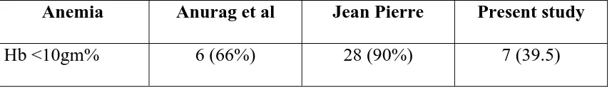
Hemoglobin and white cells counts taken on admission are highly
informative. Only a rising or marked leucocytosis indicates inflammatory
condition, Patients with hemoglobin levels below l0gm% are considered as
Serum electrolytes, sugar, urea and creatinine are important, especially if
hypovolemia is expected. Uremia may present itself with persistent vomiting
accompanied by increasing distention of the abdomen.
In patients with suspected hepato biliary disease, LFT (Serum bilirubin
alkaline phosphatase SGOT, SGPT) albumin and globulin are useful to
differentiate medical from surgical disorders, to gauge the severity of
underlying disease and to assess the nutritional status.
Urine Test:
So easily performed, yet frequently over looked. Dark urine or raised
specific gravity reflects mild dehydration in patient with normal renal function.
Hyper bilirubinemia may give rise to tea colored urine and the froth on shaking.
Microscopic proteinuria confirms diabetic nephropathy.
Radiological studies:
Plain chest X-ray: A chest x-ray is essential in all cases of acute
abdomen for preoperative assessment. COFD changes pulmonary infection has
implications on post operative events like wound dehiscence.
Plain X-ray abdomen: Plain supine and erect films of the Abdomen or
lateral decubitus view in weak patients should be obtained. Radiographic
abnormalities are present up to 40% of patients with acute abdomen but are
individuals in who clear cut physical signs mandating laparotomy already exist
or patient with only mild resolving non-specific pain.
X-Ray findings in General Pathological conditions
1. Air fluid levels due to ileus are characterized by gas being seen throughout the GI tract.
Ileus may be seen in
♦ General or local peritonitis
♦ Typhoid fever
♦ Blunt injury abdomen
♦ Fracture spine
♦ Secondary gaseous distension after
• Biliary colic
• Ureteric coli
• Torsion
• Retroperitoneal hematoma
Bowel obstruction is manifested by multiple air and fluid level with
dilation of the bowel proximal to the obstruction. A high small bowel
obstruction may be difficult to diagnose because repeated vomiting will
differentiated from large bowel obstruction by identifying circular valvulae
conniventes, folds of mucous membrane of small bowel and the haustral folds.
2. Pneumo peritoneum:
Free air in peritoneum is usually detected when X-ray is taken in erect
posture. The film includes diaphragm. If the patient is too ill, he may be placed
in lateral position, that is lying on one flank up and X-ray obtained as lateral
view of abdomen.
Free air develops in association with perforated viscus, commonest being
perforated duodenal ulcer. Here gas collects in moderate quantities usually
under the right hemi diaphragm. In contrast, in small intestine perforation only
a small quantities of air escape. In perforation of the stomach and colon may be
associated with much larger quantities of free intra peritoneal air which
commonly accumulated under the diaphragm.
Other conditions with free air under diaphragm being typhoid
perforation, diverticulitis with perforation. Pneumo peritoneum is unusual in
appendicular perforation. Free gas under hemidiaphragm is present in
RESULTS AND ANALYSIS
18 patients with wound dehiscence following mass closure of midline
laparatomy were studied. Their preoperative status was studied. Incidence of
various etiological factors in abdominal wound dehiscence were as follows.
Age and incidence
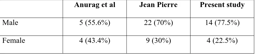
• Mean age of the study subject was 35.8 and the range was 18-90 years.
• 77.5% were male patients; 22.5% were female patients. Male female
ratio was 3.5:1.
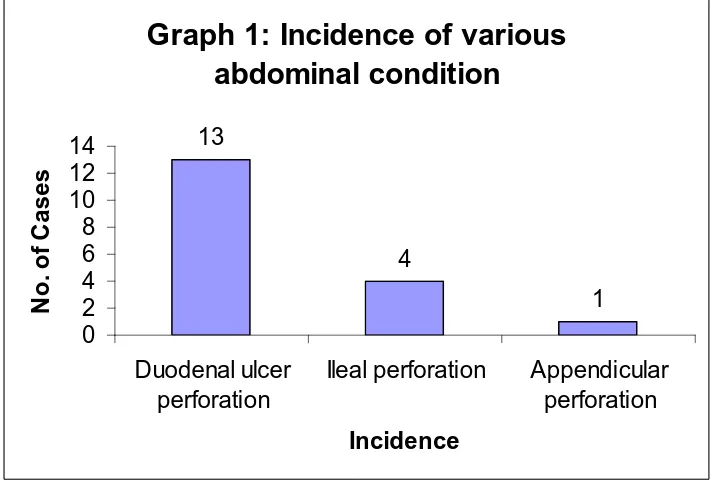
Table 1: Incidence of various abdominal condition
Incidence of various abdominal conditions in patient with wound dehiscence
was as follows
No. of cases Percentage
Duodenal ulcer perforation 13 72.2
Ileal perforation 4 22.3
History
Table 2: Duration of symptom (from on the onset of symptoms to the time of surgery)
Days No of patients
1 2
2 3
3 5
4 and above 8
Duration of symptom in cases varied from 1 to 9. Following table shows
the duration of symptoms before patients underwent surgery. More than 40% of
dehiscence were in the patients with symptoms of longer duration (>4 days).
Longer duration of symptoms was associated with more sever contamination
and hemodynamic instability.
Table 3: Co morbid condition at the time of admission
Conditions No of cases Percentage
Diabetes 3 16.5
Hypertension - -
Pulmonary disease 3 16.5
[image:57.612.149.470.522.682.2]Anemia 7 38.5
Drug history - -
CRF - -
Malignancy - -
Intra abdominal infection 18 100
Radiation - -
Table 4: General Physical Examination
No of patients Percentage
Pallor 9 50
Jaundice 4 22
Pedal edema 4 22
BP ( systolic <100- stock) 5 27.5
Pulse rate
• 7 (39.5%) patients had normal pulse rate and normal rhythm
• 11 (62.5%) patients had tachycardia (PR>100)
Blood pressure
• No patients were hypertensive
• 6 (33%) patients were in shock at the time of admission
Table 5: Anemia
HB No of patients Percentage
<8 5 27.5
8-10 2 11
>10 11 60.5
Table 6: Chest X-ray Findings
X- Ray Findings No of patients Percentage
Gas under diaphragm 17 94.5
Chronic obstructive
pulmonary disease
3 16.5
Pulmonary Koch - -
On X-ray 17 showed gas under diaphragm, 3 chronic obstructive
[image:59.612.133.485.311.468.2]Table 7: Electrocardiogram
Electrocardiogram No. of patients
Normal 17
Abnormal 1
Out of 18 patients abnormal findings were present in 1 and rest were
normal.
Table 8: Renal function test (RFT)
No of patients RFT
Normal Raised
Blood urea 14 4
Serum Creatinine 15 3
Renal function was assessed by measuring blood urea and serum
Creatinine. Of the 18 patients 4 had raised blood urea levels (>40mg/dl) and 3