Received 3 June 1997/Accepted 21 August 1997
Tumor suppressors of the retinoblastoma susceptibility gene family regulate cell growth and differentiation.
Polyomavirus large T antigens (large T) bind Rb family members and block their function. Mutations of large
T sequences conserved with the DnaJ family affect large T binding to a cellular DnaK, heat shock protein 70.
The same mutations abolish large T activation of E2F-containing promoters and Rb binding-dependent large
T activation of cell cycle progression. Cotransfection of a cellular DnaJ domain blocks wild-type large T action,
showing that the connection between the chaperone system and tumor suppressors is direct. Although they are
inactive in assays dependent on Rb family binding, mutants in the J region retain the ability to associate with
pRb, p107, and p130. This suggests that binding of Rb family members by large T is not sufficient for their
inactivation and that a functional J domain is required as well. This work connects the DnaJ and DnaK
molecular chaperones to regulation of tumor suppressors by polyomavirus large T.
The replication of papovaviruses requires that cells enter S
phase (58). Not surprisingly, the viruses have evolved abilities
to stimulate cells (37). Each early gene product has the ability
to affect cellular pathways of growth regulation; their study has
provided important insight into cellular signal transduction.
Murine polyomavirus induces both cellular DNA replication
(10) and enzymes associated with DNA synthesis (36). A single
early gene product, large T antigen (LT), can induce cellular
DNA replication in the absence of other virus transforming
genes (17, 49). Much of LT’s ability to stimulate cell cycle
progression is connected to its association with members of the
retinoblastoma susceptibility gene (Rb) family. LT
immortal-izes primary cells in a manner dependent on its binding site for
pRb, p107, and p130 (14, 31). LT also can block cell cycle
withdrawal and prevent differentiation of myoblasts, again
de-pendent on Rb family binding (33).
One very highly conserved sequence in polyomavirus T
an-tigens is the HPDKGG found between residues 42 and 47 of
murine polyomavirus LT (42). It has been noted that this
sequence connects T antigens to the DnaJ family (6, 26).
Fig-ure 1 compares some DnaJ sequences with those of
polyoma-virus LTs.
The DnaJ family is a diverse set of proteins characterized by
a J domain (3, 9, 54). This is a highly conserved region of about
70 amino acids, usually at the N terminus. The structures of the
Escherichia coli and hsp40 DnaJ domains have been
deter-mined (19, 41, 43, 57). There are four helices with a conserved
HPD sequence on a loop between helices 2 and 3. This HPD
sequence is important to DnaJ function (13, 60, 62). Genetic
evidence supports the importance of the interaction
be-tween DnaJ and DnaK (51). DnaJs bind to and stimulate the
ATPase activity of DnaK (32, 35) in a manner requiring the
HPD sequence. DnaK chaperones in turn interact with protein
substrates in an ATP-dependent manner to prevent
aggrega-tion and to promote protein folding (15, 16). In addiaggrega-tion to
roles in protein folding, DnaJ family members are involved in
protein translocation (4). Of particular interest, this family is
involved in rearrangements in protein complexes. In
l
repli-cation, for example, DnaJ and DnaK are involved in the
re-lease of DnaB helicase from
l
P (1). In plasmid replication,
they are involved in dissociating repA dimers to monomers
(63). Auxilin, a protein with a DnaJ homology, is involved in
recruitment of hsc70 as part of the process of stripping vesicle
coats (61).
The purpose of this work was to examine the importance of
the DnaJ homology to polyomavirus LT function. This work,
along with the work of Campbell et al. (5), shows a critical role
for DnaJ homology in the binding of a DnaK family member.
It also shows that the ability of polyomavirus LT to block the
function of Rb family members is dependent on the J
homol-ogy region.
MATERIALS AND METHODS
Plasmids and mutagenesis.pCMV LT (20) and pCMV-Rb2LT (21) pro-duced by the subcloning of a PCR fragment containing LXCXE mutations of Leu142to Val and Glu146to Gln have both been described previously.
Six different point mutants (H42Q, P43S, and D44N in the HPD motif and Q32E, A33S, and Y34K in helix 2) were constructed. HPD mutants (H42Q, P43S, and D44N) were made by overlap PCR. 59-TGCTACTGCAGCCAGAC AAAG-39and CTTTGTCTGGCTGCAGTAGCA were used for H42Q; 59-GC TACTGCACTCAGACAAAG-39and 59-CCTTTGTCTGAGTGCAGTAGC-39 were used for P43S; and 59-ACTGCACCCAAACAAAGGTCC-39and 59-CCA CCTTTGTTTGGGTGCAGT-39were used to create D44N. 59-GCGCGCGCT AGCTGATCATGGATAGAGTTCTGAGCAGAG-39, which is complemen-tary to the noncoding strand at the LT start codon was used as the upstream primer. 59-GCGCGCTGATCACGGGGGACCCTGATATGACGCGC-39, which is complementary to the coding strand beginning at the C-terminus, was used as the downstream primer. The template was wild-type cytomegalovirus (CMV) LT. PCR was carried out with Vent polymerase (New England Biolabs) in a Perkin-Elmer Thermal Cycler for 25 cycles of 1 min at 96°C, 1 min at 55°C, and 2 min at 72°C. After BamHI digestion, the fragment was ligated into the
BamHI site of the pCMV NeoBam parent vector (39). Mutants Q32E and A33S
were made with oligonucleotides 59-GGAAGAATGCAG(C/G)AG(G/T)CATA TAAGCAGCAGTC-39and 59-GACTGCTGCTTATATG(A/C)CT(G/C)CTGC ATTCTTCC-39. Y34N was constructed with 59-AGAATGCAGCAGGCA(T/A) AT(A/G)AGCAGCAGTCACTGC-39 and 59-GCAGTGACTGCTGCT(T/C)A T(T/A)TGCCTGCTGCATTCT-39. All mutations were verified by dideoxy se-quencing (45).
myc-tagged Hsj1 fragments were created by PCR. To create a construct with nine amino acids of myc sequence that could be recognized by 9E10 antibody
* Corresponding author. Mailing address: Department of
Biochem-istry, Tufts University School of Medicine, 136 Harrison Ave., Boston,
MA 02111. Phone: (617) 636-6876. Fax: (617) 636-6409. E-mail:
[email protected].
9410
on November 9, 2019 by guest
http://jvi.asm.org/
(53) and that contained residues 2 to 78 of Hsj1, 59-CGCGGGATCCAACAT GGAACAGAAACTCATCTCTGAAGAGGATCTGGCTAGCGCATCCTAC TACGAGATC-39 and 59-CGCGGGATCCGCTAGCCTAAGTTCCTGTCCC TGTCAG-39were the primers used on either wild-type or Hsj1 HQ mutant template. CMV vectors expressing adenovirus E1A 12S and human papilloma-virus (HPV) E7 have been described previously (15). The EC113 E2CAT (22), provided by Elliot Androphy, contains the adenovirus E2 promoter with its E2F sites; pA10-E2F-CAT plasmid had the285-to-230 adenovirus E2 promoter sequence and a minimal simian virus 40 (SV40) promoter fused to a chloram-phenicol acetyltransferase (CAT) gene (21, 52). Rous sarcoma virus (RSV) b-galactosidase (b-Gal) (2, 11) vector was provided by Amy Yee.
Cell lines and transfections.NIH 3T3 cells originally obtained from the Amer-ican Type Culture Collection (ATCC) were kindly provided by Bruce Cohen. The cells were grown in Dulbecco’s modified Eagle’s medium (DMEM; Gibco) supplemented with 10% calf serum (Hyclone).
Transfections were performed by the calcium phosphate precipitation method of Chen and Okayama (8). The cells were ordinarily used 48 h posttransfection. For serum starvation experiments, the precipitate was left on the cells for 4 to 6 h, after which the cells were washed twice with phosphate-buffered saline (PBS) and fed with DMEM containing 0.2% calf serum.
CAT assays.NIH 3T3 cells transfected at 15 to 25% confluence with 2mg of pA10-E2F-CAT or EC113 E2CAT and different LT, E1A, or E7 expression vectors were harvested 48 h posttransfection. In all cases, the amount of CMV expression vector was kept constant by the addition of empty vector containing the CMV promoter. CAT activity was measured by standard chromatographic techniques (18). Chloramphenicols on thin-layer chromatography plates were quantitated with ImageQuant software (Molecular Dynamics) to determine the percentage of total14C in acetylated forms of chloramphenicol versus in all forms.
Measurement of S-phase induction.Bromodeoxyuridine (BrdU) labeling and staining have been described previously (17). Briefly, cells incubated in 0.2% serum for approximately 48 h were labeled with 100mM BrdU for an additional 12 to 16 h. The cells were then stained for LT with a rabbit polyclonal antibody and with the monoclonal antibody BU-1 (Amersham) for BrdU. After PBS washes, the cells were stained with a combination of anti-rabbit fluorescein isothiocyanate (FITC; Kappel) and mouse TRITC (Sigma) secondary anti-bodies. Nuclear fluorescence was observed with a Zeiss microscope.
Immunoprecipitations.After washing and collection in PBS, the cells were extracted in T extraction buffer (TEB; 137 mM NaCl, 10 mM Tris-Cl [pH 8.0], 1 mM MgCl2, 1 mM CaCl2, 10% [vol/vol] glycerol, 1% [vol/vol] Nonidet P-40) for 20 min at 4°C. Cleared extracts were incubated with antibody and protein A-Sepharose (Pharmacia) for 1 h. After washing in PBS1, immunoprecipitates were boiled for 2 min in dissociation buffer (DB; 62.5 mM Tris-Cl [pH 6.8], 5% [wt/vol] sodium dodecyl sulfate [SDS], 25% [vol/vol] glycerol, 0.0075% [wt/vol] bromophenol blue, 50ml ofb-mercaptoethanol per ml) and subjected to SDS-polyacrylamide gel electrophoresis (PAGE) (30). After electrophoresis, samples were blotted onto nitrocellulose and analyzed by immunoblotting (59). The antibodies used in blotting included mouse monoclonal PN116 (20) or anti-LT polyclonal rabbit serum for LT, 12CA5 to detect hemagglutinin (HA)-tagged Rb, and p130 or p107 and 9E10 to detect myc-tagged Hsj1.
RESULTS
To test the role of the DnaJ region of LT, initial attention
focused on HPDKGG. Deletion or substitution of these
resi-dues was tried, but the mutant proteins were not stably
ex-pressed. Then, three different HPD point mutants (H42Q,
P43S, and D44N) were created by using overlap PCR. LTs
were transiently expressed in NIH 3T3 cells with vectors that
used the CMV intermediate-early promoter for expression.
Each mutant was expressed at a level similar to that of the wild
type, as shown by immunofluorescence and Western blotting.
Immunofluorescence, showing nuclear staining with nucleolar
exclusion (not shown), was indistinguishable from the wild
type.
If polyomavirus LT functions as a DnaJ protein, it should
interact with members of the DnaK family. Figure 2 shows that
this was the case. After transfection with LT expression
vec-tors, immunoprecipitations were carried out with either anti-T
serum or monoclonal antibody SP822 that recognized hs70.
After SDS-PAGE, the samples were immunoblotted with
ei-ther anti-hsp70 or anti-T. The heat shock monoclonal antibody
precipitated wild-type LT but little if any P43S LT. This heat
shock antibody did not discriminate between hsp70 and hsc70.
Isoelectric focusing suggested that hsc70 was the relevant
part-ner (not shown). This is not unexpected, since Butel and
co-workers showed association of hsc70 with SV40 LT (46, 47).
Anti-T immunoprecipitation of P43S LT showed no
coprecipi-tation of hs70, again consistent with the need for the HPD
sequence in interactions of DnaJ with DnaK. A mutant
defec-tive in Rb binding was clearly still able to bind hsc70, although
the level was occasionally somewhat reduced. These genetics
are consistent with earlier demonstrations that interaction of
SV40 LT and hsc70 occurred in the first 97 residues of LT,
which include the J domain but not the Rb-binding motif (46,
47). Our recent work also showed the need for the HPD
sequence of SV40 LT in its interaction with hsc70 (5).
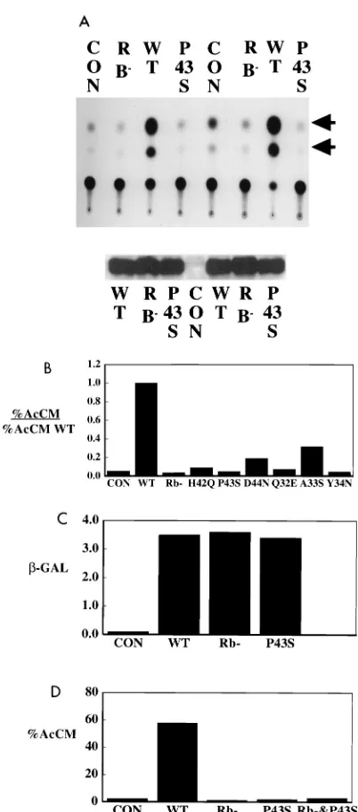
What is the role of the J region of LT? The one
character-FIG. 1. J domains and T antigens. Sequences of DnaJ proteins (top) compared to LT antigen sequences from different polyomaviruses (bottom). Residues in boldface are conserved throughout. Underlined residues of E. coli DnaJ represent the four alpha helices (57). Underlined residues in the murine polyomavirus (PY) LT sequence indicate alpha helices predicted according to Kneller and colleagues (26, 27).FIG. 2. Comparison of mutant and wild-type LT binding to hs70. Cells trans-fected with vector expressing Rb2LT (Rb2), control CMV vector (C), vector expressing wild type (WT), or the P43S HPD mutant LT (P43S) were extracted 46 h posttransfection. After extraction, immunoprecipitation was carried out with either anti-T serum (aT) or anti-hsp70 monoclonal antibody SP822 (aHS). SDS-PAGE was followed by blotting and immunostaining with either SP822 (aHS) or with anti-LT (aT).
on November 9, 2019 by guest
http://jvi.asm.org/
activation. The important result is that shown for the P43S
mutant. This mutant lacked the ability to transactivate E2F
sites (Fig. 3A). Results similar to those with P43S have been
obtained for the H42Q and D44N mutations in the conserved
HPD sequence (Fig. 3B). In many repeated experiments, no
substantial activation was observed with the HPD mutants.
Mutants defective in the HPD sequence were almost as
defec-tive as the LT mutant in the Rb binding site in this test of Rb
function. Other mutations in conserved residues in the J
do-main were also tried. Of the conserved residues in helix 2,
mutation of Q32 and Y34 gave inactive LT, while the A33
mutant gave significant activity. Mutation of leucine 17, a
res-idue expected to be involved in holding the helices together
(41, 57), yielded very low levels of protein expression (not
shown); presumably, this resulted from protein instability.
Two questions immediately arise about the role of the J
domain in the activation of E2F-containing promoters. Since
LT transactivates a variety of promoters, including ones that
are not Rb or E2F dependent, is the function of the J region
connected to the Rb interaction or is it general? The
experi-ment shown in Fig. 3C argues that the DnaJ domain was not
generally involved in all LT transactivation. LT was a potent
activator of an RSV–
b
-Gal construct. This activation was
de-pendent on neither Rb binding nor an intact J region. Similar
results have been obtained for activation of the fos promoter
(not shown).
The second question is whether the J domain is directly
connected to LT’s effects on the Rb family or whether it
rep-resents an independent activity required in addition to Rb
inactivation for E2F promoter activation. To assess these
al-ternatives, complementation experiments were performed.
Very little, if any, activation of the E2F promoter was observed
upon cotransfection of equal amounts of Rb
2and P43S LT
vectors (Fig. 3D). This failure to complement indicated that
the HPD sequence must function as a cis element for LT to act
on Rb family members.
[image:3.612.326.527.79.535.2]One concern was that N-terminal mutations simply unfolded
the N terminus, preventing large Ts interaction with the Rb
family. Cotransfection experiments with large T and individual
Rb family members were performed to rule this out.
HA-tagged Rb (HA-Rb), HA-p107, or HA-p130 was individually
cotransfected with wild-type or mutant LTs. After
immunopre-cipitation with anti-T serum, the samples were analyzed by
Western blotting. Figure 4A shows that equivalent amounts of
wild-type and mutant LTs were present after cotransfection
with p130. As expected, the LT mutant in the pRb binding site
did not bring down detectable quantities of p130 (B lane 2).
Figure 4B shows that the P43S mutant (lane 1) brought down
amounts of p130 equivalent to that for wild type (lane 3).
Figure 4C shows a coprecipitation experiment for p107, and
Fig. 4D shows one for pRb. In each case, the J domain mutant
LT bound the Rb family member at the same level as that of
the wild type. These results show that mutation in the HPD
FIG. 3. J region mutants are defective in Rb-dependent transactivation of E2F sites. (A) (Top) cells were cotransfected with A10 CAT (lanes 1 to 4) or EC113 E2 CAT (lanes 5 to 8) along with the indicated CMV expression plasmids. Cells were extracted 48 h posttransfection and assayed for CAT activity. Arrow-heads, acetylated chloramphenicols. (Bottom) Aliquots of the extracts were run on SDS-PAGE and blotted with anti-LT monoclonal pN116 to confirm protein expression. The set on the left are from the A10 E2FCAT transfections; the set on the right are from the EC113 E2CAT transfections. (B) Comparisons E2F transactivation by different J region mutants. Cells were cotransfected with A10 CAT with the indicated expression plasmids. The cells were extracted 48 h posttransfection and assayed for CAT activity. The data are presented as acti-vation relative to that of the wild type. (C) Neither the J domain nor the Rb binding site is important for LT activation of RSV–b-Gal constructs. Cells were cotransfected with RSV–b-Gal and with the indicated expression plasmids. After 48 h,b-Gal activity was measured by standard techniques with o-nitrophenyl-b -D-galactopyranoside and measurement of the optical density at 420 nm. (D) Rb2 and J2mutants of LT do not complement each other. Cells were transfected with A10 CAT and control CMV vector, wild type, Rb2, P43S, or equal amounts of P43S and Rb2. CAT activities measured approximately 48 h after transfection are expressed as percent acetylated chloramphenicols (AcCM). Con, control; WT, wild type.
on November 9, 2019 by guest
http://jvi.asm.org/
sequence was not simply preventing LT-Rb family member
interactions.
The result with p130 suggests another aspect of the
interac-tion. When p130 and HPD mutant LT were cotransfected,
p130 (Fig. 4B, lane 1) showed a mobility shift of the sort
previously attributed to phosphorylation. In fact, p130 in cells
expressing mutant LT was more heavily labeled with
32PO
4
.
This is consistent with results of the DeCaprio lab (56),
show-ing that expression of N-terminal mutants of SV40 LT resulted
in a high level of p130 phosphorylation compared with the wild
type.
LT can promote cell cycle progression. In some cells, such as
ATCC NIH 3T3 cells, this function requires interaction with
Rb family members. The ability to promote S phase after
serum withdrawal can be measured by BrdU incorporation
(Fig. 5). In these cells, which behave differently from the NIH
3T3 clone used by Gjorup et al. (17), LT lost the ability to
promote S phase when its Rb binding site was mutated. The
ability of large T to promote BrdU incorporation was also lost
when its HPD sequence was mutated. As for transactivation,
an intact HPD sequence was required for a productive
inter-action between LT and Rb family members.
The failure of Rb
2and P43S complementation (Fig. 3D)
suggested that DnaJ function and inactivation of Rb family
member function were closely connected. This was confirmed
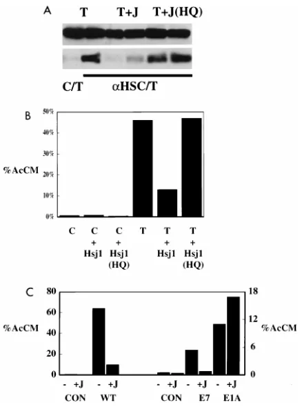
by examining the consequences of coexpression of a
heterolo-gous DnaJ domain. We (5) have shown that sequences from
Hsj1, a human DnaJ protein, could be substituted for SV40
sequences to restore LT function in DNA replication. CMV
vectors expressing residues 2 to 78 of the Hsj1 J domain in both
wild-type and H31Q mutant forms fused to a myc epitope tag
were prepared. Figure 6A showed that association of LT with
heat shock 70 could be suppressed by expression of Hsj1
se-quences 2 to 78. This result suggests that the Hsj1 fragment
titrated the DnaK interacting with LT. As expected, mutant
Hsj1 with the H of the HPD mutated to Q failed to block
association. Figure 6B shows that the Hsj1 construct also
blocked LT activation of E2F containing promoters, again
depending on an intact HPD sequence. The inhibition of LT
activation of E2F-containing promoters observed with Hsj1
has been repeated more than a dozen times in NIH 3T3 and
U2OS cells. The size of the effect varied from threefold to
FIG. 4. HPD mutant P43S binds Rb family members. Cells were [image:4.612.319.538.66.361.2]cotrans-fected with either P43S LT (lanes 1), Rb2LT (lanes 2), or wild-type LT (lanes 3) along with either tagged p130 (A and B), tagged p107 (C), or HA-tagged Rb (D). Anti-T antibody was used to immunoprecipitate LT from extracts made approximately 44 h after transfection. After SDS-PAGE and blotting, LT was detected by blotting with LT monoclonal antibody pN116 (A); Rb family members p130 (B), p107 (C), and pRb (D) were detected with 12CA5 to detect the HA tag.
FIG. 5. Mutation of the J region inactivates the ability of LT to promote cellular DNA replication. ATCC NIH 3T3 cells were transfected with the indi-cated DNAs. Transfected cells incubated in 0.2% serum for approximately 48 h were labeled with 100mM BrdU for an additional 14 h. After staining for LT with rabbit polyclonal antibody-anti-rabbit FITC and BrdU with the monoclonal antibody BU-1–anti-mouse TRITC (Amersham), nuclear fluorescence was scored with a Zeiss microscope. Percent positives represent counts of more than 200 T-positive cells in each case. Con, control; WT, wild type.
FIG. 6. Effects of coexpression of the heterologous J domain from Hsj1. (A) Exogenously expressed J domain (J) of Hsj1 blocks LT-hsc70 interaction. NIH 3T3 cells were transfected with vector expressing LT (T) and control vector (C) (lanes 1 and 2), a vector expressing Hsj1 sequences 1 to 78 (lanes 3 and 4), or a vector expressing an H-to-Q mutant of Hsj1 1 to 78 (lanes 5 and 6). Aliquots of cell extracts were run directly on SDS-PAGE (top) or were immunoprecipitated (bottom) with control monoclonal antibody (lane 1) or anti-hsp70 SP822 (lanes 2 to 6) and then run on SDS-PAGE gels. Samples were then blotted with anti-LT (PN116). (B) Exogenously expressed J domain of Hsj1 blocks LT transactivation of E2F promoters. A10 E2FCAT expression vector was cotransfected with either control vector (C) or wild-type LT (T) and with either control vector, a vector expressing myc-tagged Hsj1 wild-type sequence 1 to 78, or a vector expressing myc-tagged Hsj1 mutant H31Q sequence. CAT activity measured approximately 48 h after transfection is expressed as percent acetylated chloramphenicols (AcCM). (C) A10 E2FCAT expression vector was transfected with CMV control (Con) or with wild-type (WT) LT, adenovirus E1A 12S, or HPV E7 as well as with control or a vector expressing myc-Hsj1 sequences 2 to 78. CAT activity measured approximately 48 h after transfection is expressed as percent acety-lated chloramphenicols (AcCM). Note that the scales are different for LT acti-vation compared with E7 or E1A actiacti-vation of E2FCAT.
on November 9, 2019 by guest
http://jvi.asm.org/
[image:4.612.52.287.562.657.2]there was actually a stimulation. In five other experiments,
most showed no effect and a twofold decrease was observed
once. The difference in response to Hsj1 between LT and E1A
activation of E2F promoters showed that the inhibition
ob-served with LT was not a general effect. For HPV E7, the
ability to activate a promoter containing E2F sites could be
blocked by exogenous Hsj1. In four experiments, effects from
4- to 10-fold were observed. This suggests that E7 may utilize
chaperone pathways to reach the Rb family.
DISCUSSION
The similarity of the N-terminal sequence of polyomavirus T
antigens to those of the DnaJ family is clear. As for J domain
structures, four helical regions are predicted for the LT N
terminus with the conserved HPD motif on the loop between
helices 2 and 3. As expected for DnaJs, LT associates with
hsc70, a member of the DnaK family. The HPD motif is
im-portant for the interaction of DnaJ proteins with DnaK family
members (62). Consistent with this, LT binding to hsc70
re-quires an intact HPD sequence. There is also a functional link.
LT function genetically dependent on the integrity of its J
sequences can be blocked by coexpression of a heterologous
cellular DnaJ sequence. These results suggest that LT acts as a
DnaJ family member. Two very recent results also support the
idea that the N-terminal regions of polyomavirus T antigens
function as a DnaJ. First, Kelley and Georgopoulos (25)
showed that the N termini of SV40 and BK and JC viruses
could functionally substitute for E. coli DNA J sequences in
bacteria. Secondly, the J domain sequence of Hsj1 could be
substituted into SV40 LT to allow viral DNA replication (5).
The substitution experiments argue that approximately 80
res-idues of the T antigens are an independent folding unit
rep-resenting a domain. This is likely true for polyomavirus LT,
although proteolysis experiments demonstrated only a larger
260 amino-terminal element (17).
The DnaJ region of LT is critical for a productive interaction
between LT and members of the Rb tumor suppressor family
as measured by E2F transactivation or cell cycle assays.
Be-cause Rb
2and HPD LT mutants fail to complement one
another, hsc70 binding and Rb binding cannot represent
inde-pendent functions. LT oligomerizes (44), so failure to
comple-ment suggests that binding of Rb and hsc70 must be on the
same molecule. The suppression of LT transactivation by a
human DnaJ domain, Hsj1, can be explained by its competition
with LT for hsc70.
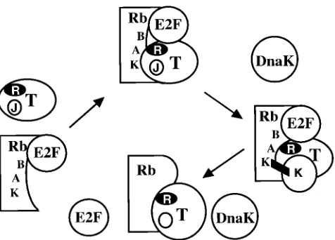
How does LT affect Rb family member function via the heat
shock binding site? Free E2F activates promoters, and
expres-sion of E2F1 can drive cells into S phase (24). Rb family
members block E2F function by forming complexes with
mem-bers of the E2F family. Ordinarily these interactions are cell
cycle dependent. When a viral oncogene is expressed,
com-plexes of Rb and E2F are disrupted (7). An interaction
be-tween Rb and hsp70 has already been shown (23). The
obser-vations suggest that the data can be explained by a simple
model (Fig. 7). Although the LXCXE motif of LT binds the
pocket of Rb family members, this interaction is not sufficient
to displace E2F family members from Rb. The J function of LT
serves to bring a substrate (Rb family-E2F family member
complexes) to a DnaK family member. Interaction between the
Rb-E2F complex and DnaK results in separation of Rb and
E2F. As with other DnaK activities, separation of Rb-E2F
complexes would require ATP. This model is simply a variation
of what has already been proposed for E1A; following the
interaction of CR2 and Rb, CR1 interacts with Rb to cause
separation of E2F and Rb (12). For E1A, which has CR1
sequences, transactivation should be insensitive to coexpressed
Hsj1 as observed.
The connection of the DnaJ region to Rb function implies
an important role in cell growth regulation by polyomaviruses.
For SV40 LT, the J domain is known to be important for
transformation (34, 40, 55). Polyomavirus LT is not a
trans-forming protein, so the ability to associate with DnaK or Rb
family members does not result in tumors. In fact, the tumor
profile of virus with a mutation blocking T-Rb interactions was
essentially the same as that of the wild type (14). However,
LT’s Rb-dependent abilities to block myoblast differentiation
(33) and to immortalize (31) reflect continuing cell cycle
pro-gression. When the ability to promote cellular DNA synthesis
depends on LT’s ability to bind Rb family members, it also
depends on an intact DnaJ sequence.
[image:5.612.311.549.66.237.2]Does the J domain serve other functions for the
polyoma-virus LTs? There have been connections between DnaK
pro-teins and translocation to the nucleus (38, 64). However,
im-munofluorescence showed that LT HPD mutants were
translocated efficiently to the nucleus (not shown); the
C-ter-minal domain (residues 264 to 785), lacking the 42 to 47
se-quences, was also nuclear (17). We have shown recently that
the J function of SV40 large T contributes significantly to SV40
DNA replication (5). This suggests an analogy to
l
replication
in which DnaJ and DnaK function in rearrangement of protein
complexes. Polyomavirus replication seems somewhat different
from SV40 replication. Clearly the J function cannot be
re-quired for polyomavirus DNA replication, since the C-terminal
domain can drive levels of replication comparable to those of
FIG. 7. Model for the role of the DnaJ domain in LT interactions with Rb family members. When LT encounters an Rb-E2F complex, it binds to the pocket (A and B) via its LXCXE motif. DnaK is recruited to the complex by the LT J domain. This DnaK acts on Rb to release active E2F. The site marked K on Rb may be the site of interaction between Rb and hsc70 reported by Inoue and coworkers (23).on November 9, 2019 by guest
http://jvi.asm.org/
the full-length molecule (17). Also, experiments with viral
DNA replication in growing cells using full-length LT showed
no striking defect for HPD mutants (not shown). Differences in
SV40 and polyomavirus LT domain interconnections could
explain the differences. For polyomavirus, the two domains
appear loosely associated, while for SV40 data have suggested
that the N- and C-terminal portions of the molecule are more
tightly linked (48, 50). The DnaJ domain in SV40 LT may
function to regulate domain interactions. Polyomavirus would
be analogous to mutants in the
l
system that weaken the
P-DnaB interaction and are less dependent on heat shock (28).
Finally, what of the relevance of DnaJs to growth situations
outside the polyomaviruses? Interestingly, in Drosophila,
mu-tation in a DnaJ homolog [1(2)tid] causes malignant growth of
the imaginal disc cells and death of the mutant larvae (29).
Since Hsj1 can also affect E7 activation of E2F-containing
promoters, the role of J domains in Rb family regulation may
extend to other viruses. An interaction between HPV E7 and
a novel human J domain protein has recently been discovered
(36a). The tantalizing question is whether normal cell cycle
progression might be critically dependent on DnaJ protein
function.
ACKNOWLEDGMENTS
This work was supported by NIH grants to B.S.S. (CA34722 and
CA50661), T.R. (CA32222 and CA50661), and J.D. (CA50661 and
CA63113).
ADDENDUM IN PROOF
Two very recent reports (A. Srinivasan, A. McClellan, J.
Vartikar, I. Marks, P. Cantalupo, Y. Li, P. Whyte, K. Rundell,
J. Brodsky, and J. Pipas, Mol. Cell. Biol. 17:4761–4773, 1997,
and H. Stubdal, J. Zalvide, K. Cambell, C. Schweitzer, T.
Roberts, and J. DeCaprio, Mol. Cell. Biol. 17:4979–4990, 1997)
have connected J domain function to growth and Rb family
regulation by SV40 LT.
REFERENCES
1. Alfano, C., and R. McMacken. 1989. Heat shock protein-mediated disassem-bly of nucleoprotein structures is required for the initiation of bacteriophage lambda DNA replication. J. Biol. Chem. 264:10709–10718.
2. Antonucci, T. K., P. Wen, and W. J. Rutter. 1989. Eukaryotic promoters drive gene expression in Escherichia coli. J. Biol. Chem. 264:17656–17659. 3. Bork, P., C. Sander, and A. Valencia. 1992. A nodule of the DnaJ heat shock
proteins found in malaria parasites. Trends Biochem. Sci. 17:129. 4. Brodsky, J. 1996. Post-translational protein translocation: not all hsc70s are
created equal. Trends Biochem. Sci. 21:122–126.
5. Campbell, K., K. Mullane, I. Aksoy, H. Stubdal, J. Pipas, P. Silver, T. Roberts, B. Schaffhausen, and J. DeCaprio.1997. DnaJ/hsp40 chaperone domain of SV40 large T promotes efficient viral DNA replication. Genes Dev. 11:1098–1110.
6. Cheetham, M. E., and J. P. Brion. 1992. Human homologues of the bacterial heat-shock protein DnaJ are preferentially expressed in neurons. Biochem. J. 284:469–476.
7. Chellapan, S., V. B. Kraus, B. Kroger, K. Munger, P. Howley, W. C. Phelps, and J. R. Nevins.1992. Adenovirus E1A, simian virus 40 tumor antigen, and human papilloma virus E7 protein share the capacity to disrupt the interac-tion between the transcripinterac-tion factor E2F and the retinoblastoma gene product. Proc. Natl. Acad. Sci. USA 89:4549–4553.
8. Chen, C. A., and H. Okayama. 1987. High-efficiency transformation of mam-malian cells by plasmid DNA. Mol. Cell. Biol. 7:2745–2752.
9. Cyr, D., T. Langer, and M. Douglas. 1994. DnaJ-like proteins: molecular chaperones and specific regulators of Hsp70. Trends Biochem. Sci. 19:176– 181.
10. Dulbecco, R., L. H. Hartwell, and M. Vogt. 1965. Induction of cellular DNA synthesis by polyoma virus. Proc. Natl. Acad. Sci. USA 53:403–408. 11. Edlund, T., M. D. Walker, P. J. Barr, and W. J. Rutter. 1985. Cell-specific
expression of the rat insulin gene: evidence for role of two distinct 59flanking elements. Science 230:912–916.
12. Fattaey, A. R., E. Harlow, and K. Helin. 1993. Independent regions of adenovirus E1A are required for binding to and dissociation of E2F-protein
complexes. Mol. Cell. Biol. 13:7267–7277.
13. Feldheim, D., J. Rothblatt, and R. Schekman. 1992. Topology and functional domains of Sec63p, an endoplasmic reticulum membrane protein required for secretory protein translocation. Mol. Cell. Biol. 12:3288–3296. 14. Freund, R., R. T. Bronson, and T. L. Benjamin. 1992. Separation of
immor-talization from tumor induction with polyoma large T mutants that fail to bind the retinoblastoma gene product. Oncogene 7:1979–1987.
15. Georgopoulos, C., and W. J. Welch. 1993. Role of the major heat shock proteins as molecular chaperones. Annu. Rev. Cell. Biol. 9:601–634. 16. Gething, M.-J., and J. Sambrook. 1992. Protein folding in the cell. Nature
355:33–44.
17. Gjørup, O., P. Rose, P. Holman, B. Bockus, and B. Schaffhausen. 1994. Protein domains connect cell cycle stimulation directly to initiation of DNA replication. Proc. Natl. Acad. Sci. USA 91:12125–12129.
18. Gorman, C., L. F. Moffat, and B. H. Howard. 1982. Recombinant genomes which express chloramphenicol acetyltransferase in mammalian cells. Mol. Cell. Biol. 2:1044–1051.
19. Hill, R. B., J. M. Flanagan, and J. H. Prestegard. 1995. 1H and 15N magnetic resonance assignments, secondary structure, and tertiary fold of Escherichia coli DnaJ(1-78). Biochemistry 34:5587–5589.
20. Holman, P., O. Gjørup, T. Davin, and B. Schaffhausen. 1994. Characteriza-tion of an immortalizing N-terminal domain of polyomavirus large T antigen. J. Virol. 68:668–673.
21. Howes, S., B. Bockus, and B. Schaffhausen. 1996. Genetic analysis of poly-omavirus large T nuclear localization: nuclear localization is required for productive association with pRb family members. J. Virol. 70:3581–3588. 22. Imperiale, M., R. Hart, and J. Nevins. 1985. An enhancer-like element in the
adenovirus E2 promoter contains sequences essential for uninduced and E1A-induced transcription. Proc. Natl. Acad. Sci. USA 82:381–385. 23. Inoue, A., T. Torigoe, K. Sogahata, K. Kamiguchi, S. Takahashi, Y. Sawada,
M. Saijo, Y. Taya, S. Ishii, N. Sato, et al.1995. 70-kDa heat shock cognate protein interacts directly with the N-terminal region of the retinoblastoma gene product pRb. Identification of a novel region of pRb-mediating protein interaction. J. Biol. Chem. 270:22571–22576.
24. Johnson, D., J. Schwarz, D. Cress, and J. Nevins. 1993. Expression of transcription factor E2F1 induces quiescent cells to enter S-phase. Nature 365:349–352.
25. Kelley, W. L., and C. Georgopoulos. 1997. The T/t common exon of simian virus 40, JC, and BK polyomavirus T antigens can functionally replace the J-domain of the Escherichia coli DnaJ molecular chaperone. Proc. Natl. Acad. Sci. USA 94:3679–3684.
26. Kelley, W. L., and S. J. Landry. 1994. Chaperone power in a virus? Trends Biochem. Sci. 19:277–278.
27. Kneller, D., F. Cohen, and R. Langridhe. 1990. Improvements in protein secondary structure prediction by an enhanced neural network. J. Mol. Biol. 214:171–182.
28. Konieczny, I., and J. Marszalek. 1995. The requirement for molecular chap-erones in lambda DNA replication is reduced by the mutation pi in lambda P gene, which weakens the interaction between lambda P protein and DnaB helicase. J. Biol. Chem. 270:9792–9799.
29. Kurzik-Dumke, U., D. Gundacker, M. Renthrop, and E. Gateff. 1995. Tumor suppression in Drosophila is causally related to the function of the lethal(2) tumorous imaginal discs gene, a dnaJ homolog. Dev. Genet. 16:64–76. 30. Laemmli, U. K. 1970. Cleavage of structural proteins during the assembly of
the head of bacteriophage T4. Nature 227:680–685.
31. Larose, A., N. Dyson, M. Sullivan, E. Harlow, and M. Bastin. 1991. Poly-omavirus large T mutants affected in retinoblastoma protein binding are defective in immortalization. J. Virol. 65:2308–2313.
32. Liberek, K., J. Marszalek, D. Ang, C. Georgopoulos, and M. Zylicz. 1991. Escherichia coli DnaJ and GrpE heat shock proteins jointly stimulate ATPase activity of DnaK. Proc. Natl. Acad. Sci. USA 88:2874–2878. 33. Maione, R., G. M. Fimia, P. Holman, B. Schaffhausen, and P. Amati. 1994.
Retinoblastoma antioncogene is involved in the inhibition of myogenesis by polyomavirus large T antigen. Cell Growth Differ. 5:231–237.
34. Marsilio, E., S. H. Cheng, B. Schaffhausen, E. Paucha, and D. M. Livingston. 1991. The T/t common region of simian virus 40 large T antigen contains a distinct transformation-governing sequence. J. Virol. 65:5647–5652. 35. McCarty, J. S., A. Buchberger, J. Reinstein, and B. Bukau. 1995. The role of
ATP in the functional cycle of the DnaK chaperone system. J. Mol. Biol. 249:126–137.
36. Mudrak, I., E. Ogris, H. Rotheneder, and E. Wintersberger. 1994. Coordi-nated transactivation of DNA synthesis- and precursor-producing enzymes by polyomavirus large T antigen through interaction with the retinoblastoma protein. Mol. Cell. Biol. 14:1886–1892.
36a.Mu¨nger, K. Personal communication.
37. Nevins, J. R. 1992. E2F: a link between the Rb tumor suppressor protein and viral oncoproteins. Science 258:424–429.
38. Okuno, Y., N. Imamoto, and Y. Yoneda. 1993. 70-kDa heat-shock cognate protein colocalizes with karyophilic proteins into the nucleus during their transport in vitro. Exp. Cell Res. 206:134–142.
39. Pasleau, F., M. J. Tocci, F. Leung, and J. J. Kopchick. 1985. Growth hor-mone gene expression in eukaryotic cells directed by the Rous sarcoma virus
on November 9, 2019 by guest
http://jvi.asm.org/
rol. 69:2842–2849.
45. Sanger, F., S. Nicklen, and A. R. Coulson. 1977. DNA sequencing with chain-terminating inhibitors. Proc. Natl. Acad. Sci. USA 74:5463–5468. 46. Sawai, E. T., and J. S. Butel. 1989. Association of a cellular heat shock
protein with simian virus 40 large T antigen in transformed cells. J. Virol. 63:3961–3973.
47. Sawai, E. T., G. Rasmussen, and J. S. Butel. 1994. Construction of SV40 deletion mutants and delimitation of the binding domain for heat shock protein to the amino terminus of large T-antigen. Virus Res. 31:367–378. 48. Scheidtmann, K. H., M. Buck, J. Schneider, D. Kalderon, E. Fanning, and
A. E. Smith. 1991. Biochemical characterization of phosphorylation site mutants of simian virus 40 large T antigen: evidence for interaction between amino- and carboxy-terminal domains. J. Virol. 65:1479–1490.
49. Schlegel, R., and T. L. Benjamin. 1978. Cellular alterations dependent upon the polyomavirus hr-t function: separation of mitogenic from transforming capacities. Cell 14:587–599.
50. Schwyzer, M., R. Weil, G. Frank, and H. Zuber. 1980. Amino acid sequence analysis of fragments generated by partial proteolysis from large SV40 tumor antigen. J. Biol. Chem. 255:5627–5634.
51. Scidmore, M. A., H. H. Okamura, and M. D. Rose. 1993. Genetic interac-tions between KAR2 and SEC63, encoding eukaryotic homologs of DnaK and DnaJ in the endoplasmic reticulum. Mol. Biol. Cell 4:1145–1159. 52. Shin, E., S. G. Tevosian, and A. S. Yee. 1996. The N-terminal Region of
E2F-1 is required for transcriptional activation of a new class of target
Sci. USA 91:11343–11347.
58. Tooze, J. E. 1980. DNA tumor viruses. The molecular biology of tumor viruses, vol. 1 and 2. Cold Spring Harbor Press, Cold Spring Harbor, N.Y. 59. Towbin, H., T. Staehelin, and J. Gordon. 1979. Electrophoretic transfer of proteins from polyacrylamide gels to nitrocellulose sheets: procedure and some applications. Proc. Natl. Acad. Sci. USA 76:4350–4354.
60. Tsai, J., and M. G. Douglas. 1996. A conserved HPD sequence of the J-domain is necessary for YDJ1 stimulation of Hsp70 ATPase activity at a site distinct from substrate binding. J. Biol. Chem. 271:9347–9354. 61. Ungewickell, E., H. Ungewickell, S. E. Holstein, R. Lindner, K. Prasad, W.
Barouch, B. Martin, L. E. Greene, and E. Eisenberg.1995. Role of auxilin in uncoating clathrin-coated vesicles. Nature 378:632–635.
62. Wall, D., M. Zylicz, and C. Georgopoulos. 1994. The NH2-terminal 108 amino acids of the Escherichia coli DnaJ protein stimulate the ATPase activity of DnaK and are sufficient for lambda replication. J. Biol. Chem. 269:5446–5451.
63. Wickner, S., D. Skowyra, J. Hoskins, and K. McKenney. 1992. DnaJ, DnaK, and GrpE heat shock proteins are required in oriP1 DNA replication solely at the RepA monomerization step. Proc. Natl. Acad. Sci. USA 89:10345– 10349.
64. Yang, J., and D. B. DeFranco. 1994. Differential roles of heat shock protein 70 in the in vitro nuclear import of glucocorticoid receptor and simian virus 40 large tumor antigen. Mol. Cell. Biol. 14:5088–5098.