The energetics of scleractinian coral larvae and implications for dispersal
121
0
0
Full text
(2) The energetics of scleractinian coral larvae and implications for dispersal. Thesis submitted by Erin Marie GRAHAM BSc (Hons), James Cook University in August 2012. For the degree of Doctor of Philosophy in the School of Marine and Tropical Biology James Cook University.
(3) Statement on the contribution of others. International tuition fees were paid for by an Endeavour International Postgraduate Research Scholarship from the Australian Government. Stipend support was provided by James Cook University (Postgraduate Research Scholarship). Intellectual and editorial support was provided by the supervisory team consisting of Professor Bette Willis (James Cook University), Dr. Andrew Baird (ARC Centre of Excellence for Coral Reef Studies) and Professor Sean Connolly (James Cook University). Funding for the research within this thesis was obtained from James Cook University, the Australian Research Council, the Great Barrier Reef Marine Park Authority, and the Environmental Futures Network. Additional intellectual, editorial, and research assistance and use of external infrastructure were provided by an external collaborator, Associate Professor Mary Sewell (University of Auckland). Mary Sewell and Angela Little (University of Auckland) developed the protocol that was used for lipid analysis.. 1.
(4) Acknowledgements. I would like to thank many people who have helped with fieldwork, including D. Abrego, L. Bay, J.C.H. Tan, K. Chong-Seng, C.M. Chua, V. Cumbo, J. Figueiredo, E. Puill-Stephan, F. Seneca, G. Torda, P. Warren, and staff from Orpheus Island Research Station. Thank you, M. Matz, for sharing your settlement cue preparation method. And very special thanks to S. Blowes, not only for coming out on every trip, but for listening to my tirades and keeping them under control. Lab assistance was generously provided by J.C.H. Tan and A. Negri (lipid analysis), C. Palmer (protein analysis), and A. Little and E. Zarate from the University of Auckland (Iatroscan). Thank you B. Leggat for -80 freezer space, and B. Bowden for use of the School of Chemistry’s freeze drier. I owe a lot of thanks to the staff at JCU, in particular P. Osmond and J. Webb (Boating and Diving); G. Jameson (Biological Stores); D. Bailey and K. Wood (Finance); S. Francis; and especially the IT guys: G. Bailey and V. Pullella. The feedback and camaraderie provided by everyone in the Ecological Modelling Lab was indispensable to my completion. An added thank you to L. Thibaut for help implementing monotonically constrained GAMs. Above all, a personal note of thanks to my supervisors, Bette, Andrew and Sean; good friends, Cassie, James, Rickard, Katerina, Mia, Vivian, Jess and Arnold; and family, who have been there for me every step of the way. I dedicate this thesis to my father, Ian Keith Graham.. 2.
(5) Abstract. Dispersal is a key process in the ecology and evolution of species. For sessile marine invertebrates like corals, the larval stage is the only means of dispersal, making this stage fundamentally important. Factors governing the dispersal potential of scleractinian corals include oceanographic conditions and the length of time coral larvae spend in the plankton. For lecithotrophic, broadcast-spawned coral larvae, the pelagic larval duration (PLD) will largely depend on larval energetics; that is, the amount of maternally-derived energy reserves available and the rate at which these reserves are used. The overall aim of this thesis was to investigate the energetics of scleractinian coral larvae to enhance understanding of the dispersal potential of corals, knowledge that is necessary for the design of marine protected areas and other management strategies needed to protect coral reefs. I first established the temporal dynamics of larval energy use by quantifying temporal changes in lipid content and respiration rates throughout the larval phase for a range of species. Building on these findings, I quantified how the dynamics of larval energetics changed under different experimental temperatures, to improve understanding of how climate change may influence larval dispersal and coral population connectivity. Finally, I considered how larval energetics affected post-settlement survival and growth to gain insights into whether time spent in the plankton might reduce a coral’s ability to contribute to postsettlement demography (i.e., “realized” dispersal). Lecithotrophic marine invertebrate larvae generally have shorter PLDs than planktotrophic larvae. However, non-feeding coral larvae have larval durations far exceeding predictions based on their energetics, raising questions about how they achieve such longevity. In this thesis, I measured temporal changes in metabolic rates 3.
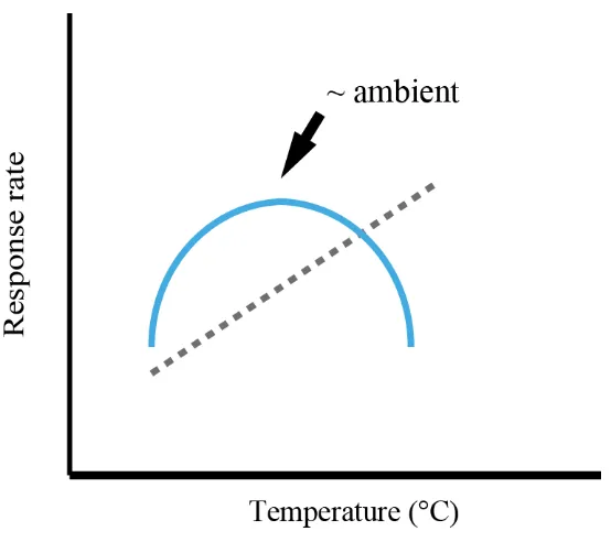
(6) and lipid content of larvae of four species of reef corals (Goniastea aspera, Acropora tenuis, A. nasuta, and A. spathulata) to determine whether changes in energy use through time contribute to their extended PLDs (Chapter 2). The temporal dynamics of both metabolic rates and lipid content were highly consistent among species. Metabolic rates pre-fertilization were low, and then increased rapidly during development to peak 1-2 days after spawning, when larvae began swimming. Rates then declined by up to two orders of magnitude over the following week, and remained low thereafter. Consistent with patterns in metabolic rates, lipid depletion was rapid during development, before slowing dramatically from about ten days onwards. Throughout this extended period of low metabolism, larvae continued to swim, complete metamorphosis, and showed no increase in mortality rates. The capacity of non-feeding coral larvae to enter a low-metabolism state soon after becoming competent significantly extends their dispersal potential, thereby accruing connectivity advantages typically associated with planktotrophy. Temperature is an important environmental variable affecting the metabolism of ectothermic organisms. Predicted increases in sea-surface temperatures due to climate change are likely to alter the energy use of coral larvae and thus influence the dispersal potential of corals. Using a regression-based approach, I quantified the effect of five temperatures on the survival and energy use of A. tenuis larvae (Chapter 3). Temperature had a significant effect on larval survival, with increasing temperature leading to a monotonic increase in mortality rates. Contrary to my expectation that metabolic rates would increase with temperature, however, temperature had a parabolic effect on peak respiration rates and lipid use during development: rates declined as temperatures either increased above or decreased below ambient. Moreover, temperature did not appear to affect larvae during the extended period of low 4.
(7) metabolism. My results suggest that even small differences in temperature from ambient affect coral larval dispersal potential. In particular, increased metabolism associated with warming temperatures leads to faster development, which increases the potential for self-recruitment, while higher mortality decreases the proportion of a larval cohort that survives for longer dispersal distances. Thus, connectivity among coral populations, which critically underpins reef resilience, is likely to decline in the near future. Demographic connectivity requires both the dispersal of individuals between sub-populations, and their subsequent contribution to population dynamics. For nonfeeding marine larvae, the capacity to delay settlement enables greater dispersal distances, but the energetic cost of delayed settlement can adversely impact postsettlement fitness. Accordingly, I assessed whether delayed settlement influences either mortality rates or growth rates for the first six weeks following settlement of A. tenuis larvae (Chapter 4). Larvae that were settled at two, four, and six weeks after spawning, and then deployed in the field, showed negligible effects of delayed settlement on postsettlement survival and time to initial budding for colony formation. Between-cohort differences in budding rate were best explained by temporal variation in the postsettlement acquisition of zooxanthellae. The potential for coral larvae to remain in the pelagic zone for increased periods of time, with little or no effect on post-settlement survival and growth, suggests that the costs of delayed settlement are largely confined to those accrued during the larval phase itself. This indicates that larvae that successfully settle after extended periods in the plankton are likely to make meaningful demographic contributions to benthic dynamics. Thus the predicted trade-off between delayed settlement and post-settlement fitness appears to be less applicable to reefbuilding scleractinian corals than other taxa with non-feeding larvae. 5.
(8) In conclusion, my research has identified a potentially novel physiological attribute of coral larvae that offers an explanation for their exceptionally long larval durations compared to other non-feeding marine invertebrate larvae. Specifically, coral larvae enter a low-metabolism state soon after competence is acquired, and they are able to maintain this state for many weeks, even at temperatures several degrees above ambient. Consistent with this, coral larvae that successfully settle after spending more time in the plankton survive and grow at rates similar to those of corals spending less time in the plankton. Nevertheless, in a warmer world, coral connectivity will likely decline due to temperature-dependent increases in larval mortality and development. The broader implications of these findings for the long-term persistence of coral metapopulations under climate change will depend on the relative importance of local population maintenance, versus replenishment from other sub-populations.. 6.
(9) Table of Contents. Statement on the contribution of others ............................................................................ 1 Acknowledgements ........................................................................................................... 2 Abstract ............................................................................................................................. 3 Table of Contents .............................................................................................................. 7 List of Tables .................................................................................................................... 9 List of Figures ................................................................................................................. 10. 1. General Introduction ................................................................................................ 12. 2. Rapid declines in metabolism explain extended coral larval longevity .................. 21 2.1. Introduction .................................................................................................. 21. 2.2. Materials and methods .................................................................................. 24. 2.2.1. Study site and larval cultures ................................................................ 24. 2.2.2. Sampling design .................................................................................... 24. 2.2.3. Respirometry ......................................................................................... 25. 2.2.4. Lipid analysis ........................................................................................ 26. 2.2.5. Survival ................................................................................................. 27. 2.2.6. Settlement assays .................................................................................. 27. 2.3. Results .......................................................................................................... 28. 2.3.1. Respirometry ......................................................................................... 28. 2.3.2. Lipid ...................................................................................................... 30. 2.3.3. Survival ................................................................................................. 32. 2.3.4. Settlement .............................................................................................. 34. 2.4. Discussion..................................................................................................... 36. 3 Temperature effects on the energetics of scleractinian coral larvae and their implications for dispersal ................................................................................................ 44 3.1. Introduction .................................................................................................. 44. 3.2. Materials and methods .................................................................................. 46. 3.2.1. Modelling approach .............................................................................. 48 7.
(10) 3.2.2. Survival ................................................................................................. 49. 3.2.3. Lipid ...................................................................................................... 50. 3.2.4. Respirometry ......................................................................................... 53. 3.3. Results .......................................................................................................... 56. 3.3.1. Survival ................................................................................................. 56. 3.3.2. Lipid ...................................................................................................... 59. 3.3.3. Respirometry ......................................................................................... 62. 3.4. Discussion..................................................................................................... 67. 4 Effects of delayed settlement on post-settlement growth and survival of scleractinian coral larvae ................................................................................................ 73 4.1. Introduction .................................................................................................. 73. 4.2. Materials and methods .................................................................................. 76. 4.2.1. Larval cultures ....................................................................................... 76. 4.2.2. Sampling design .................................................................................... 77. 4.2.3. Data analysis ......................................................................................... 78. 4.3. 4.3.1. Survival ................................................................................................. 79. 4.3.2. Growth................................................................................................... 84. 4.4. 5. Results .......................................................................................................... 79. Discussion..................................................................................................... 84. General Discussion .................................................................................................. 90 5.1. Larval energetics .......................................................................................... 91. 5.2. Temperature effects on larval energetics ...................................................... 96. 5.3. Effects of delayed settlement........................................................................ 98. 5.4. Dispersal potential of scleractinian corals .................................................... 99. 5.5. Conclusion .................................................................................................. 101. References ..................................................................................................................... 103 Appendix A ................................................................................................................... 115 Appendix B ................................................................................................................... 117. 8.
(11) List of Tables. Table 1.1 Summary of the maximum length of the competent stage and longevity for a range of scleractinian broadcast spawning species ......................................................... 20 Table 2.1 Estimates of lipid remaining after an additional 100 days in the plankton for three Acropora species.................................................................................................... 38 Table 2.2 Estimates of pelagic larval duration (PLD) for four study species using lipid content and oxygen consumption ................................................................................... 40 Table 3.1 Parameter estimates for the parabolic survival models and Akaike's Information Criterion (AIC) used for model comparison. .............................................. 57 Table 3.2 Model selection results for the effect of temperature on lipid depletion of Acropora tenuis larvae. ................................................................................................... 60 Table 4.1 Model selection results for time-to-event analyses of three cohorts of Acropora tenuis juveniles. .............................................................................................. 82 Table 4.2 Model-averaged estimates of the hazard ratio (with 95% confidence intervals) for the effect of delayed settlement on survival, time to bud, and acquisition of zooxanthellae of three cohorts of Acropora tenuis juveniles. .................................... 83. 9.
(12) List of Figures. Figure 2.1 Rates of oxygen consumption through time in four scleractinian coral species. ............................................................................................................................ 29 Figure 2.2 Depletion of energy lipids through time in four scleractinian coral species.31 Figure 2.3 Kaplan-Meier survival estimates for four scleractinian coral species. ......... 33 Figure 2.4 Proportion of larvae that are competent to settle for four species of scleractinian corals. ......................................................................................................... 35 Figure 2.5 Mass-normalized resting respiration rates for multicellular invertebrates ... 42 Figure 3.1 Illustration of two types of expected responses of metabolic rates due to increases in temperature.................................................................................................. 49 Figure 3.2 Illustration of possible outcomes tested for in the analysis of energetic lipid depletion in Acropora tenuis larvae maintained at five temperatures. ........................... 53 Figure 3.3 Estimated parametric survival function for larvae of Acropora tenuis fitted to the empirical survival data with the Weibull distribution .......................................... 58 Figure 3.4 Nonparametric Kaplan-Meier estimated median survival times for larvae of Acropora tenuis for each temperature treatment. ........................................................... 59 Figure 3.5 Best-model fits for the depletion of energy lipids through time by Acropora tenuis larvae maintained at five different temperatures. ................................................. 61 Figure 3.6 Estimated lipid depletion rates for Acropora tenuis larvae maintained at different temperatures ..................................................................................................... 62 Figure 3.7 Rates of oxygen consumption through time for Acropora tenuis larvae maintained at five different temperatures. ...................................................................... 64 Figure 3.8 Rates of oxygen consumption through time for Acropora tenuis larvae maintained at four different temperatures. ...................................................................... 65. 10.
(13) Figure 3.9 Peak rate of oxygen consumption for Acropora tenuis larvae maintained at four different temperatures. ............................................................................................ 66 Figure 3.10 Timing of peak rate of oxygen consumption for Acropora tenuis larvae maintained at four different temperatures. ...................................................................... 67 Figure 3.11 Patterns in the timing of A) peak oxygen consumption, and B) median larval lifespan larvae of Acropora tenuis maintained at four different temperatures. .... 70 Figure 4.1 Effect of delayed settlement on the survival, time to bud, and acquisition of zooxanthellae of three cohorts of Acropora tenuis settlers............................................. 81 Figure 4.2 Proportion of juveniles with zooxanthellae that also have buds for the three cohorts of Acropora tenuis settlers. ................................................................................ 87. 11.
(14) 1. General Introduction Dispersal is an important process in the ecology and evolution of species. Larval. dispersal affects the distribution and abundance of organisms, enabling replenishment of populations, colonization of new habitats and the expansion of geographic ranges (Underwood and Fairweather 1989, Hanski 1999, Clobert et al. 2001, Gaston 2003). Patterns of dispersal also have important consequences for the genetic structure of populations, affecting gene flow and rates of speciation and extinction (Bohonak 1999, Weersing and Toonen 2009). Species with limited means for dispersal generally have greater genetic structure than species with high dispersal potential (Nishikawa et al. 2003). Therefore, understanding dispersal is essential to understanding a species’ ecology and evolution. Just as importantly, understanding dispersal is essential for developing conservation and management strategies, particularly for the design of protected areas (Shanks et al. 2003) and for evaluating whether and how species can alter their geographical distributions in response to predicted changes in climate (Watkinson and Gill 2002). Despite its importance, there is considerable uncertainty about the extent to which local marine populations are self-seeded, compared to being maintained by recruits from nearby or distant habitats (Cowen et al. 2000). On coral reefs, this is normally expressed as a distinction between the extent to which recruitment consists of individuals recruiting back to the natal reef, compared to arriving from nearby or distant reefs. Like most marine invertebrates, corals have a complex life cycle consisting of a benthic adult phase and a pelagic larval phase. Most corals are broadcast spawners, releasing their gametes into the water column where fertilization and embryogenesis occurs externally (Baird et al. 2009). Initially, coral larvae are in a pre-competent stage lasting between two and five days (Nozawa and Harrison 2005), in which rapid 12.
(15) morphological and physiological changes occur before they can become competent, or capable of settlement. This is followed by a competent stage, in which the larvae are capable of metamorphosing into juvenile corals to begin their sessile phase. Thus the dispersal potential of corals will depend on the onset and duration of competence, in addition to survival during the dispersal stage. The majority of a cohort will typically acquire competence and settle within 10 days after spawning in the presence of a suitable settlement surface (Connolly and Baird 2010). In the absence of the right cue, however, settlement can be delayed many months (Table 1.1). The poor swimming capacity of coral larvae and their dependence on oceanographic processes for dispersal (Chia et al. 1984, Willis and Oliver 1990) highlight their need for adequate energy reserves to survive unpredictable planktonic durations and remain competent to settle. Accordingly, for broadcast-spawned corals, one of the most important biological characteristics affecting how long this competence period lasts is that larvae are lecithotrophic, or non-feeding. While the larvae of a few species of corals inherit symbiotic photosynthesizing dinoflagellates, or zooxanthellae, from their parents and are capable of supplementing their energy reserves, most larvae derive all of their energetic requirements from the yolk, which is inherently limited (Baird et al. 2009). Both survival and the duration of the competent period will be constrained by the amount of energy available in the egg and the rate at which this energy is expended, which makes understanding the energetics of competence and survival essential for understanding dispersal. Despite being lecithotrophic, coral larvae have remarkably long larval durations (Table 1.1). Larvae of some coral species are competent to settle for at least 100 days and several others are capable of surviving for at least 200 days (Graham et al. 2008, Connolly and Baird 2010). Moreover, because very few studies maintain larvae and conduct settlement assays for extended periods, 13.
(16) the total number of coral species capable of long larval lifespans may be considerable. Solving the puzzle of how coral larvae with limited energy reserves can have such long larval durations is therefore necessary to be able to make accurate predictions of dispersal potential. To date, there has been only one attempt to understand the length of the competent period based on an energetic approach. In the late 1980’s, Richmond (1987) measured the energy content and respiration rates of one species of brooding coral, Pocillopora damicornis, and used a simple model to predict the duration of the competent period:. 𝐶𝑝 =. 2 𝐸 (𝑂 × 𝑇𝑒 )−1 3 𝑐 𝑐. where C p = potential competency in days, E c = energy content of the larva, O c = net oxygen consumption rate in mg oxygen d-1, T e = energetic equivalency in joules used mg-1 oxygen consumed, and 2/3 is a constant for the amount of body mass Richmond believed was required for successful settlement. The main assumptions of Richmond’s model were 1) that P. damicornis planulae consisted of 17% protein, 70% lipid and 13% carbohydrate by weight; 2) these components were utilized at same rate in proportion to their occurrence within the larvae; and 3) larvae were competent until they reached 1/3 of their original size. Richmond (1988) later measured the respiration rate of Acropora tenuis larvae and used his model to predict the dispersal potential of this broadcast-spawned species. Assuming A. tenuis had the same tissue energy content as P. damicornis, Richmond’s model predicted a 20 d competent period for A. tenuis. By removing the size restriction for successful settlement, an upper bound estimate of 30 d can be made for larval longevity – this is the length of time predicted by the equation above for all of a coral larva’s initial energy content to be consumed. The 14.
(17) model estimate is substantially less than empirical observations (69 d, A. tenuis, Nishikawa and Sakai 2003) and suggests a need to revisit our understanding of larval energetics. In particular, there is a need to assess a number of larval physiological parameters that may contribute to this discrepancy, including temporal variation in biochemical composition of larvae; rates at which biochemical components are used; the capacity to supplement their energetic reserves; the capacity to regulate their energy expenditure; or some combination of these parameters. Empirically evaluating which of these alternatives contribute to coral larval energetics will improve our ability to make more accurate estimates of dispersal potential. Once competent, the length of time a larva can maintain this state will depend on its energy reserves. This is particularly true for lecithotrophic larvae, which are unable to supplement their endogenous reserves during the dispersal phase by feeding. Energy must be available for development, dispersal, and metamorphosis. A newly settled juvenile will then rely on its endogenous reserves until it develops into a selfsufficient coral with tentacles for heterotrophy and zooxanthellae for autotrophy. Long periods of time spent in the plankton should mean less energy remains for these other needs associated with metamorphosis and settlement and could have flow-on effects on the success of coral larvae post-settlement (e.g., Wendt 1998, Maldonado and Young 1999, Onituska et al. 2010). The potential for decreased growth and increased mortality after settlement should therefore increase for larvae with longer pelagic durations compared to larvae that settle immediately. This implies there may be an energetic threshold associated with the loss of competence, the onset of larval senescence, or successful settlement, which places an upper bound on dispersal potential. Ultimately, a better understanding of the energetics of coral larvae is essential for determining how reefs might cope with climate change. Temperature is one of the 15.
(18) most important environmental determinants of metabolic rate (Gillooly et al. 2001), particularly for those organisms that are unable to regulate their own body temperatures. Sessile marine invertebrates are exposed to continual fluxes in temperature that they are unable to avoid, and temperature is known to affect the physiology of marine species by increasing development and mortality rates and influencing rates of settlement (e.g., Negri et al. 2007, Nozawa and Harrison 2007, Randall and Szmant 2009a). Thus, temperature will likely be particularly important in causing variation in energy use patterns, competence dynamics, and survival in nature. Moreover, understanding how temperature affects physiological factors governing dispersal potential is relevant for understanding effects of climate change on population dynamics (O’Connor et al. 2007). For example, increased temperatures may substantially increase or decrease the number of larvae settling locally, as well as decrease the number of successful long distance dispersers. If increased temperatures lead to increased energy use, then larval durations are likely to decrease. Given the increases in sea-surface temperature predicted to occur within the next few decades, temperature will play an important role in determining which species are able to cope with climate change (IPCC 2007). The overall aim of my thesis is to investigate patterns of energy use of coral larvae to increase our understanding of the dispersal potential of scleractinian corals and the ecological implications of dispersal. In Chapter 2, I quantify temporal changes in larval lipid content and metabolic rates in order to reconcile the differences between the (short) pelagic larval durations estimated with Richmond’s competence model and the (much greater) empirical observations made in recent studies of scleractinian coral larvae. I show that energy use of coral larvae is highest during early development and remains high until larvae become competent to settle, after which larvae enter a state of 16.
(19) reduced metabolic rates which minimizes energy use and is maintained for as long as two months. These findings indicate that estimates of oxygen consumption taken early in the larval duration will lead to over-estimates of energy utilization over the entire lifespan. This temporal change in energy use offers a potential explanation for the discrepancy between Richmond’s calculations and long larval lifespans documented empirically. Chapter 2 combines data from studies of different species in two years that were originally planned as two separate chapters. However, to minimize repetition in introduction and methods sections, I have combined them into a single chapter. In Chapter 3, I examine the effect of temperature on the energetics of Acropora tenuis larvae in order to quantify the effect of temperature on survival, respiration rates, and lipid utilization. Most research in this area is designed to determine whether there are differences in survival or energy use among a small set of fixed temperatures; very few studies have actually tried to quantify the functional response of these variables to temperature. My results indicate that there are two types of responses to temperature elicited in coral larvae: 1) a monotonic response in the survival of larvae, where increased temperatures lead to progressively increasing mortality; and 2) a parabolic response in energy use, where respiration rates and lipid utilization appear to be maximized at ambient temperature at the time of spawning and performance decreases as temperatures move away from ambient. In Chapter 4, I test whether A. tenuis larvae that delay settlement for up to six weeks suffer post-settlement costs that would reduce “realized dispersal” – the extent to which larvae dispersing long distances actually contribute to post-settlement demography –compared to predictions of “potential dispersal” based on survival and competence dynamics. Although Chapter 2 indicates that coral larvae have enormous potential for dispersal given their extended competence periods and longevities, energy 17.
(20) expended during dispersal means less energy is available to complete metamorphosis, survive, and grow. I find that larvae that delay settlement for up to six weeks after spawning (five weeks after competence is acquired) do not suffer higher mortality, nor experience any decrease in growth, compared to larvae settling just two weeks after spawning. In fact, my data suggest that the acquisition of zooxanthellae is more important to the growth of coral juveniles than the length of the larval phase. This is consistent with the low energy expenditure and limited lipid depletion revealed by my study of the temporal dynamics of larval energy use, as documented in Chapter 2. My results indicate that coral larvae are equipped with ample energy reserves to survive for extended periods in the plankton, and suggest that the realized dispersal of coral larvae may be higher than predicted based on studies of other non-feeding marine invertebrates. In Chapter 5, I summarise the outcomes of the research described in the thesis,. assess its implications for understanding the ecology, evolution, conservation and management of corals and coral reefs, and suggest some productive directions for future research. In particular, my thesis shows that high dispersal potential of scleractinian coral larvae is due to the reduction in energy use that occurs during an extended period of reduced metabolism soon after larvae acquire competence. Significantly, this reduction in energy use allows coral larvae to delay settlement for up to six weeks after spawning without suffering heightened mortality or reduced post-settlement growth. Although increased temperatures lead to monotonically increasing mortality in coral larvae, respiration rates and lipid use decrease at temperatures on either side of ambient. These findings highlight the extraordinary dispersal potential of coral larvae and help to explain the large geographic range sizes of most broadcast-spawning species. They indicate that, although coral larvae have the means (energy reserves) to colonize areas outside of their. 18.
(21) present day boundaries in response to increases in sea surface temperatures, they will suffer increased mortality. Further work is needed, however, to determine the mechanism of temperature-dependent mortality in coral larvae because energy stores are not limited and therefore it is unlikely that larvae are dying of starvation. Further work is necessary to determine if coral larvae utilize other sources of energy, such as protein reserves and/or dissolved organic matter absorbed directly from seawater. Quantifying the minimum amount of energy required for a coral larva to be able to metamorphose, and how much energy is used during metamorphosis, would also help place an upper limit on dispersal potential.. 19.
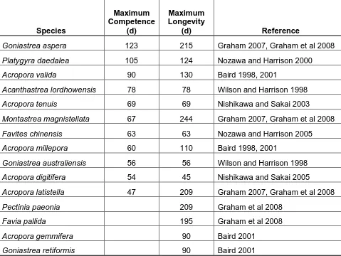
(22) Table 1.1 Summary of the maximum length of the competent stage and longevity for a range of scleractinian broadcast spawning species (updated from Graham et al. 2008). Maximum Competence (d). Maximum Longevity (d). Goniastrea aspera. 123. 215. Graham 2007, Graham et al 2008. Platygyra daedalea. 105. 124. Nozawa and Harrison 2000. Acropora valida. 90. 130. Baird 1998, 2001. Acanthastrea lordhowensis. 78. 78. Wilson and Harrison 1998. Acropora tenuis. 69. 69. Nishikawa and Sakai 2003. Montastrea magnistellata. 67. 244. Graham 2007, Graham et al 2008. Favites chinensis. 63. 63. Nozawa and Harrison 2005. Acropora millepora. 60. 110. Baird 1998, 2001. Goniastrea australiensis. 56. 56. Wilson and Harrison 1998. Acropora digitifera. 54. 45. Nishikawa and Sakai 2005. Acropora latistella. 47. 209. Graham 2007, Graham et al 2008. Pectinia paeonia. 209. Graham et al 2008. Favia pallida. 195. Graham et al 2008. Acropora gemmifera. 90. Baird 2001. Goniastrea retiformis. 90. Baird 2001. Species. 20. Reference.
(23) 2. Rapid declines in metabolism explain extended coral larval longevity. 2.1 Introduction Marine invertebrate larvae can be broadly classified into two categories, lecithotrophs or planktotrophs, depending on their source of nutrition during development. Planktotrophic larvae require external food sources to complete development, whereas lecithotrophic larvae are capable of completing development based solely on maternal provisions (Thorson 1950). The potential to feed should enable planktotrophic larvae to survive longer in the plankton (Scheltema 1986), and there are numerous examples of planktotrophic larvae that spend more time in the plankton than closely related species with lecithotrophic development (e.g. Emlet et al. 1987, Kempf and Todd 1989, Shanks et al. 2003). The longer PLDs of planktotrophic larvae are thought to confer greater dispersal potential for species with such larvae, enabling higher levels of gene flow over larger areas and potentially larger geographic ranges compared to species with non-feeding larvae (Jablonski and Lutz 1983, Pechenik 1999). Relationships between PLD, genetic population structure and range size have been documented for echinoids (Hunt 1993, Emlet 1995), gastropods (Hoskin 1997, Collin 2003, Paulay and Meyer 2006), and various other invertebrates (Foggo et al. 2007, Selkoe and Toonen 2011). However, the role of PLDs in driving such relationships is not always clear (Weersing and Toonen 2009), because population connectivity depends on many factors, including post-settlement processes (Marshall et al. 2010), that may obscure the role of larval duration alone. In reef-building scleractinian corals, larval development mode is generally a good predictor of patterns of dispersal and connectivity. Populations of brooding 21.
(24) species, whose larvae are ready to settle on release, typically have higher genetic structure than broadcast spawning species, whose larvae have an obligate planktonic period of 2-4 days (e.g., Hellberg 1996, Nishikawa et al. 2003). Corals, along with some high-latitude echinoderm taxa, are the only groups with non-feeding larvae for which extremely long PLDs have been documented (Birkeland et al. 1971, Hartnoll 1975, Sebens 1983, Bosch and Pearse 1990, Bryan 2004, Graham et al. 2008, Connolly and Baird 2010). For example, coral larvae can survive up to 200 days (Graham et al. 2008), and can complete metamorphosis up to at least 100 days after spawning (Connolly and Baird 2010). These examples indicate that species with lecithotrophic larvae, including many coral species, have developed strategies to extend larval duration, and thus accrue the advantages of dispersal traditionally associated with planktotrophy. If lecithotrophic larvae are to survive long periods in the plankton, they must possess a large supply of stored energy, have low metabolic rates, or be able to supplement their endogenous reserves. Large initial energy stores, slow development rates, and low rates of metabolism in Antarctic echinoderms imply that these larvae can persist for up to five years (Shilling and Manahan 1994). For scleractinian corals, a few species equip their propagules with photosynthetic symbionts (zooxanthellae), which may provide energy to larvae during dispersal and support PLDs over 100 days (Richmond 1987, Harii et al. 2010). However, most (>75%) coral species have larvae that lack such symbionts (Baird et al. 2009), yet even larvae of these species have exceedingly long PLDs. In Acropora tenuis larvae, initial energy content and metabolic rates observed during the first few weeks after fertilization imply larval longevities of only ~30 days (Richmond 1987, Graham et al. 2008). This estimate is less than half the 69 days observed for this species (Nishikawa et al. 2003), and many months less than 22.
(25) larval longevities reported for other Acropora species (Graham et al. 2008, Connolly and Baird 2010). Similar discrepancies between energy reserves, metabolic rates, and observed larval durations have been found in echinoderms (Bryan 2004). This suggests that corals and at least some other lecithotrophic larvae must either reduce their metabolic rates substantially as they age, take up additional energy (e.g., by absorption of dissolved organic matter (DOM)), or combine elements of both of these strategies. Although there is some evidence for uptake of DOM in soft corals (Ben-David-Zaslow and Y. Benayahu 2000), it has not, to date, been documented in scleractinian corals. Similarly, knowledge of metabolic rates for coral larvae is limited. Temporal changes in lipids over the first month after fertilization in one species suggest a slowing in the depletion of lipids after the first week, although lipids were still declining relatively rapidly towards the end of the study (e.g., by about 25% in the final week) (Harii et al. 2007). Consistent with this, Okubo and others (Okubo et al. 2008) found respiration rates of Acropora intermedia larvae declined to about one-third of peak values by seven days after spawning. These studies indicate that metabolic rates can decrease as coral larvae age, although these changes do not appear to be of sufficient magnitude to account for the extended PLDs documented in coral larvae. Determining whether long PLDs are widespread among coral species with lecithotrophic larvae, and understanding how these PLDs are attained, is critical to estimating the dispersal potential of this dominant group of reef builders. Such knowledge has important implications for understanding the ecology, evolution, and biogeography of corals and for anticipating how dispersal potential and population connectivity may be impacted by changing ocean conditions that have implications for metabolic rate, particularly increased seawater temperature. Therefore, in this chapter, I investigated physiological mechanisms underpinning extended PLDs in scleractinian 23.
(26) corals with non-zooxanthellate, lecithotrophic larvae, by quantifying respiration rates and energy use over larval lifespans. I found strong evidence in both respiration rates and lipid levels for a rapid decline in rates of energy use within approximately one week of spawning, even though larvae are still capable of settlement. I conclude that decreased metabolic rates (i.e, hypometabolism) allow non-feeding coral larvae to extend larval life by minimising depletion of their energy reserves.. 2.2 Materials and methods 2.2.1. Study site and larval cultures The study took place at Orpheus Island, Australia, in December 2008, and. November and December 2009. Gametes from a total of four broadcast spawning scleractinian species whose larvae lack zooxanthellae (Goniastrea aspera, Acropora tenuis, A. nasuta, and A. spathulata), were collected and cultured using established methods (Willis et al. 1997). Four to six adult colonies of each species were collected immediately prior to anticipated spawning dates and brought onshore. For each species, gametes were collected within an hour of release and combined. Once fertilized, developing embryos were transferred to 500 L fibreglass aquaria with flow through 0.2 µm filtered seawater (FSW) and continuous aeration, one tank per species. The aquaria were maintained in temperature controlled rooms at near-ambient temperature (27 +/1°C) and a 12 h light:dark cycle. 2.2.2. Sampling design At regular sampling intervals, subsamples of eggs, embryos, or larvae (hereafter. “propagules”) were randomly selected, for each species, and used for respiration measurements and lipid analysis. The first sample was taken from newly released 24.
(27) gametes, prior to fertilization. For respiration measurements, five replicates of 50 propagules were used. For lipid analysis of each Acropora species, the same 50 propagules used to measure respiration rates were subsequently frozen and used for lipid analysis. However, for lipid analysis of G. aspera propagules, due to their smaller size, three replicates of 600 propagules were used. Sampling took place every 12 h for the first 36-48 h to capture larval development through embryogenesis to a swimming larva, followed by daily sampling until the majority of larvae were competent to settle at 5 DAS. From 5 DAS, sampling was further reduced to every three days, unless low numbers of surviving larvae forced a further reduction to weekly sampling. Survival experiments and settlement assays, described in detail below, were conducted at each sampling point after larvae began swimming to determine whether patterns in energy use affected larval mortality rates or the larvae’s capability to metamorphose. 2.2.3. Respirometry To measure egg, embryo, and larval respiration rates, a temperature. compensated, fiber-optic oxygen meter called the Fibox was used (PreSens GmbH). Respiration chambers were custom made, with each chamber consisting of a 1.5 ml glass vial integrated with a 5 mm diameter oxygen sensitive sensor foil spot on the inside of the chamber. Prior to each experiment, the respirometer was calibrated using sodium dithionite (Na 2 S 2 O 4 ) and air-saturated FSW for a two-point (0, 100%) calibration following the manufacturer’s instructions. A sterilized miniature magnet was placed inside each chamber and magnetically stirred to ensure adequate mixing. For each of the five replicates of each species, 50 propagules were counted into the chamber and topped with fresh 0.2 µm FSW. The change in oxygen concentration in the chamber was measured for 5 min. Following each replicate, the chamber was flushed and refilled with fresh FSW, and oxygen measurements were taken of the individual25.
(28) free seawater for an additional 5 min to serve as a control. Oxygen consumption was calculated as the slope of oxygen concentration over the 5 min measuring period, and then converted into nmol O 2 larva-1 hour-1. There is no a priori theory that predicts a particular functional form to describe how oxygen consumption should change with DAS. Moreover, initial plots of oxygen consumption rate as a function of DAS suggested a complex nonlinear relationship between the two variables. A log transformation of the observed values improved the homogeneity of variances, but the underlying relationship remained highly nonlinear. Therefore, to analyse the change in oxygen consumption rate, a nonparametric generalized additive model (GAM) was fitted to the log-transformed data. GAM uses a locally-weighted smoothing function to characterize arbitrary nonlinear relationships between the response (respiration rate) and predictor (DAS) variables (Zuur et al. 2009). GAM was implemented using the mgcv package in R (Wood 1994). 2.2.4. Lipid analysis To measure the total amount of lipid in each sample, a TLC-FID detection. system was used (Iatroscan MK-5). Lipids were extracted using a modified Bligh-Dyer chloroform:methanol method, with an internal standard added to provide an estimate of lipid recovery (Sewell 2005). Two developments were used to separate the lipid sample into the following classes: aliphatic hydrocarbons, wax esters (WE), triacylglycerides (TG), free fatty acids, free aliphatic alcohols, cholesterols (ST), and phospholipids (PL) (Parrish 1999). The lipid classes were then divided into two groups -- energetic lipids (WE and TG) and structural lipids (ST and PL) -- and analysed separately. Since energetic lipids are most likely to be available for maintenance of metabolism during the larval phase, I present the energetic lipids in the results. However, I also assess the extent to which qualitative trends in total lipids reflect those of energetic lipids. 26.
(29) Like the respiration data, there is no a priori reason for favouring a particular mathematical function to describe lipid depletion over time. Moreover, visual inspection of lipid data revealed a complex nonlinear relationship with time. Therefore, after applying a square-root transformation to the lipid data (to homogenize variances), a GAM was used to characterize the nonlinearity in the change in lipid levels over time. My approach was similar to that described above for respiration. However, because coral larvae are non-feeding and lack zooxanthellae, I also applied a monotonicity constraint to the GAM (i.e., I constrained fitted lipid levels to decrease over time, as per Wood 1994). This helped to avoid over-fitting of the model, and yielded narrower confidence intervals than an unconstrained fit. I obtained 95% confidence intervals for this constrained GAM fit by bootstrapping residuals (Efron and Tibshirani 1993). 2.2.5. Survival To determine if larval survival was affected by energy use, once swimming. larvae had developed, I set up five replicate 70 ml specimen jars containing 0.2 µm FSW and 100 larvae each. At each sampling time, the number of surviving larvae was recorded and larvae were transferred into new specimen jars. Because coral larvae typically lyse within 24 hours of death, there was no need to distinguish between live and dead larvae, i.e., the larvae remaining at each interval were assumed alive (Baird et al. 2006). A Kaplan-Meier product-limit analysis was used to obtain nonparametric estimates of the median survival time and 95% confidence intervals around this estimate for each species. 2.2.6. Settlement assays To determine the onset of competence and whether this capability was. maintained throughout larval duration, at each sampling point following the onset of swimming, a subsample of 120 larvae was placed into a 6-well plate. Twenty larvae 27.
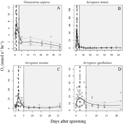
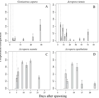
(30) were introduced into each well containing 0.2 µm FSW and a small piece of crustose coralline algae, a known settlement inducer for Acropora species (Morse et al. 1996). After 24 h, I recorded whether or not successful metamorphosis had occurred.. 2.3 Results 2.3.1. Respirometry Qualitative patterns in respiration rates through time were highly consistent. among all four study species (Figure 2.1). Oxygen consumption increased from very low levels in unfertilized eggs to a peak 1-2 days after spawning (DAS). Peak oxygen consumption coincided with the onset of larval motility (Figure 2.1, vertical dashed lines). After a week, oxygen consumption had fallen substantially, reaching levels similar to those of unfertilized eggs, and remained low until the conclusion of the experiments (up to 60 days later). Of the four study species, Acropora tenuis exhibited the most pronounced peak in oxygen consumption; respiration rates fell by approximately two orders of magnitude over the week following peak respiration (Figure 2.1B). In contrast, A. spathulata exhibited the smallest change, with an approximately two-fold decline from peak levels over approximately two weeks (Figure 2.1A, D). Unique among the four species, there was a delay until approximately 12 h after fertilization before larval respiration rates of A. nasuta began to increase, but otherwise its overall pattern of oxygen consumption was very similar to those of the other species (Figure 2.1C).. 28.
(31) Figure 2.1 Rates of oxygen consumption through time in four scleractinian coral species. Each open circle represents one replicate measurement. Solid lines represent fitted mean respiration rates from the GAM. Dashed lines show upper and lower 95% confidence intervals on the fitted GAM values. Fitted values and confidence intervals were obtained on the log-scale (on which the analysis was conducted), and have been back-transformed to the arithmetic scale for plotting. Two vertical lines show developmental stage; a dashed line for time to swim and a dotted line for the time larvae first become competent to settle. Shaded areas indicate sampling times when settlement was observed (i.e, larvae were competent).. 29.
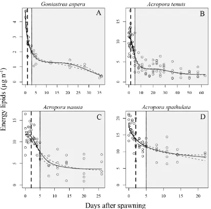
(32) 2.3.2. Lipid Total lipids consisted overwhelmingly of energy lipids, and these two quantities. exhibited quantitatively very similar temporal dynamics (Figure A.1 in Appendix A), so energetic lipids only were used for the analysis. Consistent with the trends found for respiration rates, larvae of all four species exhibited qualitatively similar patterns of energy lipid depletion (Figure 2.2). Initial lipid levels declined rapidly through embryogenesis and development approximately until larvae became competent (i.e, capable of metamorphosis) (Figure 2.2, vertical dotted line). Subsequently, energy lipid levels declined very slowly throughout the remainder of the experiment. In contrast to the three other species, lipid levels in A. nasuta larvae remained high for the first 12-24 h after fertilization, before declining rapidly (Figure 2.2C), consistent with the delayed increase in metabolic rates observed for this species (Figure 2.1C). Of the acroporids, Acropora tenuis larvae had the greatest initial decline in energy lipid levels, with an approximately three-fold reduction occurring in the first week (Figure 2.2B), consistent with its greater decline in metabolic rates after swimming commenced (Figure 2.1B). In contrast, A. spathulata exhibited the smallest decline in lipid levels, decreasing by only two-fold in the first week (Figure 2.2D), consistent with its smaller decline in metabolic rates (Figure 2.1D). Once competence was acquired, larvae from all species remained capable of settlement at every sampling point over the remainder of the experiment (Figure 2.1, Figure 2.2, shaded areas).. 30.
(33) Figure 2.2 Depletion of energy lipids through time in four scleractinian coral species. Each open circle represents one replicate measurement. Solid lines represent mean respiration rates, and dashed lines show upper and lower 95% confidence intervals. Means and confidence intervals were obtained from the GAM fits by back-transforming from the log-scale (on which fits were made) to the arithmetic scale (for plotting). Two vertical lines show developmental stage: a dashed line for time to swim, and a dotted line for the time larvae first become competent to settle. Shaded areas indicate sampling times when settlement was observed (i.e, larvae were competent).. 31.
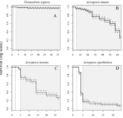
(34) 2.3.3. Survival Survival times varied among species, with estimated median lifetimes ranging. from 4 d for A. spathulata to 57 d for A. tenuis (Figure 2.3). A median lifetime for G.aspera was not estimable, due to the very high survival in this species (>98% after 35 d; Figure 2.3A). Mortality rates also varied, but in most cases, increased mortality did not occur until after larvae were competent to settle (slope of the lines in Figure 2.3, shaded areas). The exception was A. spathulata, whose survival decreased the most between the onset of swimming and the acquisition of competence (vertical dashed and dotted lines, Figure 2.3D). For each species, surviving larvae remained at the conclusion of the experiment.. 32.
(35) Figure 2.3 Kaplan-Meier survival estimates for four scleractinian coral species. Solid lines represent median estimates; dashed lines show upper and lower 95% confidence intervals. Vertical dashed lines indicate when larvae began swimming; vertical dotted lines when larvae acquired competence to settle, and gray shaded areas indicate when settlement was observed. Note the different scales on the y-axes.. 33.
(36) 2.3.4. Settlement Once competence was acquired, larvae from all species maintained competence. to settle throughout the duration of the study (Figure 2.4). The proportion of competent larvae was highest for A. nasuta and A. spathulata, with the majority of the larvae of these two species acquiring competence 8-14 days after spawning (Figure 2.4C, D). On the other hand, the proportion of G. aspera and A. tenuis larvae that had acquired competence never reached more than 50%, but a relatively large proportion of larvae was capable of settlement at the conclusion of the experiment (Figure 2.4A, B).. 34.
(37) Figure 2.4 Proportion of larvae that are competent to settle for four species of scleractinian corals. Each open circle represents a settlement assay. Error bars represent one standard error.. 35.
(38) 2.4 Discussion Temporal patterns in the metabolic rates and lipid levels of lecithotrophic, nonzooxanthellate larvae for four broadcast spawning species of corals were strikingly similar in the first three weeks after spawning. Although the specific values of oxygen consumption varied among species, in all cases, respiration rates were relatively low for eggs, followed by a rapid increase through embryogenesis to a peak, the timing of which varied among species from 12-48 h after fertilization. This spike in respiration rates is short-lived, and rates quickly fall back to initial levels within 3-6 days, where they remain for up to eight weeks. Consistent with these observations, lipids are depleted rapidly during development, until a few days after larvae become competent to metamorphose, at which time the rate of lipid utilization slows dramatically. The high concordance between these two measures of energy use strongly support the hypothesis that an extended period of reduced larval metabolism explains, at least partly, the long PLDs observed in many coral species with non-feeding larvae. Moreover, the capacity of larvae to maintain settlement competence throughout this extended period of reduced energy use implies the capacity to settle over a broad range of dispersal distances. Although embryogenesis and larval development are the most energetically demanding periods in the larval life of many marine invertebrates (e.g., Shilling and Manahan 1994, Anger 1996, Hoegh-Guldberg and Emlet 1997, Bryan 2004), the magnitude of the declines observed in some species suggests that lecithotrophic larvae have the capacity to achieve much lower levels of energy use than have previously been documented for corals. High rates of respiration correspond firstly with high rates of cell division during embryogenesis (approximately 12-36 h post-fertilization) and secondly with the development of specialised cells, such as spirocysts, that are associated with attachment and metamorphosis (36-96 h post fertilization) (Hayashibara 36.
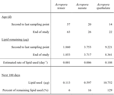
(39) et al. 2000, Okubo and Motokawa 2007). Once these energetically demanding processes are complete, larvae enter a state of substantially reduced metabolism. The magnitude of decrease in respiration varied from ~2.5-fold in A. spathulata, to about 100-fold in A. tenuis. Temporal changes in respiration have been examined previously in only one species, Acropora intermedia (Okubo et al. 2008), and were found to decrease by ~3fold, which is within, but towards the low end of the range of declines observed in this chapter. The pattern of rapid decline in lipid content during the first week of embryogenesis and larval development, followed by a period when further lipid depletion was minimal, is consistent with the trends I observed in respiration rate. Overall, larvae lost between half (A. spathulata) to ~75% (A. tenuis) of their initial lipids during the first week. This is similar to, but larger than, the ~30-40% depletion of lipids observed over a similar period in the one previous study that reports comparable data (Harii et al. 2007). For at least three of my four study species, lipid levels were stable after this period. The exception was G. aspera, which exhibited a secondary decline over the final (fifth) week. However, this decline must be treated with some caution, because it was driven entirely by samples on the final sampling date, and lipid levels were very stable over the preceding three weeks. For the three Acropora species, the rates of lipid depletion apparent at the end of the study are consistent with very long PLDs observed in other Acropora coral species, such as A. valida, which has been observed to successfully metamorphose at ~130 days (Connolly and Baird 2010). On the final sampling date (63, 26, and 22 DAS for A. tenuis, A. nasuta, and A. spathulata, respectively), the observed rates of lipid depletion in larvae of these species (the slopes of the fitted lipid line in Figure 2.2) varied from ~0.11 µg day-1 to <<0.01 µg day-1 (Table 2.1). At these rates, after a further 100 days in the plankton, A. tenuis and A. 37.
(40) nasuta larvae would have used less than approximately 6% and 16% of their remaining energetic lipids, respectively (Figure 2.2B,C), suggesting that energy reserves are consistent with the very long (100+ days) PLDs that have been documented for corals (Connolly and Baird 2010). Acropora spathulata had enough remaining lipid for an additional 77 d at their rate of consumption at the end of the study, with a total estimated PLD for this species of 99 d (Table 2.1).. Table 2.1 Estimates of lipid remaining after an additional 100 days in the plankton for three Acropora species, assuming maintenance of the lipid utilization rate observed at the end of the study (i.e., change over the last two sampling points).. Acropora tenuis. Acropora nasuta. Acropora spathulata. Age (d) Second to last sampling point. 57. 20. 14. End of study. 63. 26. 22. Second to last sampling point. 1.860. 3.753. 9.221. End of study. 1.853. 3.717. 8.361. 0.001. 0.006. 0.108. Lipid used (µg). 0.113. 0.597. 10.752. Percent of remaining lipid used (%). 6. 16. 129. Lipid remaining (µg). Estimated rate of lipid used (day-1). Next 100 days. 38.
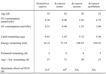
(41) In contrast to estimates based on rates of energy lipid decline, estimates of PLDs based on respiration rates prevailing at the end of the experiment still fall short of empirically observed PLDs in the literature (Table 2.2). One possible explanation for this apparent discrepancy between metabolic rates and rates of energy lipid depletion is that the larvae are supplementing their endogenous reserves by absorbing dissolved organic matter (Ben-David-Zaslow and Benayahu 2000), which would have been present both in the filtered seawater being supplied to them, and as a consequence of the lysing of dead coral larvae. Alternatively, the handling necessary to place larvae in respirometry vials for measurement of oxygen consumption may have stimulated a temporary elevation in metabolic rates for these sampled larvae, causing respirometry measures to be biased upwards, relative to average levels prevailing for larvae remaining in the tanks.. 39.
(42) Table 2.2 Estimates of pelagic larval duration (PLD) for four study species using lipid content and oxygen consumption, converted to their energetic equivalents (39.5 kJ / g lipid; 441 kJ / mol O2; (Gnaiger 1983)).. Goniastrea aspera Age (d). Acropora tenuis. Acropora nasuta. Acropora spathulata. 35. 63. 26. 22. O2 consumption (nmol/n/hr). 0.70. 0.90. 3.45. 6.79. O2 consumption (mJ/n/hr). 0.31. 0.40. 1.52. 3.00. Lipid remaining (µg). 0.41. 1.85. 3.72. 8.36. Energy remaining (mJ). 16.18. 73.19. 146.83. 330.25. Esimated remaining (d). 2. 8. 4. 5. Age + Est. remaining (d). 37. 71. 30. 27. Maximum observed PLD (d). 215a. 69b. NA. NA. a - Graham et al. 2008; b - Nishikawa et al. 2003. Even though metabolic rates measured in vials may be high relative to rates for larvae in tanks, the rates are still substantially lower than those predicted from metabolic scaling theory. This strongly suggests that metabolic rates are indeed unusually low after competence is achieved. The metabolic theory of ecology (MTE) predicts whole organism metabolic rate scales with body mass raised to ¾ power (Gillooly et al. 2001). In particular, for temperatures between 8-27°C, mass-normalized resting metabolic rates of multicellular invertebrates are predicted to lie between 0.002 and 0.135 W g-3/4, where W is metabolic rate in Watts (joules per second) (Gillooly et 40.
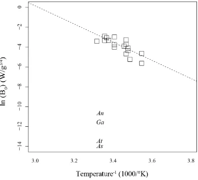
(43) al. 2001). Using egg dry weights that I measured for G. aspera (0.000012 g) and A. nasuta (0.000031 g) during an earlier study (Honours thesis; Graham 2007), and assuming A. tenuis and A. spathulata have comparable egg dry weights as similar-sized eggs of A. digitifiera (0.000043 g) and A. divaricata (0.000027 g) respectively, I calculated mass-normalized metabolic rates for larvae in this study (at 27°C). My estimates are much lower than expected (Figure 2.5), even though the larvae were actively swimming (i.e., not resting).. 41.
(44) Figure 2.5 Mass-normalized resting respiration rates for multicellular invertebrates (recreated from Gillooly et al. 2001), in comparison to mass-normalized respiration rates of the four scleractinian corals maintained at 27°C in this study. An = Acropora nasuta; Ga = Goniastrea aspera; At = Acropora tenuis; As = Acropora spathulata.. The consistency in the overall patterns of reduced oxygen consumption and lipid depletion that I observed for all four study species is striking, and implies that the energetic cost of delaying metamorphosis may be much smaller than is commonly assumed for lecithotrophic larvae. These findings help to explain large discrepancies that have been reported between energetic estimates of larval duration based on metabolic rates measured early in larval life (Richmond 1987) and the much greater. 42.
(45) durations measured empirically (Graham et al. 2008, Connolly and Baird 2010). This suggests that very low basal metabolic rates underpin the extended competence periods and larval durations of scleractinian corals, which are on par with or greater than those of most planktotrophs (Shanks et al. 2003, Shanks 2009). While there are undoubtedly costs associated with increased time in the plankton, my results suggest that some of the hypothesized post-settlement costs, such as increased mortality and decreased growth (Pechenik 2006), may be less severe for scleractinian coral larvae than might be expected based on the metabolic rates prevailing early in larval life, or typical of similar-sized invertebrates. Few studies of the temporal dynamics of metabolism in other lecithotrophic larvae have lasted more than two weeks (e.g., Okubo et al. 2008, Hoegh-Guldberg and Emlet 1997, Moran and Manahan 2003). This raises the possibility that other invertebrate lecithotrophs may extend their larval durations in a similar fashion. If reduced larval metabolism leading to extended larval duration is a common early life history trait underpinning current coral population structures, then ocean warming, which should elevate these basal metabolic rates, is likely to have significant consequences for dispersal potential and hence population connectivity, not only for the majority of reef-building corals but for other marine invertebrates as well.. 43.
(46) 3. Temperature effects on the energetics of scleractinian coral larvae and their implications for dispersal. 3.1 Introduction Temperature has a major effect on the physiology of organisms, most notably by affecting the rates of biochemical reactions that collectively govern organism functions. Typically, biochemical reactions catalysed by enzymes exhibit a parabolic response to temperature, such that reaction rates increase with temperature until an upper tolerance threshold is reached, after which rates start to decline (Sibly et al. 2012). Biological performance follows a similar trajectory, rising gradually with temperature to an optimum before dropping (Huey and Stevenson 1979). Metabolic rates, development rates and lifespans are all similarly affected by temperature (Gillooly et al. 2001, 2002). While some organisms are capable of regulating their internal body temperature to maintain optimum enzyme activity, internal temperatures of others, including most invertebrates, are dependent on their environments. Given the increases in global temperatures over the last century and the predicted increases to come in the near future (IPCC 2007), temperature will likely play a major role in determining the future distribution and abundance of animal populations. In the marine environment, most invertebrate species have a complex life-cycle consisting of a benthic, mostly sedentary, adult stage and a dispersing larval stage. Many coral species appear to live close to the upper limit of their thermal tolerances, and even small increases in temperature can lead to the loss of their symbiotic photosynthesizing dinoflagellates from the genus Symbiodinium (zooxanthellae), a condition known as bleaching (Jokiel and Coles 1990, Berkelmans and Willis 1999). If temperatures remain above this threshold for extended periods of time, death of the 44.
(47) colony can occur (Glynn 1984). Increased temperatures also affect corals during the larval stage, with increases of 3-4°C causing rates of development to increase by 1025%, the number of larvae developing abnormally to increase 20-40% (Bassim et al. 2002, Negri et al. 2007, Randall and Szmant 2009b), and mortality to increase by 2080% (Edmunds et al. 2001, Bassim and Sammarco 2003, Brooke and Young 2005, Randall and Szmant 2009a, 2009b). Short term exposure to increases in temperature of 6-8°C can increase overall settlement by 30-50% (Coles 1985, Nozawa and Harrison 2007), while long term exposure to temperatures only 3-4°C above ambient may decrease settlement during the exposure period (Randall and Szmant 2009a, 2009b). Although it is generally assumed that increased metabolic rates are the cause of such changes, very few researchers have measured the respiration rates of coral larvae at different temperatures, and no studies to date have quantified the effect of temperature on the energy content of larvae. Respiration rates of larvae from several species of brooding corals exhibit a parabolic response to temperature, with peaks occurring near ambient seawater temperatures at the time of larval release (Porites astreoides, Edmunds et al. 2001; Pocillopora damicornis, Seriatopora hystrix and Stylophora pistillata, Edmunds et al. 2011). However, brooded larvae develop internally and, considering that Symbiodinium symbionts are vertically transmitted from parents to offspring in all brooding hermatypic scleractinians except species of the genus Isopora (Baird et al. 2009), they are physiologically different from larvae of broadcast-spawned corals with horizontal Symbiodinium transmission, which make up the majority (> 80%) of coral species (Baird et al. 2009). To date, only one study has looked at the effect of temperature on respiration of broadcast-spawned larvae lacking symbionts. Rodriquez-Lanetty et al. (2009) found increased temperature increased rates of larval respiration in Acropora 45.
(48) millepora monotonically, but the larvae in this study were 10 d old and exposed for 10 hours at most. More research is needed to determine if a monotonic response of metabolic rates to temperature is typical of other broadcast spawning species, and if differences in respiration rate are correlated with increasing rates of development and mortality rates. In this chapter, I aim to quantify the effects of temperature on the survival, metabolic activity, and energy expenditure of scleractinian coral larvae over the entire larval duration. The results from Chapter 2 demonstrate that levels of oxygen consumption of coral larvae peak with the onset of larval swimming and then decline to low levels characteristic of unfertilized eggs between 7-10 days after spawning, after which respiration rates remain constant for up to two months. If temperature increases rates of early development in coral larvae, the peak in oxygen consumption should be higher and occur sooner for larvae maintained at higher temperatures. Moreover, larvae at higher temperatures should have higher rates of respiration during the extended period of low metabolism. Similarly, if respiration is increased at higher temperatures, the rate of lipid depletion, the putative energy source for coral larval metabolism (Arai et al. 1993), should also increase at higher temperatures. Higher rates of energy use should then lead to higher mortality rates. Here, I test these hypotheses using laboratory experiments on Acropora tenuis larvae, from newly fertilized gametes, through larval development, for up to two months.. 3.2 Materials and methods The study was conducted at Orpheus Island Research Station (OIRS), part of the Palm Island Group in the Central Section of the Great Barrier Reef (18.61 S, 146.48 E), 46.
(49) during the austral summer in 2009-2010. Acropora tenuis was selected as the target species because it is a locally abundant, broadcast spawning species, whose eggs lack zooxanthellae at release. Three days prior to the full moon in November, six adult colonies of A. tenuis were collected, three each from Orpheus and Pelorus Islands, and brought to OIRS where they were maintained in flow-through aquaria at ambient temperature at the time of spawning (27°C). On November 7, spawned gametes from all colonies were collected, combined, and fertilized at ambient temperature. Once cleavage had started in the majority of A. tenuis eggs, ~50,000 developing embryos were equally and randomly distributed into five 10 L aquaria containing 0.2 µm filtered seawater (FSW) maintained at different target temperatures: 25.0°C, 27.0°C (ambient), 28.5°C, 30.0°C, and 31.5°C. Five temperatures were chosen in order to analyse the data using a regression-based approach, explained below. These temperatures are within the range of temperatures larvae typically experience around Orpheus Island in the summer months. Sampling commenced immediately after transfer of larvae into the treatment tanks and was then repeated: every 12 h until all larvae were actively swimming (36 h), daily until larvae in all treatments were competent to settle (5 DAS), every 3 d in the remainder of the first month (29 DAS), and every 7 d in the second month (up to 63 DAS in treatments with larvae remaining). At each sampling time, respirometry measurements were made on a subset of larvae from each treatment, after which the same larvae were immediately frozen in liquid nitrogen for subsequent lipid analysis. A survival experiment, described below, was set up when larvae in all treatments were swimming (4 DAS).. 47.
Figure
+7
Related documents
Coumarin derivatives occurring in plants have different biological activities (Cisowski, 1983, 1984).
Earth Planets Space, 52, 927?933, 2000 Three dimensional distribution of water vapor estimated from tropospheric delay of GPS data in a mesoscale precipitation system of the Baiu
Expression of the full-length B/NS1 protein (281 amino acids), as well as either its N-terminal RNA-binding domain (amino acids 1 to 93) or C-terminal domain (amino acids 94 to 281),
Infected immature mDCs differentiated normally and enhanced CCR5-tropic but not CXCR4-tropic virus infection of T cells even in the continuous presence of neutralizing
The gastrohepatic ligament attaches to the ligamentumvenosum (sinus venosus remnant) along the left side of the left portal triad. The vascular inflow and biliary
Following the Ninth Circuit’s determination, only two circuits have addressed the narrow issue of whether a de minimis exception applies to infringement actions based upon the
Comparison of the Speed of Calculation shows that in an emergency scenario, manual dosage calculation takes around 40 seconds to compute, while the microcontroller unit
Beyond the objective he can impose a limit of advance (LOA) if he does not want the unit to conduct an exploitation or a pursuit.. and 3 discuss these control measures. They
