DINOPROSTONE GEL FOR LABOUR INDUCTION
A Dissertation Submitted to
THE TAMILNADU DR. M.G.R MEDICAL UNIVERSITY
CHENNAI
In Partial fulfilments of the Regulations for the Award of the Degree of
M.S. (OBSTETRICS & GYNAECOLOGY)
BRANCH – II
GOVERNMENT STANLEY MEDICAL COLLEGE
CHENNAI
CERTIFICATE BY THE INSTITUTION
This is to certify that dissertation entitled EFFECT OF VAGINAL PH ON EFFICACY OF DINOPROSTONE GEL FOR
LABOUR INDUCTION” is a bonafide work done by Dr.H.HUMAIRA
SAFRIN at R.S.R.M Lying in Hospital, Stanley Medical College,
Chennai. This dissertation is submitted to Tamilnadu Dr. M.G.R. Medical University in partial fulfilment of university rules and regulations for the award of M.S. Degree in Obstetrics and Gynaecology.
Prof. Dr. PONNAMBALA NAMASIVAYAM, MD., D.A., DNB.
Dean
Prof & Head of Department,
Stanley Medical College & Hospital, Chennai – 600 001
Dr. K. KALAIVANI,
M.D., D.G.O., DNB. Prof. & Head of the Department Superintendent
Dept. of Obstetrics and Gynaecology Government RSRM Lying In Hospital, Stanley Medical College,
Chennai- 600 013
CERTIFICATE BY THE GUIDE
This is to certify that this dissertation entitled “EFFECT OF VAGINAL PH ON EFFICACY OF DINOPROSTONE GEL FOR
LABOUR INDUCTION” submitted by Dr.H.HUMAIRA SAFRIN,
appearing for Part II MS, Branch II Obstetrics and Gynaecology Degree Examination in May 2018, is a Bonafide record of work done by her, under my direct guidance and supervision as per the rules and regulations of the Tamil Nadu Dr. MGR Medical university, Chennai, Tamil Nadu, India. I forward this dissertation to the Tamil Nadu Dr. MGR Medical University Chennai, India.
Dr.V.RAJALAKSHMI, M.D.,D.G.O.
Associate Professor,
Dept. of Obstetrics and Gynecology Government RSRM Lying In Hospital
Stanley Medical College, Chennai
DECLARATION
I, Dr. H. HUMAIRA SAFRIN, solemnly declare that the dissertation titled, “EFFECT OF VAGINAL PH ON EFFICACY OF
DINOPROSTONE GEL FOR LABOUR INDUCTION” is a bonafide
work done by me at R.S.R.M. Lying in Hospital. Stanley Medical College, Chennai – during December 2016–to September 2017 under the guidance and supervision of Prof. Dr. K. Kalaivani M.D., D.G.O., DNB.,
Professor and Head of the department , Obstetrics and Gynaecology. The dissertation is submitted to the Tamilnadu Dr. M.G.R. Medical University, in partial fulfilment of University rules and regulations for the award of M.S. Degree in obstetrics and Gynaecology.
Dr.H.HUMAIRA SAFRIN
Place : Chennai Date :
I am grateful to Prof. Dr. PONNAMBALA NAMASIVAYAM, M.D., D.A., D.N.B. Dean, Govt. Stanley Medical College for granting me permission to undertake this study. I take this opportunity to express my sincere and humble gratitude to Dr. K. KALAIVANI, M.D.,D.G.O.,DNB., Superintendent, Govt. R.S.R.M. Lying in Hospital who not only gave me the opportunity and necessary facilities to carry out this work but also gave me encouragement and invaluable guidance to complete the task I had undertaken. I am deeply indebted to Prof. Dr. V. RAJALAKSHMI, M.D., DGO the mover behind this study for her able guidance and inspiration and constant support without which this would not have been possible.
I am very grateful to Prof. Dr. C. SUMATHY., M.D., D.G.O. and RMO Dr.H.ANITHA VIRGIN KUMARI M.D.,D.G.O and Assistant Professor Dr. ARUNADEVI, M.D.,D.G.O., for their invaluable advice, constant guidance and supervision during this study.
I am extremely grateful to all our Assistant Professors, for their advice and support during this study.
I sincerely thank my fellow postgraduates and friends for their support and cooperation.
I owe a great many thanks to all my patients without whom this study would not have been possible.
S.NO TITLE PAGE
NO
1. INTRODUCTION 1
2. AIM OF THE STUDY 2
3. MATERIALS AND METHODS 3-5
4. REVIEW OF LITERATURE 6-44
5. RESULTS 45-76
6. DISCUSSION 77-80
7. SUMMARY 81-85
8. CONCLUSION 86-87
9 BIBLIOGRAPHY
10. ANNEXURES PROFORMA
MASTER CHART
ABBREVIATIONS
CONSENT FORM
ETHICAL COMMITTEE APPROVAL FORM
CODE DESCRIPTION
S.NO SERIAL NUMBER
IP . NO IN PATIENT NUMBER
GA GESTATIONAL AGE
40W2D 40 WEEKS 2 DAYS
5H 10M 5 HOURS 10 MINUTES
E EFFACEMENT
D DILATATION
C CONSISTENCY
P POSITION
S STATION
LSCS LOWER SEGMENT CAESAREAN
SECTION
B.WT BIRTH WEIGHT
OLIGO OLIGOHYDRAMNIOS
RH NEG RH NEGATIVE COMPLICATING
PREGNANCY
GDM GESTATIONAL DIABETES MELLITUS
IUD INTRAUTERINE DEATH
RCOG ROYAL COLLEGE OF OBSTETRICS
AND GYNAECOLOGY
ACOG AMERICAN COLLEGE OF OBSTETRICS
AND GYNAECOLOGY
HIV HUMAN IMMUNODEFICIENCY VIRUS
NST NON STRESS TEST
AFI AMNIOTIC FLUID INDEX
LN LABOUR NATURALE
LSCS LOWER SEGMENT CAESAREAN
SECTION
EPI EPISIOTOMY
PLAGIARISM CERTIFICATE
This is to certify that this dissertation work titled
EFFECT
OF VAGINAL PH ON EFFICACY OF DINOPROSTONE
GEL FOR LABOUR INDUCTION
of the candidateDr. HUMAIRA SAFRIN H. with Registration Number 221516052 for the award of MASTER OF SURGERY in the branch of
OBSTETRICS AND GYNAECOLOGY.
I personally verified the urkund.com website for the purpose of plagiarism check. I found that the uploaded thesis file contains from introduction to conclusion pages and result shows 14 percentage of plagiarism in the dissertation.
INTRODUCTION
Induction of labour can be defined as an intervention intended to artificially initiate uterine contractions resulting in progressive effacement and dilation of cervix. This should ideally result in the birth of the baby through vaginal route.
The more common indications include post term pregnancy, membrane rupture without labour, gestational hypertension, oligohydramnios, non reassuring fetal status and various maternal medical conditions such as chronic hypertension and diabetes (American College of Obstetricians and Gynaecologists, 2013b). Before induction one must ensure that the gestational age and fetal lung maturity is confirmed.
AIM OF THE STUDY
1. To evaluate the influence of vaginal pH on the efficacy of PGE2 gel for cervical ripening/labour induction
2. To improve patient selection for PGE2 induction and reduce the incidence of failed induction with PGE2 gel.
3. To asses the labour outcome in induction with PGE2 by knowing the vaginal pH prior induction.
4. To asses whether vaginal pH itself has a significant effect on the Bishop score prior induction or not.
MATERIALS AND METHODS
METHODOLOGY
The Prospective study was conducted in Govt. RSRM Lying In Hospital, Chennai during the period of December 2016 to September 2017 after getting approval from the Institutional Ethical Committee.
100 patients who underwent induction of labour for various reasons were selected for the study and examined.
Before other examinations were performed, each participant underwent a speculum examination and vaginal pH value was assessed
by using pH indicator paper (both broad & narrow spectrum).
The indicator paper was placed on the lateral vaginal wall between the two valves of Cusco’s speculum until it became wet.
Colour change of the strip was immediately compared with the
manufacturer’s colorimetric scale and the finding was recorded.
A vaginal examination was then performed to determine the Bishop’s score.
Bishop score was assessed
score of 0-2 or 0-3. The highest possible score is 13 and <5 is unfavourable that needs induction. All received intracervically placed PGE2 gel 0.5 mg
After ruling out all contraindications, All received intracervically placed PGE2 gel 0.5 mg . Following application the patient is instructed to remain recumbent for at least 30 minutes. The patient is then continuously monitored.
After 6 hrs depending on Bishop Score and uterine contraction either PGE2 gel was repeated (maximum 2 doses) or labour was augmented as per labour theatre protocol.
Inclusion criteria
(1) An unfavourable cervical Bishop score of ≤ 5,
(2) Singleton pregnancy with vertex presentation and no contraindication to vaginal delivery.
(3) Assuring fetal heart rate.
Exclusion criteria
(1) Known hypersensitivity to prostaglandins (2) Placenta previa
(3) Suspected chorioamnionitis (4) Parity of >3
(5) A previous caesarean delivery or a history of uterine surgery
REVIEW OF LITERATURE
INDUCTION OF LABOUR
Induction of labour is the initiation of contractions in a pregnant woman who is not in labour to help her achieve a vaginal birth within 24 to 48 hours.
Successful induction is defined as a vaginal delivery within 24 to 48 hours of induction of labour.
Elective induction is the induction of labour in the absence of acceptable fetal or maternal indications.
Cervical ripening is the use of pharmacological or other means to soften, efface, or dilate the cervix to increase the likelihood of a vaginal delivery.
PATIENT PREREQUISITE FOR INDUCTION
Assessment of maternal parameters
• Confirm the indication for induction
• Review for contraindication to labour and/or vaginal delivery
• Assess the shape and adequacy of bony pelvis
• Review risk and benefit of induction of labour with patient and the family
Assessment of fetal parameters
• Confirm the gestational age
• Estimate fetal weight
• Determine fetal position
• Determine fetal well being
INDICATIONS OF INDUCTION
OBSTETRIC INDICATIONS :
• Post term pregnancy
• Preeclampsia, eclampsia
• Previous unexplained IUD
• Fetal compromise (eg,Fetal growth restriction, isoimmunization)
• Preterm Premature rupture of membranes (PPROM)
• Prelabour rupture of membranes(PROM)
• Malformed fetus
• Severe hydraminos
• Oligo hydraminos
• Abruptio placentae
• Chorioamnionitis
• Fetal demise
• Cholestasis of pregnancy
MATERNAL MEDICAL CONDITIONS AGGRAVATED BY
PREGNANCY :
• Diabetes mellitus
• Chronic renal disease
• Chronic pulmonary disease
• Chronic hypertension
CONTRAINDICATIONS ABSOLUTE
• Active genital herpes infection
• Serious chronic medical condition
• Pelvic Structural abnormality
• Cephalopelvic disproportion major degree
• Abnormal fetal lie [transverse lie, oblique lie]
• Umbilical cord prolapse and cord presentation
• Previous classical Caesarean section or other transfundal uterine surgery.
• Previous Myomectomy entering the endometrial cavity.
• Contraindication specific to the inducing drug used.
• Invasive cervical cancer.
RELATIVE
• Uterine overdistension [multiple pregnancy, polyhydraminos]
• Breech
• Fetal macrosomia
• Low lying placenta
METHODS OF LABOUR INDUCTION
I-NON PHARMACOLOGIC METHODS NATURAL METHODS
• Relaxation techniques
• Sexual intercourse
• Nipple stimulation
• Hot Bath / Castor oil / Enemas
• Cumin Tea
• Several herbs
• Acupressure
• Acupuncture
MECHANICAL METHODS
• Osmotic dilators Laminaria and Dilapan
• Balloon devices Foleys .
SURGICAL METHODS
• Stripping the membranes
II- PHARMACOLOGICAL METHODS
• Oxytocin
• Prostaglandins
Misoprostol [ E1] Dinoprostone [E2]
• Mifepristone
COMPLICATIONS OF INDUCTION
MATERNAL
Uterine tachysystole Uterine Rupture
Failed Induction and Increased Caesarean Delivery Rate Sepsis
Postpartum Haemorrhage Accidental Haemorrhage Amniotic Fluid Embolism
FETAL
INDUCTION OF LABOUR
Induction of labour is defined as the process of artificially stimulating the labour. It is usually performed by administering oxytocin or prostaglandins to the pregnant woman or by manually rupturing the amniotic membranes. This should ideally result in the delivery of the baby through the vaginal route (RCOG 2001). Ideally, most pregnancies should be allowed to reach term, the onset of spontaneous labour being the sign of physiologic termination of pregnancy. It is one of the most common interventions practiced in modern obstetrics. Overall, throughout the world, up to 20 per cent of women have labour induced by one method or the other. Induction rates vary with practices and cultural backgrounds. Cervical ripening greatly facilitates labour and augments the chances of vaginal birth. The cervical state is related to the success of labour induction, duration of labour, and likelihood of vaginal delivery.
Elective inductions for the convenience of either the obstetrician or the patient are on the rise. Due to the attendant risk of severe, though infrequent, adverse maternal outcomes, elective inductions are not routinely recommended.
HISTORY OF INDUCTION OF LABOUR
Since antiquity various methods, many bizarre and some frankly dangerous, have been used in an attempt to bring on labour. Massage of the breasts and uterus are very old but inefficient methods. Something approaching the use of tents dates back to the sixth century, and stretching of the cervix digitally has been long employed. The last century brought with it more ingenuity and at one time electricity was thought of. Scanzoni used a hot carbolic acid douche in 1856, and at this time Kraus introduced his bougies, which fell into disuse by the 1930s because of their relative inefficiency, high sepsis rate and the often countered risk of harpooning or detaching the placenta.
Artificial rupture of the membranes stands in a class by itself, for it has stood a prolonged test of time, being first used by Denman in 1756 for cases of contracted pelvis, and being known since then as the “English method”. It remains to this day a widely used method in spite of the sacrifice of an intact amniotic sac that it entails. Hind water rupture with Drew Smythe catheter was introduced in 1931, but what it gains in safety, in terms of fore water preservation with reduced risk of amniotic fluid infection and cord prolapse, it loses in efficiency when compared with fore water rupture.
in 1935. It was believed to be part of prostatic secretions and was therefore called prostaglandin.
Elias Corey synthesized dinoprostone in 1970 at the Harvard University. Three biochemists, Bergstrom, Samuelsson and Vane jointly received the 1982 Nobel Prize for their discovery of prostaglandins.
The reasons for the rising rates of induction of labour can be complex and multifactorial (Rayburn and Zhang 2002).
Some of them are: -
• Improved ability of physicians to determine gestational age accurately with early dating scans, thus avoiding the possibility of iatrogenic prematurity.
• Widespread availability of cervical ripening agents.
• Improved knowledge of methods and indications for induction.
• More relaxed attitudes towards marginal/elective indications, both of the physician and the patient.
GENERAL PRINCIPLES RELATED TO INDUCTION
• The Induction of labour should be performed only when there is a clear medical indication for it and the expected benefits outweigh its potential harms.
• Induction of labour should be performed with caution since the procedure carries the risk of uterine hyperstimulation and rupture and fetal distress.
• Induction of labour is carried out, facilities should be available for assessing maternal and fetal well-being.
• Women receiving oxytocin, misoprostol or other prostaglandins should never be left unattended.
• Failed induction of labour does not necessarily indicate caesarean section.
• Wherever possible, induction of labour should be carried out in facilities where caesarean section can be performed.
Criteria of an ideal inducing agent
An ideal inducing agent is one which:
• Achieves onset of labour within the shortest possible time.
• Has low failure rate.
• Does not increase the rate of caesarean delivery or operative vaginal deliveries as compared to spontaneous labour.
• There should be a less perinatal morbidity.
We are yet to find an ideal inducing agent. Hence, the decision for induction should be well thought out and communicated to the woman concerned.
PRE INDUCTION COUNSELLING FOR THE COUPLE
It is essential to have good communication with the woman and her family prior to induction; wherever possible this should be supported by evidence-based and preferably, written information. During induction of labour, the woman has restricted mobility and the procedure itself can cause discomfort to her. To avoid potential risks associated with the procedure, the woman and her baby need to be monitored closely. According to (RCOG 2008):
- Explain the indications for induction; more specifically, the consequences associated with continuing the pregnancy
- The need for close monitoring of the fetal heart rate (including electronic fetal monitoring in labour)
- Should give multiple options.
- The risks associated inducing agent used.
- The chances of failure of induction and the options available in case of failure.
In summary, the woman and her partner should be offered to be made a part of the decision-making process. A positive attitude imparted to the woman when she is actively involved in the decision making, not only increases the chances of success of induction but also enables her to better face the consequences (Nuutila et al 1999).
WOMEN’S ATTITUDE TOWARDS INDUCTION
INDICATIONS AND CONTRAINDICATIONS FOR INDUCTION
The indications can be divided under the following headings:
1. Obstetrical conditions;
2. Medical conditions aggravated by pregnancy.
The correct selection of cases in itself predisposes certainty as to the child’s maturity. The best paediatric unit in the world is no substitute for a healthy intrauterine environment up to the time of adequate maturity and there is now no excuse for being in doubt about this, thanks to the precision afforded by modern sonar techniques.
COMMONLY ACCEPTED INDICATIONS FOR INDUCTION OF
LABOUR
- Pregnancy-induced hypertension - Premature rupture of membranes - Severe intrauterine growth restriction - Rhesus Iso immunization
- Maternal medical problems (diabetes mellitus, lupus, renal disease) - Intrauterine fetal demise
- Postdated pregnancy - Oligohydramnios
OBSTETRIC INDICATIONS
INDUCTION OF LABOUR IN WOMEN AT OR BEYOND TERM
Pregnancies that reach beyond 42 gestational weeks are defined as post-term. This is the commonest indication for induction of labour worldwide.
Evidence related to induction of labour at term and beyond term was extracted from one Cochrane systematic review of 22 randomized controlled trials (10). Most of the trials were judged by the Cochrane review authors to likely have a moderate risk of bias, largely due to unclear concealment of allocation and generation of the sequence of randomization.
Recommendations
Induction of labour is recommended for women who are known with certainty to have reached 41 weeks (> 40 weeks + 7 days) of gestation. (Low-quality evidence. Weak recommendation.)
Induction of labour is not recommended for women with an uncomplicated pregnancy at gestational age less than 41 weeks. (Low-quality evidence. Weak recommendation.)
American College of Obstetricians and Gynaecologists recommends that women who are post-term and also have unfavourable cervices can either undergo labour induction or be allowed to be managed expectantly. Many studies recommend prompt delivery in an uncomplicated post-term patient with a favourable cervix (ACOG 2004). The Department of Obstetrics and Gynaecology and Reproductive Biology at Harvard Medical School recommends routine induction at 41 weeks gestation (Rand et al 2000).
INTRAUTERINE GROWTH RESTRICTION
Chronic placental insufficiency leads to intrauterine growth restriction. Infants with growth restriction have a higher risk of perinatal morbidity and mortality, which usually results from placental insufficiency. The placental insufficiency is likely to be aggravated by labour. Due to low placental reserve as compared to normal fetus, these fetuses, as a group, might require induction of labour prior to their expected date of delivery.
PRE-ECLAMPSIA AND ECLAMPSIA
PREVIOUS UNEXPLAINED INTRAUTERINE FETAL DEATH
This peculiar entity, said to be due to placental insufficiency may, by the warning history, provide an opportunity to forestall disaster by timely induction which is usually done at 38 weeks, but may be done earlier if indicated by fetal monitoring tests.
PRELABOUR RUPTURE OF MEMBRANES
(PROM) at term complicates about 8-10% pregnancies. It has been a matter of great controversy whether women with term PROM should be induced or managed with an expectant policy, and if the latter course is opted, how long is it safe to await spontaneous labour. Results from many randomized trial to date demonstrate that expectant management was associated with an increased incidence of clinical chorioamnionitis, postpartum fever, longer hospital stay for the mother and a long stay for the baby in the neonatal intensive care unit; induction therefore seems to be a reasonable choice.
RH ISO-IMMUNISATION
MALFORMED FETUSES
The prolongation of pregnancy is profitless, and on grounds of humanity as well, pregnancy is better terminated. Besides it is better to deliver a small monster than a large one.
HYDRAMNIOS
Severe hydramnios producing marked pressure symptoms may call for relief. There is the danger of accidental haemorrhage following artificial rupture of the membranes in these cases.
ABRUPTIO PLACENTA
Minor degrees of placental abruption without any signs of fetal distress are best managed by amniotomy and oxytocin infusion.
INTRAUTERINE DEATH OF THE FETUS.
Spontaneous labour will always start eventually, but the patient can often be spared some very wretched weeks of waiting if labour is induced. Drug induction is both safe and usually efficacious.
MEDICAL INDICATIONS
CHRONIC RENAL DISEASE.
pregnancy vary between bad and disastrous. The decision and the timing of intervention must be taken considering both maternal and fetal interests.
HYPERTENSION
The risks of fetal prematurity have to be weighed against the risk of superimposed pre-eclampsia and abruption placenta.
DIABETES
Whether or not pre-eclampsia is added to this complication, induction of labour is often called for to forestall intrauterine fetal death, which is a very real risk in the third trimester, particularly in the uncontrolled diabetics and those associated with hypertension.
CONTRAINDICATIONS TO LABOUR INDUCTION
- Placenta or vasa previa
- Fetal malpresentations
- Prior classic uterine incision
- Active genital herpes infection or any other lower genital tract infections and tumors.
Disproportion that is more than borderline. It must have been made abundantly clear already that such treatment is little short of wanton folly rewarded with a high failure rate, a prohibitive fetal mortality and the likelihood of maternal morbidity.
1. Where the lie is other than longitudinal, for obvious reasons.
2. In cases of previous caesarean section for contracted pelvis or who have failed in previous trial of labour for disproportion. However, it may be added that a pelvic examination must be done to confirm the presence of cephalopelvic disproportion, as some of these cases may have been mistakenly labeled or in some cases the baby may be smaller than it was in the previous pregnancy.
3. Where a tumour occupies the pelvis.
4. When vaginal delivery is contraindicated. These include major degree placenta previa, vasa previa, cord presentation and prolapsed, invasive carcinoma cervix, and infections like active herpes genitalis and HIV.
Though not a contraindication, extreme caution is required in grand multipara because of the tumultuous precipitate labour that can follow, and cases of previous caesarean section or myomectomy because of the danger of uterine rupture.
PREINDUCTION CERVICAL RIPENING
Starting with a favourable cervix ensures the success of labour induction. Further, the time taken of labour induction is affected by parity and to a small degree by baseline uterine activity and sensitivity to oxytocic drugs. The goal of cervical ripening is to facilitate the process of cervical softening, effacement and dilatation, thus reducing the induction to-delivery time. When there is an indication for induction and the cervix is unfavourable, agents for cervical ripening may be used.
Cervical ripening is associated with the disorganization of collagen bundles which is likely to be effected by collagenase. The active area of cervical tissue remodelling is at the internal OS. The collagenase found in the cervix has been identified as neutrophil derived and the invading neutrophil plays an important role in the tissue rearrangements associated with cervical ripening.
Neutrophils represent a readily available source of collagenase, present in specific granules, which can be made available by degranulation rearrangement of extracellular matrix.
Another change is an increase in cervical decorin (dermatan sulfate proteoglycan 2), leading to collagen fiber separation.
These changes together lead to softening of the cervix. As uterine contractions ensue, the ripened cervix dilates as the presenting fetal part descends, thus leading to reorientation of the tissue fibers in the direction of the stress. The cervix passively dilates and is pulled over the presenting part.
Evidence also says that the elastin component of the cervix acts like a ratchet so that dilatation is maintained even after the contraction caeses.
this complicated series of events many changes may occur both simultaneously and sequentially.
ROLE OF THE VARIOUS HORMONES IN CERVICAL RIPENING
The hormones stimulate the complex series of chemical reactions critical for the process.
• Dilation of all the tiny vascular channels of the cervix • A rise in degradation of collagen
• Increase in hyaluronic acid
• A rise in leukocyte, chemotaxis which is the cause for collagen degradation
• And an increase in the release of interleukin (IL)
The process is associated with an increase in the activity of matrix metalloproteinases 2 and 9. Cervical collagenase and elastase also rise. At term, the degradation of collagen fibres increases, leading to a decrease in collagen content of the cervix.
PROSTAGLANDINS IN LABOUR
Since their discovery in the early 1970s, prostaglandins (PGs) have contributed significantly to the practice of obstetrics. Over the years, many PG compounds have been discovered and the importance of the role of prostaglandins in several reproductive processes including menstruation, ovulation and parturition has become apparent.
Prostaglandins are important mediators of uterine activity and play an important role in the contraction of the smooth muscle of the uterus and the biophysical changes associated with cervical ripening. It can be even said that prostaglandins seem to play a much larger role in labour than oxytocin.
The F and E series Prostaglandins are the most important for labour, delivery and the postpartum period. In contrast to oxytocin, which requires an induction of receptors that does not usually occur until the later part of pregnancy, prostaglandin receptors are always present in myometrial tissue. Thus the use of prostaglandins remains throughout pregnancy.
Although both the F and E series Prostaglandins result in uterine contractions, the E series of Prostaglandins are relatively more uteroselective and are more effective in producing cervical ripening.
The naturally-occurring prostaglandins were modified to result in products that are longer acting and effective at lower concentrations, with the potential for significant savings in cost. This has allowed their widespread use in developing countries. Problems such as intrauterine fetal death and hemorrhage from postpartum uterine atony, which earlier required surgical intervention, can be managed with prostaglandins today.
STRUCTURE AND CLASSIFICATION
Prostaglandins are members of the eicosanoid family. They are synthesized from arachidonic acid. Each molecule has 20 carbon atoms with a cyclopentane ring and two side chains. The position of the side chain and number of multiple bonds determines the group identity and its action. Prostaglandins were designated PG1, PG2, PG3, based on the number of double bonds in the polyunsaturated fatty acids from which they are formed. They were initially divided into classes E and F because of their solubility in ether and phosphate buffer. Subsequently, they have been divided into ten main groups, A to I. The subscripts (alpha, beta) were then added (Van Dorp et al 1964; Bergstrom et al 1964).
METABOLISM
hand, PGF2α causes uterine contractions. Prostaglandin is catabolised by the enzyme 15-OH PG dehydrogenase to its metabolites, several of which are bioactive. This enzyme is mainly localised in the chorion and prevents the prostaglandins from reaching the myometrium in the non-labouring state.
DISTRIBUTION IN NORMAL TISSUES
PGE2 is the main prostaglandin product of the fetal membranes. The inner membrane, the amnion, has the highest production rate (Olson et al 1993). PGE2 production by the amnion, chorion, and decidua is increased during labour (Olson et al 1993). Though PGE2 and F2α are detected in the amniotic fluid in all stages of gestation, the major increase occurs with the start of labour, and they continue to increase with dilatation of the cervix (MacDonald and Casey 1993). It has been shown that prostaglandin concentrations in amniotic fluid increase early in labour (<3 cm dilatation) before the active stage of labour is reached (Romero 1994).
Properties and clinical effects
particular. Side effects include nausea, vomiting, diarrhoea, abdominal pain, chills and fever.
Preparations and dosages of prostaglandins currently available
PGE2 Vaginal gel 1 and 2 mg
Endocervical gel 0.5 mg Timed-release vaginal insert 3 and 10mg PGF2α IM injection 250/125 mcg
Misoprostol
Oral, vaginal, rectal administration
25, 100 and 200 mcg
ROLE OF PROSTAGLANDINS IN LABOUR
The role of prostaglandins in labour includes softening of the cervix, induction of gap junctions (communication between smooth muscle cells through which conduction of electrophysiological stimuli occur) and direct stimulation of uterine contractions.
CERVICAL RIPENING
A number of functional and biochemical changes happen in the cervical connective tissue during pregnancy (Leppert 1995). Prostaglandins take part in this cervical ripening process, forming a complex network of pathways.
Prostaglandins act synergistically with interleukin-8 to stimulate the fibroblasts to produce hyaluronic acid (Ogawa et al 1998), which in turn alters the composition and structure of the cervix. Besides this, prostaglandins also have an effect on the uterine muscle, inducing contractions. Thus, prostaglandins are involved both in cervical ripening and subsequently, the process of labour.
LABOUR
to increase prostaglandin synthesis. Prostaglandins modulate myometrial cell contractility by utilizing extracellular calcium.
Prostaglandins soften the cervix, induce gap junctions and further sensitise the action of oxytocin on the myometrium, causing progressive dilatation of the cervix. At the end of the first stage of labour, there is rupture of membranes, further increasing prostaglandin synthesis, thus making it an irreversible process.
THE THIRD STAGE OF LABOUR
After the delivery of the fetus, the uterus remains tonically contracted. This helps in separation of the placenta and also prevents postpartum hemorrhage.
There is some evidence that there is considerable production of PGF2 in the decidua and the myometrium in the early postpartum period after expulsion of the fetus and placenta. (Husslein et al 1983).
PROSTAGLANDIN E2
ROUTES OF ADMINISTRATION
EXTRA-AMNIOTIC
effective routes of administration came into use, this route for administering prostaglandins has been abandoned.
ORAL TABLETS
Oral prostaglandin E2 is no more effective than oxytocin for induction of labour but the gastrointestinal side effects, particularly vomiting, has been shown to be higher (Keirse and van Oppen 1989).
This route is no longer used for the induction of labour.
INTRACERVICAL PGE2
After inserting the gel, oxytocin infusion should be delayed for 6-12 hours, because the effect of prostaglandins may be heightened with oxytocin (ACOG 2009).
Intracervical PGE2 gel not only ripens the cervix, but also induces labour and reduces the risk of failed induction. About 40 percent of women do not need further induction of labour.
A COMPARATIVE STUDY OF INTRACEVICAL PGE2 WITH
PLACEBO OR NO TREATMENT
significant in a subgroup of women with intact membranes and unfavourable cervix. While there was an increase in hyper stimulation rate, there was no significant increase in fetal heart rate changes.
COMPARISON OF TWO DIFFERENT REGIMENS OF PROSTAGLANDIN E2 IN PREINDUCTION
CERVICAL RIPENING
Three episodes of myometrial hyperstimulation occurred which required an emergency caesarean section for acute fetal distress in group 2 and none in group 1.
There were 8 caesarean sections in group 1and 13 in groups 2. The outcome of pregnancy was otherwise similar in both groups. When comparing induction of labour using either oxytocin versus PGE2 (vaginal or intracervical), induction with PGE2 was associated with (RCOG 2001):
- Increase in successful vaginal delivery within 24 hours - Reduced caesarean section rate
- Reduced risk of the cervix remaining unfavourable at 24-48 hours post induction.
- Reduced use of epidural analgesia
- An increase in the number of women satisfied with the method.
MODIFIED BISHOP’S SCORE AND VAGINAL PH
PRE-INDUCTION ASSESSMENT
delivery), BMI, maternal age, estimated fetal weight, and diabetes. The Bishop score was developed in 1964 as a predictor of success for an elective induction. The initial scoring system used 5 determinants (dilatation, effacement, station, position, and consistency) that attributed a value of 0 to 2 or 3 points each (for a maximum score of 13).
He determined that when the total score was at least 9, the likelihood of vaginal delivery following labour induction was similar to that observed in patients with spontaneous onset of labour. Although several modifications have been suggested, the Bishop score has become a classic parameter in obstetrics and has since been applied to a much wider group of patients. Nulliparous women with a Bishop score no greater than 3 have a 23-fold increased risk of induction failure and a 2- to 4- fold increased risk of caesarean delivery compared with nulliparous women with a Bishop score of at least 4.
BISHOP’S SCORE
0 1 2 3
Dilatation (cm) 0 1-2 3-4 5-6
Effacement (%) 0-30 40-60 60-70 >80
Station -3 -2 -1/0 +1/+2
Consistency Firm Medium Soft
Position Posterior Mid position Anterior
MODIFIED BISHOP’S SCORE (CALDER 1974)
0 1 2 3
Dilatation(cm) <1 1-2 2-4 >4
Length (cm) >4 2-4 1-2 <1
Station -3 -2 -1/0 +1/+2
Consistency Firm Medium Soft
Other scoring systems
1. Field system
2. Burnett modifications of bishops score. 3. Weighted Bishops score by Freidman. 4. Pelvic score by Lange
The Bishop score has become the most commonly employed pre- induction scoring system.
VAGINAL PH
In general vagina maintains a pH between 3.8-4.8, which is influenced by frequency of coitus, presence of cervical mucus and the amount of vaginal transudate. The lactic acid produced from glycogen by lactobacillus present in vagina plays an important role in maintaining acidic Ph environment. A variety of factors can alter the normal vaginal pH. Several factors such as lower genital tract infection; bacterial vaginosis, rupture of membrane, douching etc can alter the vaginal pH. The acidity of the vagina may alter the release of the drug and this could result in variable clinical response. Prostaglandins are organic acids that have diminished solubility in aqueous solution with a low pH.
Recently, vaginal pH has been investigated as a potential factor influencing the efficacy of prostaglandins for cervical ripening and labour induction but the results have been conflicting. Studies have been conducted on the effects of vaginal pH on the efficacy of controlled-release PGE2 vaginal insert and PGE2 gel for cervical priming/labour induction in which overall vaginal pH seemed to influence the PGE2 release.
Nonetheless, the effect of vaginal pH on overall efficacy of the cervical ripening/labour induction with PGE2 has not been well studied.
The vaginal pH in pregnancy is known to be acidic and not much is known about the variations in vaginal pH throughout pregnancy. There are studies that mention that pH may change the degree of ionization of a drug and affect the absorption of the drug resulting in variable clinical responses.
Vaginal pH changes also has a role in preterm delivery which suggests that it has a role in influencing cervical ripening.
RESULTS AND ANALYSIS
[image:55.595.147.474.507.749.2]AGE
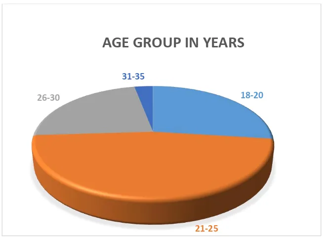
TABLE 1 : AGE DISTRIBUTION OF THE STUDY GROUP
Age Group in years Frequency Percent
18-20 27 27.0
21-25 47 47.0
26-30 23 23.0
31-35 3 3.0
Total 100 100.0
This table shows the age wise distribution of the study group. Majority (47 % )of the patients were in the age group of 21 to 25 years. The mean age of the study group was 23.49 years
CHART - 1
AGE DISTRIBUTION OF THE STUDY GROUP
18-20
21-25
26-30
31-35
GESTATIONAL AGE
TABLE : 2 GESTATIONAL AGE DISTRIBUTION OF
THE STUDY GROUP
GESTATIONAL AGE
IN WEEKS FREQUENCY PERCENT
UP TO 38 29 29.0
38-40 25 25.0
Above 40 46 46.0
Total 100 100.0
This table depicts the gestational age distribution of the study group. About 58 patients were induced at the gestational age of 40 weeks to 40 weeks 6 days interval. If the NST and AFI monitoring is normal routine induction was done at 40 weeks 3 days.
CHART - 2
0
10
20
30
40
50
Upto 38
38-40
Above 40
TABLE : 3 MODIFIED BISHOP'S SCORE DISTRIBUTION IN
THE STUDY GROUP
Bishop Score Frequency Percent
1 7 7.0
2 32 32.0
3 43 43.0
4 17 17.0
5 1 1.0
Total 100 100.0
This table shows the distribution of Modified Bishop's Score in the study group. 43 patients had a pre-induction Modified Bishop's Score of 3. The median Modified Bishop's Score was 3.
CHART : 3
0 5 10 15 20 25 30 35 40 45 50
1 2 3 4 5
TABLE : 4
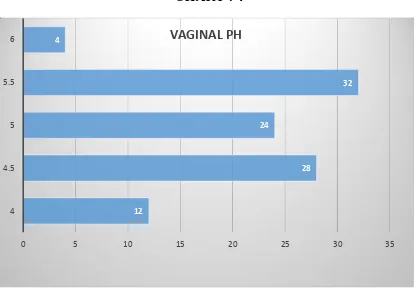
VAGINAL pH DISTRIBUTION AMONG THE STUDY GROUP
VAGINAL pH Frequency Percent
4.0 12 12.0
4.5 28 28.0
5.0 24 24.0
5.5 32 32.0
6.0 4 4.0
Total 100 100.0
The patients in the study group had vaginal pH in the range of 4 to 6.60 patients had a vaginal pH of more than 5. The mean vaginal pH in the study group was 5. In the study conducted by Ramsey et al the median vaginal pH was 5.5
CHART : 4
12
28 24
32 4
0 5 10 15 20 25 30 35
4 4.5 5 5.5
TABLE : 5 PARITY
PARITY Frequency Percent
Primi 63 63.0
Multi 37 37.0
Total 100 100.0
CHART : 5 PARITY
63 37
PARITY
TABLE – 6
INDICATION FOR INDUCTION DISTRIBUTION IN THE
STUDY GROUP
Indication for
Induction Frequency Percent
Postdated 53 53.0
Oligohydramnios 11 11.0
GHTN 25 25.0
GDM 9 9.0
RH Negative 2 2.0
Total 100 100.0
INDICATIONS FOR INDUCTION
Postdated
Oligo
GHTN
GDM
RH Negative
CHART – 6 : INDICATION FOR INDUCTION DISTRIBUTION IN
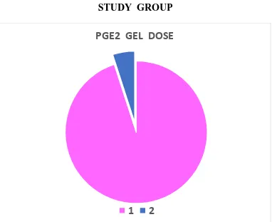
TABLE 7 : PGE2 GEL DOSE DISTRIBUTION IN THE STUDY GROUP
PGE2 GEL DOSE Frequency Percent
1 95 95.0
2 5 5.0
Total 100 100.0
This table shows the number of PGE 2 Gel doses used in the study patients.95 patients received a single dose of PGE 2 gel and 5 Patients received 2 doses of PGE 2 gel. Of these 5 patients, 1 delivered vaginally and 4 delivered by LSCS for failed induction
CHART – 7 : PGE2 GEL DOSE DISTRIBUTION IN THE
STUDY GROUP
PGE2 GEL DOSE
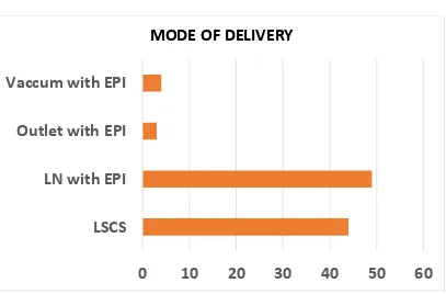
TABLE 8 : MODE OF DELIVERY DISTRIBUTION IN THE STUDY GROUP
Mode of Delivery Frequency Percent
LSCS 44 44.0
LN with EPI 49 49.0
Outlet with EPI 3 3.0
Vacuum with EPI 4 4.0
Total 100 100.0
This table shows the distribution of mode of delivery in the study group. 56 patients had normal vaginal delivery and 44 patients underwent LSCS. 3 patients delivered with Outlet forceps with episiotomy and 4 patients with vacuum with episiotomy.
CHART 8 : MODE OF DELIVERY DISTRIBUTION
IN THE STUDY GROUP
0
10
20
30
40
50
60
LSCS
LN with EPI
Outlet with EPI
Vaccum with EPI
TABLE - 9
INDICATION FOR LSCS DISTRIBUTION
INDICATION FOR
LSCS Frequency Percent
Failed Induction 30 30.0
Failure to progress 6 6.0
Fetal Distress 7 7.0
Imminent Eclampsia 1 1.0
Total LSCS 44 44.0
Normal Delivery 56 56.0
Total 100 100.0
Out of the total 100 cases, 44 cases delivered by LSCS. 7 cases were done for fetal distress and 30 cases for failed induction
CHART - 9
30 6
7
1
INDICATION FOR LSCS AMONG STUDY SUBJECTS
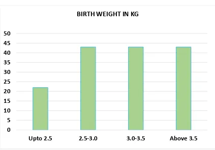
TABLE - 10 : BABY WEIGHT IN KG DISTRIBUTION IN THE STUDY GROUP
Weight in Kg Frequency Percent
Upto 2.5 22 22.0
2.5-3.0 43 43.0
3.0-3.5 29 29.0
Above 3.5 6 6.0
Total 100 100.0
In this study the mean birth weight of the babies born was found to be 2.9 kg. About 43 babies were in the range of 2.5 to 3.0 kg
CHART- 10 : BABY WEIGHT IN KG DISTRIBUTION IN THE STUDY GROUP
0 5 10 15 20 25 30 35 40 45 50
Upto 2.5 2.5-3.0 3.0-3.5 Above 3.5
TABLE 11 : ONE MINUTE APGAR DISTRIBUTION
IN THE STUDY GROUP
Frequency Percent
6 3 3.0
7 95 95.0
8 1 1.0
9 1 1.0
Total 100 100.0
In this study 95% of the babies had a 1 minute APGAR of 7.
CHART 11 : ONE MINUTE APGAR DISTRIBUTION I
N THE STUDY GROUP
0 10 20 30 40 50 60 70 80 90 100
0
2
4
6
8
10
TABLE 12 : 5 MINUTE APGAR DISTRIBUTION
IN THE STUDY GROUP
5 MIN APGAR Frequency Percent
7 4 4.0
8 94 94.0
9 2 2.0
Total 100 100.0
In this study 94 % of the babies delivered had a 5 minute APGAR of 8.
CHART 12 : 5 MINUTE APGAR DISTRIBUTION
IN THE STUDY GROUP
7
8
9
4
94
2
0
10
20
30
40
50
60
70
80
90
100
APGAR 5 MINUTES
TABLE – 13 : INDUCTION DELIVERY INTERVAL DISTRIBUTION IN THE STUDY GROUP
INDUCTION DELIVERY
INTERVAL Frequency Percent
<6 hours 14 14.0
6-10 hours 55 55.0
>10 hours 31 31.0
Total 100 100.0
This table shows induction delivery interval in the study group. The maximum induction delivery interval is around 6 – 10 hours. The average induction to delivery interval in our study group was 9 hours 52 minutes.
CHART – 13 : INDUCTION DELIVERY INTERVAL
DISTRIBUTION IN THE STUDY GROUP
0 10 20 30 40 50 60
TABLE – 15 : TIME TO ENTRY INTO ACTIVE PHASE OF LABOUR IN HOURS AMONG OUR STUDY GROUP
Time taken to enter into
active phase of labour Frequency Percent
Upto 10 52 52.0
Above 10 4 4.0
Total 56 56.0
This table shows time to entry into active phase of labour in hours among our study group. The average time to entry into active phase of labour in our study group was 7 hours 50 minutes
CHART – 15
TIME TAKEN TO ENTER INTO ACTIVE PHASE OF LABOUR IN HOURS AMONG OUR STUDY GROUP
0 10 20 30 40 50 60
Upto 10
Above 10
TABLE 16: COMPARISON OF VAGINAL pH AND
AGE GROUP IN YEARS.
Vaginal pH
Age Group in years Total
P value 18-20 21-25 26-30 31-35
4.0
Count 6 3 3 0 12
0.828 % within
Vaginal pH 50.0% 25.0% 25.0% .0% 100.0% % within Age
Group in years 22.2% 6.4% 13.0% .0% 12.0% 4.5
Count 5 15 7 1 28
% within
Vaginal pH 17.9% 53.6% 25.0% 3.6% 100.0% % within Age
Group in years 18.5% 31.9% 30.4% 33.3% 28.0%
5.0
Count 8 9 6 1 24
% within
Vaginal pH 33.3% 37.5% 25.0% 4.2% 100.0% % within Age
Group in years 29.6% 19.1% 26.1% 33.3% 24.0% 5.5
Count 7 18 6 1 32
% within
Vaginal pH 21.9% 56.3% 18.8% 3.1% 100.0% % within Age
Group in years 25.9% 38.3% 26.1% 33.3% 32.0% 6.0
Count 1 2 1 0 4
% within
Vaginal pH 25.0% 50.0% 25.0% .0% 100.0% % within Age
Group in years 3.7% 4.3% 4.3% .0% 4.0% Total Count 27 47 23 3 100
% within
Vaginal pH 27.0% 47.0% 23.0% 3.0% 100.0% % within Age
Group in years 100.0% 100.0%
100.0
Vaginal pH
6.0 5.5
5.0 4.5
4.0
Co
un
t
20
10
0
Age Group in years
18-20
21-25
26-30
31-35
This table shows the comparison of vaginal pH and age group of the study group patients which is not statistically significant (p value-0.828)
CHART : 16 : COMPARISON OF VAGINAL pH AND
TABLE 17 : COMPARISON OF VAGINAL pH AND BISHOP SCORE
Vaginal
pH Bishops score
P value
2 6 2 2
<0.05*
4.0 Count 16.7% 50.0% 16.7% 16.7% .0%
% within
Vaginal pH 28.6% 18.8% 4.7% 11.8% .0% % within
Bishops score
3 11 13 1 0
4.5 Count 10.7% 39.3% 46.4% 3.6% .0%
% within Vaginal Ph % within
Bishops score
42.9% 34.4% 30.2% 5.9% .0%
5.0 Count 2 5 15 2 0
% within
Vaginal pH 8.3% 20.8% 62.5% 8.3% .0% % within
Bishops score
28.6% 15.6% 34.9% 11.8% .0%
5.5 Count 0 9 13 9 1
% within
Vaginal pH .0% 28.1% 40.6% 28.1% 3.1% % within
Bishops score
.0% 28.1% 30.2% 52.9% 100.0%
6.0 Count 0 1 0 3 0
% within
Vaginal pH .0% 25.0% .0% 75.0% .0% % within
Bishops score
This table shows the comparison of vaginal pH and mode of delivery which is statistically significant (p value-0.019). 76.5 % of patients with a Bishops score of 4 delivered vaginally and 23.5% had LSCS. 100 % of patients with a Bishops Score of 5 delivered vaginally only 30 % of patients with a Bishops score of 3 delivered vaginally. Bishops score appears to reliably predict vaginal delivery only at values of 4 and above. For patients with a Bishops score of 3 and less than that it was difficult to predict normal vaginal delivery.
TABLE : 18 Vaginal pH * PGE 2 Dose
Vaginal
pH PGE 2 Total
P value
1 2
0.273
4.0 Count 11 1 12
% within
Vaginal pH 91.7% 8.3% 100.0%
% within
PGE 2 11.6% 20.0% 12.0%
4.5 Count 27 1 28
% within
Vaginal pH 96.4% 3.6% 100.0%
% within
PGE 2 28.4% 20.0% 28.0%
5.0 Count 21 3 24
% within
Vaginal pH 87.5% 12.5% 100.0% % within
PGE 2 22.1% 60.0% 24.0%
5.5 Count 32 0 32
% within
Vaginal pH 100.0% .0% 100.0%
% within
PGE 2 33.7% .0% 32.0%
6.0 Count 4 0 4
% within
Vaginal pH 100.0% .0% 100.0%
% within
In our study 95 patients received a single dose of PGE 2 gel and 5 Patients received 2 doses of PGE 2 gel. Of these 5 patients, 3 delivered vaginally and 2 delivered by LSCS for failed induction. The comparison between vaginal pH and number of times induced by PGE2 ( p value-0.273) which is not statistically significant.
CHART : 18 Vaginal pH * PGE 2 Dose
Vaginal pH
6.0 5.5
5.0 4.5
4.0
Co
un
t
40
30
20
10
0
TABLE 19 : COMPARISON OF VAGINAL pH AND MODE OF DELIVERY.
Mode of Delivery
P Value
Vaginal
pH LSCS
LN with
EPI
Outlet with EPI
Vacuum with EPI
4.0 Count 12 0 0 0
<0.001**
% within Vaginal pH
100.0
% .0% .0% .0% % within Mode
of Delivery 27.3% .0% .0% .0% 4.5 Count
19 8
0 1
% within
Vaginal pH 67.9% 28.6% .0% 3.6% % within Mode of
Delivery 43.2% 16.3% .0% 25.0%
5.0 Count 8 14 0 2
% within
Vaginal pH 33.3% 58.3% .0% 8.3% % within Mode
of Delivery 18.2% 28.6% .0% 50.0%
5.5 Count 5 23 3 1
% within
Vaginal pH 15.6% 71.9% 9.4% 3.1% % within Mode
of Delivery 11.4% 46.9% 100.0% 25.0%
6.0 Count 0 4 0 0
% within Vaginal pH .0% 100.0% .0% .0% % within Mode of
Delivery .0% 8.2% .0% .0%
Count 44 49 3 4
% within Vaginal pH 44.0% 49.0% 3.0% 4.0% % within Mode of
Delivery
100.0
This table shows the Comparison of vaginal pH and mode of delivery in the study group patients which is statistically significant.
100% of patients with a vaginal pH of 6 delivered vaginally. 83.4% of patients with vaginal pH 5.5, delivered vaginally and 15.6% underwent LSCS. 67.9% of patients with vaginal pH underwent LSCS, only 32.1% delivered vaginally. 100% of patients with vaginal pH of 4 underwent LSCS.
Vaginal pH in the range of 5-6 appears to predict vaginal delivery more reliably and it is a better predictor of success of induction.
CHART – 19 : : COMPARISON OF VAGINAL pH AND MODE OF DELIVERY.
Vaginal pH
6.0 5.5
5.0 4.5
4.0
Co
un
t
30
20
10
0
Mode of Delivery
LSCS
LN with EPI
Outlet with EPI
TABLE – 20: Vaginal pH * Indication for LSCS
Vaginal pH
Indication for LSCS
Total
P value
Failed Inducti
on
Failure to progress
Fetal Distress
Immine nt Eclamp
sia
4.0 Count 11 0 1 0 12 % within
Vaginal pH
91.7% .0% 8.3% .0% 100.0%
0.448 % within
Indication for LSCS
36.7% .0% 14.3% .0% 27.3%
4.5
Count 13 3 2 1 19 % within
Vaginal pH
68.4% 15.8% 10.5% 5.3% 100.0%
% within Indication for LSCS
43.3% 50.0% 28.6% 100.0% 43.2%
5.0
Count 4 2 2 0 8
% within Vaginal pH
50.0% 25.0% 25.0% .0% 100.0%
% within Indication for LSCS
13.3% 33.3% 28.6% .0% 18.2%
5.5
Count 2 1 2 0 5
% within Vaginal pH
40.0% 20.0% 40.0% .0% 100.0%
Vaginal pH
5.5 5.0
4.5 4.0
Co
un
t
14
12
10
8
6
4
2
0
Indication for LSCS
Failed Induction
Failure to progress
Fetal Distress
Imminent Eclampsia
6 % within
Vaginal pH 30 6 7 1 44 % within
Indication for LSCS
68.2% 13.6% 15.9% 2.3% 100.0% Total
100.0% 100.0% 100.0% 100.0% 100.0%
There was no statistical significance between vaginal pH and indication for LSCS. (p value > 0.05). Most of the subjects who underwent LSCS for failed induction had lower vaginal Ph.
TABLE – 21: Vaginal pH * Time Taken to enter into Active Phase of Labour in hours
Vaginal pH
Time taken to enter into Active Phase of
Labour in hours Total Value P Upto 10 Above 10
4.5 Count 8 1 9
0.909
% within Vaginal pH 88.9% 11.1% 100.0%
% within Time taken to enter into Active Phase of Labour in hours
15.4% 25.0% 16.1%
5.0 Count 15 1 16
% within Vaginal pH 93.8% 6.3% 100.0%
% within Time taken to enter into Active Phase of Labour in hours
28.8% 25.0% 28.6%
5.5 Count 25 2 27
% within Vaginal pH 92.6% 7.4% 100.0%
% within Time taken to enter into Active Phase of Labour in hours
48.1% 50.0% 48.2%
6.0 Count 4 0 4
% within Vaginal pH 100.0% .0% 100.0%
% within Time taken to enter into Active Phase of Labour in hours
7.7% .0% 7.1%
Count 52 4 56
% within Vaginal pH 92.9% 7.1% 100.0%
% within Time taken to enter into Active Phase of Labour in hours
There was no significant association found in vaginal pH influencing the time taken to enter active phase of labour. (p value > 0.05).
CHART – 21: Vaginal pH * Time Taken to enter into Active Phase of Labour in hours
Vaginal pH
6.0 5.5
5.0 4.5
Co
un
t
30
20
10
0
Time to entry into A Upto 10
TABLE – 22: Vaginal pH * Parity
Vaginal pH
Parity
Total P value Primi Multi
4.0 Count 10 2 12
<0.05* % within
Vaginal pH 83.3% 16.7% 100.0% % within Parity 15.9% 5.4% 12.0%
4.5 Count 19 9 28
% within
Vaginal pH 67.9% 32.1% 100.0% % within Parity 30.2% 24.3% 28.0%
5.0 Count 17 7 24
% within
Vaginal pH 70.8% 29.2% 100.0% % within Parity 27.0% 18.9% 24.0% 5.5 Count 17 15 32
% within
Vaginal pH 53.1% 46.9% 100.0% % within Parity 27.0% 40.5% 32.0%
6.0 Count 0 4 4
% within
Vaginal pH .0% 100.0% 100.0% % within Parity .0% 10.8% 4.0% Total Count 63 37 100
% within
Vaginal pH 63.0% 37.0% 100.0% % within Parity 100.0% 100.0% 100.0%
CHART – 22: Vaginal pH * Parity
Vaginal pH
6.0 5.5
5.0 4.5
4.0
Co
un
t
20
10
0
Parity
Primi
TABLE – 23: Bishop score*Mode of delivery
Bishops score
Mode of Delivery
Total P value LSCS LN with EPI Outlet with EPI Vacuum with EPI
1 Count 6 1 0 0 7
<0.05* % within
Bishops score 85.7% 14.3% .0% .0%
100.0 % % within
Mode of Delivery
13.6% 2.0% .0% .0% 7.0%
2 Count 21 9 2 0 32
% within
Bishops score 65.6% 28.1% 6.3% .0%
100.0 % % within
Mode of Delivery
47.7% 18.4% 66.7% .0% 32.0% 3 Count 13 26 1 3 43
% within
Bishops score 30.2% 60.5% 2.3% 7.0%
100.0 % % within
Mode of Delivery
29.5% 53.1% 33.3% 75.0% 43.0%
4 Count 4 12 0 1 17
% within
Bishops score 23.5% 70.6% .0% 5.9%
100.0 % % within
Mode of Delivery
9.1% 24.5% .0% 25.0% 17.0% 5
Count 0 1 0 0 1
% within
Bishops score .0% 100.0% .0% .0%
100.0 % % within
Mode of Delivery
.0% 2.0% .0% .0% 1.0% Total Count 44 49 3 4 100
% within
Bishops score 44.0% 49.0% 3.0% 4.0%
100.0 % % within
Mode of Delivery
76.5 % of patients with a Bishops score of 4 delivered vaginally and 23.5% had LSCS. 100 % of patients with a Bishops Score of 5 delivered vaginally. Only 30 % of patients with a Bishops score of 3 delivered vaginally. Bishops score appears to reliably predict vaginal delivery only at values of 4 and above .For patients with a Bishops score of 3 and less than that it was difficult to predict normal vaginal delivery. (P value – 0.031)
CHART – 23: Bishop score*Mode of delivery
Bishops score
5 4
3 2
1
Co
un
t
30
20
10
0
Mode of Delivery
LSCSDISCUSSION
100 patients were included in this study in the age group of 18 to 35 years. The mean age of the study group being 23.49 years. The most common indication for induction was postdatism. The other two indications were Oligohydramnios and Gestational hypertension complicating pregnancy.
In a similar study by Ramsey et al the indications for induction were prolonged pregnancy, gestational hypertension, diabetes mellitus, maternal cholestasis, pruritus, hypothyroidism, maternal renal disease, suspected fetal growth restriction, oligohydramnios, polyhydramnios etc.
The patients in the study group were induced from 37 to 42 weeks gestational age. About 58 patients were induced at the gestational age of 40 weeks to 40 weeks 6 days interval. If the NST and AFI monitoring is normal routine induction was done at 40 weeks 3 days. In the study conducted by Ramsey et al the mean gestational age at induction was 41 weeks
The patients in the study group had vaginal pH in the range of 4 to 6. 60 patients had a vaginal pH of more than 5.5. The mean vaginal pH in the study group was 5. In the study conducted by Ramsey et al the median vaginal pH was 5.5
In our study 95 patients received a single dose of PGE 2 gel and 5 Patients received 2 doses of PGE 2 gel. Of these 5 patients, 3 delivered vaginally and 2 delivered by LSCS for failed induction.
On analyzing the mode of delivery in our study 56 patients had normal vaginal delivery and 44 patients underwent LSCS. 3 patients delivered with Outlet forceps, 4 patients delivered with vacuum. 7 cases of LSCS were done for fetal distress, 6 cases for failure to progress and 30 cases for failed induction.
In this study the mean birth weight of the babies born was found to be 2.9 kg. About 36 babies were in the range of 2.5 to 3.0 kg.
76.5 % of patients with a Bishops score of 4 delivered vaginally and 23.5% had LSCS.