Anthony P. West, Jr.,aRachel P. Galimidi,aPriyanthi N. P. Gnanapragasam,aand Pamela J. Bjorkmana,b Division of Biologyaand Howard Hughes Medical Institute,bCalifornia Institute of Technology, Pasadena, California, USA
The existence of very potent, broadly neutralizing antibodies against human immunodeficiency virus type 1 (HIV-1) offers the potential for prophylaxis against HIV-1 infection by passive immunization or gene therapy. Both routes permit the delivery of modified forms of IgGs. Smaller reagents are favored when considering ease of tissue penetration and the limited capacities of gene therapy vectors. Immunoadhesin (single-chain fragment variable [scFv]-Fc) forms of IgGs are one class of relatively small reagent that has been explored for delivery by adeno-associated virus. Here we investigated the neutralization potencies of im-munoadhesins compared to those of their parent IgGs. For the antibodies VRC01, PG9, and PG16, the imim-munoadhesins showed modestly reduced potencies, likely reflecting reduced affinities compared to those of the parent IgG, and the VRC01 immunoad-hesin formed dimers and multimers with reduced neutralization potencies. Although scFv forms of neutralizing antibodies may exhibit affinity reductions, they provide a means of building reagents with multiple activities. Attachment of the VRC01 scFv to PG16 IgG yielded a bispecific reagent whose neutralization activity combined activities from both parent antibodies. Although the neutralization activity due to each component was partially reduced, the combined reagent is attractive since fewer strains escaped neutralization.
D
eveloping an effective human immunodeficiency virus type 1(HIV-1) vaccine has been a great challenge for more than 25 years. Results from the RV144 vaccine trial in Thailand suggested that a partial degree of protection from infection was achieved (32), but whether and how a more effective vaccine can be devel-oped remain open questions (39, 40). Difficulties in making an effective vaccine result in part from the humoral immune re-sponse against HIV-1, in which the antibodies produced are gen-erally strain specific and can be quickly evaded by the rapidly mutating virus (43). Highly potent cross-strain anti-HIV antibod-ies have been isolated (5, 35, 41, 42, 45), but the unresolved prob-lem is how to elicit these rare antibodies.
Although neutralizing antibodies have shown limited efficacy for controlling an established HIV-1 infection (26, 31, 38), the observation that most new infections appear to be initiated by only one or a few viral particles (17, 33, 34) highlights the potential for antibodies to provide sterilizing immunity. Passive immuni-zation studies with broadly neutralizing antibodies have demon-strated their ability to protect animals from an HIV/simian immu-nodeficiency virus (SIV) chimera challenge (2, 11–13, 23, 24, 29, 30, 36). Hence, an alternative approach to prophylaxis is to deliver the genes for potent anti-HIV proteins to provide long-lasting protection. A successful demonstration of this approach in rhesus macaques using adeno-associated virus (AAV) as the gene deliv-ery vehicle has been achieved (15). AAV is an attractive vector due to its long-term gene expression and low toxicity (10). However, the use of AAV vectors imposes a size restriction on the gene delivered: expression from AAV vectors with genomes larger than 4,900 bases is greatly attenuated (7). This can make it difficult to use AAV for delivery of large proteins, such as IgG antibodies, which include a heavy chain (HC) with four domains (the Fab
heavy (VH) and constant heavy 1 (CH1) domains and the Fc CH2
and CH3 domains) and a light chain (LC) with two domains (the
Fab variable light (VL) and constant light (CL) domains) (Fig. 1).
Self-complementary AAV vectors are one means of achieving high expression levels (25); however, size restrictions for these vectors
prevent simultaneous incorporation of conventional antibody heavy and light chain genes.
To achieve high transduction levels, a smaller immunoglob-ulin architecture was used in the AAV-mediated gene therapy experiments in rhesus macaques (15): single-chain fragment variable (scFv) units attached to an Fc domain (an scFv immu-noadhesin comprising IgG VH, VL, CH2, and CH3 domains, here referred to as an immunoadhesin or IA) (Fig. 1). A wide variety of Fc fusions have been developed over the last 20 years to take ad-vantage of this architecture’s key benefits: avidity provided by ho-modimeric Fc, serum persistence provided by the Fc region due to FcRn-mediated protection from catabolism, and a size large enough to avoid filtration by the kidneys (14). An scFv unit, in
which VHis fused to VLwith a short linking region usually
com-posed of glycines and serines, generally retains the antigen binding functionality of its parent Fab, although scFv is only about 1/2 the size of an intact Fab. Although scFv-based reagents have been under development for many years, their overall potential as drugs remains uncertain (28).
Several broadly neutralizing and highly potent antibodies against HIV-1 have recently been isolated from infected individ-uals. Two such antibodies, PG9 and PG16, target a quaternary epitope involving the V1-V2 and V3 variable loops of gp120 (42). Another class of antibodies, typified by antibody VRC01, targets the CD4-binding site of gp120 (44). The efficacy of gene therapy reagents derived from these antibodies depends on a number of
factors, including their potency, strain coverage,in vivostability,
effector function, and serum concentrations that can be achieved.
Received31 July 2011 Accepted13 October 2011
Published ahead of print19 October 2011
Address correspondence to Pamela Bjorkman, [email protected].
Supplemental material for this article may be found athttp://jvi.asm.org/.
Copyright © 2012, American Society for Microbiology. All Rights Reserved.
doi:10.1128/JVI.05848-11
on November 7, 2019 by guest
http://jvi.asm.org/
To help inform decisions relating to the architecture of potential reagents, we systematically compared the potencies of IAs with their IgG and Fab counterparts. We also explored the potential to combine VCR01 and PG9/PG16 activities to produce a single re-agent with two gp120 specificities.
MATERIALS AND METHODS
Materials.Sequences for all constructs are in Fig. S1 in the supplemental material. VRC01 IgG was expressed using plasmids VRC8551 and VRC8552, provided by Gary Nabel (Vaccine Research Center, NIH). VRC01 Fab was expressed using a truncated VRC8552 heavy chain gene sequence encoding a 7⫻-His tag and stop codon after the CH1 domain. VRC01 IgG-2A was expressed using plasmid VRC9715, which contains a picornavirus 2A peptide sequence (37) between the heavy and light chain genes. VRC01 IA was expressed using plasmid VRC9713 (provided by Gary Nabel), which encodes an IA protein in which a VRC01 scFv [VH domain connected via a (Gly3Ser)4linker to the VLdomain] is fused to the Fc region from human IgG1. A VRC01 scFv gene was constructed by truncating the VRC01 IA gene by inserting a 6⫻-His tag and stop codon after the VLdomain.
Genes encoding the variable regions (VHand VL) or the intact light chain (VL-CL) of PG9 and PG16 antibodies (Abs) were synthesized (Blue-Heron Biotechnologies or Integrated DNA Technologies) based on se-quences provided by Dennis Burton (The Scripps Research Institute). Intact IgG genes were constructed by subcloning the relevant variable sequences onto a human IgG1 sequence. The designs of the PG9 and PG16 IAs were patterned after the rhesus IAs described previously (15); thus, PG9 IA was constructed as VH-(Gly4-Ser)3-VL-Fc, and PG16 IAs were constructed as VL-(Gly4-Ser)3-VH-Fc and VH-(Gly4-Ser)3-VL-Fc; for these IAs, the Fc sequence was that of human IgG2. PG9 and PG16 Fabs were expressed using truncated heavy chains with additional 7⫻-His tags. The PG9 and PG16 constructs were subcloned into the mammalian ex-pression vector pTT5 (NRC Biotechnology Research Institute). The scFv genes for PG9 and PG16 were constructed by inserting a 6⫻-His tag and stop codon after the second variable domain.
VRC01scFv-PG16, VRC01scFv-ZAG␣3-Fc, and VRC01scFv-E51 were constructed by combining the scFv gene from VRC01 IA with IgG heavy chain or Fc fusion constructs by PCR and enzymatic ligation techniques. All gene constructs were verified by complete sequencing.
Protein expression and purification.Proteins were expressed tran-siently in suspension HEK 293-6E cells (NRC Biotechnology Research Institute) using 25-kDa linear polyethylenimine (PEI) (Polysciences) for transfection as described previously (8). When expressing heterodimeric constructs, the heavy chain (HC) and light chain (LC) plasmids were mixed at a 1:1 ratio by mass. Cell culture supernatants were collected 6 days posttransfection. For Fc-containing constructs, supernatants were passed over protein A resin (Thermo Fisher Scientific), eluted using pH 3.0 citrate buffer, and then immediately neutralized. 7⫻-His-tagged Fabs and scFvs were purified using nickel-nitrilotriacetic acid (Ni-NTA) chro-matography and eluted using 300 mM imidazole. All reagents tested in neutralization assays were purified by size exclusion chromatography us-ing a Superdex 200 10/300 GL column.
In vitro neutralization assays.A previously described pseudovirus neutralization assay was used to evaluate the neutralization potencies of the reagents (21, 27). Neutralization assays were performed either by the Collaboration for AIDS Vaccine Discovery (CAVD) core neutralization facility (Table 1; see also Tables S1 and S2 in the supplemental material) or by our laboratory (Table 2) using the same protocol (21, 27). Briefly, pseudoviruses were generated by cotransfection of HEK 293T cells with an Env expression plasmid and a replication-defective backbone plasmid. Neutralization was determined by measuring the reduction in luciferase reporter gene expression in the presence of a potential inhibitor following a single round of pseudovirus infection in TZM-bl cells. Nonlinear regres-sion analysis was used to calculate the concentrations at which half-maximal inhibition was observed (IC50s).
Biosensor affinity measurements.A Biacore 2000 biosensor system (GE Healthcare) was used to evaluate the interactions of VRC01 reagents with gp120. Approximately 750 response units (RUs) of protein A was covalently immobilized on all flow cells of a CM5 biosensor chip using standard primary amine coupling chemistry (Biacore manual). VRC01 IA, VRC01scFv-ZAG␣3-Fc, and VRC01 IgG were then bound to three of the individual flow cells (⬃1,400 RUs each), with the fourth flow cell serving as a blank. A concentration series of gp120 from strain Q259.d2.17 (expressed in baculovirus-infected insect cells as described previously [6]) was injected at room temperature in 10 mM HEPES with 150 mM NaCl, 3 mM EDTA and 0.005% surfactant P20 at pH 7.4. Equilibrium dissoci-ation constants (KDs) were determined from kinetic constants derived
from sensorgram data using simultaneous fitting to the association and dissociation phases of the interaction.
RESULTS
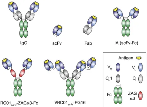
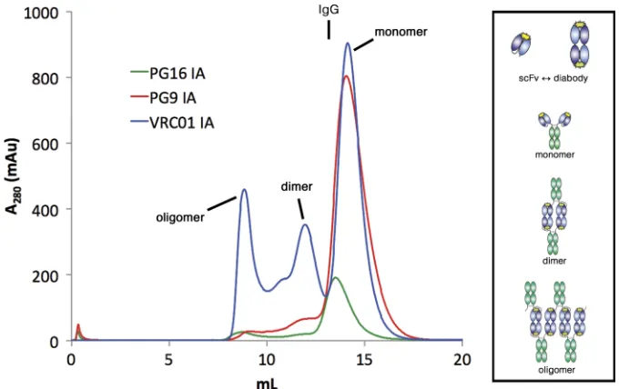
In order to compare the neutralization potencies of various anti-body forms, we produced IgG, IA, and Fab forms of the anti-HIV antibodies PG9, PG16, and VRC01 (42, 44) (Fig. 1). We also pro-duced the PG9 scFv but were unable to express either the VRC01 or PG16 scFvs in isolation in sufficient quantities for testing. Pro-teins were expressed by transient transfection of mammalian cells, and purification was by protein A or Ni-NTA chromatography followed by size exclusion chromatography. The size exclusion chromatography profiles of the PG9 and PG16 IAs revealed large peaks corresponding to the expected products (Fig. 2), i.e., a single Fc unit with two scFvs, which we will subsequently refer to as an IA monomer. During size exclusion chromatography of the VRC01 IA, however, both aggregated and apparently dimeric forms were observed in addition to the expected product (Fig. 2). These larger forms were a significant fraction of the material eluted from pro-tein A chromatography, in contrast to the PG9 and PG16 IA pu-rifications, in which only trace amounts of larger oligomers were observed.
The proteins were evaluated in an Env-pseudotyped HIV-1 neutralization assay against a panel of 30 strains (Table 1; see also Table S1 in the supplemental material). As observed for other
FIG 1Schematic depiction of antibody reagent architectures. VH, variable
domain of the IgG heavy chain (HC); VL, variable domain of the IgG light
chain (LC); CH1, constant region 1 of the HC; CL, constant region of the LC;
Fc, CH2 and CH3 domains of dimerized HCs; scFv, single-chain fragment
variable (VHand VLdomains of an IgG). The scFv shown is VHfollowed by VL;
scFvs can also be constructed as VLfollowed by VH.
on November 7, 2019 by guest
http://jvi.asm.org/
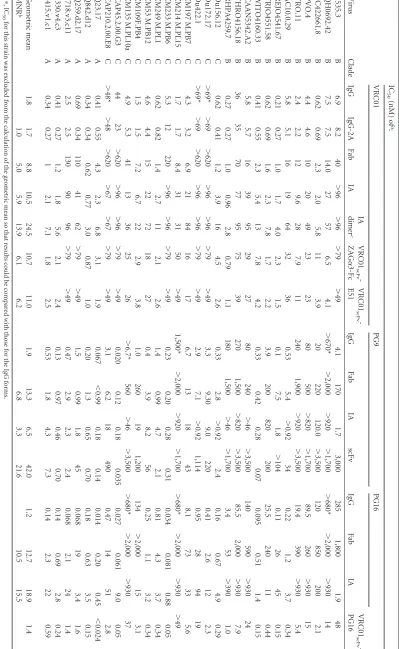
[image:2.585.47.284.67.240.2]TABLE 1 IC 50 s for IgG, Fab, IA, and scFv forms of VRC01, PG9, and PG16 a Virus Clade IC 50 (nM) of a : VRC01 PG9 PG16 VRC01 scFv -PG16 IgG IgG-2A Fab IA IA dimer c VRC01 scFv -ZAG ␣ 3-Fc VRC01 scFv -E51 IgG Fab IA scFv IgG Fab IA 6535.3 B 6.9 8.2 40 ⬎ 96 ⬎ 96 ⬎ 79 ⬎ 49 4.1 170 1.7 3,000 285 1,800 1.9 48 QH0692.42 B 7.5 7.5 14.0 27 57 6.5 4.1 ⬎ 670* ⬎ 2,000 ⬎ 920 ⬎ 1,700 ⬎ 680* ⬎ 2,000 ⬎ 930 14 SC422661.8 B 0.62 0.69 2.3 2.0 5.8 11 3.9 20 220 120.0 ⬎ 3,500 120 850 200 2.1 PVO.4 B 4.4 4.6 10 20 49 23 23 80 500 ⬎ 820 ⬎ 1,700 89.5 260 ⬎ 930 15 TRO.11 B 2.4 2.2 12 9.6 28 7.9 11 240 1,900 ⬎ 920 ⬎ 3,500 19.4 390 ⬎ 930 5.4 AC10.0.29 B 5.8 5.1 16 19 64 32 36 0.53 5.4 ⬎ 0.92 34 0.22 1.2 3.7 0.34 REJO4541.67 B 0.21 0.27 1.0 1.7 4.0 2.3 1.5 0.1 7.5 1.8 ⬎ 104 0.11 26 45 0.15 TRJO4551.58 B 0.62 0.69 1.6 2.3 7.8 1.7 2.2 3.9 200 820 200 25.5 240 11 0.44 WITO4160.33 B 0.41 0.55 2.3 5.4 13 7.8 4.2 0.33 0.42 0.28 0.07 0.095 0.51 1.4 0.15 CAAN5342.A2 B 5.8 5.7 16 39 95 29 27 80 240 ⬎ 46 ⬎ 3,500 140 590 ⬎ 930 24 THRO4156.18 B 36 35 70 77 95 75 39 270 1,500 ⬎ 820 ⬎ 3,500 85.5 2,000 ⬎ 930 7.9 RHPA4259.7 B 0.27 0.27 1.0 0.96 2.8 0.79 1.1 180 1,500 ⬎ 46 ⬎ 1,700 3.4 53 ⬎ 390 1.0 Du156.12 C 0.62 0.41 1.2 3.9 16 4.5 2.6 0.33 2.8 ⬎ 0.92 2.4 0.16 0.67 4.9 0.29 Du172.17 C ⬎ 69* ⬎ 69 ⬎ 620 ⬎ 96 ⬎ 96 ⬎ 79 ⬎ 49 3.3 9.30 9.0 220 0.41 2.6 12 2.3 Du422.1 C ⬎ 69* ⬎ 69 ⬎ 620 ⬎ 96 ⬎ 96 ⬎ 79 ⬎ 49 2.9 7.1 ⬎ 0.92 1,114 0.95 28 94 19 ZM197 M.PB7 C 4.3 3.2 6.9 21 84 16 17 6.7 13 18 43 8.1 73 33 5.6 ZM214 M.PL15 C 1.7 1.7 8.4 31 31 50 ⬎ 49 1,500* ⬎ 2,000 ⬎ 920 ⬎ 1,700 ⬎ 680* ⬎ 2,000 ⬎ 930 ⬎ 49 ZM233 M.PB6 C 5.3 12 220 ⬎ 96 ⬎ 96 ⬎ 79 ⬎ 49 0.23 0.20 0.28 0.31 0.034 0.081 0.88 0.05 ZM249 M.PL1 C 0.62 0.82 1.4 2.7 11 2.1 2.6 1.4 0.99 4.7 2.1 0.81 4.3 3.7 0.34 ZM53 M.PB12 C 4.6 4.4 15 22 72 18 27 0.4 3.9 8.2 56 0.25 1.1 3.2 0.34 ZM109F.PB4 C 1.5 1.5 7.2 6.7 22 2.9 3.8 1.0 260 19 1,200 134 ⬎ 2,000 15 3.1 ZM135 M.PL10a C 4.9 5.3 41 13 36 25 26 ⬎ 6.7* 560 ⬎ 46 ⬎ 3,500 ⬎ 680* ⬎ 2,000 ⬎ 930 37 CAP45.2.00.G3 C 44 23 ⬎ 620 ⬎ 96 ⬎ 96 ⬎ 79 ⬎ 49 0.020 0.12 0.18 0.035 0.027 0.061 9.0 0.05 CAP210.2.00.E8 C ⬎ 48* ⬎ 48 ⬎ 620 ⬎ 67 ⬎ 67 ⬎ 79 ⬎ 49 3.1 6.2 18 490 0.47 14 51 2.8 Q23.17 A 0.41 0.55 4.3 2.3 6.8 3.1 1.9 0.067 ⬍ 0.99 0.18 0.14 0.014 0.20 0.45 ⬍ 0.024 Q842.d12 A 0.34 0.34 0.62 0.77 3.0 0.87 1.0 0.20 1.3 0.65 0.70 0.18 0.63 3.5 0.15 Q259.d2.17 A 0.69 0.34 110 41 62 ⬎ 79 ⬎ 49 1.5 0.99 1.8 45 0.068 19 3.4 1.6 3718.v3.c11 A 2.5 2.5 130 90 96 ⬎ 79 ⬎ 49 0.47 2.9 2.3 2.4 0.068 2.1 24 1.4 0330.v4.c3 A 0.41 0.27 1.2 1.8 5.6 2.1 2.4 0.13 0.97 0.46 0.70 0.14 0.69 2.8 0.24 3415.v1.c1 A 0.34 0.27 1 2.1 7.1 1.8 2.5 0.53 1.8 4.3 7.3 0.14 2.3 22 0.59 Geometric mean 1.8 1.7 8.8 10.5 24.5 10.7 11.0 1.9 13.3 6.5 42.0 1.2 12.7 18.9 1.4 MNR b 1.0 5.0 5.9 13.9 6.1 6.2 6.8 3.3 21.6 10.5 15.5 a ⴱ ,I C 50 for this strain was excluded from the calculation of the geometric mean so that results could be compared with those for the IgG forms. b MNR, molar neutralization ratio of the indicated reagent compared to results for the parental IgG. c IC 50 s given for the VRC01 IA dimer fraction were calculated using the monomer molecular weight. IC 50 s (in g/ml) are presented in Table S1 in the supplemental material.
on November 7, 2019 by guest
http://jvi.asm.org/
[image:3.585.95.494.71.720.2]anti-HIV antibodies, the Fab forms were on average 5- to 10-fold less potent on a molar basis than the intact IgG (18). The 2A form of VRC01 IgG (translated from a single mRNA containing a pi-cornavirus 2A peptide sequence [37] between the heavy and light chain genes) had potency equal to that of VRC01 IgG. However, the IA forms of VRC01, PG9, and PG16 were less potent than the corresponding IgGs; the potencies were reduced overall by 5.9-fold (VRC01), 3.3-5.9-fold (PG9), and 15.5-5.9-fold (PG16 IA,
con-structed as VLfollowed by VH). Two-tailed pairedttests
demon-strated that these differences were significant (Pvalues of 0.00045,
0.047, and 0.032, respectively). A PG16 IA constructed with VH
followed by VLwas also expressed and tested on a more limited
number of strains; this reagent was also less potent than PG16 IgG (see Table S2 in the supplemental material). Although the dimeric fraction of the VRC01 IA was active in neutralization, it was
2.3-fold less potent on a mass basis (Pvalue of 0.00012) than
mono-meric VRC01 IA (Table 1).
Potential reasons for the IAs to be less potent than their parent IgGs include the following: (i) a shorter span between the two
antigen combining sites, (ii) reduced stability of IAs versus IgGs in the assay media, and/or (iii) reduced affinity of the scFv antigen binding sites compared to that of Fab binding sites. To evaluate whether the shorter arm span of the IAs diminished their activity, we expressed an scFv-containing reagent with a combining site separation more similar to that of IgG by inserting an
immuno-globulin constant region-like domain, the␣3 domain from Zn-␣2
glycoprotein (ZAG), between the scFv and Fc components to
cre-ate VRC01scFv-ZAG␣3-Fc (Fig. 1). The maximal separation
be-tween the VH-VLcombining sites should be similar in VRC01scFv
-ZAG␣3-Fc and VRC01 IgG. To permit even greater separation,
the VRC01 scFv was also attached to the N terminus of the heavy chain of an unrelated IgG to create VRC01scFv-E51. The CD4-induced (CD4i) antibody E51 was chosen for this construct be-cause E51 IgG expresses well and is weakly neutralizing or non-neutralizing in the absence of CD4 (22), as observed for other CD4i antibodies (19). Neutralization assays using these reagents
(Table 1) demonstrated an average IC50 similar to that with
VRC01 IA, indicating that the arm span of VRC01scFv-based re-agents had little impact on their neutralization potencies.
We next tested whether differences in reagent stability (i.e., survival) under our neutralization assay conditions contributed to the differences we observed between IAs and IgGs by conducting assays with a 12- or 24-h preincubation in assay medium prior to adding pseudovirus. No trend toward diminished neutralization potency over time was observed (Table 2), demonstrating that differential stability in the assay medium did not account for dif-ferences in potency.
[image:4.585.42.285.88.179.2]A weaker antigen-binding affinity of the scFv in an IA versus the Fab in an IgG was suggested by the less potent neutralization observed for PG9 scFv than for PG9 Fab (Table 1). The possibility of reduced antigen-binding affinity was directly tested by compar-ing the bindcompar-ing of gp120 to VRC01 IgG versus scFv-containcompar-ing
TABLE 2Effect of preincubation of VRC01 reagents on neutralization
potencya
Pseudovirus Reagent
IC50(nM) of reagent at
preincubation time
0 h 12 h 24 h
JR-FL VRC01 IgG 0.37 0.40 0.31
VRC01 IA 5.2 4.6 1.7
Du156 VRC01 IgG 0.67 0.60 0.47
VRC01 IA 3.1 2.6 2.0
aThe neutralization assay was modified by incubating the reagent in assay medium for
the indicated times prior to addition of pseudovirus.
FIG 2Size exclusion chromatography profiles of PG16, PG9, and VRC01 IAs. Protein A-purified IAs were injected over a Superdex 200 10/300 GL column. IgGs
normally elute at⬃13 ml (as indicated at the top of the figure), compared to the slightly smaller IA monomers, which elute at⬃14.5 ml. (Data are shown for the VL-VHversion of the PG16 IA; similar results were obtained for the VH-VLversion.) Although the PG9 and PG16 IAs were predominantly monomeric, the
VRC01 IA profile showed multimeric (presumably dimeric) and aggregate peaks in addition to the monomer. The tendency of scFv molecules to dimerize by 3-D domain swapping (shown schematically at the top in the box) may lead to the formation of dimeric and oligomeric forms of IAs (potential structures shown in the box).
on November 7, 2019 by guest
http://jvi.asm.org/
[image:4.585.122.462.451.665.2]forms of VRC01. Purified gp120 from strain Q259.d2.17 was in-jected over protein A-immobilized VRC01 IgG, VRC01 IA, or
VRC01scFv-ZAG␣3-Fc in a surface plasmon resonance
(SPR)-based binding assay (see Fig. S2 in the supplemental material). The
equilibrium dissociation constant (KD) derived for VRC01 IgG
was 160 nM, compared with affinities of 590 nM and 570 nM for
VRC01 IA and VRC01scFv-ZAG␣3-Fc, respectively. The
approxi-mately 4-fold-weaker affinity of the scFv-containing reagents was
comparable to the 6-fold-weaker neutralization IC50of VRC01 IA
compared to that of VRC01 IgG.
To explore the potential for combined reagents to provide greater neutralization breadth, we expressed a modified form of PG16 in which a VRC01 scFv was attached to the N terminus of
the PG16 light chain with a (Gly3-Ser)6linker. The neutralization
properties of VRC01scFv-PG16 are shown in Table 1. Inspection of
the measured IC50s for strains that were resistant to either VRC01 or PG16 confirmed that both components in the combined re-agent were active.
We assessed the potencies of each of the components in the bispecific VRC01scFv-PG16 reagent by a modeling procedure
us-ing the IC50s for VRC01 IgG and PG16 IgG as follows. Assume the
IC50s of the parent IgGs VRC01 and PG16 arevandp, respectively,
for a given HIV-1 strain. Consider an idealized combined reagent in which both components functioned independently with no synergy or interference. In a very simplified picture of virus neu-tralization, reagent binding is equivalent to neuneu-tralization, and the
IC50can be approximated by a single binding event with the same
equilibrium dissociation constant. Solving the equilibrium
equa-tions for 50% binding/neutralization, we found the modeled IC50
was共
冑
v2⫹6vp⫹p2⫺
v⫺p兲⁄2. In the actual bispecific reagent, we
anticipated the individual components would have reduced
activ-ity, i.e.,vreduced⫽veff⫻v, whereveffis⬎1, andpreduced⫽peff⫻p,
wherepeffis⬎1. Assuming thatveffandpeffare constant across
different strains, we solved for best-fit values of these parameters
that minimized⌺strains(log IC50 observed⫺log IC50 modeled)2. A fit
assuming only one active antibody did not fit the data as well as a fit assuming that both components were active (see Fig. S3 in the supplemental material).
Using our neutralization data for VRC01scFv-PG16 (Table 1),
we derived values of 3.16 and 3.22 forveffandpeff; thus, the VRC01
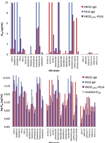
scFv component possessed about 1/3 of the potency of VRC01 IgG, and the PG16 IgG portion possessed about 1/3 of the potency of unmodified PG16 IgG. For any given strain, the combined re-agent was nearly always weaker than the stronger parent IgG. However, the combined reagent was superior on a mass basis to the weaker parent for 27 out of 30 strains (Fig. 3, bottom panel). In addition, the combined reagent neutralized more strains than
ei-ther parent; e.g., using an IC50of⬍5.0g/ml as the cutoff for
neutralization, VRC01scFv-PG16 neutralized 90% of the strains we
tested, while VRC01 IgG neutralized 83% and PG16 IgG neutral-ized 70% (Table 1 and Fig. 3, top panel). However, this depends on
the neutralization threshold chosen. For IC50s of⬍1.0g/ml, the
combined reagent neutralized 67% of strains, while VRC01 and PG16 neutralized 77% and 60%, respectively.
DISCUSSION
Provision of prophylactic or therapeutic antibodies by direct in-jection or gene therapy permits consideration of a wide range of potential reagents, building on initial discoveries of anti-HIV IgGs. Variations fall into three major categories: (i) those affecting
the antigen binding site, (ii) choice of the overall architecture (e.g., IA versus IgG), and (iii) modulations of effector function. A thor-ough evaluation of the efficacies of the full range of potential re-agents is a large task. A variety of selection strategies is available to screen variants in category i, i.e., natural antibody repertoires and antigen binding site libraries. However, full exploration of the alternatives in categories ii and iii is limited by the need to produce purified, testable quantities of reagents and the screening limita-tions of evaluating effector function in complex cell- or animal-based assays. The present studies are intended to provide insight into the potential effects of architecture on reagent potency.
The size of a delivered reagent must be considered for gene therapy efforts involving AAV. In general, constructs approaching the size limit for packaging AAV suffer reduced expression levels (7). Although the minimum serum or genital tract anti-HIV IgG concentration necessary for protection is not known, a rough
es-timate of 100 times the reagent’s IC50has been suggested (30). For
broad strain coverage with reagents such as VRC01, PG9, or PG16, this implies desired concentrations in the tens to hundreds of g/ml. Early efforts using an AAV vector with separate promoters for heavy and light chains directing expression of anti-HIV IgG
b12 yielded serum concentrations of only⬃5g/ml (20).
Alter-native AAV/IgG constructs have permitted high-level IgG ex-pression in other cases. For example, use of a single promoter with a 2A self-processing peptide inserted between the
anti-body heavy and light chains permitted⬃1-mg/ml IgG levels to
be achieved with AAV-transduced liver expression (9). Never-theless, the smaller size of IA constructs versus conventional IgG is attractive for gene therapy approaches in which vector
capacity is severely limited—in particular, for
self-complementary AAV vectors, which are more efficient at trans-duction than AAV vectors with a single-stranded genome (25). To more fully understand the potential trade-offs in vector and construct design, we compared the neutralization activities of IA versus IgG versions of three broadly neutralizing antibodies: PG9, PG16, and VRC01 (42, 44). We found that the PG9, PG16, and VRC01 IAs were severalfold less potent than their IgG forms. A reduced affinity of the scFv antigen-binding site is a likely contrib-uting factor to this difference. Although some scFvs have affinities equivalent to those of the related Fabs, it was noted in early studies that scFvs can exhibit up to 10-fold weaker binding (4). The weaker binding and neutralization by the VRC01 scFv-containing reagents is likely due to suboptimal geometry of the antigen bind-ing site and/or steric interference by the Gly-Ser linker joinbind-ing the
VRC01 VH and VLdomains. Steric interference from the scFv
linker is consistent with the VRC01 Fab-gp120 crystal structure
(45), in which the N terminus of the Fab light chain is⬃8 Å from
the gp120 backbone. This relatively close distance suggests that the
Gly-Ser linker extending from the VLdomain N terminus could
sterically interfere with antigen binding. It is possible that the scFvs in the reagents we tested were suboptimal; different designs might yield scFvs with affinities matching those of the corre-sponding Fabs. Thus, to achieve maximal efficacy, it will be
nec-essary to explore different architectures (VH-VLversus VL-VH)
and different linker lengths for each scFv used in IAs.
A potential complication of scFv reagents is their tendency to dimerize or multimerize by three-dimensional (3-D) domain swapping (3). The extent of dimerization of scFvs is variable, de-pending on linker length, antibody sequence, concentration, buf-fer conditions, and the presence or absence of antigen (1). The
on November 7, 2019 by guest
http://jvi.asm.org/
potential for IAs to form dimers or other oligomers as observed for VRC01 IA is a special concern for reagents that will be deliv-ered by gene therapy, where it is not possible to remove higher-order products once they are secreted from transduced cells. Mul-timeric forms of IAs may be less potent and potentially more immunogenic than the monomeric molecule; this may add to the immunogenicity of the artificial linker of IAs. AAV-mediated ex-pression of IAs in rhesus macaques led to various levels of IA-specific antibody response, which appeared to be correlated with reduced efficacy against viral challenge (15). The best means of
addressing this possible complication is careful biophysical char-acterization of proposed gene therapy protein products.
In the recent rhesus macaque SIV challenge experiment, deliv-ery of IAs was found to be superior to either scFv or whole IgG with respect to the serum concentrations that could be achieved (15). Although the IAs used in the challenge experiments
exhib-ited neutralization IC50s well below 1.0 g/ml, the Fabs from
which they were derived had IC50s that were 3-fold more potent
on a mass basis (16). (The IC50s of the corresponding IgGs have
not been reported). On a molar basis, the IAs were thus about
FIG 3Histograms displaying IC50s (g/ml) of VRC01 IgG (red), PG16 IgG (blue), and VRC01scFv-PG16 (purple) (top; linear scale) or VRC01 IgG (red), PG16
IgG (blue), VRC01scFv-PG16 (purple), and the modeled IC50s of VRC01scFv-PG16 (light purple) (bottom; logarithmic scale).
on November 7, 2019 by guest
http://jvi.asm.org/
[image:6.585.111.475.63.556.2]1.5-fold less potent than the Fab forms. This ratio is very similar to the average 1.2-fold- and 1.5-fold-weaker molar neutralization we observed for the VRC01 and PG16 IAs versus the corresponding Fabs (Table 1). In the rhesus challenge study, the IA potencies
(IC50s of 0.01 to 0.02 g/ml against the SIVmac316 challenge
strain) were sufficient to provide protection. However, since anti-HIV IgGs are generally 6- to 30-fold more potent than their cor-responding Fabs on a molar basis (18), the IAs evaluated here represent a significant trade-off necessitated by the lower serum IgG concentrations achievable (15) with currently available AAV technology.
While VRC01, PG9, and PG16 have very broad activities, each of these antibodies fails to neutralize 9 to 27% of HIV-1 strains
(using a cutoff IC50of 50g/ml; at 1g/ml, 28 to 49% of strains
are not neutralized) (42, 44). For passive immunization or gene therapy applications, addressing the incomplete strain coverage requires delivering either multiple antibodies or a single reagent combining two or more activities. Here we investigated the feasi-bility of one such bispecific reagent, in which a VRC01 scFv was
attached to PG16 IgG (VRC01scFv-PG16).In vitroneutralization
assays against a panel of HIV-1 strains demonstrated both VRC01 and PG16 activities. Although both potencies were reduced
com-pared to those of the parental IgGs, VRC01scFv-PG16 showed
greater breadth, suggesting the potential for a bispecific reagent to provide complete or near-complete protection against HIV-1. Further development of such a reagent is possible, for example,
given that the VRC01 scFv component of VRC01scFv-PG16 had a
reduced activity similar to that observed for the VRC01 IA, im-provement of the scFv portion could be attempted. The weaker activity of the PG16 component may result from steric factors from the scFv attached to the N terminus of the PG16 light chain. Switching the VRC01 scFv to the N terminus of the PG16 heavy chain did not improve PG16 activity (see Table S3 in the supple-mental material). Changing the size or structure of the linker or switching to the C terminus of the heavy chain may permit greater PG16 activity. Although the reduced potencies of the individual components of a bispecific reagent might increase the risk that resistance to these neutralizing activities could develop, this con-cern is arguably secondary to providing breadth of coverage in the context of infection prophylaxis (versus treatment) since the goal of infection prophylaxis is to neutralize a small viral inoculum rather than to control an established infection.
The design of gene therapy reagents for HIV-1 prophylaxis potentially involves a variety of trade-offs, including breadth of reagent, potency, expression level, and minimization of potential for immunogenicity and other side effects. The newly discovered anti-HIV antibodies (42, 44) demonstrate that breadth and great potency can be achieved simultaneously. Our results suggest that careful optimization of reagent architecture and full biophysical characterization of the oligomeric states of potential protein re-agents are important for fully exploiting the potential offered by genetic approaches to providing HIV-1 immunity. Direct conver-sion of IgGs to IAs will often entail a loss of potency due to weaker binding of the scFv compared to the Fab binding site and/or do-main swapping to create scFv multimers, which can perhaps be avoided by screening many scFv designs. Whether this loss of
po-tency is acceptable depends on relativein vivo serum levels of
AAV-expressed IAs and IgGs. For prophylaxis against a wide va-riety of circulating HIV-1 strains, a delivered reagent will face strains where its activity is far from maximal. In this situation,
optimization of the reagent will be critical to provide robust pro-tection.
ACKNOWLEDGMENTS
We thank the CAVD Neutralizing Antibody Core Laboratories for per-formingin vitroneutralization assays, Gary Nabel (Vaccine Research Cen-ter) for VRC01-related constructs and critical reading of the manuscript, Dennis Burton (Scripps Research Institute) for PG9 and PG16 sequences, Terri Lee and Erin Flanagin for DNA preparation, Timothy Feliciano, Michael Anaya, and the Caltech Protein Expression Center for protein expression and purification, Maria Politzer for subcloning and DNA preparation, Paola Marcovecchio for providing purified Q259.d2.17 gp120, Jennifer Keeffe for assistance in data collection, Marta Murphy for figure preparation, and Ron Diskin and Alex Balazs for helpful discus-sions.
This work was supported by a grant from the Bill and Melinda Gates Foundation through the Grand Challenges in Global Health Initiative.
REFERENCES
1.Arndt KM, Muller KM, Pluckthun A. 1998. Factors influencing the
dimer to monomer transition of an antibody single-chain Fv fragment. Biochemistry37:12918 –12926.
2.Baba TW, et al.2000. Human neutralizing monoclonal antibodies of the
IgG1 subtype protect against mucosal simian-human immunodeficiency virus infection. Nat. Med.6:200 –206.
3.Bennett MJ, Eisenberg D.2004. The evolving role of 3D domain
swap-ping in proteins. Structure12:1339 –1341.
4. Bird RE, Walker BW.1991. Single chain antibody variable regions.
Trends Biotechnol.9:132–137.
5.Corti D, et al.2010. Analysis of memory B cell responses and isolation of
novel monoclonal antibodies with neutralizing breadth from HIV-1-infected individuals. PLoS One5:e8805.
6.Diskin R, Marcovecchio PM, Bjorkman PJ.2010. Structure of a clade C
HIV plus CD4 and CD4-induced antibody reveals anti-CD4 polyreactiv-ity. Nat. Struct. Mol. Biol.17:608 – 613.
7.Dong JY, Fan PD, Frizzell RA.1996. Quantitative analysis of the
pack-aging capacity of recombinant adeno-associated virus. Hum. Gene Ther.
7:2101–2112.
8.Durocher Y, Perret S, Kamen A.2002. High-level and high-throughput
recombinant protein production by transient transfection of suspension-growing human 293-EBNA1 cells. Nucleic Acids Res.30:E9.
9.Fang J, et al.2005. Stable antibody expression at therapeutic levels using
the 2A peptide. Nat. Biotechnol.23:584 –590.
10. Grimm D, Kay MA.2003. From virus evolution to vector revolution: use
of naturally occurring serotypes of adeno-associated virus (AAV) as novel vectors for human gene therapy. Curr. Gene Ther.3:281–304.
11. Hessell AJ, et al.2007. Fc receptor but not complement binding is
im-portant in antibody protection against HIV. Nature449:101–104.
12. Hessell AJ, et al.2009. Effective, low-titer antibody protection against
low-dose repeated mucosal SHIV challenge in macaques. Nat. Med.15: 951–954.
13. Hessell AJ, et al.2009. Broadly neutralizing human anti-HIV antibody
2G12 is effective in protection against mucosal SHIV challenge even at low serum neutralizing titers. PLoS Pathog.5:e1000433.
14. Huang C.2009. Receptor-Fc fusion therapeutics, traps, and MIMETI
BODY technology. Curr. Opin. Biotechnol.20:692– 699.
15. Johnson PR, et al.2009. Vector-mediated gene transfer engenders
long-lived neutralizing activity and protection against SIV infection in mon-keys. Nat. Med.15:901–906.
16. Johnson WE, et al.2003. Assorted mutations in the envelope gene of
simian immunodeficiency virus lead to loss of neutralization resistance against antibodies representing a broad spectrum of specificities. J. Virol.
77:9993–10003.
17. Keele BF, et al.2008. Identification and characterization of transmitted
and early founder virus envelopes in primary HIV-1 infection. Proc. Natl. Acad. Sci. U. S. A.105:7552–7557.
18. Klein JS, Bjorkman PJ.2010. Few and far between: how HIV may be
evading antibody avidity. PLoS Pathog.6:e1000908.
19. Labrijn AF, et al.2003. Access of antibody molecules to the conserved
coreceptor binding site on glycoprotein gp120 is sterically restricted on primary human immunodeficiency virus type 1. J. Virol.77:10557–10565.
on November 7, 2019 by guest
http://jvi.asm.org/
20. Lewis AD, Chen R, Montefiori DC, Johnson PR, Clark KR. 2002. Generation of neutralizing activity against human immunodeficiency vi-rus type 1 in serum by antibody gene transfer. J. Virol.76:8769 – 8775.
21. Li M, et al.2005. Human immunodeficiency virus type 1 env clones from
acute and early subtype B infections for standardized assessments of vaccine-elicited neutralizing antibodies. J. Virol.79:10108 –10125.
22. Li M, et al.2006. Genetic and neutralization properties of subtype C
human immunodeficiency virus type 1 molecular env clones from acute and early heterosexually acquired infections in Southern Africa. J. Virol.
80:11776 –11790.
23. Mascola JR, et al. 1999. Protection of macaques against pathogenic
simian/human immunodeficiency virus 89.6PD by passive transfer of neutralizing antibodies. J. Virol.73:4009 – 4018.
24. Mascola JR, et al.2000. Protection of macaques against vaginal
transmis-sion of a pathogenic HIV-1/SIV chimeric virus by passive infutransmis-sion of neutralizing antibodies. Nat. Med.6:207–210.
25. McCarty DM, et al.2003. Adeno-associated virus terminal repeat (TR)
mutant generates self-complementary vectors to overcome the rate-limiting step to transduction in vivo. Gene Ther.10:2112–2118.
26. Mehandru S, et al.2007. Adjunctive passive immunotherapy in human
immunodeficiency virus type 1-infected individuals treated with antiviral therapy during acute and early infection. J. Virol.81:11016 –11031.
27. Montefiori DC.2005. Evaluating neutralizing antibodies against HIV,
SIV, and SHIV in luciferase reporter gene assays. Curr. Protoc. Immunol.
2005:12.11. doi:10.1002/0471142735.im1211s64.
28. Nelson AL.2010. Antibody fragments: hope and hype. MAbs2:77– 83.
29. Nishimura Y, et al.2003. Transfer of neutralizing IgG to macaques 6 h but
not 24 h after SHIV infection confers sterilizing protection: implications for HIV-1 vaccine development. Proc. Natl. Acad. Sci. U. S. A.100: 15131–15136.
30. Parren PW, et al. 2001. Antibody protects macaques against vaginal
challenge with a pathogenic R5 simian/human immunodeficiency virus at serum levels giving complete neutralization in vitro. J. Virol.75: 8340 – 8347.
31. Poignard P, et al.1999. Neutralizing antibodies have limited effects on
the control of established HIV-1 infection in vivo. Immunity10:431– 438.
32. Rerks-Ngarm S, et al.2009. Vaccination with ALVAC and AIDSVAX
to prevent HIV-1 infection in Thailand. N. Engl. J. Med. 361: 2209 –2220.
33. Ritola K, et al.2004. Multiple V1/V2 env variants are frequently present
during primary infection with human immunodeficiency virus type 1. J. Virol.78:11208 –11218.
34. Salazar-Gonzalez JF, et al.2008. Deciphering human immunodeficiency
virus type 1 transmission and early envelope diversification by single-genome amplification and sequencing. J. Virol.82:3952–3970.
35. Scheid JF, et al.2011. Sequence and structural convergence of broad and
potent HIV antibodies that mimic CD4 binding. Science333:1633–1637.
36. Shibata R, et al.1999. Neutralizing antibody directed against the HIV-1
envelope glycoprotein can completely block HIV-1/SIV chimeric virus infections of macaque monkeys. Nat. Med.5:204 –210.
37. Szymczak AL, et al.2004. Correction of multi-gene deficiency in vivo
using a single ‘self-cleaving’ 2A peptide-based retroviral vector. Nat. Bio-technol.22:589 –594.
38. Trkola A, et al.2005. Delay of HIV-1 rebound after cessation of
antiret-roviral therapy through passive transfer of human neutralizing antibodies. Nat. Med.11:615– 622.
39. Virgin HW, Walker BD.2010. Immunology and the elusive AIDS
vac-cine. Nature464:224 –231.
40. Walker LM, Burton DR.2010. Rational antibody-based HIV-1 vaccine
design: current approaches and future directions. Curr. Opin. Immunol.
22:358 –366.
41. Walker LM, et al.2011. Broad neutralization coverage of HIV by multiple
highly potent antibodies. Nature477:466 – 470.
42. Walker LM, et al.2009. Broad and potent neutralizing antibodies from an
African donor reveal a new HIV-1 vaccine target. Science326:285–289.
43. Wei X, et al.2003. Antibody neutralization and escape by HIV-1. Nature
422:307–312.
44. Wu X, et al.2010. Rational design of envelope identifies broadly
neutral-izing human monoclonal antibodies to HIV-1. Science329:856 – 861.
45. Zhou T, et al.2010. Structural basis for broad and potent neutralization
of HIV-1 by antibody VRC01. Science329:811– 817.