Cell-
Selective
Chemoproteomics
for
Biological Discovery
Thesis by
Shannon Elizabeth Stone
In Partial Fulfillment of the Requirements for the degree of
Doctor of Philosophy in Chemistry
CALIFORNIA INSTITUTE OF TECHNOLOGY Pasadena, California
2018
© 2017
Shannon Elizabeth Stone ORCID: 0000-0002-6617-3874
ACKNOWLEDGEMENTS
The greatest part of my graduate school experience has been the privilege to learn from many brilliant people and be a part of the exceedingly collaborative nature of this scientific community. First, I thank my advisor and mentor in every sense of the word, Prof. Dave Tirrell, for his continued guidance and support. In addition to his immense scientific insight and encouragement throughout the years, I am continuously impressed by his degree of integrity and thoughtfulness, both of which I strive to emulate. Thanks also to the other members of my thesis committee: Prof. Sarkis Mazmanian for many illuminating discussions, both scientific and personal, Prof. Bob Grubbs for advice and guidance, and Prof. Long Cai for astute recommendations on my project.
I have been fortunate to share my time here with many members of the Tirrell lab, whose patience, brilliance, and humor made the time fly by. I will always cherish the days of the Best Office and summer softball with the Tools of Synthetic Biology.
I greatly enjoyed working with Judy Shon, a talented Caltech undergraduate, over the past years. The Proteome Exploration Laboratory is run by the most wonderful and knowledgeable team, who all contributed to most chapters of this thesis. Thank you also to members of both the Newman and Mazmanian labs for sharing knowledge and resources. The staff at the Beckman Imaging Center has also been exceedingly helpful. Thanks to Prof. Linda Hsieh-Wilson for her support, as well as former members of the Hsieh-Wilson lab for their advice and expertise, and especially Dr. Abby Pulsipher, who continues to be a source of scientific and personal inspiration. I was also fortunate to spend a month at the University of Chicago, learning from some of the world’s top researchers in
Staphylococcus aureus, for which I am very grateful.
you. I also thank the Chemistry division for its support, especially for help in starting the Women in Chemistry group. I greatly appreciate the enthusiastic lecturers I had during my time at Caltech, especially Justin Bois, who taught me the joys of data analysis.
To my friends who remind me that there is more to the world than science, and who drink wine with me to celebrate or drink wine with me to commiserate, thank you.
I am forever grateful to my family for their love and encouragement from the other side of the country. I would not be where I am today without each of you.
ABSTRACT
Cellular protein synthesis changes rapidly in response to internal and external cues in ways
that vary from cell to cell. Global proteomic analyses of microbial communities, tissues,
and organisms have provided important insights into the behavior of such systems, but can
obscure the diversity of responses characteristic of different cellular subpopulations. Recent
advances in cell-specific proteomics—fueled in part by the development of bioorthogonal
chemistries, more sensitive mass spectrometers and more advanced mining algorithms—
have yielded unprecedented glimpses into how proteins are expressed in space and time.
Whereas previous cell-specific proteomic analyses were confined to abundant cells in
relatively simple systems, recent advances in chemoproteomics allow researchers to map
the protein expression patterns of even rare cells in complex tissues and whole organisms.
Chapter 1 highlights recently developed strategies for cell-selective proteomics, including
metabolic labeling strategies such as bioorthogonal noncanonical amino acid tagging
(BONCAT). BONCAT is a chemoproteomic technique that enables temporal labeling of
proteins using noncanonical amino acids. In the cell-selective version of BONCAT,
expressing a mutant aminoacyl-tRNA synthetase under the control of cell-specific genetic
elements affords cellular resolution; only cells of interest can selectively incorporate a
noncanonical amino acid into proteins for subsequent detection and identification. Chapter
2 details protocols to set up a cell-selective BONCAT system.
While BONCAT had previously been applied to studies of microbial pathogenesis in tissue
culture-based models of infection, we sought to further develop the method to identify the
proteome of methicillin-resistant Staphylococcus aureus (MRSA) within a mouse model of
infection, as detailed in Chapter 3. We used this technique to enrich for staphylococcal
important for infection, we also found many that had not previously been associated with
infection. Screening several of these previously unknown factors in vivo led to the
discovery of a novel protein important for MRSA infection. This unbiased approach to
cell-selectively label pathogenic proteins during infection could be used as a global discovery
tool for novel anti-infective strategies.
In Chapter 4, we combine this cell-selective BONCAT strategy with microbial
identification after passive clarity technique (MiPACT) to visualize both staphylococcal
protein synthesis and ribosomal RNA within whole skin abscesses during infection. In
Chapter 5, we continue developing cell-selective BONCAT to study microbial protein
synthesis in the context of a living mouse by extending the system to Bacteroides fragilis, a
common human gut commensal.
Finally, cell-selective BONCAT is wholly dependent on the bioorthogonal nature of the
azide and its detection reagents. Fishing out an azide-tagged molecule from the rest of the
cellular milieu requires optimization of enrichment-based strategies. In Chapter 6, we
describe the development of a peptide to quantitate the gain of our enrichments.
While innovations in mass spectrometry and computational algorithms have facilitated the
identification and quantification of thousands of proteins simultaneously from complex
samples, this abundance of data does not necessarily lead to biological insight. Cell-specific
proteomic techniques will play a key role in the identification of the mechanisms that
govern cell specialization and that allow organisms to respond to changing environments.
Overall, this work demonstrates the power of cell-selective chemoproteomics to ascertain
PUBLISHED CONTENT AND CONTRIBUTIONS
(1) Pulsipher, A., Griffin, M. E., Stone, S. E., Brown, J. M., Hsieh-Wilson, L. C. “Directing neuronal signaling through cell-surface glycan engineering.” J. Am. Chem. Soc. 2014, 136 (19), 6794-6797. DOI: 10.1021/ja5005174
S.E.S. participated in project conception, optimized the TrkA assay, and contributed to the writing of the manuscript.This article’s figures are reproduced in part within Appendix B with permission according to the ACS AuthorChoice terms of use.
(2) Pulsipher, A., Griffin, M. E., Stone, S. E., Hsieh-Wilson, L. C. “Long-lived engineering of glycans to direct stem cell fate.” Angew. Chem. Int. Ed. 2015, 54, 1466-1470. DOI: 10.1002/anie.201409258
S.E.S. participated in project conception, created the plasmids, determined conditions for cell labeling, and contributed to the writing of the manuscript. This article’s figures are reproduced in part within Appendix C with permission from the publisher.
(3) Griffin, M. E., Jensen, E. H., Mason, D. E., Jenkins, C. L., Stone, S. E., Peters, E. C., Hsieh-Wilson, L. C. “Comprehensive mapping of O-GlcNAc modification sites using a chemically cleavable tag.” Mol. Biosys. 2016, 12, 1756-1759. DOI: 10.1039/c6mb00138f
S.E.S. synthesized and characterized compound 1 in Figure 1A. This article’s figures are reproduced in part within Appendix D with permission according to the Creative Commons Attribution 3.0 Unported License terms of use.
(4) Stone, S. E., Glenn, W. S., Hamblin, G. D., Tirrell, D. A. “Cell-selective proteomics for biological discovery.” Curr. Opin. Chem. Biol. 2017, 36, 50-57. DOI: 10.1016/j.cbpa.2016.12.026
S.E.S. chose references, devised the outline, designed figures, created the table, and wrote the manuscript. This article including figures is reproduced in Chapter I with permission from the publisher.
(5) Glenn, W. S., Stone, S. E., Ho, S. H., Sweredoski, M. J., Moradian, A., Hess, S., Bailey-Serres, J., Tirrell, D. A. “Bioorthogonal noncanonical amino acid tagging (BONCAT) enables time-resolved analysis of protein synthesis in native plant tissue.” Plant Phys. 2017, 173 (3), 1543-1553. DOI: 10.1104/pp.16.01762
TABLE OF CONTENTS
Acknowledgements………..iii
Abstract ……….v
Published Content and Contributions………..vii
Table of Contents………viii
List of Figures………x
Nomenclature………xi
Chapter I: Cell-Selective Proteomics……….1
1.1 Abstract……….……….. 1
1.2 Introduction……….……… 2
1.3 Cell-Selective Translatomics and Ribosome Profiling……… 3
1.4 Separating Cells for Steady-State Proteomic Analysis……… 4
1.5 Metabolic Labeling……….……….5
1.6 Spatially Restricted & Subcellular Proteomics………11
1.7 Choosing a Cell-Selective Proteomic Method……….12
1.8 Conclusions & Future Outlook……….………14
Chapter II: How to do a cell-selective bioorthogonal noncanonical amino acid tagging (BONCAT) experiment………19
2.1 Abstract….………….………….………….………….………….………19
2.2 Introduction….………….………….………….………….………….……20
2.3 Design of a cell-selective BONCAT experiment….………….………….24
2.4 Materials….………….………….………….………….………….………26
2.5 Procedure….………….………….………….………….………….…… 27
Chapter III: Cell-Selective Proteomics in Mice Reveals Factors Important for MRSA Infection……….36
3.1 Abstract….………….………….………….………….………….………36
3.2 Introduction….………….………….………….………….……….37
3.3 Results…….………….………….………….……….………….………….40
3.4 Discussion…….…….…….…….…….…….…….…….…….…….……53
3.5 Materials & Methods…….…….…….…….…….…….…….…….…….56
Chapter IV: Visualizing Pathogenic Protein Synthesis During Infection…….67
4.2 Introduction…….………….………….………….……….…….. 68 4.3 Results…….………….………….………….……….………….. 69 4.4 Discussion & Future Directions…….………….………….………….….75 4.5 Supplemental Information…….………….………….………….………..78 4.6 Experimental Procedures…….………….………….………….…………80
Chapter V: Towards cell-specific proteomics of the gut microbiota………….87 5.1 Abstract………….………….………….………….………….………….87 5.2 Introduction….………….………….………….………….……….……..88 5.3 Results….………….………….………….………….……….…………..89 5.4 Discussion & Future Directions….………….………….………….…….94 5.5 Experimental Procedures….………….………….………….………95
Chapter VI: Quantifying Enrichment of Azide-Tagged Proteins Using
SPIQE (Spike Peptide In to Quantify Enrichment) ………. 98 6.1 Abstract….………….………….………….………….………. 98 6.2 Introduction….………….………….………….………….……….…….. 99 6.3 General Approach….………….………….………….………….………104 6.4 Results….………….………….………….………….……….……..…..106 6.5 Discussion….………….………….………….………….……….…….. 114 6.6 Future Work….………….………….………….………….……….……115 6.7 Experimental Procedures….………….………….………….…………..116
LIST OF
FIGURES
Number Page
1.1The importance of cell-type-specific proteomics ... 2
1.2Labeling strategies for cell-selective proteomics ... 10
1.3Advantages and disadvantages of cell-specific proteomic methods .... 14
2.1Cell-specific bioorthogonal noncanonical amino acid tagging ... 22
2.2Noncanonical amino acids ... 24
2.3Workflow of cell-selective BONCAT ... 26
2.4Representative BONCAT labeling gel ... 32
3.1Scheme depicting BONCAT to label MRSA in a live mouse ... 40
3.2BONCAT labels newly-synthesized MRSA proteins ... 44
3.3Comparative proteomics reveals MRSA proteins expressed ... 49
3.4Screening of mutant hits from BONCAT analysis ... 51
3.5The metabolic role of AdhE during infection ... 54
4.1 Scheme used to label skin abscess during infection ... 70
4.2 Scheme used to conjugate fluorophore to azide-tagged proteins ... 72
4.3 Wider field view of skin abscess with BONCAT and HCR ... 74
4.4 Comparison of techniques to visualize MRSA abscesses ... 75
5.1 Using BONCAT in B. fragilis ... 89
5.2 Labeling B. fragilis in the mouse gut ... 92
6.1 Scheme depicting shotgun proteomics ... 99
6.2 Scheme of SPIQE peptide method ... 104
6.3 Results of SPIQE peptide enrichments ... 109
6.4 Results of DBCO-agarose enrichments ... 110
NOMENCLATURE
aaRS. Aminoacyl-tRNA synthetase Aha. Azidohomoalanine
Anl. Azidonorleucine
APEX. Ascorbate peroxidase Azf. 4-azido-L-phenylalanine
BONCAT. Bioorthogonal noncanonical amino acid tagging CFU. Colony-forming units
Cm. Chloramphenicol
CTAP. Cell-type specific labeling with amino acid precursors CuAAC. Copper catalyzed alkyne-azide cycloaddition
DAPI. 4,6-Diamidino-2-phenylindole DBCO. Aza-dibenzocyclooctyne
Dde. N-(1-(4,4-dimethyl-2,6-dioxocyclohexylidene)ethyl) EF. 4-ethynyl-L-phenylalanine
Erm. Erythromycin
FACS. Fluorescence activated cell sorting FISH. Fluorescence in situ hybridization Gent. Gentamicin
GF. Germ-free GO. Gene ontology
H&E. Hematoxylin and eosin HCR. Hybridization chain reaction Hpg. Homopropargylglycine IP. Intraperitoneal
LB. Luria-Bertani broth LC. Liquid chromatography LFQ. Label-free quantitation
LIMMA. Linear models for microarray data MetRS. Methionyl-tRNA synthetase
MRSA. Methicillin-resistant Staphylococcus aureus
MS. Mass spectrometry
ncAA. Noncanonical amino acid OCT. Optimum cutting temperature PACT. Passive CLARITY technique PBS. Phosphate-buffered saline PCA. Principal coordinate analysis PCR. Polymerase chain reaction PEG. Polyethylene glycol PFA. Paraformaldehyde Phe. Phenylalanine
PheRS. Phenylalanyl-tRNA synthetase PMN. Polymorphonuclear leukocyte PO. Per os (Oral Gavage)
Pra. Propargylglycine
Rcf. Relative centrifugal force
RIMS. Refractive index matching solution S/C. Sub-cutaneous
SILAC. Stable isotope labeling using amino acids in culture SORT. Stochastic orthogonal recoding of translation
SPAAC. Strain-promoted azide-alkyne cycloaddition Spec. Spectinomycin
SPF. Specific-pathogen free TAMRA. Tetramethylrhodamine Tet. Tetracycline
C h a p t e r 1
CELL
-
SELECTIVE PROTEOMICS
Published as:
Stone, S. E., Glenn, W. S., Hamblin, G. D., Tirrell, D. A. “Cell-selective
proteomics for biological discovery.” Curr. Opin. Chem. Biol. 2017, 36, 50-57. DOI: 10.1016/j.cbpa.2016.12.026
1.1 Abstract
1.2 Introduction
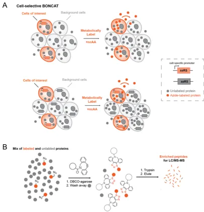
Cellular protein synthesis changes rapidly in response to internal and external cues in ways that vary from cell to cell. Global proteomic analyses of microbial communities, tissues, and organisms have provided important insights into the behavior of such systems, but can obscure the diversity of responses characteristic of different cellular subpopulations (Figure 1). Cell-selective methods for the analysis of protein synthesis are being developed to resolve proteomic changes in space and time.
Figure 1.1: The importance of cell-type-specific proteomics. Bulk measurements of complex
tissues can obscure proteomic changes that occur in specific sub-populations of cells. A protein
that is highly expressed (up arrows) in the cells of interest might be detected at low abundance
overall due to low expression (down arrows) in background cells. Cells of interest must be
physically isolated or tagged to measure the cell-specific proteome. Physical isolation measures
steady-state levels of intracellular proteins, whereas labeling methods can be time-resolved and
used to identify secreted proteins.
Cell-type-specific transcriptomics experiments have revealed mRNA expression patterns
in a wide array of biological systems, but mRNA and protein levels are often dissonant (1). Moreover, some important elements of proteome dynamics, including
post-Physically
Isolate
Metabolically
Label
Cells of interest Background
Observe Steady state
Observe Steady state
translational modification, degradation, and localization, cannot be addressed by mRNA measurements alone (2, 3). Until recently, changes in protein abundance in specific cells could be measured only in targeted, low-throughput experiments, but innovations in mass spectrometry and computational algorithms have facilitated the identification and quantification of thousands of proteins simultaneously from complex biological samples (4-6).
In this chapter, we highlight recent developments in determining cell-type-specific proteomes and recommend experimental design strategies that are guided by the question at hand.
1.3 Cell-selective translatomics and ribosome profiling
Translatomic studies, which select for ribosome-associated transcripts, have yielded stronger correlations between transcript and protein abundances than experiments that measure steady-state mRNA levels (7). Cell-type-specific studies have been enabled by translating ribosome affinity purification (TRAP), a method in which epitope-tagged ribosomes and their associated transcripts are captured, enriched and subjected to amplification and deep sequencing (8). TRAP can be rendered cell-specific by placing expression of the tagged ribosome under control of a selective promoter.
levels and locate non-canonical start sites (7). Gonzalez et al. used TRAP to cell-selectively purify ribosome-bound transcripts, and employed ribosome profiling to identify the translatome of gliomas and to reveal decreased translation in glial progenitors compared to the tumor microenvironment (9). Ribosome profiling is a powerful technique that we expect to find increasing use upon further development of cell-specific methods.
While translatomic studies provide greater depth of coverage than current proteomic measurements, ribosome binding does not ensure that a transcript is undergoing active translation (10).
1.4 Separating cells for steady-state proteomic analysis
The earliest strategies to determine cell-specific proteomes relied on separating and purifying the cells of interest prior to analysis. Cells can be sorted on the basis of expression of a transgene under control of a cell-specific promoter or by antibody staining of marker epitopes. These tools are well established and have been thoughtfully reviewed (10, 11). Physical methods have been used for years to isolate cell types from mammalian tissues for subsequent downstream analyses (12, 13). More recently these methods have been used to measure growth rates and elucidate proteomic signatures of Salmonella
during murine infection (14).
Furthermore, such methods intrinsically yield steady-state proteomic information. In contrast, metabolic labeling strategies enable cell-specific proteomic analysis to be accomplished in time-resolved fashion.
1.5 Metabolic labeling: trade-offs between sensitivity and perturbation
Metabolic labeling methods are temporally resolved and use an arsenal of amino acid isotopologs, non-canonical amino acids, and analogs of protein synthesis inhibitors (Figure 2). Each of these strategies can be placed under control of cell-specific genetic elements to afford cellular resolution. The choice of promoter(s) is key for these systems, and the degree of protein labeling needs to be weighed against the possibility of perturbing the system. Results should be validated via independent assays because labels may affect protein expression, stability, and/or function.
Cell-type-specific labeling using amino acid precursors (CTAP)
Stable isotope labeling by amino acids in cell culture (SILAC) relies on the incorporation of isotopically labeled amino acids into proteins. To make SILAC cell-selective, Gauthier
In principle, both exchange of L-lysine between cells and extracellular processing of the precursor can compromise the cell-specificity of the CTAP method. When Lavis and coworkers employed an analogous strategy to unmask fluorophores in targeted cells, they noted that the unmasked small molecule diffused through gap junctions. This effect can be exploited to study cell-cell connectivity, but would confound cell-specific protein labeling if the small molecule were to diffuse to cells lacking the decaging enzyme (16). To address these concerns, Tape et al. optimized CTAP for eukaryotic cell types and achieved ~90% cell-specific labeling in ten-day co-cultures (17). Using their optimized method, Tape et al. combined CTAP with phosphoproteomics to study heterocellular KRASG12D signaling in pancreatic ductal adenocarcinoma cells (18). By restricting their proteomic analysis to cells that expressed KRASG12D, the authors showed that the oncogene regulates AKT through reciprocal signaling – not through the accepted cell-autonomous pathway.
Bioorthogonal Noncanonical Amino acid Tagging (BONCAT)
probe proteome dynamics in bacterial (22-26) and mammalian (27) systems, and notably, to enrich and quantify secreted proteins (28). Depletion of cellular methionine is not necessary for Aha labeling; Bagert et al. showed that a 30:1 ratio of Aha to Met yielded excellent protein labeling while minimizing perturbations that might be expected to arise from methionine starvation (29). Other studies have shown that ncAA labeling for periods of up to two days do not perturb embryonic growth in live mice (30). In designing a BONCAT experiment, the investigator should choose concentrations of the ncAA label and its natural counterpart that reflect the relative rates of activation of the amino acids by the cognate synthetase.
In 2009, Ngo and coworkers developed a cell-selective version of BONCAT by engineering an E. coli methionyl-tRNA synthetase (EcMetRS) variant that activates azidonorleucine (Anl). Because Anl is a poor substrate for wild-type EcMetRS, labeling is essentially restricted to cells that express the mutant synthetase. In the first example of the cell-specific BONCAT method, Ngo et al. reported specific labeling of E. coli cells co-cultured with murine alveolar macrophages (31). Grammel et al. expanded on this method by enriching for proteins synthesized during Salmonella typhimurium infection (32), and Mahdavi and coworkers used BONCAT to determine the order in which Yersinia enterocolitica effector proteins are injected into HeLa cells in the course of infection (33).
-azidophenylalanine (Azf) as a BONCAT probe in Caenorhabditis elegans (34). Combining cell-selective BONCAT with stable isotope labeling, we used the myo-2
promoter to direct expression of the mutant synthetase to the 20 pharyngeal muscle cells of the worm. We were able to quantify 2270 proteins by this method, and to verify the pharyngeal expression patterns of several previously uncharacterized proteins.
Dieterich and coworkers have adapted cell-selective BONCAT labeling to Drosophila melanogaster through controlled expression of the DmMetRS L262G mutant (35). Chronic administration of Anl in developing flies expressing the mutant synthetase caused slight impairments in larval growth and behavior, but shorter (48 h) labeling times led to no noticeable defects. Importantly, administration of the amino acid in flies that did not express the mutant MetRS caused no discernible effect. Using this strategy, Niehues et al. measured reduced neuronal protein synthesis rates in a Drosophila model of Charcot-Marie-Tooth (CMT) neuropathy (36). Mahdavi et al. and Muller et al. have employed the analogous (L274G) mouse synthetase in mammalian cell culture and in a neuron-glia co-culture system, respectively (37, 38). The latter experiments enabled the investigators to monitor changes in the astrocytic proteome in response to treatment with brain-derived neurotrophic factor (BDNF).
Stochastic Orthogonal Recoding of Translation (SORT)
Chin and coworkers have developed a residue-specific ncAA-labeling technology termed stochastic orthogonal recoding of translation (SORT), which – like BONCAT – allows chemoselective modification and enrichment of newly synthesized cellular proteins. SORT relies on expression of a pyrrolysyl-tRNA synthetase and its cognate tRNA (40, 41). Using this method, Elliott et al. cell-selectively labeled and identified proteins made during different stages of larval growth in Drosophila. Importantly, SORT allows the anticodon of the cognate tRNA to be changed to direct the ncAA to different sets of codons in the labeled proteins. Elliott et al. have characterized the enrichment process and found that tagging at different codons leads to the enrichment of overlapping, but distinct sets of proteins (42). The authors noted that simultaneous expression of multiple tRNAs (i.e., tRNA-Ala, -Ser and -Met) increases labeling efficiency. Furthermore, Elliott et al.
found that enrichment after tagging improves detection of low-abundance proteins.
Cell-selective O-propargyl-puromycin (OP-Puro) labeling
inhibition of global translation. Furthermore, premature truncation renders this method ineffective for the identification of secreted proteins.
Figure 1.2: Labeling strategies for cell-selective proteomics. a) The process by which amino acids
are incorporated into proteins, and the step exploited by each of the labeling methods discussed in
this chapter. b) Schematic of each technique. Translating ribosome affinity purification: TRAP;
Cell type-specific labeling using amino acid precursors: CTAP; Bioorthogonal noncanonical
amino acid tagging: BONCAT; Stochastic orthogonal recoding of translation: SORT; O-propargyl
puromycin: OP-Puro; ascorbate peroxidase: APEX; Lysine racemase: Lyr; diaminopimelate
1.6 Spatially restricted & subcellular proteomics
Ting and coworkers first used a mutant ascorbate peroxidase (APEX) to selectively tag proteins localized to the mitochondrial matrix (46, 47). Unlike the cell-selective metabolic labeling methods just described, this method labels all proteins, including pre-existing proteins, within a subcellular volume. Chen et al. used this elegant strategy to characterize multiple cell types in Drosophila, including the mitochondrial matrix of muscle tissue (48). The Weissman laboratory has combined the APEX labeling method with ribosome profiling to characterize localized protein synthesis in yeast (49, 50); extension of their method to cell-selective analysis is readily imagined.
1.7 Choosing a cell-selective proteomic method
The choice of a cell-selective method of proteomic analysis should reflect careful consideration of the advantages and disadvantages of each of the available approaches (Table 1).
Physical sorting methods allow straightforward characterization of the steady-state proteome of the cell type of interest. However, removing cells from their natural environments prior to analysis raises concerns about artifacts, leads to limited temporal information, and sacrifices information about secreted proteins.
information regarding protein secretion (51). Moreover, only direct proteomic methods allow detection of post-translational modifications.
CTAP simplifies quantitative proteomic measurements for samples of relatively low complexity, but enrichment-based strategies (i.e., BONCAT, SORT or OP-Puro) are likely to be superior for short labeling times or for analysis of rare cells in complex tissues. Only APEX yields snapshots of the steady-state proteome with sub-cellular resolution. All cell-selective, enrichment-based experiments require the use of genetically tractable organisms.
reagents such as iodoacetamide or N-ethylmaleimide to avoid high background (34). Many azide and alkyne resins and linkers are commercially available, and tetrazine-based reagents are beginning to appear on the market.
If the investigator wishes to identify the sites at which protein labeling has occurred, linkers with cleavable moieties can be used (52). For many experiments, though, identification of labeling sites is not necessary, and on-bead digestion of enriched proteins is often simpler and more straightforward. In our hands, directly conjugating azide-labeled lysates to cyclooctyne resins has allowed us to identify larger numbers of relevant proteins (34). Because enrichments are never perfect, running mock enrichments of unlabeled sample along with labeled samples provides a useful indication of background reactivity and non-specific protein contamination. Samples with abundant contaminating biopolymers such as pectin, serum proteins, or mucin may need an additional step to remove or degrade these contaminants and facilitate successful enrichment.
1.8 Conclusions & future outlook
Figure 1.3: Advantages and disadvantages of cell-specific proteomic methods.
References
1. Vogel C, Marcotte EM: Insights into the regulation of protein abundance from proteomic and transcriptomic analyses. Nat Rev Gen 2012, 13:227-232.
2. Maier T, Guell M, Serrano L: Correlation of mRNA and protein in complex biological samples. FEBS Lett 2009, 583:3966-3973.
3. Kislinger T, Cox B, Kannan A, Chung C, Hu PZ, Ignatchenko A, Scott MS, Gramolini AO, Morris Q, Hallett MT, et al.: Global survey of organ and organelle protein expression in mouse: Combined proteomic and transcriptomic profiling. Cell 2006, 125:173-186.
4. Zhang YY, Fonslow BR, Shan B, Baek MC, Yates JR: Protein Analysis by Shotgun/Bottom-up Proteomics. Chem Rev 2013, 113:2343-2394.
5. Aebersold R, Mann M: Mass-spectrometric exploration of proteome structure and function. Nature 2016, 537:347-355.
6. Yuet KP, Tirrell DA: Chemical Tools for Temporally and Spatially Resolved Mass Spectrometry-Based Proteomics. Ann Biomed Eng 2014, 42:299-311.
7. Ingolia NT: Ribosome Footprint Profiling of Translation throughout the Genome. Cell
2016, 165:22-33.
8. Sanz E, Yang L, Su T, Morris DR, McKnight GS, Amieux PS: Cell-type-specific isolation of ribosome-associated mRNA from complex tissues. Proc Natl Acad Sci 2009, 106:13939-13944.
10. Handley A, Schauer T, Ladurner AG, Margulies CE: Designing Cell-Type-Specific Genome-wide Experiments. Mol Cell 2015, 58:621-631.
• This review delineates recent advances in cell-type-specific genomic methods, and thoughtfully recommends experimental approaches and controls.
11. Okaty BW, Sugino K, Nelson SB: Cell Type-Specific Transcriptomics in the Brain.
J Neurosci 2011, 31:6939-6943.
12. Sharma K, Schmitt S, Bergner CG, Tyanova S, Kannaiyan N, Manrique-Hoyos N, Kongi K, Cantuti L, Hanisch UK, Philips MA, et al.: Cell type- and brain region-resolved mouse brain proteome. Nat Neuro 2015, 18:1819-1831.
13. Azimifar SB, Nagaraj N, Cox J, Mann M: Cell-Type-Resolved Quantitative Proteomics of Murine Liver. Cell Metab 2014, 20:1076-1087.
14. Claudi B, Sprote P, Chirkova A, Personnic N, Zankl J, Schurmann N, Schmidt A, Bumann D: Phenotypic Variation of Salmonella in Host Tissues Delays Eradication by Antimicrobial Chemotherapy. Cell 2014, 158:722-733.
15. Gauthier NP, Soufi B, Walkowicz WE, Pedicord VA, Mavrakis KJ, Macek B, Gin DY, Sander C, Millerephrin ML: Cell-selective labeling using amino acid precursors for proteomic studies of multicellular environments. Nat Meth 2013, 10:768-+.
16. Tian L, Yang YL, Wysocki LM, Arnold AC, Hu A, Ravichandran B, Sternson SM, Looger LL, Lavis LD: Selective esterase-ester pair for targeting small molecules with cellular specificity. Proc Natl Acad Sci 2012, 109:4756-4761.
17. Tape CJ, Norrie IC, Worboys JD, Lim L, Lauffenburger DA, Jorgensen C: Cell-specific Labeling Enzymes for Analysis of Cell-Cell Communication in Continuous Co-culture. Mol Cell Prot 2014, 13:1866-1876.
18. Tape CJ, Ling S, Dimitriadi M, McMahon KM, Worboys JD, Leong HS, Norrie IC, Miller CJ, Poulogiannis G, Lauffenburger DA, et al.: Oncogenic KRAS Regulates Tumor Cell Signaling via Stromal Reciprocation. Cell 2016, 165:910-920.
• The authors combine cell-specific proteomic labeling (CTAP) and multivariate phosphoproteomics to study pancreatic ductal adenocarcinomas in heterocellular contexts. This study reveals reciprocal KRASG12D
signaling, which would have gone undetected if the carcinomas were studied in isolation.
19. Tape CJ: Systems Biology Analysis of Heterocellular Signaling. Trends in Biotech
2016, 34:627-637.
20. Dieterich DC, Link AJ, Graumann J, Tirrell DA, Schuman EM: Selective identification of newly synthesized proteins in mammalian cells using bioorthogonal noncanonical amino acid tagging (BONCAT). Proc Natl Acad Sci
2006, 103:9482-9487.
21. Dieterich DC, Lee JJ, Link AJ, Graumann J, Tirrell DA, Schuman EM: Labeling, detection and identification of newly synthesized proteomes with bioorthogonal non-canonical amino-acid tagging. Nat Prot 2007, 2:532-540.
23. Feng L, Rutherford ST, Papenfort K, Bagert JD, van Kessel JC, Tirrell DA, Wingreen NS, Bassler BL: A qrr noncoding RNA deploys four different regulatory mechanisms to optimize quorum-sensing dynamics. Cell 2015, 160:228-240.
24. Kramer G, Sprenger RR, Back J, Dekker HL, Nessen MA, van Maarseveen JH, de Koning LJ, Hellingwerf KJ, de Jong L, de Koster CG: Identification and quantitation of newly synthesized proteins in Escherichia coli by enrichment of azidohomoalanine-labeled peptides with diagonal chromatography. Mol Cell Prot 2009, 8:1599-1611.
25. Sinai L, Rosenberg A, Smith Y, Segev E, Ben-Yehuda S: The molecular timeline of a reviving bacterial spore. Mol Cell 2015, 57:695-707.
26. Hatzenpichler R, Connon SA, Goudeau D, Malmstrom RR, Woyke T, Orphan VJ: Visualizing in situ translational activity for identifying and sorting slow-growing archaeal-bacterial consortia. Proc Natl Acad Sci 2016, 113:E4069-E4078.
27. Howden AJ, Geoghegan V, Katsch K, Efstathiou G, Bhushan B, Boutureira O, Thomas B, Trudgian DC, Kessler BM, Dieterich DC, et al.: QuaNCAT: quantitating proteome dynamics in primary cells. Nat Meth 2013, 10:343-346. 28. Eichelbaum K, Winter M, Diaz MB, Herzig S, Krijgsveld J: Selective enrichment of
newly synthesized proteins for quantitative secretome analysis. Nat Biotech
2012, 30:984-+.
29. Bagert JD, Xie YSJ, Sweredoski MJ, Qi YT, Hess S, Schuman EM, Tirrell DA: Quantitative, Time-Resolved Proteomic Analysis by Combining Bioorthogonal Noncanonical Amino Acid Tagging and Pulsed Stable Isotope Labeling by Amino Acids in Cell Culture. Mol Cell Prot 2014, 13:1352-1358.
30. Calve S: Incorporation of non-canonical amino acids into the developing murine proteome. Sci Rep 2016.
31. Ngo JT, Champion JA, Mahdavi A, Tanrikulu IC, Beatty KE, Connor RE, Yoo TH, Dieterich DC, Schuman EM, Tirrell DA: Cell-selective metabolic labeling of proteins. Nat Chem Biol 2009, 5:715-717.
32. Grammel M, Zhang MZM, Hang HC: Orthogonal Alkynyl Amino Acid Reporter for Selective Labeling of Bacterial Proteomes during Infection. Angew Chem 2010, 49:5970-5974.
33. Mahdavi A, Szychowski J, Ngo JT, Sweredoski MJ, Graham RLJ, Hess S, Schneewind O, Mazmanian SK, Tirrell DA: Identification of secreted bacterial proteins by noncanonical amino acid tagging. Proc Natl Acad Sci 2014, 111:433-438.
34. Yuet KP, Doma MK, Ngo JT, Sweredoski MJ, Graham RLJ, Moradian A, Hess S, Schuman EM, Sternberg PW, Tirrell DA: Cell-specific proteomic analysis in Caenorhabditis elegans. Proc Natl Acad Sci 2015, 112:2705-2710.
35. Erdmann I, Marter K, Kobler O, Niehues S, Abele J, Muller A, Bussmann J, Storkebaum E, Ziv T, Thomas U, et al.: Cell-selective labelling of proteomes in Drosophila melanogaster. Nat Comm 2015, 6.
• This study utilizes a mutant MetRS to perform cell-type-specific BONCAT analysis in Drosophila.
36. Niehues S, Bussmann J, Steffes G, Erdmann I, Kohrer C, Sun LT, Wagner M,
Schafer K, Wang GX, Koerdt SN, et al.: Impaired protein translation in Drosophila models for Charcot-Marie-Tooth neuropathy caused by mutant tRNA synthetases.
Nat Comm 2015, 6.
37. Mahdavi A, Hamblin GD, Jindal GA, Bagert JD, Dong C, Sweredoski MJ, Hess S, Schuman EM, Tirrell DA: Engineered Aminoacyl-tRNA Synthetase for Cell-Selective Analysis of Mammalian Protein Synthesis. J Am Chem Soc 2016, 138:4278-4281.
38. Muller A, Stellmacher A, Freitag CE, Landgraf P, Dieterich DC: Monitoring Astrocytic Proteome Dynamics by Cell Type-Specific Protein Labeling. PLoS One 2015, 10.
39. Mahdavi A, Segall-Shapiro TH, Kou SZ, Jindal GA, Hoff KG, Liu S, Chitsaz M, Ismagilov RF, Silberg JJ, Tirrell DA: A Genetically Encoded AND Gate for Cell-Targeted Metabolic Labeling of Proteins. J Am Chem Soc 2013, 135:2979-2982.
40. Elliott TS, Townsley FM, Bianco A, Ernst RJ, Sachdeva A, Elsasser SJ, Davis L, Lang K, Pisa R, Greiss S, et al.: Proteome labeling and protein identification in specific tissues and at specific developmental stages in an animal. Nat Biotech
2014, 32:465-U186.
• This study introduces the use of SORT to label, image and identify proteins from germ cells in Drosophila melanogaster ovaries.
41. Elliott TS, Bianco A, Chin JW: Genetic code expansion and bioorthogonal labelling enables cell specific proteomics in an animal. Curr Opin Chem Biol 2014, 21:154-160.
42. Elliott TS, Bianco A, Townsley FM, Fried SD, Chin JW: Tagging and Enriching Proteins Enables Cell-Specific Proteomics. Cell Chem Biol 2016, 23:805-815. 43. Liu J, Xu YQ, Stoleru D, Salic A: Imaging protein synthesis in cells and tissues with
an alkyne analog of puromycin. Proc Natl Acad Sci 2012, 109:413-418.
44. Barrett RM, Liu HW, Jin HH, Goodman RH, Cohen MS: Cell-specific Profiling of Nascent Proteomes Using Orthogonal Enzyme-mediated Puromycin Incorporation. ACS Chem Biol 2016, 11:1532-1536.
• This study describes cell-type-specific metabolic labeling by O -propargyl-puromycin.
45. Dieck ST, Kochen L, Hanus C, Heumueller M, Bartnik I, Nassim-Assir B, Merk K, Mosler T, Garg S, Bunse S, et al.: Direct visualization of newly synthesized target proteins in situ. Nat Meth 2015, 12:411-+.
47. Hung V, Udeshi ND, Lam SS, Loh KH, Cox KJ, Pedram K, Carr SA, Ting AY: Spatially resolved proteomic mapping in living cells with the engineered peroxidase APEX2. Nat Prot 2016, 11:456-475.
48. Chen CL, Hu YH, Udeshi ND, Lau TY, Wirtz-Peitz F, He L, Ting AY, Carr SA, Perrimon N: Proteomic mapping in live Drosophila tissues using an engineered ascorbate peroxidase. Proc Natl Acad Sci 2015, 112:12093-12098.
• This study reports the development of APEX, an engineered ascorbate peroxidase that biotinylates proximal proteins. The authors further report a database that inventories mitochondrial proteins annotated at the sub-compartment level.
49. Jan CH, Williams CC, Weissman JS: Principles of ER cotranslational translocation revealed by proximity-specific ribosome profiling. Science 2014, 346:716-+. 50. Williams CC, Jan CH, Weissman JS: Targeting and plasticity of mitochondrial
proteins revealed by proximity-specific ribosome profiling. Science 2014, 346:748-751.
51. Tian RJ: Exploring intercellular signaling by proteomic approaches. Proteomics
2014, 14:498-512.
C h a p t e r 2
HOW TO DO A CELL
-
SELECTIVE PROTEOMICS
EXPERIMENT
USING BIOORTHOGONAL
NONCANONICAL AMINO A
CID
TAGGING (BONCAT)
2.1 Abstract
This protocol describes how to cell-selectively tag and identify newly-synthesized proteins using azide-containing noncanonical amino acids. Tagged proteins can be analyzed with conventional biochemical methods or liquid chromatography-tandem mass spectrometry (LC-MS/MS). The protocol involves an initial cloning step, but the tagging, detection, and enrichment steps can proceed over 3-5 days. Notably, this protocol does not require depletion of any amino acids in the media and can even be used in animals.
2.2 Introduction
Experiments that require proteomic analysis of subpopulations of cells within a complex multicellular environment often can be like looking for needles in a haystack. Whether an investigator is looking at proteins secreted by a pathogen into host cells (1), or defining the proteome of particular cell types such as neurons or glia within an entire organism (2, 3), cell-selective metabolic labeling methods are useful tools for distinguishing the needles from the hay.
of amino acids or use of minimal medium, and can be accomplished in whole organisms. Our goal is to make this method straightforward so that even non-specialized laboratories can perform a cell-selective proteomic experiment. Mutant aaRSs are available through Addgene, and all reagents are commercially available. Analyzing subpopulations of cells does not have to be nearly as daunting as our original needles and haystack analogy suggests.
Applications of the method
the kcat/ kM (a measure of substrate preference) is 400-fold higher for the natural
substrate of wild-type E. coli MetRS than for the ncAA analog incorporates Aha (6), which explains why previous BONCAT experiments needed to deplete cells of Met and add prohibitively high Aha concentrations in order to achieve a high degree of labeling. The mouse L274GMetRS was engineered to incorporate Anl about 4 times less than its natural substrate Met when the two compounds are in equal concentrations (2, 10). Cell-selective BONCAT can work under normal cell growth conditions and has even been used to label proteins inside living animals (2).
While we use strain-promoted alkyne cycloaddition (SPAAC) to enrich azide-labeled proteins in this protocol, we envision similar design strategies may also be useful for other click chemistries, such as tetrazine ligation or copper-catalyzed alkyne-azide cycloadditions (CuAAC). Furthermore, this method is compatible with other metabolic labeling methods, including stable isotope labeling with amino acids in culture (SILAC) for quantitative proteomics (3).
Comparisons with other methods
Figure 2.2: Noncanonical amino acids (ncAAs) and the corresponding residue they replace. Orange residues require the expression of a mutant synthetase in order to be incorporated into proteins, and thus, can be used for cell-selective BONCAT. Blue residues can be incorporated by cell’s endogenous protein synthesis machinery and cannot be used for cell-selective BONCAT.
Met: methionine. Aha: azidohomoalanine. Hpg: homopropargylglycine. Anl: azidonorleucine.
Pra: propargylglycine. Phe: phenylalanine. Azf: 4-azido-phenylalanine. Ef: 4-ethynyl-phenylalanine
Limitations of the method
Design of a cell-specific BONCAT experiment
Figure 2.3 illustrates the workflow of the entire procedure, and the details are described below. The investigator must first choose a unique promoter from the cell type of interest and clone the mutant synthetase to be under the control of that promoter. Next, expression of the synthetase and labeling efficiency should be tested in vitro; an appropriate negative control is required. Labeling should be compared with cells lacking the mutant synthetase by using click chemistry in combination with either in-gel fluorescence or fluorescence microscopy. After confirmation of cell-type specific expression, the experiment should be run in (at least) biological triplicate with enough material for the proteomics enrichment.
The choice of synthetase may be limited by the cell system of interest. We recommend testing both the MetRS and PheRS to see which system exhibits strong labeling without compromising cellular growth. Elliot et al. suggest that some bias in the set of proteins identified can depend on the residue chosen for noncanonical replacement and bioorthogonal reaction (11); labeling with the mutant PheRS or mutant MetRS may also exhibit a similar bias.
This approach also leaves the rest of the protein to be digested by trypsin for detection and identification in the mass spectrometer. In our experience, searching for the site of ncAA incorporation is difficult and unnecessary for identification and quantification of cell-specific proteins.
Materials
Reagents:
• DNA for cloning the mutant aaRS into cells of interest (MetRS and PheRS variants for prokaryotic and eukaryotic expression available at
https://www.addgene.org/David_Tirrell/) • Corresponding azido-noncanonical
o L-azidolysine hydrochloride (Iris Biotech cat. no. HAA1625)
o 4-azido-L-phenylalanine hydrochloride (Iris Biotech cat. no. HAA1850) • 4-12% NuPage Bis-Tris polyacrylamide gels (Thermo Fisher cat. no
NP0322BOX) or similar
• Complete EDTA-free Protease Inhibitor Tablets (Roche, cat. no. 1873580) • Dithiothreitol (DTT)
• Benzonase (Sigma, cat. no. E1014)
• Iodoacetamide (Sigma, cat. no. I1149): 0.5 M in molecular biology grade water, prepare fresh before use
• DBCO-TAMRA (Click Chemistry Tools, cat. no. A131) • 4x Laemmli Sample Buffer (Bio Rad cat. no. 161-0747) • InstantBlue (Expedeon cat. no. ISB1L)
• Protein ladder (such as SeeBlue)
• Destain solution (10% acetic acid, 40% methanol, 50% ddH2O)
• SDS wash buffer: 0.8% SDS, 0.15 M NaCl, in 100 mM Tris pH 8.0 • Urea wash buffer: 8 M urea, 0.15 M NaCl, in 100 mM Tris pH 8.0 • DBCO-Agarose (Click Chemistry Tools, 1034-2)
• Poly-Prep Chromatography Columns (Bio-Rad #7311550) • Sequencing-grade Trypsin (Promega, cat. no. V5111) • Ammonium bicarbonate
• Formic Acid
• DMSO
• Acetonitrile
• HiPPR Detergent Removal Spin Column Kit (Thermo Fisher cat. no. 88305)
● Bicinchoninic acid (BCA) protein quantification (Pierce, cat. no 23227)
Equipment:
• Protein gel electrophoresis system
• Rotator • Rocker
• Temperature-controlled shaker for microcentrifuge tubes (such as Thermomixer from Eppendorf)
• Speedvac • Orbitrap MS
Software tools:
• MaxQuant (freely available (13))
Procedure
Preparation of solutions
● DBCO-TAMRA stock solution (5 mM): Dissolve DBCO-TAMRA in DMSO to obtain a final concentration of 5 mM (1000x stock). Prepare 10 µL aliquots of the stock solution in individual tubes and store them at −20 °C for up to 2 years. DBCO-TAMRA is light sensitive and should be kept in the dark.
● Trypsin: Resuspend 500 µg in 50 µL 1 N HCl in HPLC grade water. Prepare 5 µL aliquots of this 10 µg/µL solution, which can be stored at -20 °C for up to 1 year.
Step 1: Plasmid construction and expression of the mutant aaRS
1. Obtain the desired cell line or organism required for the experiment.
2. Insert the aaRS gene in a vector of your choice under a cell-specific promoter. As a negative control, do not insert the aaRS.
Depending on the species of interest and the species of the aaRS,
codon-optimization may be necessary. If the aaRS does not express well in your species of interest, check the codon usage of each.
tetracycline-inducible promoter (Addgene #26252). Our control is this vector without the inserted MetRS.
Critical Step: The choice of promoter is very important, as it will determine the level of specificity of the experiment. It is recommended to check expression and cell specificity of the chosen promoter throughout the labeling period using a fluorescent protein or Western blot. Even a small degree of non-specific expression of the aaRS will result in off-target synthesis of tagged proteins and hinder results.
Step 2: Cell-specific labeling
5. Culture the cells as appropriate. There is no need to deplete the cells of amino acids.
6. Add the ncAA for the time period of interest.
The amount of ncAA to add depends on the labeling time, the concentration of free amino acids in the growth medium, the rate of protein synthesis, and the natural expression level of endogenous MetRS. For most systems, we generally add 30x the amount of free Met when using Anl and equimolar amounts of Azf and Phe in solution. Using higher concentrations will increase labeling but also increase the probability of deleterious effects such as inhibition of growth. Investigators may wish to determine the optimal ncAA concentration and labeling time for each cell type on a small scale first.
For example, the free methionine concentration in serum is ~30 µM; therefore, concentrations of 1 mM Anl procure sufficient cell-specific labeling for downstream analysis (13).
7. To stop labeling, add both a protein synthesis inhibitor, such as chloramphenicol (10 µg/mL) for bacterial cells or cycloheximide for mammalian cells (100
µg/mL), and protease inhibitors (1x) for 5 min.
8. Lyse cells in the presence of an alkylation agent.
For E. coli, we often spin down the cells at 3500 rcf (relative centrifugal force, g) for 5 min, then lyse in 10% of the original volume in 2% SDS in 100 mM Tris (pH 8.0) with 100 mM chloroacetamide. For mammalian cells, the addition of 2% SDS in 100 mM Tris (pH 8.0) and 100 mM chloroacetamide lyses the cells. Heat the lysates at 65 °C to ensure alkylation of thiols.
Critical: The addition of chloroacetamide will alkylate free thiols, which would nonspecifically react with cyclooctyne reagents (14). We have also had success with iodoacetamide and use the two interchangeably. Both of these solutions need to be made fresh on the day of the experiment and kept from light.
If the lysates are highly viscous due to the presence of DNA, sonication or benzonase treatment should be performed.
We recommend saving 10% of the volume of these labeled lysates in a separate small aliquot to test labeling efficiency using DBCO-TAMRA prior to enrichment. Additionally, they can be used as an “unenriched” control to approximate the degree of enrichment by using filter-aided sample preparation (FASP) (15) prior to LC-MS/MS analysis.
**Pause point: Labeled cell lysates can be stored at -80 °C for several months without any harmful effect on click chemistry enrichments.
Step 3: Check protein concentration and degree of labeling of small aliquot 9. Thaw lysates (if frozen). Spin 5 min at 12-14k rcf to clarify and keep the
supernatant.
10.Perform BCA assay or other method of determining protein concentration as per manufacturer's instructions.
11.Take 20 µg of protein from each sample and bring to 5 µM DBCO-TAMRA for 15 min.
13.Allow samples to cool to <50 °C, then load ~10 µg into protein gel wells, along with a protein ladder.
14.Run protein gel at constant voltage (170 V) for 1 hour.
15.Carefully remove protein gel from cast and submerge in Destain solution (enough to cover the gel). Leave the gel in this solution on a rocker, covered from light, for at least 4 hours at room temperature, to both remove leftover unbound dye and fix the proteins within the gel.
**Pause point: The gel can be left for up to several days in this Destain solution as long as it is kept from light prior to imaging.
16.After disposing of the Destain solution, allow the gel to rehydrate in deionized water (dH2O) for 15 min, then visualize the gel using a gel imaging system with
appropriate laser and bandpass filter settings. For TAMRA (λex = 555 nm and λem
= 580 nm), we excite with a green laser at 532 nm and detect signal with a 580 band-pass 30 nm filter. Only the cells that expressed the mutant aaRS should show labeling in the protein lanes, exemplified in Fig 2.4.
Figure 2.4: TAMRA gel of a cell-selective BONCAT experiment. Cells expressing the NLL-MetRS incorporate azides into their proteins, which can be conjugated to DBCO-TAMRA
Step 4: Enrichment
18.Thaw lysates (if frozen). Spin 5 min at 12-14k rcf to clarify and keep the supernatant.
19.Add equal volume 8 M urea/0.15 M NaCl/protease inhibitor in PBS, made fresh so the protein is resuspended in buffer with 0.5-1% SDS, and 4 M urea.
20.Wash ~25 µL per sample DBCO-agarose in 1 mL of 1% SDS three times. Resuspend in original volume of 1% SDS.
Critical: Always centrifuge the resin at 1500 rcf or less. Spinning at faster speeds can result in destruction of the resin. We generally use ~25 µL of resin / 5mg enrichment. Using high amounts of resin results in much higher proteomic background.
21.Add washed resin to samples and rotate end over end for 16-24 hours. 22.Wash resin, now with covalently bound proteins, with 1 mL wash buffer to
remove unbound proteins
We often keep the supernatants at this step, in case the proteins did not bind. 23.Reduce bound proteins with 0.5 mL 5 mM DTT in SDS wash buffer for 30 min.
Remove supernatant.
24.Alkylate bound proteins with 100 mM chloroacetamide or iodoacetamide in the dark for 45 min at 50 °C.
[image:44.612.274.354.108.224.2]26.Wash resin with the following solutions: a. 8 x 5 mL SDS wash buffer
b. 8 x 5 mL 8 M urea in 100 mM Tris pH 8.0 c. 8 x 5 mL 20% acetonitrile
27. Transfer beads to Eppendorf tubes using 10% ACN in 50 mM Ammonium Bicarbonate (AmBi).
28.Spin 5 min at 1500 rcf, remove supernatant down to 100 µL.
29.Add 100 ng trypsin to each sample and incubate overnight at 37 °C.
30.Collect supernatant, then wash resin with 150 µL 20% acetonitrile twice and combine washes with supernatant. Be careful to avoid carrying over resin during transfer steps.
31.Speedvac to dryness.
**Pause point: Peptides can be stored at -20 °C for several months without any harmful effects.
32.Follow StageTip protocol to desalt peptides (17).
33.Resuspend in 8 µL 0.2% formic acid for injection onto the LC-MS/MS
Step 5: LC-MS/MS and proteomic analysis (~8-48 hours, depending on the number of samples and replicates)
34.Inject ~100 ng of the enriched lysate onto a liquid chromatography system coupled to an Orbitrap mass spectrometer, equipped with a nano-electrospray ion source.
35.Separate the peptide using a chromatographic separation for 1-3 hours using an elution gradient from 2 to 30% acetonitrile at a flow rate of 220 nL/min, and operate the mass spectrometry in data-dependent mode (18).
trap by collision-induced dissociation (CID). We use precursor ion charge state screening to reject singly charged and unassigned charge states.
36.Take the raw files from the LC-MS/MS run and process using MaxQuant as previously described (13, 19).
Set carbamidomethylation of cysteine as a fixed modification, and protein N-terminal acetylation, N-N-terminal formylation, and methionine oxidation as variable modifications. We also include variable modifications of methionine corresponding to Anl (+23.0450) and reduced Anl (-2.9455), but often do not find many sites of Anl labeling because they are left on the resin during the digestion step.
Table 2.1: Troubleshooting Table
Step Problem Possible Reason Solution 8 Viscous samples
after lysis Insufficient genomic DNA lysis Use more Benzonase to facilitate lysis of genomic DNA, shear DNA with a syringe and a needle, or sonicate samples 17 Low levels of
tagged proteins
Labeling time too short or not enough ncAA added
Add the ncAA for a longer time or increase concentration 35 Few proteins
found, LC trace has most signal
towards end of run
Leftover detergent in
samples Use HiPPR detergent removal columns or increase the number of washes of the resin
35 PEG present in LC-MS
Timing
Step 1: Cloning of aaRS into cells of interest (variable) Varies depending on system chosen
Step 2: Cell labeling and lysis (1-24 hours)
Varies depending on time window of interest to label newly synthesized proteins Step 3: Testing labeling using in-gel fluorescence (6 hours)
Step 4: Enrichment and preparation of proteins for MS (48 hours) Step 5: LC-MS/MS and data analysis (2 days to 2 weeks)
Anticipated Results
Using this method, we often find 1000-5000 proteins from the cell-type of interest.
References
1. Mahdavi A, et al. (2014) Identification of secreted bacterial proteins by noncanonical amino acid tagging. Proc Natl Acad Sci 111(1):433-438.
2. Erdmann I, et al. (2015) Cell-selective labelling of proteomes in Drosophila melanogaster. Nat Comm 6.
3. Yuet KP, et al. (2015) Cell-specific proteomic analysis in Caenorhabditis elegans. Proc Natl Acad Sci 112(9):2705-2710.
4. Tanrikulu IC, Schmitt E, Mechulam Y, Goddard WA, & Tirrell DA (2009) Discovery of Escherichia coli methionyl-tRNA synthetase mutants for efficient labeling of proteins with azidonorleucine in vivo. Proc Natl Acad Sci
106(36):15285-15290.
5. Ngo JT, et al. (2009) Cell-selective metabolic labeling of proteins. Nat Chem Biol 5(10):715-717.
6. Kiick KL, Saxon E, Tirrell DA, & Bertozzi CR (2002) Incorporation of azides into recombinant proteins for chemoselective modification by the Staudinger ligation. Proc Natl Acad Sci 99(1):19-24.
7. Bagert JD, et al. (2014) Quantitative, Time-Resolved Proteomic Analysis by Combining Bioorthogonal Noncanonical Amino Acid Tagging and Pulsed Stable Isotope Labeling by Amino Acids in Cell Culture. Mol Cell Prot 13(5):1352-1358.
8. Wang JG, et al. (2017) Nonradioactive quantification of autophagic protein degradation with (L)-azidohomoalanine labeling. Nat Protoc 12(2):279-288. 9. Carrico IS (2004) Protein engineering through in vivo incorporation of
10. Mahdavi A, et al. (2016) Engineered Aminoacyl-tRNA Synthetase for Cell-Selective Analysis of Mammalian Protein Synthesis. J Am Chem Soc
138(13):4278-4281.
11. Elliott TS, Bianco A, Townsley FM, Fried SD, & Chin JW (2016) Tagging and Enriching Proteins Enables Cell-Specific Proteomics. Cell Chem Biol 23(7):805-815.
12. Elliott TS, et al. (2014) Proteome labeling and protein identification in specific tissues and at specific developmental stages in an animal. Nat Biotech 32(5):465-U186.
13. Stein WH & Moore S (1954) The Free Amino Acids of Human Blood Plasma. J
Biol Chem 211(2):915-926.
14. van Geel R, Pruijn GJM, van Delft FL, & Boelens WC (2012) Preventing Thiol-Yne Addition Improves the Specificity of Strain-Promoted Azide-Alkyne Cycloaddition. Bioconj Chem 23(3):392-398.
C h a p t e r 3
CELL
-
SELECTIVE PROTEOMICS
OF METHICILLIN
-
RESISTANT
STAPHYLOCOCCUS AUREU
S (MRSA) INFECTION IN MICE
IDENTIFIES A NOVEL ANTI
-
VIRULENCE TARGET
3.1 Abstract
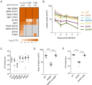
Methicillin-resistant Staphylococcus aureus (MRSA) poses a threat to human health and
is becoming increasingly resistant to current antibiotics. Characterizing the MRSA
proteome during an infection in vivo would reveal important information about the
process of infection and potentially reveal new strategies to fight the disease. Currently
available proteomic techniques, however, are not capable of efficiently measuring
pathogen proteins among the highly abundant host protein background that is inherent to
infection models. In this work we use bioorthogonal noncanonical amino acid tagging
(BONCAT) to perform cell-selective proteomic analysis of MRSA in a mouse skin
infection model, identifying 766 MRSA proteins synthesized during infection.
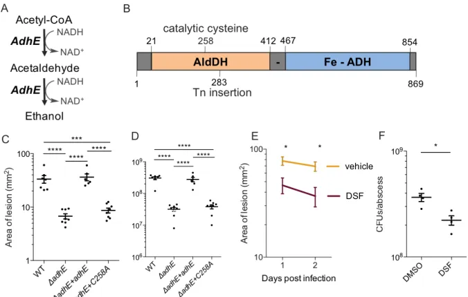
Quantitative analysis of our dataset identifies novel virulence factors that are
upregulated during the course of infection. Deletion of AdhE, one of the top BONCAT
hit proteins, led to a significant decrease in MRSA virulence phenotype during infection.
Furthermore, a point mutation within AdhE reduced virulence to levels similar to that of
the full deletion, indicating a novel site for targeted anti-virulence therapies. Overall,
this work demonstrates the importance of cell-selective chemoproteomic labeling in vivo
and provides insight into the pathogenesis of MRSA infections, revealing several
promising targets for anti-virulence therapy development.
Significance Statement
This work uses a novel chemoproteomic method to tag and detect newly synthesized
proteins by a pathogen in a mouse model of skin infection. By specifically incorporating
a label into proteins synthesized by methicillin-resistant Staphylococcus aureus (MRSA)
as it infects its mouse host, proteins important for nutrient acquisition, metabolism, and
pathogenesis were identified. We tested whether these proteins contributed to infection
by assaying strains of MRSA with single deletions in them. One deletion demonstrated a
significant decrease in infection, indicating that this protein contributes to MRSA
pathogenesis and could be a novel target for antibiotics. Overall, our findings underscore
the ability of bioorthogonal noncanonical amino acid tagging (BONCAT) to “fish out”
pathogenic proteins from more abundant host ones, suggesting that this strategy could be
used for other infectious pathogens.
3.2 Introduction
Methicillin-resistant Staphylococcus aureus (MRSA), a pathogen that causes
life-threatening infections, poses a serious human health threat due to the increasing
numbers of multidrug resistant strains and a lack of new antibiotics (1, 2). Anti-infective
drugs, which target bacteria during invasion, have been suggested as alternative or
adjunct therapies to antibiotics, as they increase bacterial susceptibility (3). By targeting
factors uniquely expressed in vivo, such antibiotics would not affect bacteria growing
outside the host, thus decreasing the possibility of evolved resistance (4). A catalog of
develop anti-infective agents. Elucidating the in vivo staphylococcal proteome during
infection, however, is challenging due to the technical limitations of shotgun
proteomics: the relative overabundance of host proteins occupy most of a mass
spectrometer’s bandwidth, which strongly represses the detection of proteins from the
pathogen. Previous studies have attempted to overcome this limitation by enriching for
bacteria using cell sorting. These approaches, however, are prone to artifacts during
sample preparation, are blind to secreted proteins, and are not amenable to certain
biological systems (5, 6). Other studies attempt to mimic the nutrient status of the host in
test tubes, but do not fully recapitulate the complex environment of the host (7, 8).
Bioorthogonal noncanonical amino acid tagging (BONCAT) is a chemoproteomic
technique that enables the cell-selective and temporal labeling of the cellular proteome
(9). With this technique, a mutant aminoacyl-tRNA synthetase expressed solely by the
cells of interest allows for selective incorporation of a noncanonical amino acid (ncAA)
with a bioorthogonal handle into proteins for subsequent detection and identification.
Because cellular proteins are only labeled during the ncAA pulse, BONCAT provides a
temporally precise overview of the cell’s proteome. The chemical handles on the ncAAs,
often azides or alkynes, can then be conjugated to fluorophores or enrichment tags using
copper-catalyzed alkyne-azide cycloaddition (CuAAC) or strain-promoted alkyne-azide
cycloaddition (SPAAC) (10, 11). Notably, all of these reagents including the ncAAs are
commercially available. BONCAT has previously been applied to studies of microbial
mouse model of skin infection and identified the proteome of MRSA in vivo (Fig 3.1).
Our findings highlight the ability of BONCAT to selectively enrich for pathogenic
proteins from the highly abundant host protein background in the complex milieu of an
active skin infection. Labeling is pathogen-specific and can be achieved within 4 hours
and subsequently used to enrich for staphylococcal proteins made within a host. Testing
potential candidates from this list for virulence defects in vivo led to the discovery of a
novel protein important for MRSA infection. We expect that this unbiased approach to
label pathogenic proteins could be used as a global discovery tool for novel
anti-infective strategies since it is compatible with other pathogens and modes of infection,
and uses commercially available reagents.
Figure 3.1: Schematic depicting BONCAT to label MRSA in a mouse model of skin infection.
Strains of MRSA that do not express NLL-MetRS are unable to incorporate azidonorleucine
(Anl) into proteins. Azide-labeled proteins are subsequently visualized using fluorescence
3.3 Results
Adapting cell-selective BONCAT for MRSA. We first needed to adapt cell-selective
BONCAT for use in MRSA. We opted to use a mutant methionyl-tRNA synthetase
(NLL-MetRS) previously employed in studies of host-pathogen interactions that allows
for the incorporation of azidonorleucine (Anl) in place of methionine residues (16). The
Escherichia coli (Ec) NLL-MetRS codon-optimized for Staphylococcus aureus was
inserted into a staphylococcal shuttle plasmid under control of the hprK/lgt promoter,
which has been shown to be constitutively active, including during infections in vivo
(17-19). This plasmid (+NLL) was transformed into the virulent MRSA strain USA300,
which accounts for up to 98% of reported skin and soft tissue infections (SSTIs) in
hospitals (1). A plasmid without the inserted NLL-MetRS (–NLL) was used as an
empty-vector control for all characterizations.
To assess the ability of the Ec NLL-MetRS to charge MRSA tRNAs with
azidonorleucine (Anl), +NLL and –NLL cultures were grown in tryptic soy broth (TSB)
and 2 mM